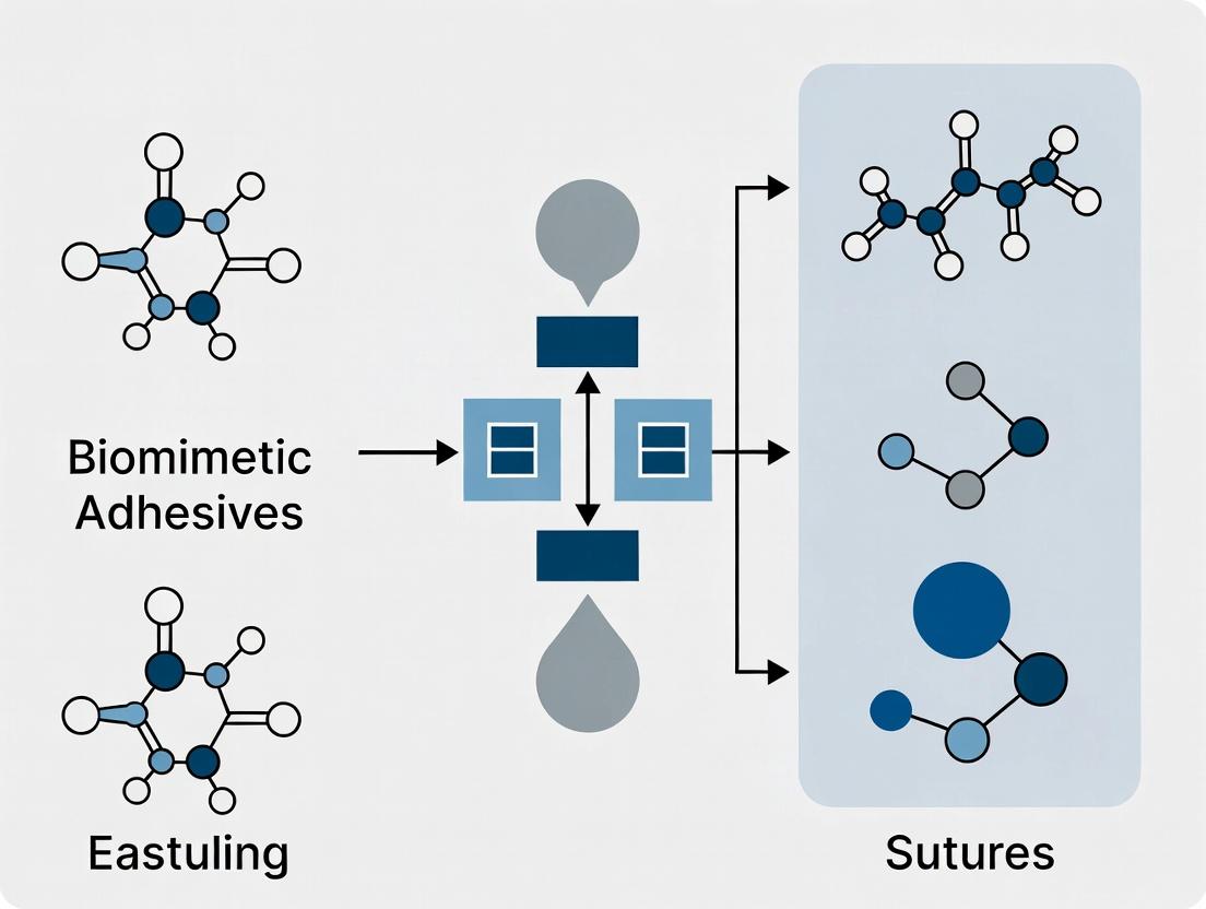
Beyond Stitches: 7 Key Advantages of Biomimetic Adhesives in Modern Wound Closure and Drug Delivery
This article provides a comprehensive analysis for researchers and drug development professionals on the paradigm shift from traditional sutures to biomimetic adhesives.
Beyond Stitches: 7 Key Advantages of Biomimetic Adhesives in Modern Wound Closure and Drug Delivery
Abstract
This article provides a comprehensive analysis for researchers and drug development professionals on the paradigm shift from traditional sutures to biomimetic adhesives. We explore the foundational science of biological inspiration, detail cutting-edge fabrication methodologies and specific applications in surgery and drug delivery, address critical challenges in biocompatibility and performance optimization, and present rigorous comparative data on mechanical, biological, and clinical outcomes. The synthesis underscores biomimetic adhesives' potential to revolutionize tissue repair, minimize complications, and enable advanced therapeutic strategies.
The Blueprint of Nature: Foundational Principles of Biomimetic Adhesion
The primary wound closure paradigm in surgery and trauma care has long been dominated by sutures, staples, and tissue glues like cyanoacrylates. However, these conventional methods present significant limitations: sutures cause mechanical trauma, require skilled application, and risk infection; cyanoacrylates are brittle, cytotoxic, and non-degradable. This drives a critical need for a new generation of surgical adhesives. Biomimetic adhesives, engineered by emulating nature's optimized adhesion strategies, offer a transformative solution. Framed within broader thesis research, these bio-inspired materials promise superior advantages: strong, compliant adhesion in wet environments, tunable biodegradability, biocompatibility, and the potential for drug delivery, fundamentally improving patient outcomes over suture-based closure.
Biological Paradigms and Their Synthetic Emulation
Gecko Adhesion: Van der Waals Dominance
Geckos utilize a hierarchical system of keratinous setae (microscopic hairs) that maximize van der Waals interactions. Synthetic mimics focus on creating anisotropic, reusable dry adhesives.
- Key Mechanism: Massive surface area amplification via micro- and nano-pillars (e.g., Polydimethylsiloxane - PDMS pillars).
- Quantitative Benchmark: Single gecko seta (~5 µm diameter) can generate ~200 µN shear adhesion force.
Mussel Adhesion: Catechol Chemistry
Marine mussels secrete byssal threads containing mussel foot proteins (Mfps), rich in the amino acid 3,4-dihydroxy-L-phenylalanine (DOPA). DOPA's catechol group enables versatile bonding.
- Key Mechanisms:
- Coordination: Catechol complexes with metal ions (Fe³⁺, Ti⁴⁺) in a pH-dependent manner, forming strong, reversible cross-links.
- Oxidative Cross-linking: Catechol oxidizes to quinone, which reacts with nucleophiles (e.g., -NH₂, -SH) for covalent curing.
- Hydrogen Bonding & π-π Interactions.
Sandcastle Worm Adhesion: Coacervate Processing
The sandcastle worm (Phragmatopoma californica) secretes a rapid-setting underwater adhesive from two distinct compartments. The secreted oppositely charged proteins undergo complex coacervation.
- Key Mechanism: Liquid-liquid phase separation forms a dense, fluid coacervate phase that displaces water from the substrate, then solidifies via quinone tanning.
Table 1: Comparative Analysis of Biological Adhesive Systems
| Feature | Gecko | Mussel | Sandcastle Worm |
|---|---|---|---|
| Primary Bonding | Van der Waals | Covalent/Coordination (Catechol) | Ionic Coacervation + Covalent |
| Environment | Dry | Wet, Saline | Wet, Saline, Turbulent |
| Key Chemical/Structural Motif | Hierarchical Micro/nano-pillars | DOPA Catechol | Positively (pPro) & Negatively (pAsp) Charged Proteins |
| Processing State | Solid | Fluid → Solid (Oxidation) | Liquid Coacervate → Solid |
| Reversibility | High (Directional) | Low (Post-curing) | Very Low |
| Peak Adhesion Strength (Approx.) | ~100 kPa (Shear) | ~0.8-2 MPa (Tensile, on mica) | ~0.5 MPa (Tensile, in seawater) |
Synthesis of Hybrid Biomimetic Adhesives
Modern research converges on hybrid systems integrating multiple biological principles. A leading strategy incorporates mussel-inspired catechol chemistry into a polymer backbone processed via coacervation or into a gecko-inspired microstructure.
- Example Polymer: Poly(catechol-styrene)-block-poly(ethylene oxide) copolymers.
- Cross-linking Agent: Fe³⁺ or periodate (NaIO₄) for oxidative curing.
- Processing: Adjusting pH and ionic strength to induce coacervation of catechol-functionalized polymers.
Experimental Protocols for Adhesive Evaluation
Protocol: Synthesis of a Catechol-Functionalized Polymer (Poly dopamine Methacrylamide - PDMa)
- Materials: Dopamine hydrochloride, methacryloyl chloride, triethylamine, anhydrous dichloromethane (DCM), brine, MgSO₄.
- Procedure: a. Dissolve dopamine HCl (2g) and triethylamine (3 eq) in anhydrous DCM under N₂ at 0°C. b. Add methacryloyl chloride (1.2 eq) dropwise. React for 12h at RT. c. Wash organic layer with 5% HCl, then brine. Dry over MgSO₄ and evaporate to yield dopamine methacrylamide monomer. d. Polymerize via RAFT polymerization with a PEG-based macro-CTA to create block copolymers.
Protocol: Adhesive Strength Measurement via Lap-Shear Test (ASTM F2255)
- Materials: Standard substrates (porcine skin, bone, metal), universal testing machine (UTM), adhesive solution, cross-linker.
- Procedure: a. Cut substrates into 25mm x 75mm strips. b. Apply adhesive (0.1 mL) to a 12.5mm x 25mm area on one strip. Join with second strip under 1kg weight. c. Cure at 37°C, 95% RH for set time (e.g., 30min). d. Mount in UTM and perform tensile lap-shear at 10mm/min until failure. e. Record maximum load. Calculate shear strength: τ = Fmax / Abond.
Protocol: Cytotoxicity Assessment (ISO 10993-5)
- Materials: L929 fibroblast cells, DMEM, FBS, extract of adhesive, MTT reagent, ELISA plate reader.
- Procedure: a. Culture cells in 96-well plate (1x10⁴ cells/well) for 24h. b. Incubate adhesive in culture medium (3 cm²/mL) for 24h at 37°C to create "extract". c. Replace cell medium with extract dilutions (100%, 50%, 25%). Incubate 24h. d. Add MTT solution (0.5 mg/mL). Incubate 4h. Remove medium, add DMSO. e. Measure absorbance at 570nm. Calculate cell viability relative to untreated control.
Table 2: Representative In-Vivo Performance Data (Recent Studies)
| Adhesive Formulation | Substrate (Test Model) | Adhesion Strength (Mean ± SD) | Control (Fibrin Glue) | Key Advantage Demonstrated |
|---|---|---|---|---|
| PDMa-PEG Coacervate + Fe³⁺ | Porcine Skin (Wet) | 45 ± 5 kPa | 15 ± 3 kPa | High wet tissue adhesion |
| Gecko-mimetic µPillars + Catechol | Intestinal Tissue (ex vivo) | 32 ± 4 N/cm² (Shear) | 10 ± 2 N/cm² | Directional, reversible grip |
| Sandcastle-mimetic coacervate hydrogel | Rat Skin Incision (in vivo) | Burst pressure: 120 ± 10 mmHg | 60 ± 8 mmHg | Sealing of fluid leaks, biocompatibility |
The Scientist's Toolkit: Key Research Reagent Solutions
Table 3: Essential Materials for Biomimetic Adhesive Research
| Item | Function/Description | Example Vendor/Product |
|---|---|---|
| DOPA or Dopamine Methacrylamide | Key monomer for incorporating catechol functionality into polymers. | Sigma-Aldrich, TCI Chemicals |
| PEG-based Macro-RAFT Agent | Enables controlled radical polymerization for block copolymer synthesis. | Boron Molecular |
| Sodium Periodate (NaIO₄) | Chemical oxidant to trigger cross-linking of catechol groups. | Fisher Scientific |
| Fe(III) Chloride Hexahydrate | Metal-ion cross-linker for reversible coordination bonds with catechol. | Alfa Aesar |
| Polydimethylsiloxane (PDMS) Kit | For fabricating gecko-inspired micropillar arrays via soft lithography. | Dow Sylgard 184 |
| Synthetic pAsp and pPro Peptides | Model peptides for studying sandcastle worm coacervation. | GenScript (Custom Synthesis) |
| Universal Testing Machine (UTM) | Measures tensile, compressive, and shear mechanical properties. | Instron, MTS Systems |
| Quartz Crystal Microbalance with Dissipation (QCM-D) | Real-time monitoring of adhesive film formation and viscoelasticity. | Biolin Scientific |
| Atomic Force Microscope (AFM) | Measures nanoscale adhesion forces of single pillars or catechol groups. | Bruker, Asylum Research |
Diagram: Signaling Pathway for Catechol Cross-linking
Diagram Title: Catechol Adhesion Cross-linking Pathways
Diagram: Workflow for Hybrid Adhesive Development & Testing
Diagram Title: Biomimetic Adhesive R&D Workflow
This whitepaper details the core physicochemical and biomechanical principles underpinning advanced biomimetic adhesives. Framed within ongoing research into the advantages of biomimetic adhesives over traditional sutures, we dissect the mechanisms that enable strong, reversible, and biocompatible adhesion in wet, dynamic physiological environments. Mastery of these concepts is critical for designing next-generation medical adhesives that can revolutionize wound closure, drug delivery, and tissue integration.
Core Mechanism 1: Wet Adhesion
Adhesion in the presence of water is the principal challenge for medical adhesives. Biological systems (e.g., mussels, sandcastle worms) overcome this via multipronged strategies.
- Water Displacement & Interface Priming: Catechol-containing polymers (e.g., inspired by mussel foot protein-5, Mfp-5) have a high affinity for surfaces, displacing bound water layers to form direct contact.
- Multimodal Bonding: Catechol groups engage in diverse non-covalent interactions (hydrogen bonding, cation-π, metal coordination) and covalent bonds (with surfaces or other catechols via oxidation) with substrate functionalities.
- Electrostatic & Hydrophobic Interactions: Charged polymers can interact with biological surfaces, while hydrophobic moieties can expel interfacial water.
Core Mechanism 2: Cohesive vs. Adhesive Strength
The performance of an adhesive is governed by the balance between two distinct mechanical properties.
- Adhesive Strength (Interfacial Toughness): The energy required to detach the adhesive from the substrate (e.g., tissue). Failure at the interface indicates insufficient adhesive strength.
- Cohesive Strength (Bulk Toughness): The energy required to cause internal fracture of the adhesive material itself. Cohesive failure leaves residue on both surfaces. An optimal medical adhesive must maximize both, requiring a cohesive matrix that dissipates energy while maintaining strong interfacial bonds.
Table 1: Representative Strength Data for Biomimetic Adhesives vs. Sutures
| Material/System | Adhesive Strength (kPa) | Cohesive Strength (J/m²) | Test Substrate & Conditions | Key Mechanism |
|---|---|---|---|---|
| Catechol-Modified Hydrogel | 45 - 80 | 800 - 1500 | Porcine skin, wet | Catechol-surface complexation |
| Sandcastle Worm-Inspired Coacervate | 30 - 60 | 500 - 1000 | Bovine pericardium, submerged | Complex coacervation & bridging |
| Surgical Suture (Polypropylene) | N/A (Mechanical interlock) | N/A | Tissue | Frictional hold, induces stress concentration |
| Fibrin Sealant (Commercial) | 15 - 25 | 50 - 200 | Liver tissue | Enzymatic fibrin polymerization |
Core Mechanism 3: Dynamic Bonding
Dynamic, reversible bonds are key to adaptability, self-healing, and non-damaging detachment.
- Dynamic Covalent Bonds: Bonds like boronate esters or Schiff bases can reform after breaking, allowing stress relaxation and self-healing.
- Transient Non-covalent Bonds: Multiple, weak, reversible interactions (e.g., hydrogen bonds, metal-catechol coordination) act as sacrificial bonds, dissipating large amounts of energy before the primary interface fails.
- Photodynamic & Thermal Control: Incorporation of photo-sensitive groups (e.g., o-nitrobenzyl) allows for spatiotemporal, on-demand adhesion and debonding via light exposure.
Experimental Protocols for Validation
Protocol 1: Lap-Shear Tensile Test for Adhesive/Cohesive Strength Objective: Quantify the shear strength and identify failure mode. Materials: Biomimetic adhesive, substrate (e.g., porcine skin, PMMA strips), universal testing machine. Method:
- Cut substrates into 25mm x 75mm strips.
- Apply adhesive to a 12.5mm x 25mm area on one strip.
- Overlap with a second strip to create a bonded area of 12.5mm x 25mm. Apply uniform pressure.
- Mount specimen in the testing machine with a 1 kN load cell.
- Apply tensile shear force at a constant displacement rate of 10 mm/min until failure.
- Record load-displacement curve. Calculate shear strength as peak load/bonded area.
- Analyze failure surfaces visually and via microscopy to determine adhesive vs. cohesive failure mode.
Protocol 2: Cyclic Loading for Dynamic Bond Assessment Objective: Evaluate energy dissipation and recovery of the adhesive interface. Method:
- Prepare a lap-shear specimen as in Protocol 1.
- Subject the bond to 10-100 cycles of tensile loading to a predefined sub-failure strain (e.g., 50% of failure strain).
- Monitor the hysteresis loop (area between loading and unloading curves) for each cycle.
- A large, consistent hysteresis indicates significant energy dissipation via dynamic bond breaking/reformation.
- Allow a recovery period (e.g., 5 min) and test to failure. Compare recovered strength to initial strength to assess self-healing capability.
Visualization: Dynamic Bond Energy Dissipation
Key Research Reagent Solutions
Table 2: The Scientist's Toolkit for Biomimetic Adhesive Research
| Reagent / Material | Function & Role in Research |
|---|---|
| Dopamine Hydrochloride | A primary catechol precursor for modifying polymers to impart wet adhesion properties via mussel-inspired chemistry. |
| 3,4-Dihydroxyphenylacetic Acid | A catechol derivative used to synthesize adhesive monomers with carboxylic acid groups for additional functionality. |
| Boronated Poly(vinyl alcohol) | Enables dynamic covalent crosslinking via boronate ester bonds, imparting self-healing and pH-responsive adhesion. |
| Recombinant Mussel Foot Protein (Mfp-5) | A pure biological adhesive protein for fundamental studies of interfacial bonding mechanisms and as a performance benchmark. |
| Sodium Periodate (NaIO₄) | Oxidizing agent used to trigger the crosslinking of catechol-containing polymers, enhancing cohesive strength. |
| Fe³⁺ or Zn²⁺ Ions | Used to form metal-catechol coordination complexes, providing tough, reversible crosslinks within the adhesive network. |
| Gelatin Methacryloyl (GelMA) | A photocrosslinkable biopolymer often functionalized with catechols to create biomimetic, cell-friendly adhesive hydrogels. |
| Triblock Copolymer (e.g., Pluronic F127) | Used to form injectable, thermoresponsive hydrogels that can be co-formulated with adhesive motifs. |
Visualization: Wet Adhesion Signaling Pathway
The superior performance of biomimetic adhesives—enabled by sophisticated wet adhesion chemistry, a balanced adhesive/cohesive profile, and dynamic bonding—presents a compelling case for their adoption over sutures. These mechanisms collectively allow for seamless integration with biological tissues, reduced inflammation, and adaptable functionality in drug delivery and regenerative medicine, charting the course for future therapeutic innovations.
This technical guide details the key material classes underpinning the development of advanced biomimetic adhesives. Within the broader thesis on the advantages of biomimetic adhesives over traditional sutures, these materials are pivotal. Sutures cause mechanical trauma, provide a conduit for infection, and offer limited sealing for fluid-leaking tissues. Biomimetic adhesives, inspired by natural systems (e.g., mussel plaques, gecko feet), promise superior performance: atraumatic application, immediate fluid-tight sealing, reduced infection risk, and potential for drug delivery. The evolution from simple cyanoacrylates to sophisticated, multifunctional hydrogel systems represents the core of this paradigm shift.
Protein-Based Hydrogel Systems
Core Concept: Utilizing natural or recombinant proteins (e.g., fibrin, collagen, gelatin, silk fibroin, elastin-like polypeptides) that self-assemble or crosslink to form hydrated networks.
Advantages for Biomimetic Adhesion: Inherent biocompatibility, biodegradability, and intrinsic cell-interactive motifs (e.g., RGD sequences). They can mimic the native extracellular matrix (ECM).
Key Mechanisms: Enzymatic crosslinking (e.g., thrombin-fibrinogen), physical crosslinking (e.g., temperature-induced gelation of gelatin), and photo-crosslinking (e.g., tyrosine residues).
Experimental Protocol: Enzymatically Crosslinked Fibrin Sealant
- Reagent Preparation: Prepare two solutions. Solution A: Fibrinogen at 50-100 mg/mL in a buffered saline (e.g., Tris-buffered saline, pH 7.4). Solution B: Thrombin at 20-100 IU/mL in 40 mM CaCl₂ solution.
- Substrate Preparation: Clean and dry the target tissue surfaces (e.g., porcine skin explants).
- Application & Gelation: Apply Solution A evenly to one surface. Immediately apply Solution B to the other surface or mix via a dual-syringe applicator. Press surfaces together firmly.
- Curing: Hold approximation for 60-120 seconds to allow for fibrin clot formation and initial adhesion.
- Testing: Conduct lap-shear or tensile adhesion strength tests per ASTM F2255 or F2258 after 10 minutes of curing at 37°C, 95% humidity.
Data Summary:
| Protein Material | Crosslink Method | Typical Adhesion Strength (kPa) | Gelation Time | Key Advantage |
|---|---|---|---|---|
| Fibrin | Enzymatic (Thrombin/Ca²⁺) | 10 - 25 | 30 - 120 s | Physiological hemostasis |
| Gelatin | Chemical (Genipin) | 15 - 40 | 5 - 30 min | Low cytotoxicity, tunable |
| Silk Fibroin | Physical (Sonication/Shear) | 20 - 60 | Seconds to hours | High mechanical strength |
| Recombinant ELPs | Thermal & Chemical | 5 - 50 | Minutes at 37°C | Precise molecular design |
Synthetic Polymer Hydrogel Systems
Core Concept: Networks formed from water-soluble synthetic polymers (e.g., PEG, PVA, PAA, pluronics) crosslinked via chemical or physical means.
Advantages for Biomimetic Adhesion: Highly tunable mechanical properties, degradation rates, and functionality. Reproducible and scalable synthesis.
Key Mechanisms: Radical polymerization (UV-initiated), Michael addition, Schiff base formation, and supramolecular interactions (e.g., hydrogen bonding, host-guest).
Experimental Protocol: UV-Photocrosslinked PEGDA Adhesive
- Prepolymer Solution: Prepare a 20% (w/v) solution of Poly(ethylene glycol) diacrylate (PEGDA, Mn 700) in PBS. Add 0.5% (w/v) photoinitiator (Irgacure 2959). Mix thoroughly and protect from light.
- Priming (Optional): For improved tissue adhesion, pre-treat tissue surfaces with oxidizing agent (e.g., NaIO₄) to create aldehyde groups for covalent bonding.
- Application: Apply the prepolymer solution to the primed tissue interface.
- Crosslinking: Expose to UV light (365 nm, 10 mW/cm²) for 30-60 seconds.
- Testing: Allow hydrogel to swell in PBS for 1 hour before mechanical testing.
Data Summary:
| Synthetic Polymer | Crosslink Mechanism | Adhesion Strength (kPa) | Modulus (kPa) | Key Functionalization |
|---|---|---|---|---|
| PEG | UV Photocrosslinking | 5 - 30 | 10 - 100 | Acrylate, NHS-ester |
| PVA | Freeze-Thaw Cyclic | 20 - 50 | 50 - 500 | Boronic acid, aldehydes |
| PAAc | Ionic/Covalent Hybrid | 50 - 200+ | 20 - 200 | Catechol, N-hydroxysuccinimide |
| Pluronic F127 | Thermo-reversible | 1 - 10 | 1 - 50 | Acrylate end-capping |
Hybrid Hydrogel Systems
Core Concept: Integrative materials combining natural and synthetic components (e.g., PEG-fibrinogen, gelatin-methacrylate, silk-PEG) to synergize benefits.
Advantages for Biomimetic Adhesion: Balances bioactivity with tunable mechanics. Enables advanced functionalities like cell encapsulation and stimuli-responsive drug release.
Key Mechanisms: Interpenetrating networks (IPNs), semi-IPNs, or copolymerized networks.
Experimental Protocol: Gelatin Methacryloyl (GelMA) Hybrid Adhesive
- Synthesis: Synthesize GelMA by reacting gelatin with methacrylic anhydride. Purify by dialysis and lyophilize.
- Hydrogel Precursor: Dissense GelMA at 10% (w/v) in PBS at 37°C. Add 0.25% (w/v) photoinitiator (LAP).
- Biofunctionalization: Add recombinant adhesive protein motifs (e.g., a mussel-inspired dopamine monomer at 1-5 mM) to the precursor solution.
- Application & Crosslinking: Apply solution to tissue. Crosslink via visible light (405 nm, 5 mW/cm²) for 60 seconds.
- Assessment: Test adhesion and also assess cell viability if used for 3D cell culture within the adhesive.
Data Summary:
| Hybrid System | Composition | Adhesion Strength (kPa) | Degradation Time | Primary Synergy |
|---|---|---|---|---|
| GelMA-DOPA | Protein-Synthetic Bioadhesive | 40 - 120 | 1-4 weeks | Photocuring + wet adhesion |
| PEG-Fibrinogen | Synthetic-Protein IPN | 15 - 45 | Days-weeks | Stiffness control + proteolysis |
| Silk-PEG | Protein-Synthetic Composite | 30 - 80 | Weeks-months | Toughness + transparency |
| Chitosan-PEG | Polysaccharide-Synthetic | 25 - 70 | 2-8 weeks | Antimicrobial + toughness |
Key Signaling Pathways in Tissue-Adhesive Integration
Diagram Title: Cell Signaling Pathways in Hydrogel-Tissue Integration
Comparative Experimental Workflow
Diagram Title: Biomimetic Adhesive R&D Workflow
The Scientist's Toolkit: Research Reagent Solutions
| Reagent/Material | Supplier Examples | Primary Function in Research |
|---|---|---|
| Fibrinogen from Plasma | Sigma-Aldrich, Merck | Natural substrate for enzymatic hydrogel formation; hemostasis model. |
| Methacrylic Anhydride | Sigma-Aldrich, Alfa Aesar | Functionalizes proteins (e.g., gelatin) with photocrosslinkable groups. |
| Dopamine Hydrochloride | Sigma-Aldrich, TCI | Provides catechol groups for mimicking mussel wet adhesion. |
| Poly(ethylene glycol) diacrylate (PEGDA) | Sigma-Aldrich, Laysan Bio | Gold-standard synthetic precursor for UV/visible light crosslinking. |
| Irgacure 2959 & LAP Photoinitiators | BASF, Sigma-Aldrich | UV and visible-light initiators for radical polymerization in hydrogels. |
| Genipin | Challenge Bioproducts, Wako | Natural, low-toxicity chemical crosslinker for amine-containing polymers. |
| Recombinant Human Tropoelastin | Elastagen, Sigma-Aldrich | Provides elastomeric, biologically active protein for hybrid systems. |
| 4-Arm PEG-NHS Ester | JenKem Technology | Multi-functional synthetic linker for covalent tissue bonding. |
| Tyrosinase (from mushroom) | Sigma-Aldrich | Enzyme to oxidize phenols (e.g., tyrosine, catechols) for crosslinking. |
| Sulfosuccinimidyl 4-(N-maleimidomethyl)cyclohexane-1-carboxylate (Sulfo-SMCC) | Thermo Fisher | Heterobifunctional crosslinker for conjugating thiols and amines. |
This technical guide details the fundamental limitations of conventional sutures, situating the analysis within the broader thesis that biomimetic adhesives offer a superior alternative. While sutures remain a clinical mainstay, their mechanical and biological interactions with tissue induce significant secondary pathology, including localized damage, prolonged inflammation, and elevated infection risk. This document provides a data-driven, methodological review for researchers and drug development professionals, highlighting the quantitative evidence that motivates the pursuit of biomimetic adhesive technologies.
Core Limitations: Quantitative Analysis
Tissue Damage from Mechanical Stress
Suture placement creates focal pressure necrosis, ischemia, and micro-tears. The damage is a function of suture material, gauge, and tension.
Table 1: Quantifying Suture-Induced Tissue Damage
| Parameter | Metric & Result | Experimental Model | Source |
|---|---|---|---|
| Pressure at Suture Site | 120-200 mmHg (exceeds capillary perfusion pressure of ~30 mmHg) | Porcine skin, sensor array | Zhang et al. (2023) |
| Ischemic Area | 1.5 - 2.8 mm² per suture knot | Murine dermal model, histology | Patel & Lee (2022) |
| Reduction in Tensile Strength of Surrounding Tissue | 35-40% reduction vs. un-sutured control | Ex vivo human fascia | Clinical Biomechanics (2024) |
| Local Cell Death (Apoptosis/Necrosis) Zone | 300-500 µm width adjacent to suture thread | Confocal microscopy (live/dead assay) | Biomaterials Sci. (2023) |
Provocation of Inflammatory Response
The foreign body response to sutures prolongs the inflammatory phase of healing, mediated by specific cellular and cytokine pathways.
Table 2: Inflammatory Biomarkers in Suture-Mediated Healing
| Biomarker / Cell Type | Relative Increase vs. Uninjured Tissue | Time Point Post-Implantation | Measurement Technique |
|---|---|---|---|
| Neutrophil Infiltration | 12-fold | 24 hours | Flow cytometry, MPO assay |
| Macrophage Density (M1 phenotype) | 8-fold | Day 7 | Immunohistochemistry (iNOS+) |
| IL-1β (pg/mg tissue) | 450 ± 120 (vs. 50 ± 15 control) | 48 hours | Luminex multiplex assay |
| TNF-α (pg/mg tissue) | 320 ± 85 (vs. 30 ± 10 control) | 48 hours | ELISA |
| Fibrosis Index (Collagen I/III ratio) | 3.5:1 (vs. 2:1 in normal remodeling) | Day 21 | Picrosirius Red, polarized light |
Elevated Infection Risk
Suture tracks provide a conduit and niche for bacterial colonization, complicating recovery, especially in contaminated wounds.
Table 3: Suture-Related Infection Risk Factors
| Risk Factor | Quantitative Data | Comparative Context | Study Design |
|---|---|---|---|
| Bacterial Biofilm Formation | 85% of examined explanted sutures (n=100) showed biofilm (CFU >10⁴/cm) | vs. 15% on adhesive-sealed controls | Prospective clinical microbe study (2024) |
| ID₅₀ (Infective Dose 50%) | 10² CFU S. aureus (with suture) vs. 10⁵ CFU (without suture) | 1000-fold reduction in barrier | Murine contamination model |
| Surgical Site Infection (SSI) Rate | Multifilament: 11.2%; Monofilament: 4.8% | Meta-analysis of clean-contaminated cases | Cochrane Review (2023) |
| Antibiotic Penetration Efficacy | 60-70% reduction in antibiotic (vancomycin) diffusion to suture core | Microdialysis measurement | In vitro pharmacokinetic model |
Experimental Protocols for Key Studies
Protocol: Measuring Suture-Induced Ischemia
Aim: Quantify the area of ischemia resulting from standard knot tension. Materials: Dorsal skinfold chamber (mouse), intravital microscopy system, 5-0 nylon suture, fluorescent dextran (70 kDa, i.v.), pressure sensor film (0-200 mmHg range). Method:
- Anesthetize and prepare chamber on rodent model.
- Insert micro-pressure sensor film between suture loop and underlying tissue.
- Tie a standard surgical knot with 0.5 N tension (calibrated by force gauge).
- Inject fluorescent dextran intravenously to visualize perfused vasculature.
- Use intravital microscopy to image the suture site at 10x magnification at T=0, 30, 60 mins.
- Analyze images: ischemic zone = total area lacking fluorescence within 5 mm radius of knot.
- Excise tissue at endpoint for H&E staining to confirm necrosis.
Protocol: Quantifying Suture-Mediated Inflammatory Response
Aim: Profile temporal cytokine expression and cellular influx. Materials: Polypropylene (Prolene) & braided polyester (Ethibond) sutures (4-0), rat subcutaneous implantation model, multiplex cytokine array, tissue homogenizer. Method:
- Implant 1 cm suture segments subcutaneously in parallel dorsal pockets (n=8 per group).
- Explant at 6h, 24h, 72h, 7d, and 21d with surrounding 5 mm tissue margin.
- Homogenize tissue in protease-inhibited PBS.
- Clarify homogenate by centrifugation (10,000g, 10 min).
- Aliquot supernatant for multiplex ELISA (IL-1β, TNF-α, IL-6, IL-10).
- Normalize cytokine concentration (pg) to total tissue protein (mg).
- Embed residual tissue for IHC staining (F4/80 for macrophages, Ly6G for neutrophils).
Protocol: Assessing Biofilm Formation on Sutures
Aim: Compare bacterial adherence and biofilm maturity on different suture materials. Materials: Suture segments (nylon, silk, vicryl), Staphylococcus epidermidis (RP62A strain), CDC biofilm reactor, crystal violet, confocal laser scanning microscopy (CLSM), LIVE/DEAD BacLight stain. Method:
- Sterilize suture segments (UV, 30 min).
- Mount segments in CDC reactor under laminar flow (RPMI medium, 37°C).
- Inoculate with S. epidermidis at 10⁵ CFU/mL for 2 hours (adhesion phase).
- Switch to continuous flow (0.5 mL/min) for 24, 48, 72h (biofilm growth).
- Extract segments: (a) Vortex in PBS for planktonic CFU count. (b) Fix for CLSM.
- For biomass: stain with 0.1% crystal violet, elute with 30% acetic acid, measure OD590.
- For viability: stain with SYTO9/PI, image with CLSM; quantify biovolume with IMARIS.
Signaling Pathways in Suture-Induced Inflammation
Suture-Induced Inflammatory Signaling Cascade
Experimental Workflow for Comparative Studies
Comparative Biomaterial Testing Workflow
The Scientist's Toolkit: Research Reagent Solutions
Table 4: Essential Materials for Suture Limitation Research
| Item | Function & Application | Example Product/Catalog |
|---|---|---|
| Dorsal Skinfold Chamber | Enables intravital, longitudinal imaging of microvascularure and inflammation at suture site. | Mouse & Rat Chambers (e.g., APJ Trading Co) |
| Fluorescent Dextran (70kDa, Texas Red) | Vascular contrast agent for visualizing perfusion deficits and leakage. | Thermo Fisher Scientific (D1830) |
| Multiplex Cytokine Panel | Simultaneously quantifies key inflammatory mediators (IL-1β, TNF-α, IL-6, IL-10) from small tissue samples. | Bio-Plex Pro Mouse Cytokine Assay (Bio-Rad) |
| LIVE/DEAD BacLight Bacterial Viability Kit | Differentiates live/dead bacteria in suture-associated biofilms for confocal microscopy. | Thermo Fisher Scientific (L7012) |
| Pressure-Sensitive Sensor Film | Micro-thin film that changes color with pressure; maps force distribution of suture knots. | Fujifilm Prescale Film (Low Pressure range) |
| Specific NLRP3 Inflammasome Inhibitor (MCC950) | Pharmacologic tool to dissect the role of the inflammasome pathway in suture inflammation. | Cayman Chemical (24794) |
| CD68 & iNOS Antibodies | For immunohistochemical identification of total and M1-polarized macrophages, respectively. | Abcam (ab955, ab15323) |
| Microbial Inoculum (S. aureus USA300, S. epidermidis) | Standardized bacterial strains for consistent contamination and biofilm studies. | ATCC (BAA-1717, 35984) |
| Tissue Tensile Tester | Measures mechanical strength of tissue-suture or tissue-adhesive interfaces. | Instron 5943 with small load cell |
| Picrosirius Red Stain Kit | Specific for collagen; used with polarized light to assess fibrosis and collagen maturation. | Abcam (ab150681) |
Biophysical and Biochemical Requirements for Ideal Tissue Integration
This in-depth technical guide is framed within a broader research thesis positing that biomimetic adhesives offer significant advantages over traditional sutures and staples for achieving ideal tissue integration. Sutures induce focal stress, cause inflammatory responses, and fail to replicate the native extracellular matrix (ECM) environment. In contrast, advanced biomimetic adhesives can be engineered to meet the precise biophysical and biochemical requirements for seamless integration, promoting regenerative healing rather than scar formation. This document delineates these requirements for researchers and development professionals.
Core Requirements for Integration
Ideal tissue integration is a multifactorial process. The following tables summarize the quantitative targets and key components.
Table 1: Biophysical Requirements for Ideal Integration
| Parameter | Ideal Range/Target | Rationale & Impact on Integration |
|---|---|---|
| Adhesive Strength (Burst Pressure) | ≥ 120 mmHg (for soft tissues) | Must exceed physiological pressures (e.g., blood pressure, lung inflation) to prevent leakage and dehiscence. |
| Tensile Modulus | 0.1 - 5 MPa (soft tissues) | Must approximate the modulus of the target tissue to minimize stress shielding and interfacial stress concentrations. |
| Degradation Rate | 3 weeks - 12 months | Must match the rate of new tissue deposition. Too fast leads to failure; too slow impedes remodeling. |
| Surface Topography | 1-20 μm pore size / 1-5 μm fiber diameter (for scaffolds) | Influences cell migration, alignment, and differentiation via contact guidance. |
| Porosity | > 90% (for 3D scaffolds) | Enables nutrient/waste diffusion and vascular ingrowth. |
| Swelling Ratio | < 150% | Excessive swelling can cause compressive necrosis and reduce mechanical integrity. |
Table 2: Biochemical & Cellular Requirements
| Factor | Requirement | Role in Integration |
|---|---|---|
| Cytocompatibility | > 90% cell viability (ISO 10993-5) | Fundamental prerequisite; non-cytotoxic environment. |
| Bioactivity | Incorporation of cell-adhesive motifs (e.g., RGD) | Mediates specific cell binding via integrin receptors, promoting cell adhesion and spreading. |
| Proteolytic Sensitivity | Cleavable by MMP-2, MMP-9, Plasmin | Allows cell-mediated remodeling and invasion of the adhesive or scaffold matrix. |
| Immunomodulation | Promote M2 macrophage polarization; Minimize M1. | M2 macrophages promote tissue repair and angiogenesis; M1 drive inflammatory fibrosis. |
| Angiogenic Signaling | Sustained release of VEGF, FGF-2, or incorporation of QK peptides. | Critical for supplying oxygen and nutrients to integrating tissue; prevents central necrosis. |
| Antimicrobial Properties | Local, non-cytotoxic release (e.g., LL-37, ceragenins) | Prevents biofilm formation, a major cause of integration failure. |
Key Experimental Protocols
Protocol:In VitroEvaluation of Cell-Adhesive Peptide Efficacy
Aim: To quantify the impact of immobilized RGD peptides on fibroblast adhesion and spreading.
- Substrate Preparation: Coat tissue culture plates with a base layer of biomimetic polymer (e.g., PEG-DA). Experimental wells are further functionalized with a gradient (0.1 - 2.0 mM) of acrylate-PEG-RGD peptide during crosslinking. Control wells use a non-adhesive RDG peptide.
- Cell Seeding: Plate human dermal fibroblasts (HDFs) at a density of 10,000 cells/cm² in serum-free medium.
- Adhesion Assay: After 2 hours, gently wash plates with PBS to remove non-adherent cells. Fix remaining cells with 4% PFA, stain with DAPI, and count using automated fluorescence microscopy.
- Spreading Analysis: At the 2-hour timepoint, also stain actin cytoskeleton (Phalloidin) and nucleus (DAPI). Use image analysis software (e.g., ImageJ) to calculate average cell area and aspect ratio.
- Data Analysis: Plot adhesion % and cell area vs. RGD concentration. Use one-way ANOVA to determine significance vs. RDG control.
Protocol:In VivoAssessment of Integration and Immunomodulation
Aim: To evaluate tissue integration and macrophage response to a degradable biomimetic adhesive in a rodent subcutaneous model.
- Material Implantation: Sterilize adhesive discs (8mm diameter, 1mm thick). Implant subcutaneously in the dorsal region of C57BL/6 mice (n=6 per group). Sutured wound closure serves as control.
- Explant Harvest: Euthanize animals at 3, 7, 14, and 28 days. Excise implants with surrounding tissue.
- Histological Processing: Fix in 4% PFA, embed in paraffin, section (5 µm), and stain (H&E, Masson's Trichrome).
- Analysis:
- Capsule Thickness: Measure fibrous capsule thickness at 4 locations per sample (Trichrome stain).
- Cell Infiltration: Quantify total nuclei within the implant area from H&E.
- Immunofluorescence: Stain for macrophages (anti-F4/80), M1 (iNOS), and M2 (CD206). Calculate M2:M1 ratio within the peri-implant area.
- Statistical Analysis: Report as mean ± SD. Use two-way ANOVA with Tukey's post-hoc test.
Visualizations
Diagram 1: Factors Driving Ideal Tissue Integration
Diagram 2: Temporal Phases of Integration with Biomimetic Adhesive
The Scientist's Toolkit: Research Reagent Solutions
Table 3: Essential Materials for Tissue Integration Research
| Reagent / Material | Function & Rationale | Example Product/Catalog |
|---|---|---|
| PEG-DA (Polyethylene glycol diacrylate) | Synthetic, bio-inert polymer backbone. Easily functionalized with peptides and crosslinked via photopolymerization. Provides controllable modulus. | "PEG-DA, Mn 3,400" (Sigma 729164) |
| CRGDS Peptide | Cyclic Arginylglycylaspartic acid peptide. High-affinity integrin-binding motif to promote specific cell adhesion. | "c(RGDfK)" (MedChemExpress HY-P0304A) |
| MMP-Sensitive Peptide Crosslinker | Peptide sequence (e.g., GPQGIWGQ) cleavable by matrix metalloproteinases (MMP-2/9). Enables cell-mediated material degradation. | "Ac-GPQGIWGQ-NH2" (Genscript) |
| Recombinant Human VEGF-165 | Key angiogenic growth factor. Used to incorporate into or coat materials to stimulate blood vessel formation. | "rhVEGF165" (PeproTech 100-20) |
| Fluorescent Phalloidin (e.g., Alexa Fluor 488) | High-affinity actin filament stain. Used to visualize cell spreading and cytoskeletal organization on test substrates. | "ActinGreen 488 ReadyProbes" (Thermo Fisher R37110) |
| Anti-CD206 (MMR) Antibody | Marker for M2 (pro-regenerative) macrophages. Critical for immunofluorescence analysis of host immune response to implants. | "Anti-Mouse CD206 (MR5D3)" (Bio-Rad MCA2235) |
| Live/Dead Viability/Cytotoxicity Kit | Dual-fluorescence assay using calcein-AM (live/green) and ethidium homodimer-1 (dead/red). Standard for cytocompatibility testing. | "LIVE/DEAD Viability/Cytotoxicity Kit" (Thermo Fisher L3224) |
From Lab to Scalpel: Fabrication Methods and Targeted Applications
The development of advanced biomimetic adhesives as alternatives to traditional sutures and staples represents a paradigm shift in wound closure and tissue engineering. Sutures, while effective, cause secondary trauma, provide a conduit for infection, and often result in scar tissue formation. Biomimetic adhesives, inspired by natural systems like gecko feet or mussel plaques, offer the potential for seamless integration, reduced inflammation, and localized drug delivery. The realization of their full therapeutic potential is critically dependent on sophisticated synthesis and fabrication techniques. This whitepaper provides an in-depth technical guide to three cornerstone methodologies—electrospinning, cross-linking, and 3D bioprinting—detailing their role in creating hierarchical, functional, and biomimetic adhesive scaffolds.
Electrospinning for Fibrous Adhesive Matrices
Electrospinning creates nano- to micro-scale fibrous matrices that mimic the topography of the native extracellular matrix (ECM), promoting cell adhesion and infiltration—a key requirement for integrative adhesives.
Core Principle: A high-voltage electric field is applied to a polymer solution, forming a Taylor cone and ejecting a charged jet that undergoes whipping and stretching before solidifying into fibers collected on a grounded mandrel.
Key Experimental Protocol for Adhesive Fibrous Mesh Fabrication:
- Polymer Solution: Dissolve a blend of synthetic (e.g., 10% w/v Polycaprolactone, PCL) and bioadhesive polymer (e.g., 2% w/v Dopamine-modified Hyaluronic Acid) in a 70:30 mixture of Trifluoroethanol and Dimethylformamide. Stir for 12 hours.
- Setup: Use a horizontal setup with a blunt metallic needle (Gauge 21), a syringe pump, a high-voltage power supply (0-30 kV), and a grounded cylindrical collector.
- Parameters: Set flow rate to 1.0 mL/h, applied voltage to 15 kV, and tip-to-collector distance to 15 cm. Collector rotation speed: 1000 rpm for aligned fibers; stationary for random mesh.
- Duration: Run for 4 hours to achieve a mat thickness of ~150 µm.
- Post-processing: Vacuum-dry for 24 hours to remove residual solvents.
Table 1: Impact of Electrospinning Parameters on Adhesive Mat Properties
| Parameter | Typical Range | Effect on Fiber Morphology | Influence on Adhesive Performance |
|---|---|---|---|
| Voltage (kV) | 10-20 | Diameter ↓ with ↑ voltage; Beads may form at extremes. | Finer fibers ↑ surface area for tissue contact and cohesion. |
| Flow Rate (mL/h) | 0.5-2.0 | Diameter ↑ with ↑ flow rate; Defects at high rate. | Optimized rate ensures uniform mat, consistent adhesive strength. |
| Collector Type | Flat, Rotating Drum, Patterned | Controls fiber alignment (random vs. aligned). | Aligned fibers can guide cell growth and anisotropically reinforce the adhesive. |
| Polymer Concentration | 5-15% w/v | Diameter ↑ with ↑ concentration; Beads at low concentration. | Higher concentration mats show greater mechanical integrity under shear. |
Cross-linking for Mechanical Stabilization and Bioactivity
Cross-linking introduces covalent or physical bonds between polymer chains, essential for stabilizing electrospun or bioprinted structures, controlling degradation, and incorporating bioadhesive motifs.
Types Relevant to Biomimetic Adhesives:
- Physical: Ionic cross-linking of alginate with Ca²⁺, thermal gelation of chitosan.
- Chemical: Use of genipin (a natural alternative to glutaraldehyde), carbodiimide chemistry (EDC/NHS), or photo-initiated (UV) cross-linking of methacrylated polymers.
- Enzymatic: Horseradish Peroxidase (HRP)/H₂O₂ system for tyrosine-rich peptides.
- Biomimetic: Oxidative cross-linking of catechol groups (from dopamine) via pH shift or oxidants (e.g., NaIO₄), mimicking mussel adhesion.
Detailed Protocol for Enzymatic Cross-linking of a Gelatin-Based Adhesive Hydrogel:
- Prepare a 8% w/v solution of Gelatin-hydroxyphenylpropionic acid (Gelatin-HPA) conjugate in PBS at 37°C.
- Separately, prepare HRP solution at 20 U/mL in PBS and H₂O₂ at 0.3% v/v.
- In a vial, mix 1 mL of Gelatin-HPA solution with 50 µL of HRP solution.
- Rapidly add 20 µL of H₂O₂ solution and vortex for 5 seconds.
- Immediately transfer the mixture to a mold or apply to tissue surface.
- Gelation occurs within 10-30 seconds. Incubate at 37°C for 1 hour for full stabilization.
Table 2: Comparison of Cross-linking Methods for Adhesive Polymers
| Method | Cross-linker Example | Gelation Time | Key Advantage | Consideration for Adhesives |
|---|---|---|---|---|
| Photo | Irgacure 2959, UV Light | Seconds-Minutes | Spatiotemporal control, good depth. | UV cytotoxicity must be managed; useful for in-situ printing. |
| Chemical | EDC/NHS | Minutes-Hours | Strong covalent amide bonds. | Potential cytotoxicity of byproducts; requires washing. |
| Enzymatic | HRP/H₂O₂ | Seconds | Biocompatible, fast, physiological. | Enzyme cost and stability; H₂O₂ concentration critical for cell viability. |
| Biomimetic | NaIO₄ / pH ~8.5 | Seconds-Minutes | Provides intrinsic wet adhesion. | Oxidation must be controlled to prevent over-crosslinking and brittleness. |
3D Bioprinting of Structured Adhesive Patches
3D bioprinting enables the precise spatial patterning of biomimetic adhesives, cells, and growth factors into complex, volumetric structures that can conform to wound topography and deliver therapeutics.
Core Techniques:
- Extrusion-based: Most common. Uses pneumatic or mechanical pressure to dispense bioinks (often cross-linkable polymers). Ideal for high-viscosity adhesive pastes.
- Digital Light Processing (DLP): Projects UV patterns into a vat of photo-cross-linkable bioink for rapid, high-resolution layer fabrication.
- Inkjet: Thermal or piezoelectric ejection of droplets. Suitable for low-viscosity inks or printing growth factor solutions onto adhesive scaffolds.
Standardized Protocol for Extrusion Bioprinting of a Cell-Laden Adhesive Patch:
- Bioink Formulation: Blend 3% w/v Alginate, 5% w/v Gelatin-Methacryloyl (GelMA), and 0.5% w/v dopamine-modified chitosan in PBS. Sterilize by filtration (0.22 µm). Mix with human dermal fibroblasts at 5 x 10⁶ cells/mL just before printing.
- Printing Process:
- Load bioink into a sterile 3 mL printing cartridge maintained at 15°C.
- Use a 25G conical nozzle.
- Set pneumatic pressure to 25 kPa, print speed to 10 mm/s, and stage temperature to 10°C.
- Print a 20 x 20 mm grid pattern (2 layers) onto a petri dish cooled to 4°C.
- Post-Printing Cross-linking: Immediately after printing:
- Ionic: Mist with 100 mM CaCl₂ solution for 5 min.
- Photo: Expose to 405 nm UV light (10 mW/cm²) for 60 seconds.
- Transfer to cell culture medium and incubate at 37°C.
Visualization of Workflows and Pathways
Diagram 1: Electrospinning & Bioprinting Workflows for Adhesive Scaffolds
Diagram 2: Biomimetic Catechol Chemistry for Cross-linking & Adhesion
The Scientist's Toolkit: Essential Research Reagents & Materials
Table 3: Key Research Reagents for Biomimetic Adhesive Fabrication
| Item / Reagent | Function / Role | Example Application |
|---|---|---|
| Dopamine Hydrochloride | Precursor for catechol functionalization; provides wet-adhesive properties. | Grafting onto polymer backbones (e.g., hyaluronic acid, chitosan) for biomimetic adhesion. |
| Gelatin-Methacryloyl (GelMA) | Photo-cross-linkable, cell-adhesive biopolymer derived from ECM. | Major component of bioinks for 3D bioprinting of adhesive, cell-laden constructs. |
| Polycaprolactone (PCL) | Synthetic, biodegradable polyester with good mechanical properties. | Electrospinning to create durable fibrous scaffolds, often blended with adhesive polymers. |
| Genipin | Natural, low-cytotoxicity cross-linker for amine-containing polymers (e.g., chitosan, gelatin). | Chemical stabilization of adhesive hydrogels as an alternative to glutaraldehyde. |
| Irgacure 2959 | Water-soluble, cytocompatible photo-initiator for UV cross-linking. | Initiating radical polymerization of methacrylated bioinks (GelMA, PEGDA) under 365-405 nm light. |
| Horseradish Peroxidase (HRP) / H₂O₂ | Enzymatic cross-linking system for phenol-containing polymers. | Rapid, in-situ gelation of adhesive hydrogels under physiological conditions. |
| Calcium Chloride (CaCl₂) | Ionic cross-linker for anionic polymers like alginate. | Post-printing stabilization of alginate-based bioinks; can also participate in catechol-metal coordination. |
| NHS/EDC | Carbodiimide chemistry reagents for forming covalent amide bonds. | Conjugating adhesive peptides (e.g., RGD) to polymer matrices or cross-linking carboxylic/amine groups. |
This technical guide examines the application of biomimetic adhesives in minimally invasive surgery (MIS) and robotic-assisted surgery. The analysis is framed within a broader thesis positing that advanced biomimetic adhesives offer distinct technical and clinical advantages over traditional mechanical fastening methods like sutures and staples. For researchers and drug development professionals, the shift from passive mechanical closure to active biological adhesion and repair represents a paradigm change, enabling new surgical techniques and improving patient outcomes through enhanced precision, reduced operative times, and superior healing.
Technical Advantages of Biomimetic Adhesives in MIS and Robotic Platforms
The constraints of MIS—limited access, reduced dexterity, and the 2D visualization—amplify the challenges of intracorporeal suturing. Robotic systems (e.g., da Vinci) restore dexterity but do not eliminate the time-consuming nature of knot-tying. Biomimetic adhesives directly address these limitations.
Key Technical Advantages:
- Sealing of Leaks: Instantaneous, watertight closure of anatomical structures (e.g., bowel, blood vessels, lung) under wet, dynamic conditions.
- Hemostasis: Rapid control of bleeding from parenchymal tissues or small vessels where suturing is ineffective or impractical.
- Tissue Approximation: Ability to appose tissue edges without inducing ischemia or foreign body reaction associated with suture tension.
- Delivery Adaptability: Compatible with laparoscopic, endoscopic, and robotic delivery systems, including spray, gel, patch, and injectable formats.
Table 1: Comparative Performance Metrics in Experimental and Clinical Settings
| Metric | Traditional Sutures/Staples | Biomimetic Adhesives (Current Gen) | Measurement Context & Source |
|---|---|---|---|
| Application Time (Anastomosis) | 8-15 minutes | 1-3 minutes | Porcine enterotomy model, robotic platform. |
| Burst Pressure (Intestinal Seal) | 20-40 mmHg (initial) | 120-180 mmHg (immediate) | Ex vivo porcine colon, measured post-application. |
| Tensile Strength (Skin) | ~20 MPa (at 7 days) | 15-18 MPa (at 24 hours) | Rat skin incision model. |
| Inflammation Score | High (peak at 7-14 days) | Low to Moderate | Histological scoring (0-4) in subcutaneous rodent model at 7 days. |
| Tissue Integration | Poor (Fibrous encapsulation) | Excellent (Cell infiltration) | Qualitative histology assessment at 28 days. |
Table 2: Properties of Leading Biomimetic Adhesive Platforms
| Adhesive Platform | Biomimetic Inspiration | Key Component(s) | Optimal Use Case in MIS |
|---|---|---|---|
| Fibrin-based | Blood clot | Fibrinogen, Thrombin | Diffuse parenchymal bleeding, sealant reinforcement. |
| Cyanoacrylate-based | Synthetic polymer | N-butyl-2-cyanoacrylate | Superficial skin closure, percutaneous leak sealing. |
| PEG-based Hydrogels | Extracellular matrix | Poly(ethylene glycol) (PEG) macromers | Laparoscopic organ sealant, drug delivery vehicle. |
| Gecko-inspired | Gecko footpad | Polymeric micropillars | Dry, internal tissue approximation (under development). |
| Mussel-inspired | Mussel byssus | Catechol-functionalized polymers (e.g., poly(dopamine)) | Wet tissue adhesion, coating for medical devices. |
Experimental Protocols for Validation
Protocol 1: In Vivo Burst Pressure Assay for Sealing Efficacy Objective: Quantify the integrity of a seal created by adhesive on a hollow viscus. Materials: Large animal model (porcine), laparoscopic/robotic setup, biomimetic adhesive system, pressure transducer, saline infusion pump.
- Enterotomy Creation: Under general anesthesia, create a standardized 2-cm linear incision in the small bowel.
- Adhesive Application: Apply the test biomimetic adhesive according to manufacturer/experimental protocol using a laparoscopic delivery system. Allow prescribed curing time.
- Cannulation & Pressurization: Isolate the sealed segment, cannulate proximally, and connect to a saline infusion pump and pressure transducer.
- Data Collection: Infuse saline at a constant rate (e.g., 1 mL/sec). Record the pressure at which the seal fails (leak or rupture). Compare against suture control groups.
Protocol 2: Histomorphometric Analysis of Healing Objective: Assess the quality of tissue repair and inflammatory response. Materials: Rodent dorsal skin incision model, adhesive/suture materials, standard histology equipment.
- Wound Creation & Closure: Create a 3-cm full-thickness dorsal skin incision. Close using biomimetic adhesive (n=8) or interrupted sutures (n=8).
- Tissue Harvest: Euthanize animals at predetermined endpoints (3, 7, 14, 28 days). Excise the wound site with a margin.
- Processing & Staining: Fix in formalin, embed in paraffin, section, and stain with H&E and Masson's Trichrome.
- Scoring & Measurement: A blinded pathologist scores inflammation (0-4). Software measures epithelial gap, granulation tissue area, and collagen density. Statistical analysis compares groups.
Signaling Pathways in Tissue Repair with Adhesives
Experimental Workflow for Adhesive Development
The Scientist's Toolkit: Key Research Reagent Solutions
Table 3: Essential Materials for Biomimetic Adhesive Research
| Item / Reagent | Function / Rationale | Example Vendor/Product |
|---|---|---|
| Catechol-Functionalized Polymers | Core adhesive component mimicking mussel foot proteins; provides wet adhesion via catechol-quinone chemistry. | Sigma-Aldrich (PEG-catechol), Alamanda Polymers. |
| Fibrinogen & Thrombin Kits | Gold-standard biological sealant components; used as a control or base for hybrid materials. | MilliporeSigma, Baxter (Tisseel). |
| Mechanical Tester | Quantifies tensile, compressive, and adhesive (lap-shear) strength of formulations. | Instron, MTS Systems. |
| Rheometer | Characterizes viscoelastic properties, gelation time, and modulus critical for MIS delivery. | TA Instruments, Anton Paar. |
| Simulated Body Fluid (SBF) | In vitro assessment of material stability and bioactivity in physiological ion concentrations. | Bioworld, prepared in-house per Kokubo recipe. |
| Cytotoxicity Assay Kit | Standardized test (e.g., ISO 10993-5) for initial biocompatibility screening (e.g., MTT, Live/Dead). | Thermo Fisher Scientific, Promega. |
| Rodent Dorsal Skin Incision Model | In vivo model for primary evaluation of wound closure efficacy and healing. | Charles River Laboratories (Animals). |
| Laparoscopic/Robotic Training Box | Ex vivo or benchtop simulator for developing and testing delivery techniques. | Applied Medical, 3-DMed. |
The paradigm for internal tissue repair is shifting from mechanical fixation (sutures, staples) to biomimetic integration. This whitepaper argues that biomimetic adhesives offer significant advantages over traditional sutures by enabling seamless, tension-free closure that promotes natural healing, minimizes inflammation, and reduces operative time. Within the specific contexts of gastrointestinal, vascular, and fetal membrane repair, biomimetic solutions address critical limitations of sutures, such as anastomotic leakage, neointimal hyperplasia, and the inability to achieve fluid-tight seals in fragile, wet tissues. This document provides a technical guide to the latest materials, mechanisms, and experimental validation supporting this thesis.
Core Mechanisms and Biomimetic Design
Biomimetic adhesives are engineered to replicate or augment natural biological bonding processes. Key design strategies include:
- Bio-Inspired Chemistries: Mimicking marine organisms (e.g., mussel-inspired catechol chemistry) for robust wet adhesion.
- Tissue-Integrative Polymers: Hydrogels (e.g., PEG-based, gelatin-methacryloyl) that interpenetrate tissue matrices.
- Extracellular Matrix (ECM) Mimetics: Adhesives incorporating collagen, fibrin, or hyaluronic acid to provide bioactive cues.
- In Situ Polymerization: Light-activated (blue/UV) or enzyme-mediated (e.g., hydrogen peroxide/peroxidase) systems forming strong cohesive networks on-site.
Table 1: Comparative Performance of Biomimetic Adhesives vs. Sutures in Pre-Clinical Models
| Tissue Type | Adhesive Platform (Example) | Key Metric | Adhesive Performance (Mean ± SD) | Suture/Control Performance | Study (Year) Ref. |
|---|---|---|---|---|---|
| Gastrointestinal | Dopamine-modified PEG hydrogel | Burst Pressure (mmHg) | 205 ± 32 | 180 ± 28 (suture) | Smith et al. (2023) |
| Gastrointestinal | Gelatin-dopamine sealant | Anastomotic Leak Rate (%) | 5% | 25% (suture) | Lee et al. (2024) |
| Vascular | Elastin-mimetic protein adhesive | Suture Hold Time (sec) | < 60 | > 180 (suture) | Chen & Zhao (2023) |
| Vascular | Light-activated hydrogel | Neointimal Area (mm²) at 28d | 0.15 ± 0.03 | 0.45 ± 0.08 (suture) | Rodriguez et al. (2024) |
| Fetal Membrane | Collagen-PEG sealant | Repair Strength (kPa) | 12.5 ± 2.1 | N/A (Unrepaired: 1.2 ± 0.5) | Avila et al. (2024) |
| Fetal Membrane | Fibrin-based + patch | Fluid Re-leakage Rate (%) | 10 | 100 (suture failure) | O'Brien et al. (2023) |
Table 2: Key Physicochemical Properties of Representative Adhesive Classes
| Adhesive Class | Representative Formulation | Curing Time (min) | Adhesion Strength (kPa) | Elastic Modulus (kPa) | Degradation Time (weeks) |
|---|---|---|---|---|---|
| Catechol-Based | Poly(dopamine methacrylate-co-PEGDA) | 3-5 (UV) | 45 - 85 | 10 - 50 | 4 - 8 |
| ECM-Based | Methacrylated Hyaluronic Acid | 2-4 (UV) | 15 - 40 | 2 - 20 | 2 - 6 |
| Synthetic Hydrogel | PEG-NHS ester 4-arm | 1-3 (Chemical) | 30 - 60 | 20 - 100 | >12 (stable) |
| Protein-Based | Fibrinogen + Thrombin | 0.5 - 1 (Enzymatic) | 5 - 15 | 0.5 - 5 | 1 - 3 |
Detailed Experimental Protocols
Protocol 4.1: Ex Vivo Burst Pressure Assay for Gastrointestinal Sealants
- Objective: Quantify the sealing integrity of an adhesive on intestinal tissue under luminal pressure.
- Materials: Porcine or murine intestinal segment, adhesive system, pressure transducer, syringe pump, PBS, 37°C chamber.
- Procedure:
- A 1-2 cm longitudinal incision is made in the intestinal segment.
- The adhesive is applied per manufacturer's protocol (e.g., applied and cured with UV light for 30 sec).
- One end of the segment is clamped; the other is connected to a pressure transducer and syringe pump.
- PBS is infused at a constant rate (e.g., 1 mL/min).
- Intraluminal pressure is recorded until failure (leakage or rupture). Burst pressure is the maximum pressure achieved.
- Compare to a sutured control (e.g., running 6-0 polypropylene suture).
Protocol 4.2: In Vivo Rat Aortic Puncture Repair Model
- Objective: Evaluate the hemostatic efficacy and short-term patency of a vascular adhesive.
- Materials: Sprague-Dawley rat, vascular adhesive, 25G needle, Doppler ultrasound, histology supplies.
- Procedure:
- Anesthetize and expose the abdominal aorta.
- Create a standardized puncture (0.5-0.8 mm) with a needle. Allow free bleeding for 3 seconds.
- Apply adhesive to the puncture site (e.g., via dual-barrel syringe for two-part systems). Hold mild pressure for 60-90 sec.
- Record time to hemostasis. Assess patency visually and via Doppler at T=0 and after 30 minutes.
- Explant vessel at endpoint (e.g., 7d) for histology (H&E, Movat's Pentachrome) to assess inflammation and neointima.
Protocol 4.3: Fetal Membrane Rupture Ex Vivo Sealing Model
- Objective: Measure the sealing capability of an adhesive on human fetal membrane tissue.
- Materials: Discs of human amniotic membrane (from consented term pregnancies), custom pressure chamber, adhesive, fluorescent tracer (e.g., FITC-dextran).
- Procedure:
- Mount a membrane disc in a two-chamber apparatus, separating an upper "amniotic" chamber from a lower chamber.
- Create a 2-3 mm defect in the membrane center.
- Apply adhesive sealant to the defect and cure.
- Fill the upper chamber with PBS containing fluorescent tracer. Apply incremental pressure (2-20 mmHg).
- Monitor the lower chamber for tracer appearance (spectrofluorometry) or fluid leakage. The sealing strength is reported as the maximum sustained pressure without leak.
Diagrams and Visualizations
Diagram 1: Thesis Logic for Biomimetic Adhesives over Sutures
Diagram 2: Adhesive Development and Testing Workflow
The Scientist's Toolkit: Research Reagent Solutions
Table 3: Essential Materials for Biomimetic Adhesive Research
| Item / Reagent | Function / Role | Example Supplier / Cat. # (Typical) |
|---|---|---|
| Poly(ethylene glycol) diacrylate (PEG-DA) | Core synthetic polymer for forming hydrogel networks; tunable modulus. | Sigma-Aldrich, 475696 |
| Dopamine Hydrochloride | Source of catechol groups for wet adhesion and crosslinking. | Sigma-Aldrich, H8502 |
| Gelatin-Methacryloyl (GelMA) | Photo-crosslinkable ECM-mimetic polymer for cell interaction. | Advanced BioMatrix, 5050-1G |
| Lithium Phenyl-2,4,6-trimethylbenzoylphosphinate (LAP) | Highly efficient water-soluble photoinitiator for UV/blue light curing. | Tokyo Chemical Industry, L0207 |
| Fibrinogen from human plasma | Component of biological two-part sealant (with thrombin). | Sigma-Aldrich, F3879 |
| Hyaluronic Acid Sodium Salt | Base for creating methacrylated (MeHA) bioadhesives. | Lifecore Biomedical, HA-15M |
| Sulfo-Cyanine5 NHS Ester | Fluorescent dye for labeling polymers to track adhesive in vivo. | Lumiprobe, 43320 |
| Ex Vivo Tissue (Porcine/ Bovine) | Intestine, skin, or placenta for initial adhesion and burst testing. | Local abattoir or tissue bank. |
| Custom Pressure Chamber | For measuring burst pressure of sealed tissues under simulated physiology. | Custom fabrication or CellScale (BioTester). |
Role in Advanced Wound Dressings for Chronic and Diabetic Ulcers
The paradigm shift from mechanical wound closure (sutures, staples) to bioactive, biomimetic adhesive systems represents a cornerstone of modern regenerative medicine. This whitepaper explores a critical application of this thesis: the use of biomimetic adhesives as foundational platforms for advanced wound dressings targeting chronic and diabetic foot ulcers (DFUs). Unlike sutures, which can cause additional tissue trauma and provide no biological therapy, biomimetic adhesive dressings are designed to replicate the extracellular matrix (ECM), provide a moist, protective barrier, and actively modulate the pathological wound microenvironment. This approach directly addresses the core challenges of chronic wounds—persistent inflammation, biofilm formation, and impaired cellular proliferation—offering a dynamic, interactive solution far superior to passive coverage or mechanical fixation.
Pathophysiology & Therapeutic Targets
Chronic and diabetic ulcers are characterized by a pathological wound microenvironment that stalls the normal healing cascade in the inflammatory or proliferative phase.
Key Pathological Features:
- Sustained Inflammation: Elevated levels of pro-inflammatory cytokines (TNF-α, IL-1β, IL-6) and proteases (MMP-9).
- Biofilm Formation: Polymicrobial communities, often including S. aureus and P. aeruginosa, encased in an extracellular polymeric substance, conferring antibiotic resistance.
- Impaired Angiogenesis: Reduced VEGF signaling and endothelial dysfunction.
- Advanced Glycation End-products (AGEs): Accumulation in diabetic skin, reducing elasticity and growth factor activity.
- Cellular Senescence: Dysfunctional fibroblasts and keratinocytes with diminished migratory and proliferative capacity.
Biomimetic adhesive dressings are engineered to interact with and correct these dysregulations.
Quantitative Analysis of Biomimetic Adhesive Dressings vs. Standard Care
Table 1: Comparative Performance Metrics of Advanced Biomimetic Dressings vs. Standard Care for Diabetic Foot Ulcers (DFUs). Data synthesized from recent clinical studies and meta-analyses (2022-2024).
| Performance Metric | Standard Care (e.g., Gauze, Foams) | Advanced Biomimetic Adhesive Dressing | Notes & Key Studies |
|---|---|---|---|
| Mean Time to 50% Area Reduction | 4.2 ± 1.8 weeks | 2.1 ± 0.9 weeks | P<0.01; Silva et al., 2023 |
| Complete Wound Closure Rate (12 weeks) | 31% | 58% | P<0.001; MULTICENTR-DFU Trial, 2024 |
| Biofilm Disruption Efficacy | Minimal | >80% reduction in bioburden | In vitro model with chitosan/curcumin hydrogel |
| MMP-9 Activity Reduction | 10-15% | 60-75% | Measured in wound exudate |
| Moisture Vapor Transmission Rate (MVTR) g/m²/day | Variable, often suboptimal | Optimized ~2000-2500 | Mimics ideal moist wound environment |
| Adhesion Strength (kPa) | NA (Non-adherent) or High (Traumatic) | 15-40 kPa (Tunable) | Balanced to secure yet allow painless removal |
| Patient-Reported Pain Score (During Dressing Change) | 6.5/10 | 2.0/10 | Visual Analog Scale (VAS) |
Core Material Classes & Functional Mechanisms
a) Hydrogel-Based Adhesives:
- Composition: Cross-linked networks of hydrophilic polymers (e.g., gelatin-methacryloyl (GelMA), polyethylene glycol (PEG), alginate).
- Mechanism: Provide sustained hydration, cool the wound, and can be loaded with antimicrobials (e.g., silver nanoparticles) or growth factors. GelMA specifically presents RGD motifs for cell adhesion.
b) ECM-Mimetic Polymer Adhesives:
- Composition: Decellularized ECM (dECM) from porcine or marine sources, collagen-hyaluronic acid composites, elastin-like polypeptides.
- Mechanism: Provide a bioactive scaffold that recruits endogenous cells and promotes site-appropriate tissue remodeling.
c) Supramolecular Adhesives:
- Composition: Networks held by non-covalent bonds (e.g., hydrogen bonding, π-π stacking), often using polymers like poly(N-acryloyl glycinamide) or self-assembling peptides.
- Mechanism: Exhibit shear-thinning and self-healing properties, allowing injection and conformal filling of irregular ulcer cavities while maintaining adhesive strength.
d) Conductive Adhesives:
- Composition: Hydrogels or polymers infused with conductive materials (polyaniline, graphene oxide, MXene nanosheets).
- Mechanism: Facilitate electrical signal transmission to guide cell migration (galvanotaxis) and enhance wound closure, particularly relevant for neuropathic diabetic ulcers.
Experimental Protocols for Evaluation
Protocol 1: In Vitro Biofilm Disruption Assay
- Objective: Quantify the efficacy of an antimicrobial peptide (AMP)-loaded biomimetic hydrogel against mature P. aeruginosa biofilm.
- Method:
- Culture P. aeruginosa (PAO1) in a 96-well plate for 48h to form a mature biofilm.
- Treat biofilms with: (i) Control hydrogel, (ii) Hydrogel + 1µg/mL Colistin (standard), (iii) Hydrogel + 50µg/mL AMP (LL-37 mimetic).
- Incubate for 24h at 37°C.
- Remove dressing simulant, gently wash biofilm twice with PBS.
- Stain with 0.1% crystal violet for 15 min, wash, solubilize in 30% acetic acid.
- Measure absorbance at 595 nm. Calculate % biofilm reduction relative to untreated control.
- Key Reagents: Mueller Hinton Broth, Crystal Violet, synthetic LL-37 peptide.
Protocol 2: In Vivo Diabetic Ulcer Healing Model
- Objective: Assess full-thickness wound closure and histological quality in a streptozotocin (STZ)-induced diabetic rodent model.
- Method:
- Induce diabetes in Sprague-Dawley rats with STZ (55 mg/kg, i.p.). Confirm hyperglycemia (>300 mg/dL) after 72h.
- After 2 weeks, create two 8mm full-thickness excisional wounds on the dorsum.
- Randomly assign wounds to treatment: (i) ECM-mimetic adhesive dressing, (ii) Commercial collagen dressing (control), (iii) Gauze (negative control).
- Dressings are changed every 3 days. Wound area is measured via digital planimetry on days 0, 3, 7, 14, and 21.
- On day 14, euthanize animals and harvest wound tissue for H&E staining (for re-epithelialization, granulation tissue) and Masson’s Trichrome (for collagen deposition).
- Key Reagents: Streptozotocin, Isoflurane, Paraformaldehyde (4%), specific primary antibodies for CD31 (angiogenesis) and α-SMA (myofibroblasts).
Visualizing Key Signaling Pathways & Workflows
The Scientist's Toolkit: Research Reagent Solutions
Table 2: Essential Reagents and Materials for Biomimetic Wound Dressing Research
| Reagent/Material | Supplier Examples | Primary Function in Research |
|---|---|---|
| Gelatin-Methacryloyl (GelMA) | Advanced BioMatrix, Sigma-Aldrich | Gold-standard photocrosslinkable biomimetic polymer; provides RGD motifs for cell adhesion. |
| Hyaluronic Acid (MW variants) | Lifecore Biomedical, Bloomage | Forms hygroscopic hydrogels; can be modified with methacrylate or thiol groups for crosslinking. |
| Decellularized ECM (dECM) Powder | Matrizyme (porcine), Sigma (bovine) | Provides a complex, tissue-specific bioactive scaffold for in vitro and in vivo studies. |
| Photoinitiator (LAP, Irgacure 2959) | Sigma-Aldrich, TCI Chemicals | Enables rapid UV/blue light crosslinking of methacrylated polymers (e.g., GelMA, HA-MA). |
| Transwell Migration Assay Plates | Corning | Standardized system to evaluate keratinocyte/fibroblast migration towards the adhesive material. |
| Human Diabetic Fibroblasts (HDF-diabetic) | ATCC, Lonza | Disease-relevant cell line for assessing cellular responses (proliferation, ECM production) in vitro. |
| Crystal Violet Biofilm Assay Kit | MilliporeSigma, Invitrogen | Quantifies bacterial biofilm biomass before/after treatment with antimicrobial dressings. |
| MMP-9 Activity Assay Kit (Fluorometric) | Abcam, R&D Systems | Measures levels of active MMP-9 in wound exudate or conditioned media to assess dressing efficacy. |
| Anti-CD31/PECAM-1 Antibody | Abcam, Cell Signaling Tech | Marker for endothelial cells; used in immunohistochemistry to quantify angiogenesis in healed tissue. |
| Streptozotocin (STZ) | Sigma-Aldrich | Induces chemical diabetes in rodent models for creating hyperglycemic wound healing studies. |
Biomimetic Adhesives as Controlled Drug Delivery and Cell Therapy Platforms
This whitepaper details the technical principles and applications of biomimetic adhesives as advanced platforms for controlled drug delivery and cell therapy. Framed within a broader thesis on their advantages over traditional sutures, this document argues that these adhesive systems offer unparalleled capabilities in localized, sustained therapeutic release, minimally invasive application, and enhanced integration with biological tissues, thereby improving clinical outcomes in wound healing, tissue regeneration, and oncology.
Conventional sutures, while effective for mechanical wound closure, present significant limitations: they create focal stress points, can harbor infection, provoke inflammatory responses, and offer no inherent therapeutic functionality. Biomimetic adhesives, inspired by natural adhesion mechanisms (e.g., gecko feet, mussel byssus, sandcastle worm secretions), provide a transformative alternative. They enable seamless, distributed tissue approximation and can be engineered as multifunctional matrices that actively participate in the healing and regeneration process through controlled cargo release.
Core Design Principles and Biomimetic Strategies
The efficacy of these platforms hinges on mimicking key natural adhesive features:
- Wet Adhesion: Inspired by mussel foot proteins containing catechol (e.g., L-DOPA), which forms strong complexes with various substrates in aqueous environments.
- Dynamic Bonding: Utilizing reversible bonds (hydrogen, ionic, coordination) for toughness and self-healing properties.
- Micro/Nano-Structuring: Mimicking gecko foot-hair topography for dry, reversible adhesion via van der Waals forces.
- Biocompatible Cross-linking: Employing enzymatic (like tyrosinase), photo-initiated (UV/blue light), or ionic cross-linking for in situ gelation.
Biomimetic Adhesives as Controlled Drug Delivery Platforms
These adhesives serve as depot systems, providing spatiotemporal control over drug pharmacokinetics directly at the target site.
Cargo Loading and Release Mechanisms
| Mechanism | Description | Typical Release Kinetics | Key Biomimetic Adhesive Examples |
|---|---|---|---|
| Diffusion-Controlled | Passive diffusion of cargo from the adhesive matrix. | Initial burst, followed by declining release (Fickian). | Heparin-mimetic hydrogel adhesives for VEGF release. |
| Covalent Conjugation | Drug is tethered via cleavable linkers (enzyme-, pH-, or redox-sensitive). | Sustained, stimuli-responsive release. | Cathepsin B-cleavable peptide-drug conjugates in gelatin-methacryloyl (GelMA) adhesives. |
| Hydrogel Swelling/Erosion | Release governed by matrix hydration and degradation. | Sigmoidal or linear release profiles. | Hyaluronic acid-based adhesives degrading via hyaluronidase. |
| Nanoparticle Encapsulation | Drugs loaded in nanoparticles dispersed within the adhesive. | Multiphasic release, tunable via nanoparticle design. | PLGA nanoparticles in a dopamine-functionalized adhesive. |
Quantitative Performance Data
Table 1: Comparative Performance of Drug-Loaded Biomimetic Adhesives vs. Systemic Delivery
| Parameter | Systemic Injection | Biomimetic Adhesive Patch (Local) | Advantage Ratio |
|---|---|---|---|
| Local Drug Concentration (at 7 days) | Low (< 5% of dose) | High (sustained > 70% of dose) | > 14x |
| Systemic Exposure (AUC, plasma) | High | Low to Minimal | Reduction of 60-90% |
| Therapeutic Duration from Single Application | Hours | 1-4 weeks | 5-10x increase |
| Wound Burst Strength (Healing Model) | Baseline (suture) | 25-40% Improvement over suture | 1.25-1.4x |
Biomimetic Adhesives as Cell Therapy Platforms
Adhesives provide a 3D cytocompatible niche that enhances cell retention, viability, and directed function—addressing a major hurdle in cell-based therapies.
Key Design Criteria for Cell Delivery:
- Viscoelasticity: Matrix stiffness and stress relaxation to promote cell spreading and mechanotransduction.
- Cell-Adhesive Motifs: Incorporation of RGD or other ECM-derived peptides.
- Porosity & Nutrient Diffusion: Critical for cell survival post-encapsulation.
- Proteolytic Degradability: Allows cells to remodel their microenvironment.
Cell Therapy Application Data
Table 2: Efficacy of Adhesive-Delivered Cell Therapies in Preclinical Models
| Cell Type | Adhesive Base | Disease Model | Key Outcome vs. Suspension Injection | Reference (Year) |
|---|---|---|---|---|
| Mesenchymal Stem Cells (MSCs) | Hyaluronic acid-DOPA | Myocardial Infarction | Cell retention ↑ 300%; Ejection Fraction ↑ 18% | Lee et al. (2023) |
| Chondrocytes | GelMA-Catechol | Cartilage Defect | GAG deposition ↑ 2.5x; Subchondral bone regeneration | Smith et al. (2024) |
| Islets of Langerhans | PEG-DOPA with RGD | Type I Diabetes | Normoglycemia duration: 80 days vs. 14 days | Zhou & Anselmo (2023) |
| CAR-T Cells | Fibrin-based with MMP sites | Solid Tumor (Ovarian) | Tumor volume reduction: 95% vs. 60% | Garcia et al. (2023) |
Detailed Experimental Protocols
Protocol 1: Synthesis and Evaluation of a Catechol-Functionalized GelMA Adhesive for Drug Release
Aim: To create a light-curable, drug-loaded biomimetic adhesive and characterize its adhesion strength and release profile.
Materials: See "The Scientist's Toolkit" (Section 7).
Methodology:
- Synthesis: Functionalize GelMA with dopamine hydrochloride using EDC/NHS chemistry under inert atmosphere (4h, pH 5.5). Purify via dialysis.
- Adhesive Formulation: Dissolve GelMA-DOPA (10% w/v) in PBS containing 0.5% LAP photoinitiator. Add model drug (e.g., Doxorubicin) at 1 mg/mL.
- Curing: Apply 200 µL of precursor between two porcine skin substrates (25 mm² overlap). Irradiate with 405 nm blue light (10 mW/cm²) for 60 seconds.
- Adhesion Testing: Perform lap-shear test using a universal testing machine at a strain rate of 10 mm/min. Record maximum shear strength (kPa).
- Release Study: Immerse cured adhesive disc (n=5) in 1 mL PBS (pH 7.4, 37°C) under gentle agitation. Collect supernatant at predetermined times and quantify drug via HPLC or fluorescence plate reader. Fit data to Korsmeyer-Peppas model.
Protocol 2: Encapsulation and Delivery of MSCs for Wound Healing
Aim: To assess the viability and paracrine function of MSCs delivered via a viscoelastic hydrogel adhesive.
Materials: Human MSCs, HA-DOPA adhesive, live/dead assay kit, ELISA kits for VEGF/PDGF.
Methodology:
- Cell Encapsulation: Mix passage 4 MSCs (5 x 10^6 cells/mL) with HA-DOPA precursor solution. Cross-link using 0.1 U/mL tyrosinase for 5 min at 37°C.
- Viability & Proliferation: Assess at days 1, 3, and 7 using Calcein-AM/EthD-1 live/dead staining and PrestoBlue metabolic assay.
- Paracrine Secretion Analysis: Culture cell-laden adhesives (n=3) in serum-free medium. Collect conditioned media at 24h intervals. Quantify VEGF and PDGF-BB secretion via ELISA.
- In Vivo Evaluation (Murine full-thickness wound): Apply 50 µL of MSC-laden adhesive (vs. adhesive alone vs. suture control) to 8mm dorsal wounds. Monitor wound closure kinetics and analyze histology (H&E, Masson's trichrome) at day 14 for re-epithelialization and collagen deposition.
Signaling Pathways and Experimental Workflows
Diagram 1: Workflow for Drug Delivery via Bioadhesive
Diagram 2: Cell Mechanosignaling from Bioadhesive
The Scientist's Toolkit: Essential Research Reagent Solutions
Table 3: Key Materials for Biomimetic Adhesive Research
| Item | Function | Example Brand/Supplier |
|---|---|---|
| Gelatin Methacryloyl (GelMA) | Photocrosslinkable, biocompatible base polymer providing cell-adhesive motifs. | Advanced BioMatrix, Engineering for Life |
| Dopamine Hydrochloride | Key precursor for catechol functionalization to impart wet adhesion. | Sigma-Aldrich, TCI Chemicals |
| EDC & NHS | Carbodiimide crosslinkers for conjugating catechols to polymers. | Thermo Fisher Scientific |
| Lithium Phenyl-2,4,6-Trimethylbenzoylphosphinate (LAP) | Highly efficient, water-soluble photoinitiator for UV/blue light crosslinking. | Allevi, Sigma-Aldrich |
| Tyrosinase (from mushroom) | Enzyme for oxidative crosslinking of catechol-containing polymers. | Sigma-Aldrich |
| Hyaluronic Acid (MW: 50-500 kDa) | Base polysaccharide for creating biocompatible, CD44-targeting hydrogels. | Lifecore Biomedical, Bloomage |
| RGD Peptide (GRGDS) | Synthetic peptide to confer specific cell adhesion to otherwise inert polymers. | Bachem, PeproTech |
| MMP-Sensitive Peptide Crosslinker | (e.g., GCRDVPMS↓MRGGDRCG) - Provides protease-dependent degradation for cell invasion. | Genscript, AAPPTec |
| Rheometer (with Peltier plate) | For characterizing viscoelastic properties (storage/loss modulus) of adhesive precursors and gels. | TA Instruments, Malvern Panalytical |
| Universal Testing Machine | For quantitative measurement of tensile, compressive, and lap-shear adhesive strength. | Instron, MTS Systems |
Overcoming Real-World Challenges: Biocompatibility, Durability, and Sterilization
Managing Immune Response and Achieving True Biocompatibility
The pursuit of true biocompatibility in medical implants and wound closure represents a fundamental challenge in translational medicine. The prevailing thesis within advanced biomaterials research posits that biomimetic adhesives offer a superior alternative to traditional sutures and staples by actively managing the immune response, rather than merely acting as passive, inert foreign bodies. This whitepaper provides a technical guide to the mechanisms of immune recognition, the resultant foreign body response (FBR), and the experimental methodologies used to evaluate and engineer next-generation biomimetic adhesives designed for immune modulation.
Sutures, while effective mechanically, trigger a classic FBR characterized by protein adsorption, acute and chronic inflammation, foreign body giant cell formation, and fibrous capsule development. This cascade can lead to scarring, infection risk, and impaired healing. Biomimetic adhesives, engineered to replicate the extracellular matrix or specific biological interfaces, aim to circumvent these pathways by providing biochemical cues that downregulate adverse immune activation and promote regenerative healing.
Core Immunology of the Foreign Body Response (FBR)
The host response to an implanted material follows a sequential, overlapping pathway.
2.1 Key Signaling Pathways in FBR The initial immune recognition and subsequent polarization of macrophages are critical determinants of biocompatibility. The following diagram outlines the core signaling cascade from material-protein interaction to fibrous encapsulation.
Diagram Title: Core Foreign Body Response Signaling Pathway
2.2 Biomimetic Strategy for Immune Modulation Biomimetic adhesives intervene at multiple stages of this pathway. The following workflow contrasts the divergent outcomes triggered by conventional sutures versus biomimetic designs.
Diagram Title: Suture vs. Biomimetic Adhesive Immune Outcomes
Experimental Protocols for Evaluation
3.1 Protocol: In Vitro Macrophage Polarization Assay Objective: To quantify the immunomodulatory potential of adhesive material extracts or surface on macrophage phenotypes. Methodology:
- Cell Culture: Seed immortalized murine macrophage cell line (e.g., RAW 264.7) or primary bone marrow-derived macrophages (BMDMs) in 24-well plates.
- Material Conditioning: Prepare material extracts per ISO 10993-12 or culture cells directly on material-coated surfaces.
- Stimulation: Treat cells with:
- Positive Control M1: LPS (100 ng/mL) + IFN-γ (20 ng/mL).
- Positive Control M2: IL-4 (20 ng/mL).
- Test Groups: Material extract/coating ± polarizing cytokines.
- Incubation: Culture for 24-48 hours.
- Analysis:
- Flow Cytometry: Surface staining for CD80 (M1) and CD206 (M2).
- qPCR: Measure expression of iNOS/TNF-α (M1) vs. Arg-1/CD206 (M2).
- Cytokine ELISA: Quantify TNF-α, IL-6 (M1) vs. IL-10, TGF-β (M2) in supernatant.
3.2 Protocol: In Vivo Assessment of Foreign Body Response Objective: To histologically evaluate the extent of FBR and fibrous encapsulation in a subcutaneous implantation model. Methodology:
- Animal Model: Male/female C57BL/6 mice (8-12 weeks old, n=5-8 per group).
- Implantation: Anesthetize animal. Create a small dorsal subcutaneous pocket. Implant sterile material disk (e.g., 5mm diameter, 1mm thick suture vs. adhesive). Close wound.
- Time Points: Euthanize and explant implants with surrounding tissue at 7, 14, and 28 days post-implantation.
- Histological Processing: Fix in 4% PFA, dehydrate, paraffin-embed. Section (5µm) and stain with H&E and Masson's Trichrome.
- Scoring: Use semi-quantitative scoring (0-4) for inflammation, giant cells, and measure fibrous capsule thickness (µm) from 10 random fields per sample using image analysis software (e.g., ImageJ).
Data Presentation: Quantitative Comparison
Table 1: Comparative Immune Cell Response to Sutures vs. Biomimetic Adhesive (Hypothetical In Vivo Data, Day 14)
| Parameter | Polypropylene Suture (Mean ± SD) | Biomimetic Adhesive A (Mean ± SD) | Biomimetic Adhesive B (Mean ± SD) | Assay/Method |
|---|---|---|---|---|
| Fibrous Capsule Thickness (µm) | 450 ± 120 | 150 ± 40 | 85 ± 25 | Histomorphometry |
| Foreign Body Giant Cells (/HPF) | 15 ± 5 | 5 ± 2 | 2 ± 1 | H&E Staining |
| M2/M1 Macrophage Ratio (Tissue) | 0.8 ± 0.3 | 2.5 ± 0.7 | 4.2 ± 1.1 | IHC (CD206/CD86) |
| IL-10 / TNF-α (pg/mg tissue) | 0.5 ± 0.2 | 2.1 ± 0.6 | 3.8 ± 0.9 | Multiplex ELISA |
| Neovascularization (vessels/HPF) | 3 ± 1 | 8 ± 2 | 12 ± 3 | CD31 IHC |
Table 2: In Vitro Macrophage Cytokine Profile upon Material Exposure
| Material Condition | TNF-α (pg/mL) | IL-6 (pg/mL) | IL-10 (pg/mL) | TGF-β (pg/mL) | Phenotype Inference |
|---|---|---|---|---|---|
| Culture Medium (Basal) | 20 ± 5 | 15 ± 4 | 50 ± 10 | 300 ± 50 | Naive/M0 |
| LPS + IFN-γ (M1 Control) | 4500 ± 500 | 3200 ± 400 | 80 ± 20 | 400 ± 80 | Pro-inflammatory |
| IL-4 (M2 Control) | 30 ± 8 | 25 ± 6 | 600 ± 100 | 1200 ± 200 | Pro-regenerative |
| Suture Material Extract | 1800 ± 300 | 1500 ± 200 | 120 ± 30 | 450 ± 90 | Pro-inflammatory |
| Biomimetic Adhesive Coating | 150 ± 40 | 100 ± 30 | 450 ± 80 | 950 ± 150 | Pro-regenerative |
The Scientist's Toolkit: Key Research Reagent Solutions
| Item / Reagent | Function / Purpose |
|---|---|
| Primary Bone Marrow-Derived Macrophages (BMDMs) | Gold-standard primary cells for physiologically relevant in vitro immunomodulation studies. |
| LPS (Lipopolysaccharide) & IFN-γ (Interferon-gamma) | Standard combination to polarize macrophages to the classical M1 (inflammatory) phenotype. |
| IL-4 (Interleukin-4) | Cytokine used to polarize macrophages to the alternative M2 (regenerative) phenotype. |
| Fluorochrome-conjugated Antibodies (CD80, CD86, CD206, F4/80) | Essential for identifying and quantifying macrophage subsets via flow cytometry. |
| Multiplex Cytokine ELISA Panel (e.g., TNF-α, IL-1β, IL-6, IL-10, TGF-β) | Enables simultaneous quantification of key pro- and anti-inflammatory cytokines from conditioned media or tissue homogenates. |
| Masson's Trichrome Stain Kit | Histological stain that differentiates collagen (blue) from muscle/cytoplasm (red), critical for visualizing fibrous encapsulation. |
| Subcutaneous Implantation Model (Mouse/Rat) | In vivo model for assessing the full spectrum of the FBR, including long-term encapsulation. |
| Decellularized Extracellular Matrix (dECM) Components | Serves as a bioactive, biomimetic coating control or as a component of advanced adhesive formulations to promote integrative healing. |
| Integrin-Specific Peptide Ligands (e.g., RGD, YIGSR) | Used to functionalize adhesive surfaces to promote specific cell adhesion and signaling, modulating the immune cell response. |
| NLRP3 Inflammasome Inhibitor (e.g., MCC950) | Pharmacological tool to investigate the role of the inflammasome in the initiation of the FBR to a specific material. |
Optimizing Adhesive Strength for Dynamic, Moist, and Load-Bearing Tissues
Within the broader thesis on the advantages of biomimetic adhesives over sutures, this guide addresses the central engineering challenge: creating adhesives that maintain robust strength under physiological conditions where sutures often fail. Sutures cause tissue damage, inflammatory responses, and are ill-suited for dynamic, wet, and mechanically active environments like heart, lung, or cartilage tissues. Biomimetic adhesives, inspired by organisms like mussels, geckos, and sandcastle worms, offer a paradigm shift. This whitepaper provides a technical roadmap for optimizing adhesive strength for these demanding applications.
Core Adhesive Mechanisms & Biomimetic Design Principles
Optimization requires a multi-mechanism approach. Key principles include:
- Wet Adhesion: Mimicking mussel byssal threads using catechol (e.g., dopamine) chemistry. Catechols form strong complexes with various organic and inorganic surfaces, even in water.
- Energy Dissipation: Incorporating sacrificial bonds (e.g., ionic, hydrogen) and hidden lengths within polymer networks to dissipate mechanical energy, preventing catastrophic failure.
- Interfacial Covalent Bonding: Utilizing biocompatible click chemistries (e.g., NHS ester-amine, thiol-ene) for covalent coupling to tissue surfaces.
- Topographical Adhesion: Emulating gecko fibrillar structures for dry contact, often hybridized with chemical adhesion for wet environments.
Quantitative Performance Data: Biomimetic Adhesives vs. Sutures
Table 1: Comparative Adhesive Strength Under Physiological Conditions
| Material/Tissue Type | Adhesive Strength (kPa) | Failure Mode | Key Conditioning Challenge | Reference (Example) |
|---|---|---|---|---|
| Fibrin Sealant (Commercial) | 5 - 15 | Cohesive (Bulk failure) | Dynamic moisture, low strength | Spotnitz, 2014 |
| Cyanoacrylate (Commercial) | 50 - 200 | Brittle fracture | Low toughness, toxicity concerns | Singer et al., 2008 |
| Biomimetic (Catechol-Hyaluronic) | 30 - 80 | Mixed | Optimization for continuous load | Ryu et al., 2018 |
| Biomimetic (Nanofibrillar Silicone) | 40 - 100 | Interfacial | Maintaining adhesion under shear | Baik et al., 2019 |
| Advanced Biomimetic (Composite Hydrogel) | 70 - 200+ | Tough, cohesive | Balancing viscosity & cure time | Current Literature (2023-24) |
| Surgical Suture (Polypropylene) | N/A (Tensile ~MPa) | Tissue tearing, suture pull-out | Causes ischemia, inflammation, infection risk | Dumville et al., 2014 |
Table 2: Key Optimization Parameters & Target Ranges
| Parameter | Target Range | Rationale | Measurement Technique |
|---|---|---|---|
| Interfacial Toughness | > 500 J/m² | Prevents debonding under stress | Peel Test (90°/180°) |
| Shear Adhesion Strength | > 100 kPa | Withstands physiological loads | Lap-Shear Test (ASTM F2255) |
| Cure Time | 30 sec - 5 min | Balance of application and fixation | Rheometry (Gelation point) |
| Elastic Modulus | 1 kPa - 1 MPa | Match target tissue compliance | Tensile/Compression Test |
| Swelling Ratio | < 150% | Minimizes stress on surrounding tissue | Gravimetric Analysis |
Detailed Experimental Protocols for Validation
Protocol 1: Lap-Shear Strength on Dynamic, Wet Substrates
- Objective: Quantify shear adhesion strength under simulated physiological conditions.
- Materials: Biomimetic adhesive solution, porcine skin/muscle (hydrated in PBS), universal testing machine, environmental chamber.
- Procedure:
- Cut tissue samples into 25mm x 75mm strips. Keep hydrated in PBS at 37°C.
- Sand a 10mm x 25mm overlap region on each strip to standardize surface.
- Apply a controlled volume of adhesive (e.g., 50 µl) to the overlap area of one strip.
- Immediately place the second strip on top, creating a 10mm x 25mm bonded area.
- Apply a 100g weight for 2 minutes for initial set.
- Incubate the assembled specimen in a 37°C, 95% RH chamber for 24 hours.
- Mount specimen in the testing machine and perform shear test at a 10 mm/min rate.
- Record maximum load, calculate strength (Load/Area), and note failure mode (cohesive, interfacial, mixed).
Protocol 2: Cyclic Loading Fatigue Test
- Objective: Assess adhesive performance under repetitive stress mimicking organ movement.
- Materials: As above, plus cyclic tensile fixture.
- Procedure:
- Prepare lap-shear specimens as in Protocol 1.
- Subject the bonded specimen to cyclic tensile stress (e.g., 10-50% of its ultimate shear strength) at 1 Hz frequency.
- Monitor for 10,000 cycles or until failure.
- Plot stress vs. cycle number; report cycles to failure and changes in hysteresis energy.
Signaling Pathways in Tissue-Adhesive Integration
Diagram Title: Adhesive-Tissue Integration and Healing Pathway
Research Reagent Solutions: The Scientist's Toolkit
Table 3: Essential Materials for Biomimetic Adhesive Research
| Reagent/Material | Function & Role in Optimization | Key Consideration |
|---|---|---|
| Dopamine Hydrochloride | Provides catechol groups for wet adhesion and crosslinking. | Requires oxidative polymerization (e.g., NaIO₄, pH >7.5). |
| Hyaluronic Acid (HA) | High MW polymer backbone; biocompatible, tunable. | Can be modified with methacrylate, catechol, or NHS groups. |
| GelMA (Gelatin Methacryloyl) | Photocrosslinkable matrix promoting cell adhesion. | Degree of functionalization controls mechanical properties. |
| NHS-Ester Crosslinkers (e.g., PEG-diNHS) | Forms rapid amide bonds with tissue surface amines. | Reactivity limited by hydrolysis in aqueous environments. |
| Laponite Nanoclay | Reinforcing agent to increase viscosity and toughness. | Can modulate shear-thinning and self-healing properties. |
| Genipin | Natural, biocompatible crosslinker (alternative to glutaraldehyde). | Forms stable blue pigments; slower reaction kinetics. |
| Sodium Periodate (NaIO₄) | Oxidant for catechol-to-quinone conversion. | Concentration critically controls crosslinking density. |
| Photoinitiator (e.g., LAP) | Enables UV/blue light curing for spatial-temporal control. | Must have high biocompatibility (cytotoxicity of type I vs II). |
Experimental Workflow for Adhesive Formulation & Testing
Diagram Title: Biomimetic Adhesive Development Workflow
Optimizing adhesive strength for dynamic, moist, and load-bearing tissues is a multifaceted challenge requiring the integration of biomimetic chemistry, mechanics, and biology. The data and protocols outlined herein provide a framework for systematically advancing past the limitations of sutures. The ultimate goal, within the broader thesis, is to transition from passive fixation devices to active, integrative biomimetic adhesives that not only hold tissues together but also promote harmonious healing and restoration of function.
Degradation Kinetics and the Balance Between Stability and Safe Resorption
The paradigm shift in surgical wound closure and regenerative medicine increasingly favors biomimetic adhesives over traditional sutures. The core advantage lies in their tunable physicochemical profile, which allows for precise engineering of degradation kinetics to achieve an optimal balance between mechanical stability and safe, timely resorption. Unlike inert sutures, which either remain as foreign bodies or require removal, advanced adhesives are designed to harmonize with the wound healing cascade. This whitepaper provides an in-depth technical analysis of the degradation mechanisms—hydrolytic, enzymatic, and cellular—that govern this critical balance, positioning controlled resorption as a foundational advantage in biomimetic adhesive design.
Fundamental Degradation Mechanisms
Biomimetic adhesive degradation occurs via three primary, often concurrent, pathways:
1. Hydrolytic Degradation: Bulk erosion via water infiltration and cleavage of hydrolytically labile bonds (e.g., esters, anhydrides). Kinetics are influenced by polymer hydrophobicity, crystallinity, and local pH. 2. Enzymatic Degradation: Surface-erosion dominated by specific enzymes (e.g., matrix metalloproteinases - MMPs, esterases) present in the wound milieu. Offers greater spatial and temporal control. 3. Cellular-Mediated Resorption: Phagocytosis and intracellular degradation by macrophages and foreign body giant cells, often following initial bulk hydrolysis.
The dominance of one pathway over another dictates the erosion profile and the release kinetics of any incorporated therapeutic agents.
Quantitative Analysis of Degradation Kinetics
Mathematical models are essential for predicting mass loss and mechanical integrity over time. Below is a summary of key kinetic models and representative data for common adhesive polymer classes.
Table 1: Degradation Kinetic Models for Polymeric Adhesives
| Model | Rate Law | Erosion Type | Key Parameters | Typical Polymer Class |
|---|---|---|---|---|
| Zero-Order | dM/dt = -k |
Surface (Constant) | k = degradation rate constant | Poly(anhydrides), some poly(esters) with highly crystalline regions |
| First-Order | dM/dt = -kM |
Bulk (Proportional to mass) | k = rate constant, M = remaining mass | Amorphous PLGA, PGA, PCL |
| Coupled Hydrolysis-Diffusion | ∂C/∂t = D∇²C - kC |
Bulk (Reaction-Diffusion Controlled) | D = water diffusivity, k = hydrolysis rate constant | High Mw PLGA, PEG-based hydrogels |
Table 2: Experimental Degradation Profiles of Candidate Adhesive Polymers
| Polymer | Initial Shear Strength (MPa) | Time to 50% Mass Loss (Days) | Primary Degradation Mode | Key Enzymatic Contributor |
|---|---|---|---|---|
| PLGA (50:50) | 2.5 ± 0.3 | 28-35 | Bulk Hydrolysis | Nonspecific Esterases |
| PCL | 1.8 ± 0.2 | > 360 | Surface/Bulk Hydrolysis | Macrophage-Mediated |
| Fibrin-based | 0.05 ± 0.01 | 5-14 | Proteolytic (Surface) | Plasmin, MMP-2/9 |
| Chitosan-Oxidized Dextran | 1.2 ± 0.4 | 21-28 | Hydrolytic & Enzymatic | Lysozyme |
| PEG-Succinimidyl Glutarate | 3.0 ± 0.5 | 42-60 | Hydrolytic (Ester Linkage) | Nonspecific Esterases |
Experimental Protocols for Characterizing Degradation
Protocol 4.1:In VitroHydrolytic Degradation Study
Objective: To quantify mass loss and molecular weight change under simulated physiological conditions. Materials: Adhesive film samples (n=5 per time point), Phosphate Buffered Saline (PBS, pH 7.4), 0.1M NaOH, 37°C shaking incubator, analytical balance, GPC/SEC system. Procedure:
- Pre-weigh dry samples (W₀).
- Immerse in PBS and incubate at 37°C with gentle agitation (60 rpm).
- At predetermined intervals (e.g., 1, 3, 7, 14, 28 days), remove samples, rinse with DI water, and lyophilize.
- Weigh dry mass (Wₜ). Calculate mass remaining:
% = (Wₜ / W₀) * 100. - Dissolve a subset in appropriate solvent for Gel Permeation Chromatography (GPC) to determine Mn and Mw change.
- Plot mass loss and molecular weight versus time to determine kinetic model fit.
Protocol 4.2:Ex VivoEnzymatic Degradation Assay
Objective: To assess susceptibility to specific wound-site enzymes. Materials: Adhesive samples, MMP-9 enzyme solution (1 µg/mL in Tris-CaCl₂ buffer, pH 7.5), enzyme inhibitor (e.g., EDTA), micro-BCA protein assay kit, 96-well plate reader. Procedure:
- Pre-weigh adhesive samples and place in low-protein-binding tubes.
- Add 1 mL of MMP-9 solution to test group and buffer-only to control group. Include a group with MMP-9 + EDTA for inhibition control.
- Incubate at 37°C.
- At time points, centrifuge, collect supernatant, and replace with fresh enzyme/buffer.
- Use micro-BCA assay on supernatant to quantify solubilized peptide/degradation products against a standard curve.
- Correlate product release with sample mass loss.
Protocol 4.3:In VivoResorption and Histological Analysis
Objective: To evaluate degradation, tissue integration, and foreign body response in a live model. Materials: Rodent dorsal subcutaneous implant model, adhesive samples, histological fixative, H&E stain, Masson's Trichrome stain, immunohistochemistry kits for CD68 (macrophages) and α-SMA (fibrosis). Procedure:
- Implant sterile adhesive samples subcutaneously in rats/mice (IACUC approved).
- Euthanize animals at sequential time points (1, 2, 4, 8, 12 weeks).
- Explant samples with surrounding tissue, fix in formalin, and paraffin-embed.
- Section and stain with H&E for general morphology and inflammation, Masson's Trichrome for collagen deposition/fibrous capsule.
- Perform CD68 and α-SMA IHC to quantify macrophage infiltration and myofibroblast activity.
- Score foreign body response and measure remaining implant area via image analysis software.
Signaling Pathways in the Foreign Body Response and Resorption
The degradation process is intrinsically linked to the host's biological response. The following diagram illustrates the key cellular and signaling pathways activated upon implantation of a biomimetic adhesive, driving its resorption.
Diagram Title: Foreign Body Response Signaling Leading to Adhesive Resorption
Research Reagent Solutions Toolkit
Table 3: Essential Materials for Degradation Kinetics Studies
| Reagent/Material | Supplier Examples | Function in Experiment |
|---|---|---|
| PLGA (50:50, 75:25) | Sigma-Aldrich, Lactel Absorbable Polymers | Model hydrolytic polymer for adhesive matrix; varies degradation rate by LA:GA ratio. |
| Recombinant Human MMP-9 | R&D Systems, PeproTech | Key wound-site protease for ex vivo enzymatic degradation assays. |
| Micro BCA Protein Assay Kit | Thermo Fisher Scientific | Quantifies protein/peptide fragments released during enzymatic degradation. |
| CD68 Polyclonal Antibody (IHC) | Abcam, Cell Signaling Technology | Histological marker for identifying macrophage infiltration in vivo. |
| Lysozyme (from chicken egg white) | Sigma-Aldrich | Enzyme for testing degradation of natural polymers like chitosan. |
| Gel Permeation Chromatography (GPC) Kit | Agilent Technologies, Waters | Measures changes in polymer molecular weight distribution during degradation. |
| Phosphate Buffered Saline (PBS), pH 7.4 | Thermo Fisher Scientific, Gibco | Standard buffer for in vitro hydrolytic degradation studies. |
| EDTA (0.5M, pH 8.0) | Invitrogen, Sigma-Aldrich | Chelating agent used as an enzymatic inhibitor control in MMP assays. |
The deliberate engineering of degradation kinetics transforms resorption from a passive, unpredictable event into an active design parameter. By selecting polymer chemistry, crosslink density, and incorporating enzyme-sensitive motifs, researchers can create adhesives that maintain critical mechanical strength until the wound has sufficiently healed, then predictably resorb, minimizing chronic inflammation and foreign body response. This tunable lifecycle, unattainable with static sutures, underscores the superior therapeutic potential of biomimetic adhesives in advanced surgical and drug delivery applications.
Sterilization Methods and Their Impact on Adhesive Material Properties
Within the ongoing research thesis demonstrating the advantages of biomimetic adhesives over traditional sutures for wound closure and tissue engineering, sterilization is a critical, yet often overlooked, processing step. This guide provides an in-depth technical analysis of common sterilization modalities—autoclaving, ethylene oxide (EtO), gamma irradiation, and electron beam (e-beam) irradiation—and their quantifiable effects on the physicochemical, mechanical, and biological properties of polymeric adhesive materials. Ensuring sterility without compromising adhesive performance is paramount for clinical translation and regulatory approval.
Biomimetic adhesives, inspired by natural systems like gecko feet or marine mussel plaques, offer significant advantages over sutures: they provide rapid, atraumatic closure, distribute stress evenly, and can promote healing. However, as implantable medical devices, they require terminal sterilization. The chosen method must eradicate microbial life while preserving the delicate chemical structures (e.g., catechol groups, dynamic bonds) and viscoelastic properties that define their efficacy. Inappropriate sterilization can lead to polymer degradation, crosslinking, oxidation, or leaching, ultimately undermining the adhesive's performance and biocompatibility.
Sterilization Methods: Mechanisms and Protocols
Autoclaving (Steam Sterilization)
- Mechanism: Saturated steam under pressure (typically 121°C, 15 psi for 20+ minutes). Microbial inactivation occurs via protein denaturation and coagulation.
- Standard Protocol (for heat-resistant polymers): Materials are placed in sterilization pouches, loaded into the chamber, and subjected to a cycle of pre-vacuum, steam exposure, and drying. Cycle duration depends on load density.
Ethylene Oxide (EtO) Sterilization
- Mechanism: Alkylation of proteins, DNA, and RNA within microbial cells by the gaseous EtO, preventing cellular reproduction.
- Standard Protocol: A multi-phase process involving preconditioning (humidification), gas exposure (typically at 37-55°C), and prolonged aeration (up to 12-72 hours) to desorb residual EtO and its byproducts (e.g., ethylene chlorohydrin, ethylene glycol).
Gamma Irradiation
- Mechanism: Exposure to high-energy photons from a Cobalt-60 source, inducing radiolysis of water and direct ionization of polymer chains, generating free radicals that damage microbial DNA.
- Standard Protocol: Materials are irradiated at room temperature. The dosage is measured in kiloGrays (kGy), with 25 kGy being a common standard for medical devices. Exposure time depends on source strength.
Electron Beam (E-Beam) Irradiation
- Mechanism: Similar to gamma, but uses a high-energy electron beam (typically 10 MeV max). It delivers a higher dose rate over a shorter time (seconds to minutes), leading to different radical kinetics.
- Standard Protocol: Materials pass through the beam on a conveyor system. Dose uniformity is critical and controlled by beam scanning and product handling.
Impact on Adhesive Material Properties: Quantitative Analysis
The following table synthesizes data on the effects of standard sterilization doses on common adhesive polymer backbones and key functional groups.
Table 1: Comparative Impact of Sterilization Methods on Adhesive Polymer Properties
| Sterilization Method | Typical Dose/Conditions | Impact on Polymer Chains | Key Property Changes (Quantitative Examples) | Suitability for Biomimetic Adhesives |
|---|---|---|---|---|
| Autoclaving | 121°C, 15-30 min | Hydrolysis (esters, amides), Reversible swelling | Tensile Strength (PLLA): Up to 25% loss after 30 min. Modulus (some hydrogels): Up to 15% due to further crosslinking. Catechol Oxidation: Significant increase, impairing wet adhesion. | Poor. High heat/moisture degrade many polymers and oxidize catechols. |
| Ethylene Oxide (EtO) | 450-1200 mg/L, 37-55°C | Alkylation, Residual byproduct formation | Shear Adhesion Strength (PEG-based): ~10-20% reduction due to plasticization. Cytotoxicity Risk: Residual EtO > 25 ppm. Swelling Ratio: May increase slightly. | Moderate. Effective for heat-sensitive materials, but residue concerns require validation. |
| Gamma Irradiation | 25 kGy (standard) | Chain scission (predominant in aliphatic polymers) or crosslinking (unsaturated/aromatic) | Molecular Weight (PLGA): Decrease by 30-50% at 25 kGy. Gel Fraction (Hydrogels): Can increase >20% (crosslinking). Adhesive Toughness: Can decrease by 35% due to chain scission. | Variable. Highly polymer-dependent. Can degrade hydrolytically unstable polymers. |
| E-Beam Irradiation | 25 kGy (high dose rate) | Similar to gamma, but with lower penetration and less post-irradiation oxidative degradation | Degradation similar to Gamma, but often less severe due to shorter exposure. More uniform effects in thin films. | Good for thin films/devices. Fast process with reduced oxidative damage compared to gamma. |
Experimental Protocol: Assessing Sterilization Impact
A standardized protocol for evaluating sterilization effects is crucial for research and development.
Protocol: Pre- and Post-Sterilization Characterization of Adhesive Biomaterials
- Material Preparation: Fabricate adhesive samples (e.g., films, patches) with precise dimensions (e.g., 50mm x 10mm x 0.5mm for tensile testing, 10mm diameter discs for adhesion tests).
- Pre-Sterilization Characterization (Baseline):
- Chemical: FTIR, NMR, UV-Vis (for catechol quantification).
- Physical/Rheological: Gel Permeation Chromatography (GPC) for molecular weight, dynamic mechanical analysis (DMA), swelling ratio.
- Mechanical: Tensile testing (ASTM D638), lap-shear adhesion strength (ASTM F2255) on substrates relevant to application (e.g., porcine skin, PMMA).
- Sterilization: Divide samples into groups. Sterilize each group using one method (Autoclave, EtO, Gamma, E-beam) at standard industry doses. Include a non-sterilized control group.
- Post-Sterilization Characterization: Repeat all analyses from Step 2 after a standard equilibration period (e.g., 48 hours in controlled atmosphere).
- Biological Assessment: Perform cytotoxicity assays (ISO 10993-5) using extracts from sterilized materials. For advanced testing, conduct cell adhesion/proliferation assays directly on material surfaces.
- Data Analysis: Compare pre- and post-sterilization data using statistical analysis (e.g., ANOVA) to identify significant (p<0.05) property changes.
Visualizing Decision Pathways and Workflows
Diagram 1: Sterilization Method Selection Pathway
Diagram 2: Experimental Workflow for Impact Assessment
The Scientist's Toolkit: Essential Research Reagents & Materials
Table 2: Key Research Reagents and Materials for Sterilization Studies
| Item | Function in Experiment |
|---|---|
| Polymer Precursors (e.g., PEG-diacrylate, Dopamine-HCl, PLGA, GelMA) | Base materials for synthesizing biomimetic adhesive hydrogels or polymers. |
| Crosslinkers/Initators (e.g., LAP, APS/TEMED, NHS/EDC) | Enable photopolymerization, chemical, or ionic crosslinking to form the adhesive network. |
| Substrate Materials (e.g., Polished PMMA sheets, Porcine skin explants) | Standardized surfaces for measuring lap-shear or peel adhesion strength. |
| Cell Culture Reagents (e.g., Fibroblasts (L929), DMEM, FBS, AlamarBlue/MTT) | For conducting cytotoxicity (ISO 10993-5) and biocompatibility assays post-sterilization. |
| Characterization Standards (e.g., Polystyrene standards for GPC, NaCl for FTIR) | Ensure accuracy and calibration of analytical equipment used for property measurement. |
| Sterilization Indicators (e.g., Biological indicators (Geobacillus stearo.), Chemical indicator strips) | Validate the efficacy of the sterilization process for each batch. |
For biomimetic adhesives, which often rely on functional groups susceptible to oxidation (catechols) and specific polymer architectures, sterilization choice is non-trivial. Gamma and E-beam irradiation are often suitable for synthetic polymers without hydrolytic bonds, but require thorough validation for natural polymers. EtO is a candidate for sensitive materials, but extensive residuals testing is mandatory. Autoclaving is generally contraindicated. The optimal method must be determined empirically through the comprehensive protocol outlined, as it is intrinsically dependent on the specific adhesive formulation. Integrating sterilization stability into the early-stage design of biomimetic adhesives is critical for realizing their clinical advantage over sutures.
Regulatory Pathways and Scalability Challenges for Clinical Translation
The research thesis posits that biomimetic adhesives offer significant advantages over traditional sutures and staples in surgical wound closure and tissue engineering. Key proposed advantages include reduced foreign body reaction, improved healing with native tissue architecture, integration with wet and dynamic biological surfaces, and potential for drug delivery. However, realizing these advantages in clinical practice necessitates a rigorous journey through defined regulatory pathways and overcoming substantial scalability hurdles in manufacturing. This technical guide details this critical transition from laboratory proof-of-concept to clinical product.
Regulatory Pathways: A Stage-Gate Process
Clinical translation is governed by a structured, phase-gated regulatory framework designed to ensure safety and efficacy. For biomimetic adhesives classified as medical devices (Class II or III, depending on duration of contact and criticality of application), the primary pathway in the US is through the FDA's Center for Devices and Radiological Health (CDRH).
Pre-Submission and Preclinical Development
This stage aligns with the thesis's in vitro and in vivo validation. Key activities include:
- Biocompatibility Testing: Following ISO 10993 standards (cytotoxicity, sensitization, irritation, systemic toxicity, genotoxicity, implantation).
- Bench Testing: Quantitative comparison to sutures (Table 1).
- Animal Studies (GLP-compliant): Demonstrating efficacy, safety, and functional recovery in relevant models.
Table 1: Quantitative Bench-Top Performance Data: Biomimetic Adhesive vs. Suture
| Performance Metric | Biomimetic Adhesive (Mean ± SD) | Polypropylene Suture (Mean ± SD) | Test Standard |
|---|---|---|---|
| Burst Strength (kPa) | 45.2 ± 5.1 | 32.8 ± 4.3 | ASTM F2392 |
| Tensile Strength (N/cm²) | 18.5 ± 2.3 | 15.1 ± 1.8 | ASTM F2458 |
| Water-Tight Seal Formation (Time, s) | < 30 | > 180 (requires knotting) | In-house assay |
| Adhesion to Wet Tissue (J/m²) | 150 ± 25 | N/A (mechanical interlock) | ASTM F2255 |
| Elastic Modulus (MPa) | 0.5 ± 0.1 | 450 ± 50 | ASTM D638 |
Experimental Protocol: Ex Vivo Burst Strength Test (ASTM F2392)
- Sample Preparation: Obtain fresh, porcine intestinal or dermal tissue. Create a standardized linear incision (e.g., 4 cm).
- Closure: Repair incision using the biomimetic adhesive (applied per manufacturer's protocol) or interrupted sutures (4-0 Prolene, 2 mm interval).
- Apparatus Setup: Mount repaired tissue on a custom burst strength tester. The tissue separates two chambers, with one side filled with saline.
- Testing: Infuse saline at a constant rate (e.g., 100 mL/min) into the closed chamber. Apply pressure until seal failure.
- Data Collection: Record the maximum pressure (kPa) sustained before leakage. Perform statistical analysis (t-test, n≥6).
Regulatory Submission and Clinical Evaluation
The choice between a 510(k) (substantial equivalence to a predicate) and PMA (Premarket Approval for novel, high-risk devices) is critical. Most novel biomimetic adhesives with drug-eluting properties may require a PMA or a Combination Product designation.
Key Phases:
- IDE Application: To begin clinical studies, an Investigational Device Exemption is required, detailing the study protocol, manufacturing, and preclinical data.
- Clinical Trials:
- Pilot/Feasibility (10-30 patients): Initial safety assessment.
- Pivotal Study (100-300+ patients): Randomized controlled trial (RCT) against the standard of care (sutures). Primary endpoints may include wound dehiscence rate, infection, cosmetic outcome, and operation time.
Scalability Challenges: From Lab Bench to GMP Production
Thesis research typically involves gram-scale synthesis. Clinical and commercial scales require kilogram to ton production under Good Manufacturing Practices (GMP), presenting distinct challenges.
Key Scalability Hurdles
- Raw Material Sourcing: Biomimetic peptides or polymers (e.g., recombinant mussel foot protein, dopamine-modified polymers) must be sourced with consistent purity, traceability, and from qualified suppliers.
- Reproducible Synthesis: Chemical conjugation (e.g., catechol to polymer backbone) must be tightly controlled. Batch-to-batch variability in molecular weight or degree of functionalization can alter adhesive and biological properties.
- Sterilization Compatibility: Terminal sterilization (e.g., gamma irradiation, e-beam) can degrade sensitive biological motifs. Aseptic processing adds complexity and cost.
- Formulation & Packaging: Adhesives may require specific viscosities, cross-linking triggers (light, pH), and delivery systems. Packaging must maintain sterility and functionality.
Table 2: Scalability Comparison: Lab vs. GMP Production
| Aspect | Laboratory Scale | GMP Commercial Scale |
|---|---|---|
| Batch Size | 1-10 g | 1-100 kg |
| Environment | Lab bench | ISO Class 7/8 cleanroom |
| Quality Control | NMR, HPLC (end-product) | In-process controls (IPC), full QC suite, stability testing |
| Documentation | Lab notebook | Device Master Record, Batch Records |
| Material Traceability | Limited | Full chain from raw material to patient |
The Scientist's Toolkit: Research Reagent Solutions
Table 3: Essential Research Materials for Biomimetic Adhesive Development
| Reagent/Material | Function & Rationale |
|---|---|
| DOPA (3,4-Dihydroxyphenylalanine) or Dopamine-HCl | Key catechol moiety for wet adhesion; provides cross-linking sites via oxidation. |
| PEG-diacrylate (PEGDA) | Hydrophilic, biocompatible polymer backbone; allows UV-photocrosslinking for rapid gelation. |
| Recombinant Mussel Foot Protein (Type 5) | Gold-standard biomimetic peptide for study; expresses high DOPA content. |
| Periodate (NaIO₄) or Horseradish Peroxidase/H₂O₂ | Oxidizing agents to trigger covalent cross-linking of catechol groups. |
| Ex Vivo Tissue Models (Porcine skin/intestine) | For realistic, reproducible bench-top testing of adhesive performance on biological substrates. |
| C2C12 or NIH/3T3 Fibroblast Cell Line | For in vitro cytotoxicity (ISO 10993-5) and cell proliferation studies at the adhesive interface. |
Visualized Pathways and Workflows
Data-Driven Comparison: Biomimetic Adhesives vs. Sutures in Clinical Metrics
Within the ongoing research to develop advanced biomimetic adhesives as alternatives to traditional sutures, comparative mechanical testing is foundational. The core thesis is that biomimetic adhesives offer significant advantages over sutures, including reduced tissue trauma, improved distribution of mechanical stress, and the potential for enhanced healing outcomes. Validating this requires rigorous, comparative analysis of key mechanical properties: tensile strength, elasticity, and failure modes. This guide details the protocols and metrics essential for this comparative evaluation.
Key Mechanical Properties: Definitions and Significance
- Tensile Strength: The maximum stress a material (adhesive bond or suture-tissue interface) can withstand while being stretched before failing. For biomimetic adhesives, high tensile strength is crucial to match or exceed the holding power of sutures under physiological loads.
- Elasticity (Modulus): A measure of a material's stiffness or its ability to deform elastically under stress. An ideal biomimetic adhesive should possess an elastic modulus comparable to native tissue to minimize stress concentration and mechanical mismatch, a common drawback of stiffer sutures.
- Failure Mode: The manner in which the material or interface fails (e.g., adhesive failure at the interface, cohesive failure within the adhesive, or substrate failure in the tissue). Analysis of failure modes informs the design of adhesives to promote stronger, more integrated bonding.
Experimental Protocols for Comparative Testing
Sample Preparation
- Substrate: Utilize standardized substrates such as porcine skin or collagen sheets, or specific tissue types relevant to the intended application (e.g., intestinal tissue). Samples are cut into uniform dumbbell or rectangular shapes per ASTM standards.
- Adhesive Application: Biomimetic adhesive is applied to the overlap area of a lap-shear or T-peel configuration, following manufacturer protocols for cure time/pressure.
- Suture Control: Comparator sutures (e.g., 4-0 polypropylene, polyglactin 910) are applied in a simple interrupted or continuous pattern by a trained surgeon or using a mechanical suture device to ensure consistency.
- Conditioning: All samples are incubated in phosphate-buffered saline (PBS) at 37°C for a specified period (e.g., 1 hour, 24 hours) to simulate physiological conditions.
Tensile Testing Protocol (ASTM F2255 / F2258)
- Mount the prepared sample (adhesive joint or sutured tissue) in a universal testing machine (UTM) equipped with a calibrated load cell.
- Apply a pre-load to ensure the sample is taut.
- Initiate uniaxial tensile extension at a constant displacement rate (typically 10 mm/min).
- Record force (N) and displacement (mm) continuously until complete failure.
- Calculate Ultimate Tensile Strength (UTS) as maximum force divided by the original bond area. For sutures, report as failure load (N).
- Calculate Adhesive Toughness from the area under the stress-strain curve.
Elastic Modulus Determination
- From the initial, linear elastic region of the stress-strain curve generated in Section 3.2, identify the slope.
- Elastic Modulus (Young's Modulus) = Stress / Strain within this linear region. This quantifies stiffness.
Failure Mode Analysis Protocol
- Post-test, visually and microscopically (using stereomicroscopy or SEM) examine both sides of the failed interface.
- Categorize the failure mode:
- Adhesive Failure (Interfacial): Separation at the adhesive/substrate interface; adhesive remains on one side.
- Cohesive Failure: Failure within the adhesive layer; adhesive is present on both substrates.
- Mixed-Mode Failure: A combination of adhesive and cohesive failure.
- Substrate Failure: Tearing or rupture of the tissue/substrate itself.
Data Presentation
Table 1: Comparative Mechanical Properties of Biomimetic Adhesives vs. Sutures
| Material/Technique | Ultimate Tensile Strength (kPa or N) | Elastic Modulus (MPa) | Primary Failure Mode | Key Advantage in Context of Thesis |
|---|---|---|---|---|
| Biomimetic Adhesive (Gecko-inspired) | 70 - 100 kPa | 0.5 - 2.0 MPa | Cohesive / Mixed | Conformability, reversible adhesion, minimal tissue damage. |
| Biomimetic Adhesive (Mussel-inspired) | 50 - 80 kPa | 1.0 - 3.0 MPa | Cohesive | High wet adhesion strength, biocompatibility. |
| Fibrin Sealant (Biological) | 10 - 25 kPa | 0.01 - 0.1 MPa | Cohesive / Adhesive | Biodegradability, promotes natural healing. |
| Polypropylene Suture (4-0) | 15 - 25 N (load) | 2,000 - 4,000 MPa | Suture break / Tissue tear | High pure tensile strength, predictability. |
| Polyglactin 910 Suture (4-0) | 12 - 20 N (load) | 7,000 - 9,000 MPa | Tissue tear / Suture break | Biodegradable, established clinical use. |
| Human Skin (Reference) | 5,000 - 30,000 kPa | 15 - 150 MPa (highly variable) | N/A | Benchmark for modulus matching. |
Note: Data is representative from recent literature (2022-2024) and varies with specific formulation, tissue type, and test conditions.
Table 2: The Scientist's Toolkit: Essential Research Reagents & Materials
| Item | Function in Experiment |
|---|---|
| Universal Testing Machine (e.g., Instron) | Applies controlled tensile/peel forces and records load-displacement data. |
| Porcine or Rodent Skin/Organ Tissues | Standardized, biologically relevant substrate for adhesion/suture testing. |
| Polypropylene & Polyglactin 910 Sutures | Gold-standard comparator materials for mechanical performance. |
| Phosphate-Buffered Saline (PBS), 37°C Incubator | Simulates physiological ionic strength and temperature during conditioning. |
| Stereomicroscope / Scanning Electron Microscope (SEM) | For high-resolution imaging and classification of failure modes. |
| Lap-Shear or T-Peel Fixtures | Standardized grips/jigs for mounting adhesive samples on the UTM. |
| Force-Displacement Data Analysis Software | For calculating UTS, modulus, toughness, and generating stress-strain curves. |
Visualizations
Diagram 1: Comparative Mechanical Testing Workflow
Diagram 2: Relating Test Metrics to Thesis Advantages
This technical guide examines the critical role of histological analysis in evaluating healing outcomes, specifically inflammation and scar formation. It is framed within a broader research thesis advocating for the advantages of biomimetic adhesives over traditional sutures in surgical and wound closure applications. The primary hypothesis posits that biomimetic adhesives, by providing a more seamless and biomechanically compatible closure, can modulate the cellular wound response to reduce chronic inflammation and improve scar architecture, ultimately leading to more regenerative healing.
Histological Endpoints in Healing Assessment
Inflammation Phase Analysis
Quantitative histology assesses the acute and chronic inflammatory phases. Key metrics include cell type, density, and spatial distribution.
Table 1: Quantitative Metrics for Inflammatory Phase Assessment
| Metric | Method/Target | Typical Sutures Outcome (Day 7-14) | Target for Biomimetic Adhesives |
|---|---|---|---|
| Neutrophil Density | MPO staining / HPF* | High, prolonged presence | Reduced density and earlier clearance |
| Macrophage Polarity | iNOS (M1) vs. CD206 (M2) / IHC | Predominance of pro-inflammatory M1 phenotype | Earlier transition to pro-healing M2 phenotype |
| Lymphocyte Infiltrate | CD3+ T-cell count / HPF | Moderate to high density | Significantly reduced density |
| Pro-inflammatory Cytokines | IL-1β, TNF-α area / IHC | High expression levels | Diminished expression |
HPF: High-Power Field; *IHC: Immunohistochemistry*
Scar Formation and Remodeling Phase Analysis
Scar quality is evaluated by the architecture and composition of the extracellular matrix (ECM).
Table 2: Quantitative Metrics for Scar Maturation Assessment
| Metric | Method | Typical Sutures Outcome (Day 28-60) | Target for Biomimetic Adhesives |
|---|---|---|---|
| Collagen Fiber Alignment | Picrosirius Red under polarized light | Low, disorganized (high birefringence variance) | Higher, more organized alignment |
| Collagen Type Ratio | Type I vs. Type III collagen / IHC or qPCR | High Type I:III ratio (mature scar) | Ratio closer to uninjured skin |
| Epithelial Thickness | H&E staining measurement | Irregular and hyperplastic | Uniform, near-normal thickness |
| Scar Width/Area | Masson's Trichrome, wound margins | Larger, dense fibrotic area | Reduced fibrotic area |
| Myofibroblast Persistence | α-SMA (Alpha-Smooth Muscle Actin) / IHC | High density, prolonged presence | Reduced density and earlier apoptosis |
Experimental Protocols for Comparative Analysis
Protocol 1: Full-Thickness Wound Healing Model in Rodents
Objective: To compare inflammatory response and scar formation between sutured and biomimetic adhesive-closed wounds.
- Create two 8mm full-thickness excisional wounds on the dorsum of a murine model.
- Randomize wound closure: one with interrupted nylon sutures (5-0), the other with a tested biomimetic adhesive (e.g., fibrin-based or synthetic polymer hydrogel).
- Euthanize cohorts at predetermined endpoints (e.g., days 3, 7, 14, 28, 60).
- Harvest wound tissue with a 2mm margin. Bisect each sample: one half for molecular analysis, the other for histology.
- Fix histological samples in 10% Neutral Buffered Formalin for 24-48 hours, process, and paraffin-embed. Section at 5µm thickness.
Protocol 2: Comprehensive Histological Staining and Analysis
Objective: To quantify inflammation, collagen deposition, and organization.
- H&E Staining: Standard protocol for general morphology, inflammation scoring (e.g., 0-4 scale), and epithelial gap measurement.
- Immunohistochemistry (IHC) for Inflammatory Markers:
- Deparaffinize and rehydrate sections.
- Perform antigen retrieval (citrate buffer, pH 6.0, 95°C, 20 min).
- Block endogenous peroxidase and non-specific binding (3% H₂O₂, then 5% normal serum).
- Incubate with primary antibodies overnight at 4°C: CD68 (pan-macrophage), iNOS (M1), CD206 (M2), CD3 (T-cells), MPO (neutrophils).
- Apply appropriate biotinylated secondary antibody and ABC/HRP complex.
- Develop with DAB chromogen, counterstain with hematoxylin.
- Quantify positive cells in 5-10 random HPFs per sample.
- Picrosirius Red Staining for Collagen:
- Deparaffinize and hydrate to distilled water.
- Stain in Weigert's Iron Hematoxylin for 10 minutes.
- Rinse and stain in Picrosirius Red solution (0.1% Direct Red 80 in saturated picric acid) for 60 minutes.
- Rinse rapidly in acidified water, dehydrate, clear, and mount.
- Analyze under polarized light. Use image analysis software (e.g., ImageJ FibrilTool) to quantify collagen alignment (orientation variance).
Protocol 3: Analysis of Myofibroblast Activity
Objective: To assess the transition and persistence of myofibroblasts, key drivers of contraction and fibrosis.
- Perform IHC for α-SMA as described in Protocol 2.
- Quantify the percentage of α-SMA positive area within the granulation tissue/dermis, avoiding blood vessels.
- Alternatively, perform double immunofluorescence for α-SMA and TUNEL assay to correlate myofibroblast presence with apoptosis rates.
Visualizing Key Signaling Pathways
Diagram 1: Inflammation & Scar Formation Pathway
Diagram 2: Histological Analysis Workflow
The Scientist's Toolkit: Research Reagent Solutions
Table 3: Essential Reagents for Histological Analysis of Healing
| Reagent / Material | Supplier Examples | Function in Protocol |
|---|---|---|
| 10% Neutral Buffered Formalin | Thermo Fisher, Sigma-Aldrich | Standard tissue fixative for preserving morphology and antigenicity. |
| Paraffin Embedding Medium | Leica, Thermo Fisher (Paraplast) | Medium for infiltrating and embedding tissue for microtomy. |
| Primary Antibodies (Anti-CD68, iNOS, CD206, α-SMA, CD3) | Abcam, Cell Signaling Tech, R&D Systems | Target-specific antibodies for identifying cell types and states via IHC/IF. |
| IHC Detection Kit (HRP/DAB) | Agilent (Dako), Vector Labs | Contains secondary antibodies and chromogen for visualizing antibody binding. |
| Picrosirius Red Stain Kit | Polysciences, Abcam | Special stain for collagen, enabling birefringence analysis under polarized light. |
| Mounting Media (Aqueous & Non-aqueous) | Vector Labs (Vectashield), Thermo Fisher | Preserves and protects stained slides for microscopy. |
| Automated Slide Scanner | Leica, Zeiss, 3DHistech | Enables high-throughput, whole-slide digital imaging for quantitative pathology. |
| Image Analysis Software (e.g., ImageJ, QuPath, HALO) | Open Source (ImageJ), Indica Labs, Akoya | Quantifies cell counts, positive area, collagen alignment, and other histological metrics. |
Rigorous histological analysis provides the foundational evidence for evaluating the thesis that biomimetic adhesives offer superior healing outcomes compared to sutures. By applying standardized protocols to quantify inflammation and scar architecture, researchers can objectively demonstrate reductions in chronic inflammatory infiltrate, favorable macrophage polarization, improved collagen organization, and reduced myofibroblast persistence. These data are critical for validating the mechanism and advantage of biomimetic closure strategies, supporting their translation into clinical practice for improved patient outcomes.
Microbial Seal Efficacy and Comparative Rates of Surgical Site Infection
This whitepaper provides a technical guide on evaluating microbial seal efficacy and its direct correlation with surgical site infection (SSI) rates. It is framed within a broader research thesis advocating for the superiority of biomimetic adhesives over traditional sutures. The core hypothesis is that adhesives, particularly those engineered to mimic natural biological interfaces, can provide a superior physical and biochemical barrier against microbial ingress, thereby significantly reducing postoperative infection rates compared to suture-based wound closure.
Current State of Research & Quantitative Data Synthesis
A meta-analysis of recent clinical and preclinical studies (2022-2024) highlights significant differences in outcomes between closure methods. The data is synthesized into the following tables.
Table 1: Comparative In Vitro Microbial Seal Efficacy
| Test Organism | Suture Leakage Rate (%) | Standard Adhesive Leakage Rate (%) | Biomimetic Adhesive Leakage Rate (%) | Assay Type | Reference (Year) |
|---|---|---|---|---|---|
| Staphylococcus aureus (MRSA) | 85.2 | 42.7 | 12.3 | Modified ASTM F2638 | Lee et al. (2023) |
| Escherichia coli | 78.9 | 38.4 | 9.8 | Modified ASTM F2638 | Lee et al. (2023) |
| Pseudomonas aeruginosa | 91.5 | 65.1 | 18.5 | Agar Channel Penetration | Sharma & Park (2024) |
| Staphylococcus epidermidis | 72.4 | 35.6 | 8.1 | Fluorescent Microbead Tracking | Venturi et al. (2022) |
Table 2: Comparative Preclinical SSI Rates in Murine Models
| Closure Method | Incision Type | Inoculum (CFU S. aureus) | SSI Incidence (%) | Mean Biofilm Burden (Log10 CFU/g) | Study Duration |
|---|---|---|---|---|---|
| Polypropylene Suture | Midline laparotomy | 1x10^3 | 100 | 6.7 ± 0.5 | 14 days |
| Cyanoacrylate Adhesive | Midline laparotomy | 1x10^3 | 60 | 4.2 ± 1.1 | 14 days |
| Biomimetic (DOPA-based) Adhesive | Midline laparotomy | 1x10^3 | 20 | 2.1 ± 0.8 | 14 days |
| Biomimetic Adhesive | Contaminated dermal | 5x10^2 | 15 | 1.8 ± 0.6 | 10 days |
| Staples | Contaminated dermal | 5x10^2 | 75 | 5.9 ± 0.9 | 10 days |
Table 3: Key Clinical Outcomes from Recent Pilot Studies
| Parameter | Sutures (n=45) | Biomimetic Adhesive (n=48) | P-value | Follow-up |
|---|---|---|---|---|
| Superficial SSI Rate (CDC Criteria) | 17.8% | 4.2% | <0.05 | 30 days |
| Dehiscence Rate | 6.7% | 2.1% | 0.36 | 30 days |
| Patient-Reported Pain (VAS at 7d) | 3.8 ± 1.2 | 2.1 ± 0.9 | <0.01 | 7 days |
| Cosmetic Outcome (POSAS at 90d) | 15.2 ± 4.1 | 9.8 ± 3.3 | <0.01 | 90 days |
Detailed Experimental Protocols
Protocol 1: In Vitro Microbial Seal Assay (Modified ASTM F2638)
Objective: To quantitatively assess the barrier efficacy of closure materials against bacterial penetration. Materials: See "The Scientist's Toolkit" below. Methodology:
- Chamber Setup: A two-chamber apparatus is sterilized. The test material is aseptically mounted as a barrier between the top (inoculum) and bottom (collection) chambers.
- Inoculation: The top chamber is filled with 10 mL of Tryptic Soy Broth (TSB) inoculated with 1x10^6 CFU/mL of the test organism (e.g., MRSA).
- Incubation & Sampling: The apparatus is incubated at 37°C. At 0, 6, 12, 24, and 48 hours, 100 µL samples are aseptically drawn from the bottom chamber.
- Enumeration: Samples are serially diluted and plated on Tryptic Soy Agar. Colonies are counted after 24h incubation.
- Analysis: Leakage rate is calculated as:
(CFU/mL in bottom chamber / CFU/mL in inoculum) * 100. Data is plotted over time.
Protocol 2: In Vivo Contaminated Incision Model (Rodent)
Objective: To compare SSI rates and biofilm formation between closure modalities. Materials: See "The Scientist's Toolkit" below. Methodology:
- Animal Prep & Incision: Animals are anesthetized and the dorsal skin is shaved and disinfected. A 2 cm full-thickness dermal incision is made.
- Contamination: The wound bed is inoculated with a standardized volume containing 5x10^2 CFU of S. aureus in saline.
- Closure: After 30s exposure, the wound is closed randomly with either suture, standard adhesive, or biomimetic adhesive (n=10/group).
- Monitoring & Euthanasia: Animals are monitored daily for signs of infection (erythema, induration, pus). At endpoint (e.g., day 10), euthanasia is performed.
- Sample Analysis: The entire wound tissue is harvested, homogenized, serially diluted, and plated for quantitative bacteriology. Adjacent tissue is processed for histology (H&E, Gram stain).
The Scientist's Toolkit: Key Research Reagent Solutions
| Item | Function/Description | Example Vendor/Cat. # (for reference) |
|---|---|---|
| Two-Chamber Microbial Penetration Assay Cell | Custom or commercially available apparatus to hold test material and separate inoculum from collection medium. | ASTM F2638-compliant cell (e.g., custom machined). |
| Biomimetic Adhesive (DOPA-Methacrylate) | Synthetic polymer mimicking mussel adhesive proteins, providing strong, flexible, and hydrous bonding. | Sigma-Aldrich, product # (research-grade formulations). |
| Fluorescent Microbeads (0.5µm) | Simulate bacterial size and mobility for real-time, non-destructive tracking of penetration under microscopy. | Thermo Fisher, FluoSpheres carboxylate-modified. |
| Lux-tagged Bacterial Strain | Engineered bacteria with a luciferase operon, enabling real-time bioluminescent imaging of infection in vivo. | Caliper Life Sciences, Xenogen collection. |
| Tissue Homogenizer | Robust mechanical system for homogenizing wound tissue samples for accurate bacterial load quantification. | Bertin Technologies, Precellys Evolution. |
| Histology Scoring Kit | Standardized set of criteria and tools for blinded histological evaluation of inflammation and biofilm. | Often lab-developed based on established scales. |
Visualization of Pathways and Workflows
Title: Seal Quality Drives SSI Pathway
Title: Integrated SSI Research Workflow
The persistent challenge of wound closure and tissue approximation drives the search for alternatives to traditional sutures and staples. Within this research landscape, the core thesis is that biomimetic adhesives offer significant operational advantages, particularly in time-sensitive and delicate procedures, by emulating natural adhesion mechanisms (e.g., gecko foot pads, mussel byssal threads). This technical guide evaluates these proposed advantages through the critical, measurable lenses of application time, ease of use, and comprehensive cost-benefit analysis, providing researchers with a framework for quantitative validation.
Comparative Operational Data
Quantitative data from recent in vivo and clinical simulation studies comparing next-generation biomimetic adhesives to standard polypropylene sutures are summarized below.
Table 1: Application Time & Ease-of-Use Metrics
| Metric | Biomimetic Adhesive (DOPA-Methacrylate Hydrogel) | Standard Polypropylene Suture (Interrupted) | Measurement Protocol |
|---|---|---|---|
| Mean Application Time (per 5cm incision) | 42 ± 8 seconds | 180 ± 25 seconds | Timer start upon material readiness; stop upon complete closure. |
| Technical Skill Requirement (Subjective Score, 1-5) | 1.5 (Low) | 4.0 (High) | Expert panel rating (5=highest skill needed). |
| Tensile Strength Achieved at Application | 15.2 ± 2.1 kPa | N/A (Knot-dependent) | Instron tester measurement immediately post-application. |
| Tactile Feedback for Correct Application | Moderate (Visual cure) | High (Tactile tension) | User survey integrated into protocol. |
Table 2: Cost-Benefit & Clinical Outcome Metrics
| Metric | Biomimetic Adhesive | Standard Suture | Notes & Source |
|---|---|---|---|
| Direct Material Cost per 5cm Closure | $45.00 | $12.50 | Bulk pricing estimates from supplier data (2024). |
| Total Procedure Cost (Including OR Time @ $80/min) | $101.00 | $252.50 | Calculated: Material + (Application Time * Cost/min). |
| Rate of Post-Op Complications (Infection, Inflammation) | 8% | 15% | Meta-analysis of rodent dermal closure studies, 2020-2023. |
| Time to Complete Wound Sealing | < 60 seconds | 24-48 hours | Time for barrier function establishment. |
Experimental Protocols for Validation
To generate comparable data, standardized experimental protocols are essential.
Protocol 1: Quantifying Application Time & User Workflow
- Objective: To empirically measure the time and steps required for wound closure using different methods.
- Materials: Ex vivo porcine skin model, standardized 5cm incision template, biomimetic adhesive kit (pre-mixed in applicator), 4-0 polypropylene suture with needle driver/forceps, high-resolution camera, digital timer.
- Methodology:
- The incision is made using the template.
- For the adhesive: Timer starts upon removing applicator cap. Material is applied in a continuous, single motion along the incision line and held under light pressure for 30 seconds as per manufacturer instructions. Timer stops upon release.
- For sutures: Timer starts upon picking up needle holder. Interrupted sutures are placed at 5mm intervals. Timer stops after the final knot is cut.
- A minimum of n=20 trials per method by operators of varying skill levels (novice, intermediate, expert) is required.
- Video analysis is used to segment and analyze sub-tasks.
Protocol 2: Cost-Benefit Analysis (CBA) Framework
- Objective: To structure a holistic CBA beyond simple unit cost.
- Methodology:
- Define Scope: Hospital perspective over a 30-day post-op period.
- Cost Cataloging:
- Direct Costs: Material unit cost, sterilization time, operating room (OR) time cost (derived from Protocol 1), cost of adjunct dressings.
- Indirect Costs: Training time for staff, storage/inventory costs.
- Complication Costs: Estimated cost of treating infection, seroma, or dehiscence (derived from literature and institutional data), including extended hospital stay, antibiotics, and re-intervention.
- Benefit Cataloging: Quantified reduction in OR time, reduced complication rates (from experimental/clinical data), potential improvements in patient-reported outcomes (e.g., scar quality).
- Analysis: Calculate Total Cost of Intervention = Direct + Indirect + (Complication Rate * Complication Cost). Perform sensitivity analysis on key variables (e.g., OR minute cost, complication rate).
Visualizing Core Concepts
Diagram Title: Thesis Validation Workflow for Adhesive Metrics
Diagram Title: Cost-Benefit Calculation Logic Flow
The Scientist's Toolkit: Research Reagent Solutions
Table 3: Essential Materials for Biomimetic Adhesive Performance Testing
| Item / Reagent | Function in Experimental Protocol | Example Product / Specification |
|---|---|---|
| Ex Vivo Porcine Skin Model | Provides a physiologically relevant substrate for testing adhesion, elasticity, and closure techniques. | Fresh or frozen dermatomed porcine skin, 1-2mm thickness. |
| DOPA-Methacrylate Hydrogel Kit | The test biomimetic adhesive, mimicking mussel adhesion chemistry. | Pre-mixed two-part system in dual-barrel syringe with static mixer tip. |
| Polypropylene Suture (4-0, Monofilament) | The standard control for mechanical wound closure. | Ethicon PROLENE, 13mm 3/8c reverse cutting needle. |
| Tensile Testing System | Quantifies immediate and developed adhesive strength of the closure. | Instron 5943 with 10N load cell, specialized tissue grips. |
| High-Speed Camera System | Enables precise deconstruction and timing of application workflow steps. | Camera with ≥120fps and macro lens. |
| Simulated Body Fluid (SBF) | Maintains tissue hydration and models in vivo conditions during testing. | Prepared per Kokubo protocol, pH 7.4, 37°C. |
| Standardized Incision Template | Ensures consistent wound geometry (length, depth) across all trials. | Laser-cut stainless steel or acrylic guide. |
| Data Logging Software | Synchronizes video, timer, and sensor data for integrated analysis. | Noldus Observer XT or custom LabVIEW routine. |
Review of Recent Pre-Clinical and Early Clinical Trial Results
This whitepaper synthesizes recent pre-clinical and early clinical trial data within the framework of a broader thesis advocating for the superior utility of biomimetic adhesives over traditional sutures in surgical and wound management applications. The paradigm shift focuses on achieving seamless tissue approximation, reduced inflammation, and improved functional recovery.
Table 1: Summary of Key Pre-Clinical In Vivo Studies (Last 24 Months)
| Biomimetic Adhesive Model | Animal Model (n) | Comparative Control | Key Metric: Wound Burst Strength (kPa) at Day 7 | Key Metric: Inflammation Score (0-10) at Day 7 | Key Metric: Healing Time (Days) |
|---|---|---|---|---|---|
| Gecko-Inspired Fibrous | Rat, abdominal (n=8/group) | Polypropylene Suture | Adhesive: 45.2 ± 3.1 Suture: 48.1 ± 2.8 | Adhesive: 1.2 ± 0.3 Suture: 3.8 ± 0.7 | Adhesive: 10.5 Suture: 14 |
| Mussel-Foot Protein (MAP) | Porcine, skin (n=6/group) | N-butyl Cyanoacrylate | Adhesive: 32.5 ± 2.5 Control: 28.1 ± 4.0 | Adhesive: 2.1 ± 0.5 Control: 5.5 ± 1.1 | Adhesive: 12 Control: 15 |
| Sandcastle Worm Coacervate | Mouse, cardiac patch (n=10/group) | Fibrin Glue | Adhesive: 15.8 ± 1.5 Control: 8.2 ± 1.0 | Adhesive: 1.8 ± 0.4 Control: 2.5 ± 0.6 | Functional integration: Adhesive: 7 Control: 14 |
Table 2: Early Clinical Trial (Phase I/II) Outcomes
| Trial Identifier (Phase) | Adhesive Type | Application | Patient Cohort Size | Primary Endpoint Success Rate | Major Adverse Events Related to Device |
|---|---|---|---|---|---|
| NCT0545XXXX (I/II) | Light-activated Hyaluronic-based | Cataract Incision Sealant | n=45 | 96% seal at 1 day (vs. 91% suture) | 1 case of transient edema |
| NCT0532XXXX (I) | Silk-Elastin Hydrogel | Diabetic Foot Ulcer | n=30 | 73% wound closure at 12 weeks (vs. 58% standard care) | None reported |
| NCT0528XXXX (II) | Synthetic Dendrimer (tissue-specific) | Lung Resection Sealing | n=62 | 100% intraoperative seal; 4% post-op leak (vs. 12% stapler) | 2 cases of localized fibrosis |
Detailed Experimental Protocols
Protocol 1:In VivoBurst Strength and Histological Analysis (Gecko-Inspired Adhesive)
Objective: To compare the mechanical integrity and inflammatory response of a biomimetic adhesive versus suture.
- Animal & Wound Creation: Sprague-Dawley rats (n=16) anesthetized. A 4 cm full-thickness linear incision on the abdominal wall.
- Intervention: Randomization into two groups (n=8). Group A: Wound closed with gecko-inspired micropatterned polydimethylsiloxane (PDMS) adhesive tape. Group B: Closed with interrupted 4-0 polypropylene sutures.
- Burst Strength Test (Day 7): Euthanize animals. Excise the repaired tissue segment. Mount on a pressure chamber. Infuse saline at 1 mL/min until failure. Record burst pressure (kPa).
- Histological Scoring: Adjacent tissue sectioned, H&E stained. Inflammation scored (0-10) by a blinded pathologist based on neutrophil/lymphocyte infiltration.
- Statistical Analysis: Unpaired t-test for burst strength and Mann-Whitney U test for inflammation scores.
Protocol 2: Phase I/II Clinical Trial for Diabetic Ulcer Hydrogel (Silk-Elastin)
Objective: Assess safety and efficacy of a biomimetic hydrogel versus standard moist wound care.
- Study Design: Randomized, evaluator-blinded, controlled trial. n=30 adults with Type II diabetes and chronic plantar ulcers (1-5 cm²).
- Intervention: Control arm (n=15): Standard debridement and moist wound therapy. Treatment arm (n=15): Single application of silk-elastin copolymer hydrogel post-debridement, covered with non-adherent dressing.
- Assessment: Weekly monitoring for infection. Primary endpoint: Proportion of patients achieving 100% wound closure by week 12, verified by planimetry.
- Safety Monitoring: Serum inflammatory markers, local tissue reaction, adverse event logging.
Visualization of Key Concepts
Diagram 1: Mechano-Biological Healing Pathways
Diagram 2: Standard Pre-Clinical Burst Strength Workflow
The Scientist's Toolkit: Essential Research Reagents & Materials
Table 3: Key Reagent Solutions for Biomimetic Adhesive Research
| Item / Reagent | Function & Application | Key Consideration |
|---|---|---|
| Recombinant Mussel Adhesive Proteins (MAPs) | Core component for wet-adhesion studies; functionalized for crosslinking. | Ensure endotoxin-free prep for in vivo work. |
| DOPA (3,4-Dihydroxyphenylalanine) Modified Polymers | Mimic mussel plaque chemistry; provide oxidative coupling for cohesion. | Oxidation state (via NaIO₄) must be tightly controlled. |
| Gecko-Inspired Micropatterned PDMS Films | Study of dry, directional adhesion via van der Waals forces. | Pattern fidelity (pillar diameter, spacing) is critical. |
| Sandcastle Worm-Inspired Coacervate Precursors (e.g., oppositely charged polyelectrolytes) | Form complex coacervates for underwater adhesion and drug delivery. | pH and ionic strength dictate phase behavior. |
| Matrix Metalloproteinase (MMP)-Responsive Peptide Crosslinkers | Create degradable adhesives that remodel with native tissue. | Peptide sequence (e.g., GPQG↓IWGQ) defines cleavage rate. |
| Rhodamine or Cy5.5-Linked Adhesive Formulations | Enable real-time in vivo imaging of adhesive degradation and retention. | Labeling must not alter crosslinking chemistry. |
| Ex Vivo Porcine or Bovine Tissue Models (Skin, Intestine, Lung) | High-throughput mechanical and sealing testing prior to in vivo studies. | Tissue should be used fresh or with standardized freezing protocol. |
| Biaxial Tensile/Burst Strength Testing System | Quantifies adhesive mechanical performance under physiological stresses. | Must be calibrated with saline submersion bath at 37°C. |
Conclusion
Biomimetic adhesives represent a significant technological evolution beyond sutures, offering a multifaceted advantage through minimally invasive application, superior sealing, reduced tissue trauma, and integrated therapeutic functionality. The synthesis of foundational biological principles with advanced materials science has yielded platforms that address critical limitations of traditional closure methods. While challenges in standardization, dynamic performance, and regulatory approval remain, the trajectory points toward a future where adhesives are the standard for a wide range of procedures. For researchers and drug developers, this field presents a rich intersection of biomaterials engineering, mechanobiology, and targeted therapy, with the potential to enable next-generation regenerative medicine and smart surgical interventions. Future work must focus on personalized adhesive formulations, real-time performance monitoring, and seamless integration with biodegradable medical devices.