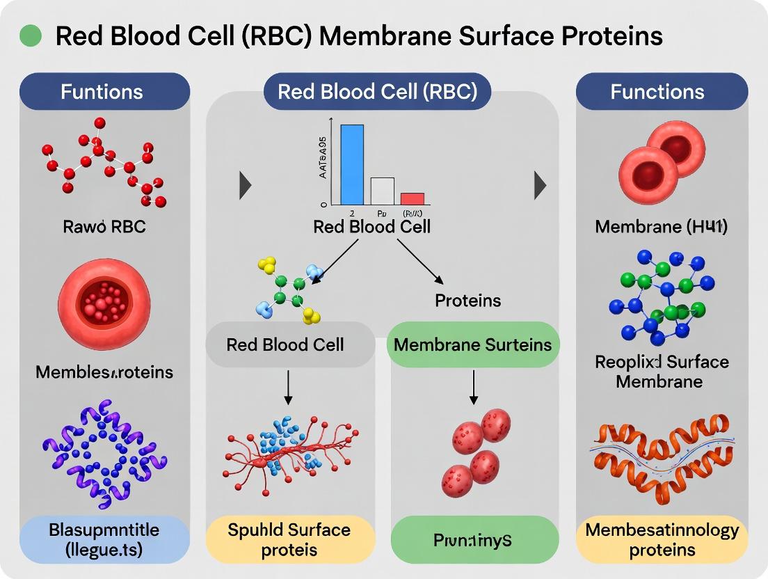
Beyond the Lipid Bilayer: A Comprehensive Guide to RBC Membrane Surface Proteins, Their Functions, and Clinical Implications
This article provides a detailed examination of red blood cell (RBC) membrane surface proteins, essential components that confer structural integrity, flexibility, and vital physiological functions.
Beyond the Lipid Bilayer: A Comprehensive Guide to RBC Membrane Surface Proteins, Their Functions, and Clinical Implications
Abstract
This article provides a detailed examination of red blood cell (RBC) membrane surface proteins, essential components that confer structural integrity, flexibility, and vital physiological functions. Targeting researchers, scientists, and drug development professionals, the content progresses from foundational knowledge of key protein complexes (e.g., Band 3, Glycophorins, RhAG) and their roles in gas exchange, antigenicity, and signaling, to advanced methodologies for their isolation, characterization, and manipulation. We explore common challenges in experimental workflows, compare validation techniques, and discuss translational applications in diagnosing hematological disorders (like hereditary spherocytosis and malaria pathogenesis) and in the engineering of novel therapeutic platforms, including drug delivery systems and antigenically modified RBCs for transfusion.
The Molecular Architects of the Erythrocyte: A Deep Dive into RBC Membrane Protein Structure and Core Functions
Within the broader thesis on erythrocyte membrane surface proteins and their functions, this technical guide establishes the mature red blood cell (RBC) membrane not as a simple, inert lipid bilayer but as a highly complex, densely packed, and dynamic interface. Its protein-rich composition, dominated by the spectrin-based cytoskeleton and a diverse array of integral and peripheral proteins, is essential for RBC deformability, stability, antigen presentation, and systemic signaling. Current research focuses on how disruptions in this interface contribute to hematologic pathologies and offer novel targets for therapeutic intervention, including for malaria, sickle cell disease, and in the engineering of RBC-mimetic drug delivery systems.
Quantitative Composition of the RBC Membrane
Table 1: Major Protein Classes in the Human RBC Membrane
| Protein Class | Key Examples | Approximate Copies per Cell | Primary Function |
|---|---|---|---|
| Cytoskeletal | Spectrin (α/β), Actin (short filaments), Protein 4.1R | ~200,000 spectrin heterodimers | Provides structural integrity and deformability |
| Integral (Band 3) | Anion Exchanger 1 (AE1) | ~1.2 million | Anion transport, CO2 exchange, anchors cytoskeleton via Ankyrin |
| Integral (Glycophorins) | Glycophorin A, B, C | ~500,000 - 1,000,000 | Sialic acid carriers, contributes to surface charge, adhesion receptors |
| Linker/Adapter | Ankyrin-1, Protein 4.1R, Protein 4.2 | ~100,000 Ankyrin copies | Connects integral proteins (Band 3) to the spectrin cytoskeleton |
| Lipid Raft-Associated | Stomatin, Flotillins | Variable | Organize membrane microdomains, signaling platforms |
Table 2: Key Mechanical & Biophysical Properties
| Property | Typical Value/Measurement | Method | Functional Significance |
|---|---|---|---|
| Membrane Bending Modulus | ~2 x 10⁻¹⁹ J | Micropipette Aspiration, Flicker Spectroscopy | Determines resistance to curvature; impacts deformability |
| Shear Modulus | ~6 µN/m | Optical Tweezers, Ektacytometry | Measure of in-plane elasticity; critical for capillary passage |
| Membrane Lifespan | ~120 days | In vivo biotinylation, cohort labeling | Reflects resilience to mechanical and oxidative stress |
Detailed Experimental Protocols
Protocol: Isolation and Ghost Preparation of Human RBC Membranes
Objective: To prepare intact, hemoglobin-free RBC membranes (ghosts) for biochemical and proteomic analysis.
- Blood Collection & Wash: Collect venous blood in heparin or EDTA. Centrifuge at 800xg for 10 min at 4°C. Aspirate plasma and buffy coat. Wash RBCs 3x in isotonic phosphate-buffered saline (PBS), pH 7.4.
- Lysis & Ghost Preparation: Resuspend packed RBCs in 40 volumes of ice-cold lysis buffer (5mM Sodium Phosphate, pH 8.0, with protease inhibitors). Incubate on ice for 30 min.
- Pellet Ghosts: Centrifuge at 20,000xg for 20 min at 4°C. The pink pellet contains ghosts.
- Wash to Whiteness: Repeat lysis and centrifugation (steps 2-3) until the ghost pellet appears white or pale pink.
- Membrane Resuspension: Resuspend final ghost pellet in appropriate buffer (e.g., 5mM Sodium Phosphate, pH 7.4) for downstream assays. Protein concentration can be determined via BCA assay.
Protocol: Analysis of Membrane Protein Complexes via Blue Native PAGE (BN-PAGE)
Objective: To separate and identify native protein complexes from the RBC membrane.
- Membrane Solubilization: Incubate ghost preparation (1-2 mg/mL protein) with 1-2% digitonin or dodecyl maltoside for 30 min on ice. Avoid harsh SDS for native analysis.
- Clarification: Centrifuge at 100,000xg for 30 min at 4°C. Retain the supernatant containing solubilized complexes.
- BN-PAGE Gel: Load supernatant onto a 4-16% gradient native PAGE gel. The cathode buffer contains Coomassie G-250.
- Electrophoresis: Run at 4°C, starting at 100V, then 500V max, until the dye front reaches the bottom.
- Analysis: Visualize complexes. Excise bands for mass spectrometry or transfer to PVDF for immunoblotting with specific antibodies (e.g., anti-spectrin, anti-Band 3, anti-Glycophorin C).
Protocol: Measuring RBC Deformability via Laser-Assisted Optical Rotational Cell Analyzer (LORRCA)
Objective: To quantitatively assess RBC membrane flexibility under shear stress.
- Sample Prep: Wash and resuspend RBCs at a low hematocrit (~0.5%) in a high-viscosity polyvinylpyrrolidone (PVP) solution.
- Loading: Inject sample into the LORRCA's Couette system, where a laser beam passes through the suspension.
- Shearing & Measurement: The outer cylinder rotates, applying defined shear stress (0.3 to 30 Pa). The laser diffraction pattern of the deformed RBCs is captured.
- Data Analysis: The instrument calculates the Elongation Index (EI) at each shear stress. The resulting deformability curve and the maximum EI (EImax) are key parameters. Reduced EImax indicates decreased membrane flexibility.
Visualization: Signaling and Experimental Workflows
Title: RBC Membrane Signaling Under Mechanical Stress
Title: RBC Membrane Proteomics Analysis Pipeline
The Scientist's Toolkit: Essential Research Reagents
Table 3: Key Research Reagents for RBC Membrane Studies
| Reagent/Material | Function/Application | Key Notes |
|---|---|---|
| Digitonin | Mild, cholesterol-dependent detergent for solubilizing membrane protein complexes while preserving native interactions. | Preferred over Triton X-100 for BN-PAGE; concentration critical. |
| Ektacytometer (LORRCA) | Instrument to measure RBC deformability and osmotic fragility as a function of shear stress. | Gold-standard for functional assessment of membrane mechanical properties. |
| Biotinylation Reagents (e.g., Sulfo-NHS-SS-Biotin) | Label surface-exposed membrane proteins for isolation, trafficking, or proteomic studies. | Cleavable linker allows elution under reducing conditions. |
| Anti-Band 3 (AE1) Antibody | Immunoprecipitation, western blotting, and immunofluorescence to study the major integral protein complex. | Essential for probing the ankyrin-based linkage to the cytoskeleton. |
| Spectrin Extraction Buffer (Low Ionic Strength) | Selectively extracts the spectrin-actin cytoskeleton from ghosts for purity assessment. | Contains 0.1-0.3 mM Sodium Phosphate, pH 7.6, overnight at 4°C. |
| Protease Inhibitor Cocktail (without EDTA) | Prevents proteolytic degradation of membrane proteins during ghost preparation and analysis. | EDTA omitted for calcium-dependent process studies. |
| Polyvinylpyrrolidone (PVP) Solution | High-viscosity medium for deformability measurements in ektacytometry. | Mimics plasma viscosity; allows application of defined shear stress. |
Within the broader thesis on RBC membrane surface proteins and functions, the vertical linkage system is paramount for maintaining erythrocyte integrity, elasticity, and survival. This whitepaper delves into the core molecular complex centered on Ankyrin-R (ANK1), the primary vertical linker tethering the lipid bilayer—via integral proteins like Band 3 and RhAG—to the underlying spectrin-actin cytoskeleton. Disruption of this network underpins hereditary spherocytosis and other hemolytic anemias, making it a critical target for mechanistic research and therapeutic intervention.
Core Molecular Architecture and Quantitative Relationships
The stoichiometry and biophysical properties of the key components dictate membrane mechanical stability. The following table summarizes core quantitative data.
Table 1: Core Component Properties and Interactions
| Component | Gene | Copy Number per RBC* | Key Binding Partner(s) | Binding Affinity (Kd) | Functional Consequence of Deficiency |
|---|---|---|---|---|---|
| Ankyrin-R | ANK1 | ~100,000 | Spectrin β-chain, Band 3, RhAG | 10-50 nM (Spectrin) | Loss of vertical linkage, membrane vesiculation, spherocytosis |
| Spectrin (αβ dimer) | SPTA1, SPTB | ~200,000 | Ankyrin-R, Actin, Protein 4.1R | 120 nM (Ankyrin-R) | Reduced structural lattice, elliptocytosis/poikilocytosis |
| Band 3 (AE1) | SLC4A1 | ~1.2 million | Ankyrin-R, Protein 4.2, Hemoglobin | 20-100 nM (Ankyrin-R) | Impaired anion exchange, weakened anchorage, acanthocytosis |
| Protein 4.2 | EPB42 | ~200,000 | Band 3, Ankyrin-R | ~50 nM (Band 3) | Reduced membrane stability, mild spherocytosis/hemolysis |
| Actin (Protofilament) | ACTB | ~300,000-500,000 | Spectrin, Protein 4.1R, Adducin | N/A | Disrupted junctional complexes, mechanical fragility |
| β-adducin | ADD2 | ~30,000-60,000 | Spectrin-actin junction, Actin capping | N/A | Increased membrane fragility, altered morphology |
Note: Copy numbers are approximate and can vary between sources and methodologies.
Detailed Experimental Protocols for Key Assays
Protocol: Co-Immunoprecipitation of Ankyrin-R Complex from RBC Ghosts
Objective: To identify and validate direct protein-protein interactions within the Ankyrin-R vertical linkage complex.
Materials: Fresh or frozen packed human RBCs, hypotonic lysis buffer (5 mM sodium phosphate, pH 8.0), membrane wash buffer (with 150 mM NaCl), solubilization buffer (1% Triton X-100, 150 mM NaCl, 25 mM Tris-HCl pH 7.5, protease inhibitors), anti-Ankyrin-R antibody (mouse monoclonal), species-matched control IgG, Protein A/G magnetic beads, SDS-PAGE and Western blot apparatus.
Method:
- Prepare RBC ghosts by lysing packed RBCs in 20 volumes of ice-cold hypotonic lysis buffer. Centrifuge at 20,000 x g for 15 min at 4°C. Repeat wash until ghosts are pale.
- Solubilize ghost membranes in solubilization buffer (1 mg protein/mL) for 1 hour at 4°C with gentle rotation. Centrifuge at 16,000 x g for 20 min to remove insoluble material.
- Pre-clear supernatant with 20 μL Protein A/G beads for 30 min.
- Incubate pre-cleared lysate with 2-5 μg of anti-Ankyrin-R antibody or control IgG overnight at 4°C.
- Add 50 μL bead slurry and incubate for 2 hours.
- Wash beads 4x with solubilization buffer.
- Elute proteins with 2X Laemmli buffer at 95°C for 5 min.
- Analyze by SDS-PAGE and Western blot, probing for Band 3, Spectrin, Protein 4.2, and Ankyrin-R.
Protocol: Microscale Thermophoresis (MST) for Binding Affinity Measurement
Objective: To quantitatively determine the binding affinity (Kd) between purified Ankyrin-R ZU5-UPA domain and a fluorescently-labeled Spectrin β-chain fragment.
Materials: Recombinant human ANK1 ZU5-UPA domain, recombinant SPTB N-terminal domain (labeled with red fluorescent dye, e.g., NT-647), MST-optimized buffer (e.g., PBS with 0.05% Tween-20), standard capillary tubes, Microscale Thermophoresis instrument.
Method:
- Label the Spectrin β-chain fragment according to the dye manufacturer's protocol. Purify labeled protein.
- Prepare a constant concentration of labeled Spectrin (e.g., 20 nM) in MST buffer.
- Perform a 1:1 serial dilution of the unlabeled Ankyrin-R domain in MST buffer (16 concentrations, starting from high μM range).
- Mix each Ankyrin-R dilution 1:1 with the constant labeled Spectrin solution. Incubate 15 min in the dark.
- Load samples into MST capillaries.
- Run MST measurement (LED power, MST power optimized). Monitor thermophoresis + T-jump.
- Analyze data using instrument software. Plot normalized fluorescence (Fnorm) vs. Ankyrin-R concentration. Fit curve to derive Kd.
Protocol: Ektacytometry for Membrane Mechanical Stability
Objective: To assess the functional consequence of disrupted vertical linkages on RBC deformability and stability.
Materials: Laser-assisted optical rotational cell analyzer (Lorrca), PBS with 4% polyvinylpyrrolidone (PVP, viscosity ~30 cP), patient or treated RBC samples.
Method:
- Wash RBCs 3x in isotonic PBS. Resuspend to ~2% hematocrit in the PVP solution.
- Load sample into the ektacytometer chamber.
- Run the osmoscannning protocol: Apply a constant shear stress (e.g., 30 Pa) while the osmolarity is linearly decreased from 500 to 0 mOsm/kg over ~10 min.
- The instrument measures laser diffraction (elongation index, EI). Plot EI vs. Osmolarity.
- Key parameters: Omin (osmolarity at which EI starts to decrease, indicates surface-area-to-volume ratio), Ohyper (osmolarity at maximum EI, indicates cellular hydration), and Elmax (maximum deformability). Reduced Elmax at isotonic osmolarity indicates decreased membrane stability from cytoskeletal defects.
The Scientist's Toolkit: Essential Research Reagents
Table 2: Key Research Reagent Solutions
| Reagent / Material | Supplier Examples | Function in Research |
|---|---|---|
| Anti-Ankyrin-R (clone 8B11) | Santa Cruz, Sigma-Aldrich | Immunoprecipitation, Western blot, and immunofluorescence detection of ANK1. |
| Anti-Band 3 (extracellular domain) | Bio-Rad, Invitrogen | Flow cytometry of RBC surface expression, IF, Co-IP studies. |
| Recombinant ANK1 (ZU5-UPA-SH3) | Novus, Abcam | In vitro binding assays (SPR, MST), crystallization studies. |
| Spectrin Actin Binding Kit | Cytoskeleton, Inc. | In vitro reconstitution of junctional complexes, binding inhibition assays. |
| 4,4'-Diisothiocyanostilbene-2,2'-disulfonate (DIDS) | Tocris, Sigma | Band 3 anion transport inhibitor; used to study Band 3 conformation's role in Ankyrin binding. |
| Protein 4.2 Knockout Mouse Model | Jackson Laboratory | In vivo model for studying compensated hemolysis and membrane organization. |
| Hereditary Spherocytosis RBC Panel | NIH, ProMetabolon | Patient-derived samples for comparative biomechanical and biochemical studies. |
Visualization of Pathways and Relationships
Diagram 1: Ankyrin-R Mediated Vertical Linkage in the RBC Membrane
Diagram 2: Co-IP Workflow for Ankyrin Interactome Analysis
Within the context of a broader thesis on red blood cell (RBC) membrane surface proteins, Band 3, also known as Anion Exchanger 1 (AE1), emerges as the quintessential horizontal anchor. This integral membrane protein constitutes the central hub of the RBC membrane skeleton, forming the critical link between the lipid bilayer and the underlying spectrin-actin cytoskeleton. This whitepaper details its dual, interdependent roles in facilitating rapid chloride/bicarbonate exchange essential for systemic CO2 transport and providing the mechanical resilience necessary for the RBC's 120-day circulatory lifespan. Dysfunction in Band 3 is implicated in hereditary spherocytosis, Southeast Asian ovalocytosis, and distal renal tubular acidosis, highlighting its physiological significance.
Structural and Functional Domains of Band 3
Band 3 is a homodimeric multipass membrane protein with two primary domains:
- The N-terminal Cytoplasmic Domain (cdb3, ~43 kDa): This domain is intrinsically disordered and serves as the primary mechanical tether. It binds ankyrin, which in turn links to the spectrin-actin network. It also provides high-affinity binding sites for hemoglobin, glycolytic enzymes, and protein 4.2.
- The C-terminal Membrane-Spanning Domain (msd, ~55 kDa): This domain contains 14 transmembrane segments and forms the anion exchange pore. It operates via a "ping-pong" mechanism, where a single substrate-binding site is alternately exposed to the cytoplasm or the extracellular space.
Table 1: Key Structural and Quantitative Features of Human Band 3 (AE1)
| Feature | Value / Detail | Functional Implication |
|---|---|---|
| Gene | SLC4A1 | Located on chromosome 17q21-q22. |
| Protein Mass | ~95-110 kDa (glycosylated) | Dimerizes to form functional unit. |
| Copy Number per RBC | ~1.2 x 10^6 copies/cell | Represents ~25% of total membrane protein mass. |
| Anion Exchange Turnover (Cl⁻) | ~50,000 ions/sec/molecule at 37°C | Facilitates rapid CO2 transport as HCO₃⁻. |
| Binding Partners (Cytoplasmic) | Ankyrin-1, Protein 4.1R, Protein 4.2, Hb, GAPDH, Aldolase | Integrates membrane with cytoskeleton & metabolism. |
| Known Pathogenic Mutations | >50 documented | Cause hereditary spherocytosis, ovalocytosis, dRTA. |
Detailed Experimental Methodologies
Protocol: Assessing Anion Exchange Function via Eosin-5-Maleimide (EMA) Binding and Flow Cytometry
- Principle: EMA covalently labels Lys-430 on the extracellular loop of Band 3. Its fluorescence is quenched upon binding of the inhibitor DIDS (4,4'-Diisothiocyano-2,2'-stilbenedisulfonic acid) or by conformational changes during transport.
- Procedure:
- Wash fresh or washed RBCs in PBS (pH 7.4).
- Incubate 5 µL of packed RBCs with 1 mL of EMA working solution (0.5 mg/mL in PBS) for 1 hour at 4°C in the dark.
- Quench the reaction by adding 1 mL of 1% BSA in PBS. Wash cells 3x in PBS.
- For inhibition assays, pre-incubate an aliquot of EMA-labeled cells with 100 µM DIDS for 30 min at 37°C.
- Analyze by flow cytometry (FL1/FL2 channel). Mean fluorescence intensity (MFI) correlates with functional Band 3 expression. A decrease in MFI post-DIDS confirms specific labeling.
Protocol: Co-Immunoprecipitation of the Band 3 Macrocomplex
- Principle: To isolate and identify proteins physically associated with Band 3 in the RBC membrane.
- Procedure:
- Prepare RBC ghost membranes by hypotonic lysis (5 mM NaPi, pH 8.0) and strip peripheral proteins with high-salt (1 M NaCl) or alkaline (pH 11) treatment as needed.
- Solubilize ghosts in 1% Triton X-100 or Digitonin in TBS (with protease inhibitors) for 1 hour at 4°C.
- Pre-clear lysate with protein A/G beads for 30 min.
- Incubate lysate with anti-Band 3 monoclonal antibody (e.g., BRIC 6, BRIC 170) or isotype control overnight at 4°C.
- Add protein A/G beads for 2 hours. Pellet beads and wash stringently with solubilization buffer.
- Elute bound proteins with Laemmli sample buffer, separate by SDS-PAGE, and analyze by western blot (for ankyrin, 4.1, 4.2, spectrin) or mass spectrometry.
Protocol: Measurement of RBC Membrane Deformability by Ektacytometry
- Principle: Assesses the contribution of Band 3-ankyrin-spectrin linkage to global membrane mechanical stability.
- Procedure:
- A laser diffraction viscometer (e.g., Lorrca) is used. A dilute RBC suspension is sheared in a Couette system.
- The laser beam diffracted by the deformed cells produces an ellipsoidal pattern. The elongation index (EI) is calculated as (A - B) / (A + B), where A and B are the long and short axes.
- Shear stress is progressively increased (0.3 to 30 Pa), generating a deformability curve.
- Key Parameter: The maximum elongation index (EImax) and the shear stress required for half-maximal deformation. RBCs with disrupted Band 3-cytoskeleton linkages show reduced EImax at physiological osmolality.
Visualizing the Band 3 Hub: Pathways and Workflows
Diagram Title: Band 3 Interaction Network: Transport, Stability, and Metabolism
Diagram Title: Key Experimental Workflows for Band 3 Research
The Scientist's Toolkit: Essential Research Reagents
Table 2: Key Research Reagent Solutions for Band 3 Studies
| Reagent / Material | Supplier Examples | Function in Experiment |
|---|---|---|
| Eosin-5-Maleimide (EMA) | Thermo Fisher, Sigma-Aldrich | Covalent fluorescent probe for extracellular loop of Band 3; used in flow cytometry for expression & function. |
| DIDS (4,4'-Diisothiocyano-2,2'-stilbenedisulfonic acid) | Sigma-Aldrich, Tocris | Irreversible, high-affinity inhibitor of Band 3-mediated anion exchange; control for transport studies. |
| Anti-Band 3 Monoclonal Antibodies (e.g., BRIC 6, BRIC 170) | IBGRL, Santa Cruz Biotechnology | Specific detection for western blot, immunofluorescence, and co-immunoprecipitation of Band 3. |
| Digitonin / Triton X-100 | Sigma-Aldrich, Thermo Fisher | Mild (digitonin) or harsh (Triton) detergents for solubilizing membrane protein complexes while preserving interactions. |
| Protein A/G Magnetic Beads | Pierce, Millipore | For efficient immunoprecipitation of antigen-antibody complexes; enables rigorous washing. |
| Lorrca Ektacytometer | RR Mechatronics | Gold-standard instrument for measuring RBC deformability as a function of shear stress, reflecting cytoskeletal integrity. |
| Protease Inhibitor Cocktail (EDTA-free) | Roche, Sigma-Aldrich | Essential for preventing protein degradation during ghost preparation and complex isolation. |
| Recombinant cdB3 (Cytoplasmic Domain) | Abcam, custom synthesis | Used in binding assays (SPR, ITC) to study interactions with ankyrin, Hb, or enzymes. |
The red blood cell (RBC) membrane is a sophisticated bilayer, stabilized by a spectrin-based cytoskeleton and populated by integral and peripheral proteins that govern cellular mechanics, signaling, and interfacial biology. Among these, the glycophorin family (GPA, GPB, GPC, GPD) stands out as a critical interface module. This whitepaper positions glycophorins within the broader thesis of RBC membrane surface protein research, elucidating their roles as primary carriers of surface negative charge (via sialic acid), as modulators of cellular adhesion, and as polymorphic carriers of essential blood group antigens. Their study is fundamental to understanding malaria pathogenesis, blood transfusion compatibility, and novel therapeutic targeting.
Structural & Functional Characteristics
Glycophorins are single-pass transmembrane sialoglycoproteins. The following table summarizes their core quantitative and functional attributes.
Table 1: Comparative Analysis of Human Glycophorins
| Feature | Glycophorin A (GPA, CD235a) | Glycophorin B (GPB, CD235b) | Glycophorin C (GPC, CD236c) | Glycophorin D (GPD) |
|---|---|---|---|---|
| Gene | GYPA | GYPB | GYPC | GYPC (Alternative splicing) |
| Amino Acids | 131 | 72 | 128 | 107 |
| Molecular Weight (kDa) | ~36-45 (Heavy glycosylation) | ~20-30 | ~32-40 | ~23-30 |
| O-Linked Glycans | ~15 | ~11 | ~12 | ~6 |
| N-Linked Glycans | 1 | 0 | 1 | 1 |
| Sialic Acid Residues | ~15 | ~11 | ~12 | ~6 |
| Blood Group System | MN | Ss | Gerbich | (Part of Gerbich) |
| Key Antigens | M/N (aa1-5) | S/s (aa29) | Ge2, Ge3, Ge4 | Ge2, Ge3 |
| Copy Number per RBC | ~0.5-1.0 x 10⁶ | ~0.1-0.2 x 10⁶ | ~0.05-0.1 x 10⁶ | ~0.05-0.1 x 10⁶ |
| Cytoskeletal Linkage | Weak, via band 3 | Weak | Strong, via 4.1R-p55 complex | Strong, via 4.1R-p55 complex |
| Malaria Ligand | P. falciparum EBA-175 | - | P. falciparum EBA-140 (BAEBL) | - |
Functional Roles & Experimental Pathways
Sialic Acid Shield: Electrostatic Repulsion & Quantification
The dense presentation of sialic acid confers a high negative charge (zeta potential ~ -15 to -20 mV), preventing RBC aggregation and adhesion to vascular endothelium.
Experimental Protocol 1: Sialic Acid Quantification via Thiobarbituric Acid (TBA) Assay
- Principle: Periodic acid oxidizes sialic acid to formylpyruvate, which reacts with TBA to form a chromogen.
- Procedure:
- Wash RBCs 3x in PBS.
- Lyse 100 µL packed RBCs in 1 mL 0.01M H₂SO₄. Incubate 1h at 80°C to release sialic acid.
- Centrifuge (10,000g, 10 min). Collect supernatant.
- Mix 100 µL supernatant with 50 µL 0.2M periodic acid in 9M phosphoric acid. Incubate 20 min at 37°C.
- Add 250 µL 10% sodium arsenite in 0.5M sodium sulfate to reduce excess periodate (until brown color disappears).
- Add 750 µL 0.6% TBA in 0.5M sodium sulfate. Heat at 100°C for 15 min.
- Cool, extract chromogen with 1 mL cyclohexanone. Measure absorbance at 549 nm.
- Calculate concentration using a N-acetylneuraminic acid standard curve.
Adhesion Modulation: Signaling & Cytoskeletal Linkage
GPC/GPD play a structural role by linking the membrane to the cytoskeleton via the 4.1R-p55 complex. Disruption causes hereditary elliptocytosis. This pathway is summarized in Diagram 1.
Diagram 1: Glycophorin C - Cytoskeletal Linkage Pathway
Experimental Protocol 2: Co-Immunoprecipitation of GPC-4.1R-p55 Complex
- Principle: Isolate native protein complexes from RBC membranes using specific antibodies.
- Procedure:
- Prepare RBC ghost membranes by hypotonic lysis (20 mOsm phosphate buffer, pH 7.4) and extensive washing.
- Solubilize ghosts in 1% Triton X-100, 150 mM NaCl, 5 mM EDTA, 25 mM Tris-HCl (pH 7.5) + protease inhibitors for 1h at 4°C.
- Clear lysate by centrifugation (16,000g, 30 min).
- Pre-clear supernatant with Protein G Sepharose beads for 1h.
- Incubate supernatant with 2 µg of anti-GPC (e.g., clone BRIC 4) or control IgG overnight at 4°C.
- Add Protein G beads for 2h. Pellet and wash beads 4x with solubilization buffer.
- Elute proteins with 2X Laemmli buffer at 95°C for 5 min.
- Analyze by SDS-PAGE and western blot using anti-4.1R and anti-p55 antibodies.
Blood Group Antigens & Molecular Genotyping
Glycophorin polymorphisms are major causes of blood group alloimmunization.
Experimental Protocol 3: PCR-RFLP for S/s (Glycophorin B) Genotyping
- Principle: A single nucleotide polymorphism (T>C) at codon 29 (Met29Thr) defines the S/s antigens. This creates a loss of an MspI restriction site in the 's' allele.
- Procedure:
- Primers: Forward: 5'-CAG GCT GGA CTT GCT GTC TC-3'; Reverse: 5'-GCA GGA GTC AAC CAG GAC TC-3' (amplicon: ~200 bp).
- Perform PCR on genomic DNA: 35 cycles of 94°C (30s), 60°C (30s), 72°C (30s).
- Digest 10 µL PCR product with 5U MspI at 37°C overnight.
- Run on a 3% agarose gel.
- Interpretation: 'S' allele cut: ~120 + 80 bp fragments. 's' allele uncut: ~200 bp.
Pathogen Interaction:Plasmodium falciparumInvasion
Glycophorins serve as receptors for malaria parasite ligands. The invasion pathway via GPA is a key model.
Diagram 2: P. falciparum Invasion via GPA (EBA-175 Pathway)
The Scientist's Toolkit: Key Research Reagent Solutions
Table 2: Essential Reagents for Glycophorin Research
| Reagent / Material | Function & Application | Example (Research Grade) |
|---|---|---|
| Monoclonal Anti-GPA (CD235a) | Flow cytometry, immunofluorescence, IP for RBC identification & quantification. | Clone BRIC 256 (anti-M) / BRIC 157 (anti-N) |
| Monoclonal Anti-GPC (CD236c) | Co-IP studies, cytoskeletal linkage analysis, Gerbich blood group typing. | Clone BRIC 4 / BRIC 10 |
| Neuraminidase (Sialidase) | Enzymatic removal of sialic acid to study charge-mediated functions & pathogen adhesion. | Clostridium perfringens neuraminidase (e.g., Sigma N2876) |
| Protein 4.1R Antibody | Western blot, IP to investigate integrity of the GPC-4.1R-p55 membrane skeleton junction. | Rabbit polyclonal (e.g., Proteintech 55139-1-AP) |
| Recombinant EBA-175 RII | Binding inhibition assays, structural studies of malaria invasion pathway. | Produced in HEK293 or E. coli systems. |
| GYPA/GYPB/GYPC Genotyping Kits | PCR-SSP or sequencing-based kits for high-throughput blood group antigen profiling. | In-house designed PCR-RFLP or commercial BLOODchip. |
| Triton X-100 (Detergent) | Solubilization of RBC membrane proteins for analysis of protein complexes (e.g., cytoskeletal linkages). | For membrane protein extraction. |
Within the broader thesis on red blood cell (RBC) membrane surface proteins, the Rh complex represents a paradigmatic multifunctional structure. It is integral not only to the most immunogenic blood group system in transfusion and perinatal medicine but also to the physiological transport of ammonium and the structural integrity of the erythrocyte membrane. This guide synthesizes current mechanistic understanding of the Rh-associated glycoprotein (RhAG), the RhD protein, and the RhCE proteins as a core complex, highlighting their dual roles in solute transport and antigenicity.
Molecular Composition and Core Functions
The Rh complex is a heterotetramer embedded in the RBC lipid bilayer. It consists of two core subunits—RhAG and either RhD or RhCE—and is associated with accessory proteins (CD47, LW, glycophorin B) that facilitate its trafficking and stability. The central functions are:
- Ammonium/NH₃ Transport: RhAG, a member of the Amt/Mep/Rh superfamily, functions as a gas channel for ammonia (NH₃), a critical byproduct of nitrogen metabolism.
- Blood Group Antigen Expression: The polymorphic RhD and RhCE proteins carry the D, C/c, and E/e antigens, which are primary targets for alloimmune hemolytic reactions.
- Membrane Skeletal Linkage: The complex, via its accessory proteins, connects to the underlying spectrin-actin cytoskeleton, contributing to erythrocyte mechanical properties.
Diagram: Structure and function of the Rh membrane complex.
Quantitative Data on Expression and Transport
Table 1: Quantitative Characteristics of the Human Rh Complex Components
| Protein | Copy Number per RBC (Mean ± SD) | Key Function | Gene Location | Polymorphic Sites |
|---|---|---|---|---|
| RhAG | 100,000 - 200,000 | NH₃/CO₂ channel, complex assembly | 6p21.1 | Limited (regulatory) |
| RhD | 100,000 - 200,000* | D antigen, structural role | 1p36.11 | Multiple (presence/absence of gene) |
| RhCE | 100,000 - 200,000* | C/c and E/e antigens, structural role | 1p36.11 | Multiple (single nucleotide polymorphisms) |
| CD47 | ~30,000 - 50,000 | Complex stability, "don't eat me" signal | 3q13.12 | Limited |
*RhD and RhCE expression is mutually exclusive per complex; total Rh protein copies are ~100,000-200,000.
Table 2: Transport Kinetics of RhAG in Model Systems
| Experimental System | Substrate | Apparent Km (mM) | Estimated Permeability | Inhibition by |
|---|---|---|---|---|
| Xenopus Oocyte (hRhAG) | NH₄⁺/NH₃ | ~5-10 mM | 10-50 x control oocytes | Cu²⁺, PCMBS |
| RBC Ghosts (Native) | NH₃ | N/A | Accounts for ~50% of total RBC NH₃ flux | Methazolamide (weak) |
| Proteoliposomes (Reconstituted) | Methylammonium (CH₃NH₃⁺) | ~1-2 mM | Direct electrophysiological measurement | Low external pH |
Detailed Experimental Protocols
Protocol 4.1: Assessment of Ammonium/Methylammonium Uptake in Rh-Expressing Xenopus Oocytes
- Objective: To characterize the transport function of RhAG and its mutants.
- Materials: See Scientist's Toolkit.
- Method:
- cRNA Synthesis: Linearize plasmid containing human RHAG cDNA. Generate capped cRNA using an in vitro transcription kit (e.g., mMessage mMachine).
- Oocyte Preparation & Injection: Isolate stage V-VI oocytes from Xenopus laevis. Manually defolliculate and incubate in ND96 solution (96 mM NaCl, 2 mM KCl, 1.8 mM CaCl₂, 1 mM MgCl₂, 5 mM HEPES, pH 7.4). Inject 25-50 ng of cRNA per oocyte. Incubate at 16°C in ND96 supplemented with penicillin/streptomycin and 2.5 mM sodium pyruvate for 3-5 days.
- Uptake Assay: Wash oocytes with uptake solution (e.g., 100 mM NaCl or Choline Cl, 2 mM KCl, 1 mM CaCl₂, 1 mM MgCl₂, 5 mM HEPES, pH 7.4). For [¹⁴C]-Methylammonium (CH₃NH₃⁺) uptake, incubate oocytes in uptake solution containing 0.1-1.0 μCi/mL tracer and varying cold substrate concentrations (0.1-10 mM) for 15-60 minutes at room temperature.
- Termination & Measurement: Wash oocytes 3x rapidly with ice-cold uptake solution. Transfer individual oocytes to scintillation vials, lyse with 1% SDS, add scintillation fluid, and count radioactivity. Normalize uptake to water-injected control oocytes.
- Data Analysis: Calculate net Rh-mediated uptake (cRNA-injected minus water-injected). Determine kinetic parameters (Vmax, Km) using non-linear regression (e.g., Michaelis-Menten).
Protocol 4.2: Co-Immunoprecipitation of the Native Rh Complex from RBC Membranes
- Objective: To validate protein-protein interactions within the Rh complex.
- Method:
- Membrane Preparation: Wash packed human RBCs (RhD+ phenotype) with PBS. Lyse in 20 volumes of hypotonic lysis buffer (5 mM sodium phosphate, pH 8.0, with protease inhibitors). Centrifuge at 20,000 x g, 30 min, 4°C. Wash the resulting ghost membranes repeatedly in lysis buffer until pale.
- Solubilization: Solubilize ghosts in 1% n-dodecyl-β-D-maltoside (DDM) in TBS (50 mM Tris, 150 mM NaCl, pH 7.4) with inhibitors for 1 hour at 4°C with gentle agitation. Clarify by ultracentrifugation (100,000 x g, 30 min).
- Immunoprecipitation: Pre-clear supernatant with Protein A/G beads for 30 min. Incubate with 5 μg of monoclonal anti-RhAG (or anti-RhD, anti-RhCE) antibody overnight at 4°C. Add fresh beads and incubate 2 hours.
- Wash & Elution: Wash beads 4x with 0.1% DDM in TBS. Elute proteins with 2x Laemmli buffer at 37°C for 30 min (avoid boiling to prevent aggregation).
- Analysis: Resolve eluates by SDS-PAGE (4-12% gradient gel). Perform Western blotting with antibodies against RhAG, RhD, RhCE, CD47, and glycophorin B to confirm co-precipitation.
Diagram: Workflow for RhAG functional assay in oocytes.
The Scientist's Toolkit: Research Reagent Solutions
Table 3: Essential Reagents for Rh Complex Research
| Reagent / Material | Supplier Examples | Primary Function in Experiments |
|---|---|---|
| Monoclonal Anti-RhAG (clone 2D10) | Beckman Coulter, IBGRL | Immunoprecipitation, Western blot, flow cytometry to isolate/complex. |
| Monoclonal Anti-RhD (clone LOR15C9) | Diagast, IBGRL | Rh phenotyping, complex IP, blocking studies. |
| cDNA Clones: Human RHAG, RHD, RHCE | GeneOracle, Origene | Functional expression in heterologous systems (oocytes, HEK293). |
| [¹⁴C]-Methylammonium Chloride | American Radiolabeled Chemicals | Tracer for direct measurement of RhAG-mediated transport kinetics. |
| n-Dodecyl-β-D-Maltoside (DDM) | Anatrace, Sigma | Mild detergent for solubilizing native Rh complex from RBC membranes. |
| Rh-null Erythrocytes (Regulator type) | Rare blood banks, research repositories | Critical negative control for Rh complex function/structure studies. |
| Xenopus laevis Oocytes | Nasco, Xenopus 1 | Gold-standard expression system for electrophysiology & uptake assays. |
| CRISPR/Cas9 RhAG Knockout K562 | ATCC, or custom generation | Model for studying complex assembly and trafficking in an erythroid background. |
Within the comprehensive study of red blood cell (RBC) membrane surface proteins, Aquaporin-1 (AQP1), the Duffy Antigen/Receptor for Chemokines (DARC), and CD47 represent three critical non-transport, non-cytoskeletal proteins with pivotal and distinct functions. AQP1 facilitates rapid water permeability, DARC is a key chemokine scavenger and malarial receptor, and CD47 delivers an essential "self" signal to phagocytes. This whitepaper provides an in-depth technical guide to their structure, function, and experimental analysis, contextualized within modern RBC membrane proteomics and pathophysiology research.
Protein-Specific Analysis
Aquaporin-1 (AQP1)
Function: AQP1 is a constitutively active, bidirectional water channel essential for RBC osmotic stability and volume regulation. It facilitates high-capacity water transport (pf ~2x10^-14 cm³/s per channel) driven by osmotic gradients. Structure: Homotetrameric integral membrane protein. Each monomer contains six transmembrane helices forming a pore with the conserved NPA (Asn-Pro-Ala) motifs. Clinical Relevance: AQP1-null individuals are phenotypically normal but exhibit reduced osmotic water permeability. It is a potential drug target for edema and certain cancers.
Duffy Antigen/Receptor for Chemokines (DARC)
Function: A nonspecific, promiscuous receptor for multiple inflammatory chemokines (e.g., IL-8, MCP-1, RANTES). Acts as a chemokine sink, modulating systemic inflammation. It is also the portal for Plasmodium vivax and Plasmodium knowlesi mercozoite invasion. Structure: 7-transmembrane domain protein, atypical G-protein-coupled receptor (GPCR) that does not signal internally. Polymorphism: The FYB/A polymorphism (Gly42Asp) determines antigenicity. The FYBES/AES null phenotype (Duffy-negative) confers resistance to P. vivax malaria.
CD47
Function: Integrin-associated protein that binds Signal Regulatory Protein Alpha (SIRPα) on macrophages and dendritic cells, delivering a potent inhibitory "don't eat me" signal that prevents phagocytosis of healthy RBCs. Structure: Single-pass transmembrane immunoglobulin superfamily protein with an extracellular IgV domain, five membrane-spanning segments, and a short cytoplasmic tail. Role in Aging & Clearance: CD47 expression decreases on aged or damaged RBCs, contributing to their removal by splenic macrophages.
Table 1: Core Biophysical & Expression Data
| Protein | Copy Number per RBC | Gene Locus | Key Ligands/Binding Partners | Binding Affinity (Kd) |
|---|---|---|---|---|
| AQP1 | 120,000 - 160,000 | 7p14.3 | H₂O, CO₂ (?), ions (?) | N/A (Channel) |
| Duffy (DARC) | 10,000 - 12,000 | 1q23.2 | CXC & CC Chemokines (e.g., IL-8), P. vivax Duffy Binding Protein (DBP) | IL-8: ~5 nM; DBP: <10 nM |
| CD47 | 15,000 - 25,000 | 3q13.12 | SIRPα, Thrombospondin-1, Integrins | SIRPα: ~0.2 - 1 µM |
Table 2: Phenotypic & Clinical Associations
| Protein | Null/Mutant Phenotype in Humans | Associated Diseases/Therapeutic Target Potential |
|---|---|---|
| AQP1 | Reduced RBC osmotic fragility; generally normal physiology. | Aquaporin modulator development for edema, glaucoma, cancer. |
| Duffy | Duffy-negative (Fy(a-b-)): Resistance to P. vivax malaria; altered inflammatory response. | Malaria vaccine/blockade target; modulator of inflammation and chemokine biology. |
| CD47 | Not viable (embryonic lethal in mice). On RBCs: increased basal phagocytosis, accelerated clearance. | Cancer immunotherapy ("Don't eat me" blockade); Aging/storage-related RBC transfusion efficacy. |
Key Experimental Protocols
Protocol: Stopped-Flow Light Scattering for AQP1 Water Permeability
Objective: Measure osmotic water permeability (Pf) of RBCs or proteoliposomes reconstituted with AQP1. Materials: Stopped-flow apparatus, light scatter detector, hyperosmotic solution (e.g., sucrose in PBS). Procedure:
- Prepare a 1% (v/v) suspension of intact RBCs or AQP1-proteoliposomes in isosmotic buffer.
- Rapidly mix 1:1 with a hyperosmotic buffer (e.g., 2x osmolality) in the stopped-flow chamber.
- Monitor 90° light scatter intensity over time (ms-scale). Cell/proteoliposome shrinkage increases scatter.
- Fit the scatter time course to a single exponential. Calculate Pf using the equation: ( Pf = k/(V_0 * A * \Delta Osm) ), where k is the rate constant, V₀ is initial volume, A is surface area, and ΔOsm is the osmotic gradient. Key Control: Use AQP1-null RBCs or inhibitors (e.g., Hg²⁺, now largely obsolete) to establish baseline.
Protocol: Flow Cytometry Binding Assay for Duffy-Chemokine Interaction
Objective: Quantify chemokine (e.g., IL-8) binding to Duffy on intact RBCs. Materials: Fresh RBCs, recombinant biotinylated chemokine, fluorescent streptavidin (e.g., SA-FITC), flow cytometer. Procedure:
- Wash RBCs 3x in PBS/0.1% BSA (binding buffer).
- Incubate RBCs (1% hematocrit) with serial dilutions of biotinylated IL-8 (0-100 nM) for 60 min at 4°C.
- Wash cells twice to remove unbound ligand.
- Incubate with saturating concentration of SA-FITC for 30 min at 4°C in the dark.
- Wash and resuspend. Analyze 50,000 events per sample by flow cytometry.
- Plot Median Fluorescence Intensity (MFI) vs. [IL-8] to derive binding parameters. Use Duffy-null RBCs for nonspecific binding.
Protocol:In VitroPhagocytosis Assay for CD47 Function
Objective: Measure macrophage phagocytosis of RBCs with modulated CD47 expression. Materials: Primary human macrophages or THP-1-derived macrophages, test RBCs (e.g., aged, CD47-blocked, or genetically modified), fluorescent cell linker (e.g., PKH26), flow cytometer. Procedure:
- Label RBCs with PKH26 per manufacturer's protocol. Wash thoroughly.
- Differentiate THP-1 cells with PMA (e.g., 100 nM, 48 hr) and rest for 24 hr.
- Pre-incubate labeled RBCs with anti-CD47 blocking antibody (e.g., B6H12) or isotype control (30 min, 37°C).
- Co-culture macrophages and RBCs (effector:target ~1:10) in serum-containing medium for 2 hours at 37°C.
- Vigorously wash to remove non-phagocytosed RBCs. Detach macrophages and fix.
- Analyze by flow cytometry. Phagocytic index = (% PKH26+ macrophages) * (MFI of PKH26+ macrophages) / 100.
Diagrams
Diagram 1: Aquaporin-1 Mediated Water Transport Pathway
Diagram 2: Duffy Receptor Dual Function in Inflammation and Malaria
Diagram 3: CD47-SIRPα Signaling Dictates RBC Phagocytic Fate
The Scientist's Toolkit: Research Reagent Solutions
Table 3: Essential Reagents for Featured Experiments
| Reagent | Supplier Examples (for citation) | Function in Research |
|---|---|---|
| Human AQP1 ELISA Kit | Abcam, R&D Systems | Quantifies soluble or membrane-extracted AQP1 protein levels. |
| Recombinant Human DARC Protein (His-tag) | Sino Biological, Novus Biologicals | Positive control for binding assays; structural studies. |
| Anti-CD47 Blocking Antibody (Clone B6H12) | BioLegend, Thermo Fisher | Inhibits CD47-SIRPα interaction in functional phagocytosis assays. |
| Biotinylated IL-8 (CXCL8) | PeproTech, R&D Systems | Ligand for flow cytometry-based Duffy binding/competition assays. |
| PKH26 Red Fluorescent Cell Linker Kit | Sigma-Aldrich | Lipophilic dye for stable, long-term labeling of RBC membranes for phagocytosis tracking. |
| Protease-Free BSA | New England Biolabs, MilliporeSigma | Essential for blocking and background reduction in sensitive ligand-binding assays. |
| SIRPα-Fc Chimera Protein | ACROBiosystems, R&D Systems | Decoy receptor to quantify CD47 binding affinity via SPR or ELISA. |
| AQP1 Inhibitor (Tetrakis-4-[(2-methoxyethoxy)methoxy]phthalocyanato) cobalt(II) | Tocris (custom synthesis) | Specific, non-toxic small molecule inhibitor for AQP1 functional studies. |
This whitepaper serves as a technical guide within the broader thesis on Red Blood Cell (RBC) Membrane Surface Proteins and Functions Research. The central thesis posits that the RBC's extraordinary functional repertoire is not the product of isolated proteins but emerges from the integrated, multi-functional collaboration of specialized protein complexes. This document will dissect the mechanistic synergy between the membrane cytoskeleton (primarily the spectrin-based network) and transmembrane adhesive complexes (like the Band 3 complex) in achieving three critical outcomes: deformability, mechanical stability, and immune evasion.
Core Protein Complexes and Their Integrated Roles
The Spectrin-Actin Cytoskeletal Network: The Scaffold
- Primary Components: α- and β-spectrin heterodimers, actin protofilaments (short, ~37 nm), protein 4.1R, adducin, tropomyosin, tropomodulin.
Quantitative Structure:
Parameter Value Significance Spectrin dimer contour length ~200 nm Provides elastic spring-like properties. Spectrin tetramer persistence length ~10-20 nm Measures chain flexibility; key for network fluidity. Hexagonal lattice edge length (junction-to-junction) ~60-80 nm Determines mesh density and lateral mobility. Number of spectrin tetramers per junction complex ~6 Defines network connectivity. Membrane thickness ~4.5 nm (lipid bilayer only) Context for vertical linkages. Function in Integration: This network forms a quasi-2D elastic mesh underlying the lipid bilayer, providing the mechanical foundation for deformability and stability.
The Band 3 Multiprotein Complex: The Transmembrane Hub
- Primary Components: Band 3 (AE1, anion exchanger 1), glycophorin A (GPA), protein 4.2, ankyrin-R, carbonic anhydrase II.
Quantitative Interactions:
Interaction Binding Partner Affinity (Kd) / Notes Function in Integration Band 3 - Ankyrin-R β-spectrin tail ~10-100 nM Primary vertical linkage, couples membrane to cytoskeleton. Band 3 - Protein 4.2 Cytoplasmic domain of Band 3 Stabilizes Band 3-Ankyrin complex Strengthens vertical linkage. Band 3 - Glycophorin A Transmembrane helices ~1.4 x 10⁴ molecules/RBC Assists in Band 3 trafficking/stability. Band 3 - Carbonic Anhydrase II Cytoplasmic domain of Band 3 Enhances CO₂ transport efficiency Metabolic "metabolon" function. Function in Integration: Serves as the central transmembrane anchor, linking the lipid bilayer and extracellular environment to the spectrin cytoskeleton. It is also a critical site for immune evasion via glycan masking.
Mechanistic Integration of Functions
Deformability & Stability: A Dynamic Equilibrium
Deformability (ability to stretch and bend) and stability (resistance to fragmentation) are two sides of the same coin, governed by the spectrin network's entropic elasticity and its regulated connectivity.
- Mechanism: Under shear stress, spectrin tetramers reversibly unfold from a compact, folded state to an extended chain. This allows massive deformation without plasticity. Protein 4.1R strengthens the junction by stabilizing spectrin-actin binding. Adducin caps actin filament ends, controlling protofilament length. Disruption of this balance (e.g., spectrin or protein 4.1 deficiency in hereditary spherocytosis) increases rigidity and reduces stability.
Immune Evasion: The Glycocalyx and Antigen Masking
The RBC surface must avoid both autoimmune attack and clearance by macrophages, while still performing gas exchange.
- Mechanism: The extracellular domains of Glycophorin A (GPA) and other glycoproteins are heavily glycosylated, creating a dense, negatively charged glycocalyx. This acts as a physical shield, sterically hindering access to underlying antigens, such as those on Band 3. Furthermore, components like CD47 (integrated into the Band 3 complex via RhAG) send an inhibitory "don't eat me" signal to macrophage SIRPα receptors, preventing phagocytosis.
Collaborative Crosstalk: The Integrated Response
For example, during cyclical deformation in circulation, the strain on the spectrin network is transmitted via ankyrin to the Band 3 complex. This physical coupling may regulate Band 3's anion exchange activity or its organization into higher-order clusters, potentially influencing gas exchange efficiency and antigen presentation. Conversely, oxidative damage to Band 3 can lead to neoantigen formation, complement binding, and vesiculation—a process that requires cytoskeletal remodeling.
Experimental Protocols for Studying Integration
Micropipette Aspiration for Mechanical Testing
Objective: Quantify membrane elasticity (shear modulus) and viscosity. Protocol:
- RBC Preparation: Whole blood is washed 3x in isotonic PBS (pH 7.4, 290 mOsm) with 0.5% bovine serum albumin (BSA).
- Chamber Setup: A dilute RBC suspension is introduced into a chamber mounted on an inverted microscope with a micromanipulated glass micropipette (diameter 1-1.5 µm).
- Aspiration: Negative pressure (ΔP) is applied in stepwise increments (0.1-5 pN/µm²).
- Imaging & Analysis: The aspirated tongue length (Lp) is measured at each pressure. The shear modulus (µ) is calculated from the linear slope of ΔP vs. (Lp/Rp) * (1 - Rp/Rc), where Rp is pipette radius and Rc is cell radius. Membrane viscosity is derived from the time-dependent deformation.
Quantitative Fluorescence Imaging of Protein Dynamics (FRAP/FLIP)
Objective: Measure lateral mobility and binding kinetics of complexes (e.g., Band 3, spectrin). Protocol:
- Labeling: RBC ghosts or intact RBCs are labeled with a fluorescent probe (e.g., eosin-5-maleimide for Band 3 or Alexa Fluor-conjugated antibodies).
- Photobleaching: A high-intensity laser pulse bleaches a defined region (strip for FRAP, spot for FLIP).
- Recovery/Decay Monitoring: Time-lapse imaging tracks fluorescence recovery into the bleached zone (FRAP, indicates diffusion/rebinding) or loss from adjacent zones (FLIP, indicates connectivity).
- Analysis: FRAP curves are fitted to a diffusion-binding model to extract diffusion coefficient (D) and immobile fraction.
Co-Immunoprecipitation (Co-IP) and Crosslinking for Complex Analysis
Objective: Identify and quantify protein-protein interactions within native complexes. Protocol:
- Membrane Solubilization: Isolated RBC ghosts are solubilized in a mild, non-denaturing detergent (e.g., 1% Triton X-100, 1% C12E8) in isotonic buffer with protease inhibitors.
- Immunoprecipitation: The lysate is incubated with antibody-conjugated beads (e.g., anti-Band 3, anti-protein 4.1R). Controls use IgG isotype beads.
- Crosslinking (Optional): For weak/transient interactions, a reversible crosslinker (e.g., DSP) is applied to intact ghosts prior to lysis.
- Analysis: Beads are washed, bound proteins eluted, and analyzed by SDS-PAGE and Western blotting with a panel of antibodies to identify co-precipitating partners.
Signaling and Functional Relationship Diagrams
Title: Integrated RBC Membrane Protein Function Map
Title: Experimental Workflows for RBC Membrane Study
The Scientist's Toolkit: Key Research Reagent Solutions
| Reagent / Material | Function / Application in RBC Membrane Research |
|---|---|
| Eosin-5-Maleimide (EMA) | Fluorescent dye that covalently labels Lys-430 on the extracellular loop of Band 3. Used for Band 3 quantification, FRAP, and diagnosis of hereditary spherocytosis. |
| Triton X-100 / C12E8 (Octaethylene Glycol Monododecyl Ether) | Non-ionic detergents for solubilizing RBC membranes. Triton X-100 extracts lipids and non-cytoskeleton-bound proteins; C12E8 is milder, preserving the spectrin-actin network integrity. |
| Dithiobis(succinimidyl propionate) (DSP) | Cleavable, membrane-permeable amine-reactive crosslinker. Used to "freeze" transient protein-protein interactions in intact cells before lysis and Co-IP. |
| Anti-Glycophorin A Antibody (e.g., MEM-06) | Common marker for human RBCs. Used for flow cytometry, immunofluorescence, and immunoprecipitation of the GPA-containing complex. |
| Micropipette Aspiration System | Custom or commercial setup featuring a pressure transducer, micromanipulator, and inverted microscope. The gold standard for direct measurement of RBC membrane mechanical properties. |
| Protein 4.1R-Deficient RBCs (from patients) | A critical disease model system for studying the specific role of protein 4.1R in stabilizing the junctional complex and its impact on membrane mechanical stability. |
From Bench to Bedside: Techniques for Isolating, Analyzing, and Harnessing RBC Surface Proteins
This whitepaper details critical techniques for the isolation and analysis of red blood cell (RBC) membrane proteins. These methodologies are foundational for research framed within a broader thesis investigating the structure, function, and dynamic interactions of RBC surface proteins, which are essential for understanding cellular mechanics, antigen presentation, and drug target discovery.
Gentle Hemolysis and Ghost Isolation
Gentle hemolysis is the controlled rupture of the RBC plasma membrane to release cytoplasmic content while preserving the structural and functional integrity of the membrane "ghost."
Detailed Protocol: Hypotonic Lysis for Ghost Preparation
Principle: A rapid reduction in extracellular osmolarity causes water influx, swelling, and rupture of the RBC membrane at its weakest point, releasing hemoglobin while leaving a resealed, right-side-out membrane vesicle (ghost).
Materials:
- Fresh or anticoagulant-treated whole blood.
- Isotonic Phosphate Buffered Saline (PBS), pH 7.4: 137 mM NaCl, 2.7 mM KCl, 10 mM Na₂HPO₄, 1.8 mM KH₂PO₄.
- Hypotonic Lysis Buffer: 5 mM Sodium Phosphate, pH 7.4 - 8.0 (on ice). pH influences ghost sidedness; pH 8.0 favors right-side-out vesicles.
- Protease Inhibitor Cocktail (PIC).
- High-Salt Wash Buffer: 5 mM Sodium Phosphate, pH 8.0, 150 mM NaCl (optional, for peripheral protein stripping).
Procedure:
- Blood Wash: Centrifuge whole blood at 800 x g for 10 min at 4°C. Aspirate plasma and buffy coat. Resuspend RBC pellet in 10x volume of isotonic PBS containing 1x PIC. Repeat wash 3 times.
- Hypotonic Lysis: Resuspend packed, washed RBCs in 40x volume of ice-cold Hypotonic Lysis Buffer with PIC. Mix gently and incubate on ice for 20-30 minutes. The solution will become translucent red.
- Ghost Isolation: Centrifuge the lysate at 20,000 x g for 20 min at 4°C. A tight, pink pellet (ghosts with trapped hemoglobin) will form under a red supernatant. Carefully aspirate the supernatant.
- Ghost Washing: Resuspend the pellet in a large volume of Hypotonic Lysis Buffer. Centrifuge at 20,000 x g for 20 min. Repeat this wash until the ghost pellet becomes off-white to white (typically 3-5 washes). The final supernatant should be clear.
- (Optional) High-Salt Wash: For studies focusing on integral membrane proteins, resuspend white ghosts in High-Salt Wash Buffer, incubate on ice for 15 min, and centrifuge to remove peripherally associated proteins.
Key Considerations: Lysis buffer osmolarity, pH, temperature, and the presence of divalent cations (e.g., Mg²⁺) are critical variables affecting ghost yield, sidedness, and protein composition.
Table 1: Typical Yield and Purity Metrics for RBC Ghost Preparation via Hypotonic Lysis
| Parameter | Typical Value/Range | Measurement Method | Notes |
|---|---|---|---|
| Protein Yield | 1.0 - 1.5 mg ghost protein / mL packed RBCs | Bicinchoninic Acid (BCA) Assay | Varies with donor and lysis efficiency. |
| Hemoglobin Removal | >99% | Spectrophotometry (A₄₁₀/A₂₈₀ ratio) | A₄₁₀/A₂₈₀ < 0.01 indicates high-purity ghosts. |
| Phospholipid Recovery | ~95% | Phospholipid Phosphorus Assay | Majority of membrane lipid is retained. |
| Right-Side-Out (RSO) Vesicles | 70 - 90% (at pH 8.0) | Acetylcholinesterase Accessibility Assay | pH 7.4 yields more mixed orientation. |
Protease Protection Assay
This assay determines the transmembrane topology and cytosolic vs. exoplasmic domain localization of RBC membrane proteins using intact ghosts and proteolytic digestion.
Detailed Protocol: Topology Mapping
Principle: Proteases (e.g., Trypsin, Proteinase K) are added to intact right-side-out ghost preparations. Proteins or domains exposed on the external surface are cleaved, while domains protected within the membrane bilayer or on the cytoplasmic face remain intact. Comparison with lysed ghosts (where all domains are exposed) confirms localization.
Materials:
- Preparation of white, right-side-out RBC ghosts.
- Protease: Trypsin (e.g., 0.1 - 1.0 mg/mL) or Proteinase K.
- Protease Inhibitors: PMSF, Aprotinin, or specific inhibitor cocktails.
- Isotonic Incubation Buffer: e.g., PBS, pH 7.4.
- Detergent (for "Lysed" control): 1% (v/v) Triton X-100 or SDS.
Procedure:
- Sample Setup: Prepare four aliquots of equivalent ghost membrane protein (e.g., 50 µg).
- Sample A (Intact, No Protease): Ghosts in isotonic buffer.
- Sample B (Intact + Protease): Ghosts in isotonic buffer + protease.
- Sample C (Lysed + Protease): Ghosts lysed with 1% detergent + protease.
- Sample D (Protease Only): Protease in buffer alone.
- Digestion: Incubate all samples for a defined time (e.g., 30 min) on ice (for stringent control) or at 37°C.
- Reaction Stop: Add excess, specific protease inhibitors (e.g., PMSF for serine proteases) to Samples B & C. For trypsin, soybean trypsin inhibitor is effective.
- Protein Precipitation: Precipitate proteins with ice-cold acetone or TCA to remove salts and inhibitors.
- Analysis: Resuspend pellets in SDS-PAGE sample buffer, boil, and analyze by SDS-PAGE and Western blotting using antibodies against target proteins (e.g., Band 3, Glycophorin A, cytoplasmic proteins like Ankyrin-R).
Data Interpretation & Quantitative Metrics
Table 2: Interpretation of Protease Protection Assay Results
| Target Protein (Example) | Intact Ghosts + Protease | Lysed Ghosts + Protease | Interpretation (Topology) |
|---|---|---|---|
| Glycophorin A | Cleaved (smaller fragment) | Cleaved (smaller fragment) | Single-pass TM protein. Large exoplasmic domain is accessible and cleaved in both conditions. |
| Band 3 (Anion Exchanger 1) | Cytoplasmic domain intact; exoplasmic domain cleaved. | Fully degraded or significantly fragmented. | Multi-pass TM protein. Cytoplasmic domain is protected in intact ghosts but exposed upon lysis. |
| Spectrin (α/β) | Intact | Degraded | Peripheral protein on the cytoplasmic face. Protected in intact right-side-out ghosts. |
| No Protein Target | No cleavage | No cleavage | Control for protease activity failure. |
The Scientist's Toolkit: Research Reagent Solutions
Table 3: Essential Materials for RBC Membrane Protein Studies
| Reagent/Material | Function & Rationale |
|---|---|
| Protease Inhibitor Cocktail (PIC) | Broad-spectrum inhibition of serine, cysteine, metallo-proteases, and aminopeptidases to prevent artefactual proteolysis during ghost preparation. |
| Phenylmethylsulfonyl fluoride (PMSF) | Specific, irreversible serine protease inhibitor (e.g., against residual trypsin). Used to quench proteolysis assays. Note: Short half-life in aqueous solution. |
| Sodium Dodecyl Sulfate (SDS) | Ionic detergent for complete membrane solubilization and protein denaturation for SDS-PAGE controls in protease assays. |
| Triton X-100 | Non-ionic detergent for mild membrane solubilization, useful for differential extraction of membrane proteins. |
| Dithiothreitol (DTT) or β-Mercaptoethanol | Reducing agents to break disulfide bonds in protein complexes, essential for SDS-PAGE analysis. |
| Primary Antibodies (e.g., anti-Band 3, anti-Glycophorin, anti-Spectrin) | For specific detection of target membrane and cytoskeletal proteins via Western blot or immunofluorescence. |
| Magnetic Beads Conjugated to Lectins (e.g., WGA) | For selective isolation of glycosylated membrane proteins or vesicles from complex mixtures. |
Experimental Workflow & Pathway Diagrams
Diagram 1 Title: RBC Ghost Prep & Protease Assay Workflow
Diagram 2 Title: Protease Protection of Band 3 Topology
Within the broader thesis of red blood cell (RBC) membrane surface protein function, comprehensive proteomic profiling is foundational. The RBC membrane, a simplified yet critical model for studying membrane organization, is governed by a specialized proteome. Key proteins like Band 3 (anion exchanger 1), glycophorins, and the spectrin-based cytoskeleton define cellular integrity, gas exchange, antigenicity, and signaling. Dysregulation of this proteome is implicated in hereditary spherocytosis, malaria pathogenesis, and storage lesion development in transfusion medicine. This whitepaper details an integrated technical pipeline for the separation, differential analysis, and identification of the RBC membrane proteome, serving as a core methodology for functional discovery and therapeutic target identification.
Core Methodologies: A Technical Guide
RBC Membrane Isolation (Ghost Preparation)
- Protocol: Fresh or frozen packed RBCs are washed 3-5 times in isotonic phosphate-buffered saline (PBS, pH 7.4) to remove plasma proteins and buffy coat. Hemolysis is induced by hypotonic lysis in 5-10 volumes of cold 5mM sodium phosphate buffer (pH 7.4-8.0) with protease inhibitors (e.g., 1mM PMSF, protease cocktail). The mixture is centrifuged at high speed (e.g., 20,000-40,000 x g, 30 min, 4°C). The resulting pink pellet of membrane "ghosts" is repeatedly washed with hypotonic buffer until the supernatant is clear, yielding white ghosts. For stripped membranes, a high-pH (0.1M NaOH) or chaotropic salt (e.g., 1M KI) wash removes peripherally associated proteins.
- Rationale: This step isolates the total membrane fraction, crucial for differentiating integral from peripheral membrane proteins.
SDS-PAGE for Initial Separation
- Protocol: Isolated membrane proteins are solubilized in Laemmli buffer containing 2% SDS and 50-100mM DTT, heated at 70-95°C for 5-10 minutes. Proteins are separated on a discontinuous polyacrylamide gel (e.g., 4-20% gradient or 10% constant). A classic setup uses 80-120 V through the stacking gel and 120-150 V through the resolving gel. Gels are stained with Coomassie Brilliant Blue, Silver stain, or fluorescent stains (e.g., Sypro Ruby).
- Application: Provides a first-pass view of protein complexity, molecular weight distribution, and purity. Key markers include Band 3 (~95 kDa), spectrin α/β (~240/220 kDa), and glycophorin A/C (~40/32 kDa).
Two-Dimensional Differential Gel Electrophoresis (2D-DIGE)
- Protocol: Membrane proteins are solubilized in a chaotropic urea/thiourea-based lysis buffer (e.g., 7M urea, 2M thiourea, 4% CHAPS, 30mM Tris). Protein extracts from different conditions (e.g., healthy vs. diseased) are minimally labeled with distinct, mass- and charge-matched cyanine dyes (Cy2, Cy3, Cy5). An internal pooled standard, labeled with Cy2, is included in all gels. Equal amounts of labeled samples are mixed and co-separated on the same 2D gel: First Dimension: Isoelectric focusing (IEF) using immobilized pH gradient (IPG) strips (pH 3-10 or pH 4-7 for greater resolution). Second Dimension: SDS-PAGE as above.
- Imaging & Analysis: Gels are scanned at each dye's specific excitation/emission wavelength. Dedicated software (e.g., DeCyder) performs spot detection, in-gel normalization, and statistical analysis to identify protein spots with significant abundance changes (>1.5-fold, p<0.05).
Advanced Mass Spectrometry (MS) for Identification and Profiling
- Protocol: Protein spots/bands of interest are excised, destained, and digested in-gel with trypsin. The resulting peptides are extracted.
- LC-MS/MS Analysis: Peptides are separated by nano-flow reverse-phase liquid chromatography (LC) and analyzed by tandem MS. A high-resolution Orbitrap or Q-TOF mass spectrometer operates in data-dependent acquisition (DDA) mode: a full MS1 scan is followed by MS2 fragmentation of the most intense precursor ions.
- Data Processing: MS/MS spectra are searched against the human UniProt database using engines (e.g., Mascot, Sequest, Andromeda). Criteria: trypsin specificity, 1 missed cleavage, fixed modification (carbamidomethylation of Cys), variable modifications (Met oxidation, N-terminal acetylation, Cy3/Cy5 labeling), and a false discovery rate (FDR) <1%.
Data Presentation: Quantitative Proteomic Findings
Table 1: Representative RBC Membrane Proteins Identified via Integrated Pipeline
| Protein Name (Gene) | Approx. MW (kDa) | pI | Relative Abundance* | Key Function |
|---|---|---|---|---|
| Band 3 / AE1 (SLC4A1) | 95-102 | ~7.2 | High (25-30%) | Anion transport, cytoskeletal anchor |
| Spectrin α-chain (SPTA1) | 280 | ~5.2 | High (15-20%) | Cytoskeletal scaffold, flexibility |
| Spectrin β-chain (SPTB) | 246 | ~5.5 | High (15-20%) | Cytoskeletal scaffold, flexibility |
| Ankyrin-1 (ANK1) | 206 | ~5.5 | Medium (5-8%) | Links spectrin to Band 3 |
| Protein 4.1 (EPB41) | 66/80 | ~6.5 | Medium (4-6%) | Stabilizes spectrin-actin junction |
| Glycophorin A (GYPA) | 40 | ~8.2 | Medium (5-7%) | Sialic acid carrier, MN blood group |
| Aquaporin-1 (AQP1) | 28 | ~8.2 | Low (1-2%) | Water channel |
| Glucose transporter 1 (SLC2A1) | 54 | ~6.9 | Low (<1%) | Glucose uptake |
*Abundance estimates as % of total membrane protein.
Table 2: Example 2D-DIGE Results: RBC Membrane from Stored Blood vs. Fresh
| Spot ID | Protein Identified (Gene) | Fold Change (Day 42/Fresh) | p-value | Implication |
|---|---|---|---|---|
| 427 | Peroxiredoxin-2 (PRDX2) | +3.5 | 0.002 | Oxidative stress response |
| 215 | Band 3 (SLC4A1) Fragments | -2.1 | 0.01 | Proteolytic degradation during storage |
| 118 | Dematin (EPB49) | -1.8 | 0.03 | Cytoskeletal remodeling |
The Scientist's Toolkit: Research Reagent Solutions
Table 3: Essential Materials for RBC Membrane Proteomics
| Item | Function/Application | Example/Notes |
|---|---|---|
| Protease Inhibitor Cocktail | Prevents protein degradation during ghost preparation. | EDTA-free cocktail for metal-chelate sensitive steps. |
| Cyanine Dyes (CyDye DIGE Fluor) | Minimal labeling for multiplexed, differential 2D gel analysis. | Cy3 and Cy5 for samples, Cy2 for internal standard. |
| Immobilized pH Gradient (IPG) Strips | First-dimension IEF for 2D-based separation. | pH 3-10 NL for broad view, pH 4-7 for enhanced resolution. |
| CHAPS Detergent | Non-ionic, zwitterionic solubilizer for membrane proteins in IEF buffer. | Maintains protein solubility without interfering with IEF. |
| Sequencing-Grade Modified Trypsin | Specific proteolytic digestion for LC-MS/MS sample prep. | Cleaves C-terminal to Lys/Arg, generating peptides ideal for MS. |
| C18 StageTips / Columns | Desalting and concentration of peptide mixtures prior to LC-MS/MS. | Critical for removing salts and buffers that suppress ionization. |
Visualization of Experimental Workflows and Pathways
Title: Integrated Workflow for RBC Membrane Proteomics
Title: RBC Membrane Integrity Disruption Pathway
Within the broader thesis on erythrocyte membrane surface proteins and their functions, this guide details three critical functional assays. The red blood cell (RBC) membrane, a complex composite of lipids and proteins like Band 3 (AE1), glycophorins, and the spectrin-based cytoskeleton, dictates cell integrity, flexibility, and specialized transport. These assays quantitatively link specific protein functions—anion exchange via Band 3, membrane stability via cohesive protein-lipid interactions, and deformability via the vertical and horizontal interactions of the cytoskeleton—to overall cellular physiology and pathophysiological states. Their measurement is paramount for research in hemoglobinopathies, membranopathies, and drug discovery targeting RBC disorders.
Anion Transport (Band 3) Assay
Band 3, the major anion exchanger protein, facilitates the chloride-bicarbonate exchange critical for CO2 transport. Its function is assessed by measuring the rate of sulfate influx or efflux.
Experimental Protocol: Sulfate Influx Stopped-Flow Assay
Principle: The rate of extracellular pH change, monitored via a pH-sensitive fluorescent dye, reflects Band 3-mediated HSO₃⁻/Cl⁻ exchange as SO₄²⁻ influx is coupled with H⁺.
Detailed Methodology:
- RBC Ghost Preparation: Wash fresh RBCs in 310 mOsm phosphate-buffered saline (PBS, pH 7.4). Lyse in 20 mOsm phosphate buffer (pH 7.4) with 0.1 mM EDTA. Reseal ghosts by incubating in 310 mOsm KCl-phosphate buffer (pH 7.4) with 1 mM MgATP at 37°C for 45 min.
- Dye Loading: Incubate resealed ghosts with 2 μM BCECF-AM (a pH-sensitive fluorophore) for 30 min at 37°C. Remove excess dye via centrifugation and washing.
- Stopped-Flow Measurement: Rapidly mix equal volumes (typically 50 μL each) of:
- Syringe A: BCECF-loaded ghosts in 150 mM NaCl, 10 mM HEPES (pH 7.4).
- Syringe B: 150 mM Na₂SO₄, 10 mM HEPES (pH 7.4), with 0.1 mM DIDS (4,4'-Diisothiocyanostilbene-2,2'-disulfonic acid) for control runs.
- Monitor fluorescence emission at 535 nm (excitation 440/500 nm) over time. The initial rate of fluorescence change is proportional to sulfate influx.
- Data Analysis: Calculate Vmax and apparent Km for sulfate. Inhibitory constants (Ki) for drugs like DIDS can be derived from dose-response curves.
Table 1: Typical Kinetic Parameters for Band 3-Mediated Sulfate Transport in Human RBCs
| Parameter | Normal RBC Value (Mean ± SD) | DIDS-Treated Control | Notes / Conditions |
|---|---|---|---|
| Vmax (mmol/L cells x h) | 50.2 ± 5.8 | < 5.0 | Highly temperature and pH dependent |
| Km for SO₄²⁻ (mM) | 8.5 ± 1.2 | N/A | Measured at pH 7.4, 25°C |
| Ki for DIDS (μM) | 0.05 - 0.15 | N/A | Irreversible, covalent binding |
| Optimal pH | 6.5 - 7.0 | N/A | Reflects H⁺ coupling |
| Activation Energy (Ea) | ~100 kJ/mol | N/A | From Arrhenius plot |
Osmotic Resistance (Fragility) Assay
This assay measures the RBC's ability to withstand hypotonic stress, reflecting the cohesive strength of the membrane lipid bilayer and its anchor to the underlying spectrin network.
Experimental Protocol: Serial Saline Lysis
Principle: RBCs are exposed to a graded series of hypotonic NaCl solutions. Hemoglobin release, proportional to lysed cell fraction, is measured spectrophotometrically to determine the osmotic fragility curve.
Detailed Methodology:
- Solution Preparation: Prepare a series of 20 tubes with NaCl concentrations ranging from 0.1% to 0.9% (w/v) in 0.05% increments in distilled water. Include a 0% (water) tube for 100% lysis.
- Sample Addition: Add 50 μL of washed, packed RBCs to 5 mL of each hypotonic solution. Run in duplicate.
- Incubation: Mix gently and incubate at room temperature for 30 minutes.
- Centrifugation: Centrifuge tubes at 1200 x g for 5 minutes to pellet intact cells and ghosts.
- Spectrophotometric Measurement: Transfer 200 μL of supernatant from each tube to a 96-well plate. Measure absorbance at 540 nm (Hb absorbance peak).
- Data Analysis: Calculate % hemolysis for each tube: (Abssample - Abs0.9%NaCl) / (Abs0%NaCl - Abs0.9%NaCl) * 100. Plot % hemolysis vs. NaCl concentration. Report [NaCl] at 50% lysis (OF₅₀).
Table 2: Representative Osmotic Fragility Data for Normal and Diseased RBCs
| Sample Type | OF₅₀ (% NaCl) | Curve Shape (MCHC Correlation) | Clinical/Research Context |
|---|---|---|---|
| Normal Adult RBC | 0.48 ± 0.02 | Sigmoidal | Reference standard |
| Hereditary Spherocytosis | 0.60 - 0.75 | Shifted right, steeper | Membrane surface area deficit |
| Iron Deficiency Anemia | 0.35 - 0.45 | Shifted left | Flattened, hypochromic cells |
| β-Thalassemia Trait | 0.40 - 0.48 | Often left-shifted | Target cells with excess membrane |
| Oxidatively Stressed RBC | 0.55 ± 0.05 | Shifted right | Band 3 clustering & vesiculation |
Membrane Deformability Assay
RBC deformability, essential for microvascular transit, depends on membrane shear elasticity, cytoplasmic viscosity, and surface-area-to-volume ratio. Ektacytometry is the gold-standard measurement.
Experimental Protocol: Laser Diffraction Ektacytometry
Principle: A dilute RBC suspension is subjected to constant, increasing shear stress in a Couette system. A laser beam passed through the sample produces a diffraction pattern from which the elongation index (EI) is calculated.
Detailed Methodology:
- Sample Preparation: Wash RBCs and resuspend at a very low hematocrit (~0.5%) in an iso-osmotic, viscous medium (e.g., 35 cP polyvinylpyrrolidone (PVP) solution).
- Instrument Calibration: Standardize the ektacytometer (e.g., Lorrca) with latex beads and verify laser alignment.
- Shear Stress Sweep: Load the sample into the gap of the rotating cylinder. Apply a linearly increasing shear stress (e.g., from 0.3 to 30 Pa over 2 minutes).
- Image Capture & Analysis: At each shear stress, capture the diffraction pattern. The elongation index (EI) is calculated as EI = (L - W) / (L + W), where L and W are the length and width of the diffraction pattern.
- Parameter Extraction: Generate a deformability curve (EI vs. Shear Stress). Key parameters include:
- EImax: Maximum elongation at high shear.
- SS₁/₂: Shear stress required to achieve half of EImax (indicates membrane rigidity).
- Area Under Curve (AUC): Global deformability index.
Table 3: Ektacytometry Parameters for RBC Deformability Assessment
| Parameter | Normal RBC Value (Mean ± SD) | Significance & Correlates |
|---|---|---|
| EI at 3 Pa | 0.45 ± 0.05 | Deformability under physiological shear |
| EI_max | 0.55 ± 0.04 | Maximum extensibility (spectrin network) |
| SS₁/₂ (Pa) | 1.8 ± 0.3 | Membrane rigidity; increases with spherocytosis |
| AUC (a.u.) | 450 ± 30 | Integrative deformability score |
| Osmotic Gradient EI_max | 0.52 ± 0.03 | From Osmoscan; indicates optimal deformability osmolality |
The Scientist's Toolkit: Research Reagent Solutions
Table 4: Essential Materials for RBC Functional Assays
| Item | Function / Application | Example Product / Specification |
|---|---|---|
| BCECF-AM | Cell-permeant pH-sensitive fluorescent dye for stopped-flow anion transport assays. | Thermo Fisher Scientific, Catalog #B1170, >95% purity. |
| DIDS (4,4'-Diisothiocyanostilbene-2,2'-disulfonic acid) | Irreversible, covalent inhibitor of Band 3; critical for generating negative controls in transport assays. | Sigma-Aldrich, Catalog #D3514, disodium salt. |
| High-Purity PVP (Polyvinylpyrrolidone) | Viscous medium for ektacytometry; inert polymer that imposes shear stress on RBCs without cellular adhesion. | MW ~360,000, 35 cP solution at 37°C. |
| Spectrin Antibodies (e.g., anti-αI, anti-βI) | Used in correlative studies (e.g., ELISA, flow cytometry) to quantify membrane protein content or clustering linked to deformability defects. | Available from Santa Cruz Biotechnology or Abcam, clone-specific. |
| LORRCA (Laser Optical Rotational Red Cell Analyzer) | Integrated instrument for measuring deformability (ektacytometry) and osmotic gradient deformability (Osmoscan). | RR Mechatronics. The standard for clinical research. |
| Stopped-Flow Spectrofluorometer | Instrument for rapid kinetic measurements (millisecond scale) of anion exchange or other transport phenomena. | Applied Photophysics or KinTek models with temperature control. |
Visualized Pathways and Workflows
Diagram 1: Band 3 Anion Transport Assay Workflow
Diagram 2: Osmotic Fragility Logical Pathway & Outcomes
Diagram 3: Determinants of RBC Deformability & Assay Output
The study of red blood cell (RBC) membrane surface proteins is foundational to understanding hematological physiology and pathology. This research thesis posits that precise immunological phenotyping of these proteins is critical for diagnosing rare blood disorders, elucidating disease mechanisms, and developing targeted therapeutics. This technical guide focuses on the application of flow cytometry—the gold-standard methodology—for the detailed analysis of RBC membrane deficiencies, with specific emphasis on rare blood types and Paroxysmal Nocturnal Hemoglobinuria (PNH). PNH serves as a paradigm for a somatic mutation affecting glycosylphosphatidylinositol (GPI)-anchored proteins, directly linking membrane protein expression to clinical disease.
Immunological Basis for RBC Membrane Phenotyping
RBC membrane integrity and function are governed by a complex array of surface proteins and glycans. Their absence or alteration defines specific pathological or rare phenotypic states.
Key Protein Categories:
- GPI-Anchored Proteins (e.g., CD55, CD59): Deficient in PNH due to PIG-A gene mutations.
- Blood Group Antigens (e.g., Rh, Kell, Duffy): Proteins and carbohydrates defining blood types; rare absences constitute rare blood types.
- Transporters and Adhesion Molecules.
Flow Cytometric Protocols for High-Sensitivity Analysis
High-Sensitivity PNH Clone Detection (RBC Assay)
This protocol is designed to detect very small PNH clones (<0.1%) with high precision.
Materials & Reagents:
- Patient EDTA whole blood: <72 hours old, kept at 2-8°C.
- Monoclonal Antibodies:
- FITC-conjugated anti-CD59 (clone MEM-43/5 or equivalent).
- PE-conjugated anti-CD235a (Glycophorin A; clone GA-R2) – serves as an RBC identifier.
- Isotype Controls: Mouse IgG1-FITC and IgG1-PE.
- Phosphate-Buffered Saline (PBS) with 0.1% BSA.
- 1X Ammonium Chloride-based Lysing Solution.
- Flow Cytometer: Calibrated for low fluorescence detection (e.g., BD FACSLyric, Beckman Coulter Navios).
Detailed Protocol:
- Labeling: For each test and control tube, add 50 µL of well-mixed whole blood.
- Add 20 µL of the pre-titrated antibody cocktail (anti-CD59 + anti-CD235a) to the test tube. Add isotype controls to the control tube. Vortex gently.
- Incubate: Protect from light for 30 minutes at room temperature (20-25°C).
- Lysis: Add 2 mL of 1X lysing solution directly to each tube. Vortex immediately and incubate for 10-15 minutes in the dark until solution is clear.
- Wash: Centrifuge at 500 x g for 5 minutes. Carefully aspirate supernatant.
- Resuspend: Wash cell pellet with 2 mL PBS/0.1% BSA. Centrifuge and aspirate. Resuspend in 0.5 mL PBS for acquisition.
- Acquisition: Acquire immediately on flow cytometer. Collect a minimum of 500,000 events in the RBC gate defined by CD235a positivity and forward/side scatter characteristics.
- Analysis: Analyze CD59 expression on the gated RBC population. PNH RBCs (Type III) show complete absence of CD59. A continuum of expression (partial deficiency, Type II) may also be observed. Report clone size as a percentage of total RBCs.
Phenotyping for Rare Blood Types (e.g., Rh null)
This protocol assesses the absence of high-frequency antigens.
Materials & Reagents:
- Patient and control (known positive) EDTA whole blood.
- Monoclonal Antibodies: Against the target high-frequency antigen (e.g., anti-Rh29 for total Rh protein).
- Secondary Antibody (if needed): Fluorochrome-conjugated anti-mouse Ig.
- PBS/BSA, Lysing Solution as above.
Detailed Protocol:
- Indirect Staining (if using unconjugated primary): Incubate 50 µL blood with 50 µL primary antibody for 30 min at RT. Wash x2 with PBS/BSA. Incubate with fluorochrome-conjugated secondary antibody for 20 min in the dark. Lyse, wash, and resuspend as in 2.1.
- Direct Staining (if conjugated antibody available): Follow protocol 2.1.
- Acquisition & Analysis: Acquire ≥100,000 RBC events. Compare histogram fluorescence intensity of patient sample with positive control. A significant negative shift confirms absence of the antigen.
Table 1: Characteristic Phenotypes in PNH and Rare Blood Types
| Condition | Genetic Defect | Key Deficient Proteins (RBC) | Flow Cytometric Finding | Typical Clinical Clone Size (RBC) |
|---|---|---|---|---|
| PNH | Somatic PIG-A mutation | CD55, CD59, CD16b, others | Discrete CD59- population | 1% to >90% |
| Rhnull (Regulator Type) | RHAG mutations | All Rh antigens (D, C/c, E/e) | Absence of Rh protein staining | 100% of RBCs |
| K0 (Kell null) | KEL gene mutations | All Kell system antigens | Absence of Kell protein staining | 100% of RBCs |
| Duffy null (Fy(a-b-)) | DARC promoter mutation | Fya, Fyb antigens | Absence of Duffy protein staining | 100% of RBCs |
Table 2: High-Sensitivity PNH Assay Performance Metrics (Current Guidelines)
| Parameter | Requirement/Specification |
|---|---|
| Sensitivity | Ability to detect ≥0.01% (1 in 10,000) PNH RBCs |
| Recommended Events | ≥500,000 CD235a+ RBC events |
| Key Antibodies (RBC) | CD59 (FITC), CD235a (PE or PerCP-Cy5.5) |
| GPI-Anchor Link | Fluorescent Aerolysin (FLAER) – Not applicable to RBCs (WBC only) |
| Reporting | Type III (complete deficit) and Type II (partial deficit) percentages |
Research Reagent Solutions Toolkit
Table 3: Essential Reagents for RBC Membrane Phenotyping Research
| Reagent/Category | Specific Example | Function in Research |
|---|---|---|
| High-Purity Monoclonal Antibodies | Anti-CD59 (clone MEM-43/5), Anti-CD235a (Glycophorin A) | Specific tagging of target membrane proteins for quantification. |
| Fluorescent Aerolysin (FLAER) | Alexa Fluor 488-FLAER (Protox Biotech) | Binds directly to GPI anchor; gold standard for PNH WBC analysis. |
| Standardized Beads | Flow Cytometry Absolute Count Standard Beads (e.g., from Thermo Fisher) | Enables absolute cell counting and assay standardization across runs. |
| Ammonium Chloride Lysing Solution | BD Pharm Lyse | Gently removes RBCs from whole blood while preserving WBCs and target RBCs (in PNH assay). |
| Cell Fixation/Preservation Buffer | 1% Paraformaldehyde in PBS or commercial stabilizers | Preserves sample integrity for delayed analysis. |
| Flow Cytometry Setup & Tracking Beads | BD CS&T Beads or equivalent | Daily instrument performance tracking and calibration for reproducibility. |
| DNA Sequencing Kits | Next-Generation Sequencing panels for PIG-A, RHAG, KEL | Molecular confirmation of phenotypes identified by flow cytometry. |
Visualized Workflows and Pathways
Title: High-Sensitivity PNH RBC Detection Flow Cytometry Workflow
Title: PIG-A Mutation to PNH Phenotype Biochemical Pathway
Advanced flow cytometric phenotyping provides an indispensable, high-resolution window into RBC membrane biology. Its application in diagnosing PNH and rare blood types is a direct clinical translation of fundamental research on membrane protein function. This methodology not only enables precise diagnosis but also allows for monitoring clonal evolution and response to novel therapies (e.g., complement inhibitors for PNH). Future research integrating these phenotypic assays with genomic and proteomic analyses will further unravel the complexities of RBC membrane disorders, driving forward the broader thesis of structure-function relationships in the erythrocyte membrane.
This technical guide details the application of Atomic Force Microscopy (AFM) and Single-Molecule Force Spectroscopy (SMFS) for high-resolution mapping and functional analysis of proteins on the red blood cell (RBC) membrane. Framed within a thesis on RBC membrane surface proteins, this whitpaper provides methodologies for probing the nanoscale organization, mechanical properties, and molecular interactions of key proteins like Band 3, Glycophorins, and the membrane skeleton, offering insights into their roles in health and disease.
Understanding the nanoscale architecture and function of RBC membrane surface proteins—such as the anion exchanger (Band 3), glycophorins, and the underlying spectrin-actin skeleton—is crucial for deciphering RBC mechanics, antigen presentation, and pathogenesis in disorders like hereditary spherocytosis and malaria. AFM and SMFS offer unparalleled capabilities for direct, label-free, in situ investigation of these proteins at the single-molecule level under near-physiological conditions.
Core Principles
Atomic Force Microscopy (AFM)
AFM operates by scanning a sharp tip (radius ~1-20 nm) attached to a flexible cantilever across a sample surface. Forces between the tip and the sample cause cantilever deflection, measured via a laser spot on a photodetector, generating topographical images with sub-nanometer resolution. In liquid, it enables imaging of biological samples in their native state.
Single-Molecule Force Spectroscopy (SMFS)
SMFS is an AFM modality where the tip is functionalized to specifically interact with a target molecule. By performing force-distance (F-D) cycles, it measures the unbinding forces (piconewtons, pN) and mechanical unfolding pathways of individual proteins, providing insights into ligand-receptor kinetics, protein folding, and molecular elasticity.
Table 1: Characteristic Mechanical Properties of Key RBC Membrane Proteins Measured by SMFS
| Protein/Complex | Unfolding/Unbinding Force (pN) | Characteristic Step Size (nm) | Key Functional Insight | Reference Context |
|---|---|---|---|---|
| Spectrin Repeat | 25 - 50 | ~25 - 31 | Modular unfolding under shear stress provides membrane elasticity. | [Rief et al., 1999] |
| Band 3 Anion Exchanger | ~50 - 70 (for specific antibody binding) | N/A | Mapping extracellular epitope accessibility. | [Cai et al., 2018] |
| Glycophorin A | 40 - 100 (for ligand binding) | N/A | Force-dependent interaction with Plasmodium falciparum proteins. | [Zhang et al., 2015] |
| Spectrin-Actin Junction | 150 - 300 (rupture) | N/A | High mechanical stability of the membrane skeleton node. | [Purohit et al., 2011] |
Table 2: Typical AFM Imaging Parameters for RBC Membrane Mapping
| Parameter | Value Range | Rationale |
|---|---|---|
| Scan Mode | Contact or Oscillating (in fluid) | Minimizes lateral force, preserves soft samples. |
| Scan Rate | 0.5 - 2 Hz | Balances resolution and thermal drift. |
| Resolution | 512 x 512 to 1024 x 1024 pixels | For resolving ~20 nm protein complexes on membrane. |
| Setpoint/Cantilever Deflection | 50 - 200 pN (in fluid) | Maintains gentle contact to avoid sample deformation. |
| Tip Radius | < 20 nm (sharpened) | Required for high-resolution imaging of protein topography. |
Experimental Protocols
Protocol: Sample Preparation for AFM/SMFS of RBC Membranes
Objective: To obtain intact, clean RBC membranes (ghosts) firmly attached to a substrate for AFM analysis.
- RBC Isolation: Draw venous blood in heparin tube. Centrifuge (800 x g, 5 min, 4°C). Remove buffy coat and plasma. Wash RBCs 3x in PBS (pH 7.4).
- Ghost Preparation: Lyse washed RBCs in 20 volumes of hypotonic lysis buffer (5 mM sodium phosphate, pH 8.0, 1 mM EDTA, protease inhibitors) on ice for 30 min. Pellet ghosts (20,000 x g, 15 min, 4°C). Repeat until supernatant is clear (typically 3-4 times).
- Substrate Immobilization: Incubate freshly cleaved mica or glass coverslips with 0.01% poly-L-lysine for 10 min, rinse. Adsorb 20-50 µL of ghost suspension on substrate for 20 min at 4°C. Gently rinse with imaging buffer (e.g., PBS).
Protocol: SMFS for Mapping Specific RBC Protein Interactions
Objective: To measure specific unbinding forces between a functionalized AFM tip and a target RBC membrane protein.
- Tip Functionalization: Clean cantilever (Si₃N₄) in UV/Ozone. Incubate with PEG-crosslinker containing an aldehyde or NHS-ester terminus. Subsequently, incubate with the specific ligand (e.g., antibody, lectin, or recombinant malaria protein) at ~0.1 mg/mL in appropriate buffer for 1 hour. Quench with ethanolamine or glycine.
- SMFS Measurement: Mount functionalized tip and RBC sample in AFM liquid cell. Approach the surface at a controlled rate (e.g., 1000 nm/s). Upon contact, apply a defined contact force (100-300 pN) and time (0.1-1 s) to allow bond formation. Retract the tip at a constant velocity (typically 500-4000 nm/s) while recording the F-D curve.
- Data Analysis: Use Worm-Like Chain (WLC) or Freely Jointed Chain (FJC) models to fit the force-extension curves. Identify specific unbinding events by their characteristic force signature and block with free ligand as a control. Compile 1000+ curves to generate force histograms and calculate kinetic off-rates.
The Scientist's Toolkit: Research Reagent Solutions
Table 3: Essential Materials for RBC Membrane AFM/SMFS Studies
| Item | Function/Application |
|---|---|
| Si₃N₄ AFM Cantilevers | Standard probes for imaging and force spectroscopy in liquid (spring constant: ~0.01-0.1 N/m). |
| PEG-Based Crosslinkers | Heterobifunctional linkers (e.g., NHS-PEG-aldehyde) for tethering ligands to the AFM tip, providing molecular flexibility. |
| Anti-Band 3 Monoclonal Antibody | Specific ligand for mapping Band 3 distribution and measuring extracellular domain binding forces. |
| Wheat Germ Agglutinin (WGA) | Lectin for mapping glycophorin distribution via sialic acid binding. |
| Poly-L-Lysine Coated Mica | Positively charged substrate for electrostatic immobilization of negatively charged RBC ghosts. |
| Protease Inhibitor Cocktail | Preserves protein integrity during ghost preparation. |
| PBS (Phosphate Buffered Saline) | Standard isotonic imaging buffer for maintaining native protein conformation. |
Visualizations
Title: AFM/SMFS Workflow for RBC Membrane Protein Analysis
Title: Key RBC Membrane Proteins & AFM Probing
1. Introduction
This whitepaper explores the emerging frontier of red blood cell (RBC)-mediated therapeutics, framed within the context of our broader thesis on the functional proteomics of the RBC membrane. The RBC, historically viewed as an inert oxygen carrier, is now recognized as a sophisticated, long-circulating biogenic particle with a rich and diverse surface proteome. This guide details the technical principles, methodologies, and applications for exploiting these surface proteins to engineer RBCs as targeted drug delivery vehicles and novel vaccine platforms.
2. RBC Surface Proteome: The Targeting Scaffold
The RBC membrane hosts over 300 proteins, categorized by function. Key classes relevant to drug targeting include:
- Transporters (e.g., Band 3, GLUT1): For cargo loading and potential homing.
- Complement regulators (e.g., CD35, CD55, CD59): For immune evasion and prolonged circulation.
- Adhesion molecules (e.g., ICAM-4): For potential targeting to specific endothelial beds.
- Ectoenzymes: For surface-mediated biochemical reactions.
Table 1: Key RBC Surface Proteins for Engineering
| Protein | Gene Name | Primary Function | Exploitation for Drug Delivery |
|---|---|---|---|
| Band 3 (AE1) | SLC4A1 | Anion exchange, structural anchor | Covalent attachment via lysine residues; natural sink for anti-RBC antibodies. |
| Glycophorin A (GPA) | GYPA | Sialic acid carrier, structural role | Provides negative charge for stealth; common antigen for ligand conjugation. |
| CD47 | CD47 | "Don't eat me" signal via SIRPα | Critical for preventing phagocytosis by macrophages. |
| CR1 (CD35) | CR1 | Complement receptor 1 | Binds complement-opsonized immune complexes; used for antigen docking. |
| GLUT1 | SLC2A1 | Glucose transporter | Potential for loading glucose-conjugated prodrugs. |
3. Core Engineering Strategies
3.1. Ex Vivo RBC Cargo Loading
- Hypotonic Dialysis/Osmotic Pulse: Entrapment of drugs, proteins, or nanoparticles in the RBC cytosol.
- Electroporation: For loading nucleic acids or larger macromolecules.
- Chemical Conjugation: Covalent linkage of drugs or targeting ligands to surface proteins (e.g., via NHS-ester or maleimide chemistry targeting lysines or cysteines).
- Non-covalent Affinity Coupling: Utilizing high-affinity interactions like streptavidin-biotin or antibody-antigen (e.g., anti-GPA).
3.2. In Vivo Targeting via the Innate Immune System This approach leverages endogenous RBC clearance pathways. For example, engineering RBCs to present antigens that bind naturally occurring anti-glycan antibodies (e.g., anti-Gal) directs them to antigen-presenting cells in the spleen upon opsonization.
4. Experimental Protocols
4.1. Protocol: Covalent Conjugation of a Peptide Ligand to Mouse RBCs via Surface Lysines Objective: Attach a targeting ligand to RBCs for tissue-specific delivery. Materials: Fresh mouse RBCs in PBS/EDTA, Ligand-NHS ester conjugate, Quenching Buffer (100mM Tris-HCl, pH 7.4), Hanks' Balanced Salt Solution (HBSS). Procedure:
- Wash 1x10^9 RBCs three times with cold HBSS.
- Resuspend RBCs in 1 mL ice-cold, ligand-free conjugation buffer (e.g., PBS, pH 7.4).
- Add the Ligand-NHS ester to a final concentration of 10-100 µM. Incubate on a rotary mixer for 30 min at 4°C.
- Quench the reaction by adding 10 volumes of cold Quenching Buffer. Incubate for 10 min at 4°C.
- Wash RBCs three times with HBSS + 0.5% HSA. Resuspend in appropriate buffer for in vitro/in vivo use.
- Validate conjugation via flow cytometry using a fluorescently tagged ligand or an antibody against an epitope tag.
4.2. Protocol: RBC-Driven Antigen Presentation for Vaccine Development Objective: Generate antigen-coated RBCs to induce a targeted immune response. Materials: Biotinylated antigen, streptavidin, biotinylated anti-CR1 (CD35) antibody, human or transgenic mouse RBCs expressing human CR1. Procedure:
- Form a pre-complex by incubating biotinylated anti-CR1 antibody with streptavidin at a 4:1 molar ratio (antibody:streptavidin) for 30 min at RT.
- Add biotinylated antigen in excess to saturate remaining streptavidin binding sites. Incubate 30 min at RT.
- Wash 1x10^8 RBCs and resuspend in PBS/BSA.
- Incubate RBCs with the pre-formed immune complex (anti-CR1/streptavidin/antigen) for 60 min at 4°C with gentle agitation.
- Wash RBCs three times to remove unbound complexes. Antigen loading is confirmed by flow cytometry.
5. Visualizing Pathways and Workflows
6. The Scientist's Toolkit: Research Reagent Solutions
Table 2: Essential Reagents for RBC Drug Targeting Research
| Reagent/Material | Supplier Examples | Function in Research |
|---|---|---|
| Anti-human CD235a (Glycophorin A) Antibody, Biotinylated | BioLegend, BD Biosciences | Primary anchor for covalent or affinity-based conjugation to human RBCs. |
| EZ-Link NHS-PEG4-Biotin | Thermo Fisher Scientific | Labels surface primary amines (lysines) on RBC proteins for streptavidin-based coupling. |
| Sulfo-SMCC (Sulfosuccinimidyl 4-(N-maleimidomethyl)cyclohexane-1-carboxylate) | Thermo Fisher Scientific | Heterobifunctional crosslinker for coupling thiol-containing drugs to RBC surface amines. |
| Streptavidin, AF647 Conjugate | Jackson ImmunoResearch | Detection of biotinylated ligands on RBCs via flow cytometry. |
| Mouse Erythrocyte Lysing Kit | R&D Systems | For isolating leukocytes from mouse blood samples post-injection of engineered RBCs. |
| Human CR1 (CD35) Recombinant Protein | Sino Biological | For blocking studies and in vitro binding assays to validate targeting complexes. |
| PKH26 Red Fluorescent Cell Linker Kit | Sigma-Aldrich | Lipophilic dye for stable, long-term membrane labeling to track RBC fate in vivo. |
| Hypotonic Lysis/Resealing Buffer Kit | Encapsula NanoSciences | Standardized reagents for loading cargo into RBC ghosts via osmotic pulse. |
This whitepaper, framed within a broader thesis on red blood cell (RBC) membrane surface proteins and functions, provides an in-depth technical guide for biomarker discovery linking specific protein alterations to hereditary spherocytosis (HS), hereditary elliptocytosis (HE), and malaria pathogenesis. The focus is on quantitative alterations in key structural and functional proteins, their diagnostic utility, and the experimental frameworks for their identification and validation. This resource is intended for researchers, scientists, and drug development professionals.
The RBC plasma membrane, a composite structure of a lipid bilayer linked to a spectrin-based cytoskeleton via junctional complexes, is essential for deformability and stability. Primary components include spectrin (α and β), ankyrin, band 3 (AE1), protein 4.1R, protein 4.2, and glycophorins. Genetic mutations or pathogenic interactions altering the quantity, structure, or interactions of these proteins lead to loss of membrane integrity, manifesting as hereditary hemolytic anemias (HS, HE) or facilitating malaria parasite invasion. Precise quantification of these alterations serves as the cornerstone for diagnostic and prognostic biomarkers.
Quantitative Protein Alterations in Target Diseases
Table 1: Core RBC Membrane Protein Alterations in Hereditary Spherocytosis, Hereditary Elliptocytosis, and Malaria
| Disease / Condition | Primary Proteins Affected | Typical Quantitative Alteration (vs. Normal) | Primary Consequence |
|---|---|---|---|
| Hereditary Spherocytosis (HS) | Ankyrin-1 | Deficiency: 50-90% reduction (most common) | Vertical (lipid bilayer to skeleton) uncoupling, vesiculation |
| Band 3 (AE1) | Deficiency: 20-65% reduction | Reduced anion exchange, reduced ankyrin binding sites | |
| β-Spectrin | Deficiency: 50-80% reduction | Weakened cytoskeletal meshwork | |
| Protein 4.2 | Deficiency: Near absence (P4.2 deficiency HS) | Stabilization of Band 3-Ankyrin complex impaired | |
| Hereditary Elliptocytosis (HE) | α-Spectrin | Partial deficiency in some alleles | |
| β-Spectrin | Mutations affecting dimer self-association | ||
| Protein 4.1R | Deficiency: 40-100% reduction (in 4.1R-deficient HE) | Horizontal (spectrin-actin) junction weakened, shear fragility | |
| Glycophorin C | Deficiency in Leach phenotype | Weakened junctional complex via 4.1R link | |
| Malaria (Plasmodium falciparum) | Band 3 (AE1) | Oxidation and clustering on RBC surface | Neo-ligand for PfEMP1 mediating rosetting & cytoadherence |
| Glycophorins (A, B, C) | Variant expression/modification | Primary receptors for merozoite invasion ligands (EBA-175, etc.) | |
| CR1 (Complement Receptor 1) | Reduced expression due to cleavage | Impaired immune complex clearance; altered rosetting | |
| PfEMP1 (Parasite protein) | De novo expression on infected RBC surface | Mediates cytoadherence to endothelial cells (key virulence factor) |
Table 2: Potential Biomarker Metrics and Detection Platforms
| Biomarker Candidate | Detection Method | Sample Type | Key Metric (Quantitative Output) |
|---|---|---|---|
| Spectrin (α/β) Content | SDS-PAGE + Densitometry, LC-MS/MS | RBC Ghosts | μg spectrin / mg membrane protein; Spectrin-to-Band 3 ratio |
| Ankyrin-1 or Protein 4.1R Level | Quantitative Western Blot, ELISA | RBC Ghosts, Whole Lysate | % of normal control value |
| Band 3 Oligomerization State | Non-denaturing PAGE, Fluorescence Imaging | Intact RBCs or Ghosts | Monomer:Dimer:Tetramer ratio |
| Surface Glycophorin A/C | Flow Cytometry | Whole Blood | MFI (Mean Fluorescence Intensity) |
| PfEMP1 Expression | Immunofluorescence, qRT-PCR | P. falciparum culture | Transcript level, % iRBCs positive, surface MFI |
Core Experimental Protocols for Biomarker Discovery & Validation
Protocol 1: Quantitative Analysis of RBC Membrane Proteins by SDS-PAGE and Densitometry
Purpose: To quantify relative deficiencies of major membrane skeletal proteins (spectrin, ankyrin, band 3, protein 4.1/4.2).
- RBC Ghost Preparation: Wash EDTA-blood 3x in PBS (pH 7.4). Lyse RBCs in 40 volumes of ice-cold 5mM Sodium Phosphate buffer (pH 8.0) with protease inhibitors. Centrifuge at 20,000g for 20min at 4°C. Repeat lysis until ghost pellet is white.
- Protein Quantification & Normalization: Resuspend ghosts in Laemmli buffer. Determine total protein concentration (BCA assay). Load equal protein masses (e.g., 20 μg) per lane on a 5-10% gradient SDS-polyacrylamide gel.
- Electrophoresis & Staining: Run gel at constant voltage. Stain with Coomassie Brilliant Blue R-250 or Sypro Ruby for total protein.
- Densitometric Analysis: Scan gel. Using image analysis software (ImageJ), measure integrated density for each band. Normalize target band intensity to an internal control band (e.g., actin in whole membranes or a stable protein like Band 3 in some analyses). Express as a percentage of a normal control sample run on the same gel.
Protocol 2: Ektacytometry for Functional Biomechanical Phenotyping
Purpose: To assess deformability and osmotic fragility as functional biomarkers correlating with protein alterations.
- Sample Preparation: Wash RBCs and resuspend to a standardized viscosity in an isotonic, viscous polyvinylpyrrolidone (PVP) solution.
- Laser Diffraction Analysis: Load suspension into a laser diffraction viscodiffractometer (e.g., Lorrca). The sample is sheared in a Couette system. A laser beam passing through creates a diffraction pattern (ellipsoid).
- Osmotic Gradient Scan: The PVP medium's osmolality is gradually increased from hypo- to hyper-osmotic. At each point, the Elongation Index (EI = (L - W)/(L + W)) is calculated from the diffraction pattern.
- Data Output: The curve (EI vs. Osmolality) provides key parameters: O
min(osmolality at minimum EI, indicates surface-area-to-volume ratio), Ohyper(point of hemolysis, indicates cellular hydration), and EImax(maximum deformability at isotonic point, indicates cytoskeletal integrity).
Protocol 3: Flow Cytometric Assay for Surface Protein Quantification & Malaria Adhesion
Purpose: To quantify surface expression of proteins (e.g., Glycophorins, CR1, Band 3 clusters) and measure rosetting/cytoadherence.
- Antibody Staining: Incubate washed RBCs (healthy, HS/HE, or P. falciparum-infected) with fluorochrome-conjugated primary antibodies (e.g., anti-GPA, anti-CR1, anti-Band 3 cluster). Use isotype controls.
- For Rosetting Assay: Stain infected RBCs (iRBCs) with DNA dye (Hoechst 33342) to identify parasitized cells. Mix with fresh uninfected RBCs. Gently resuspend and mount on slide.
- Acquisition & Analysis: Analyze on flow cytometer. For rosetting, visualize by fluorescence microscopy. A rosette is defined as an iRBC bound to ≥2 uninfected RBCs. Report % iRBCs forming rosettes.
- Metrics: Calculate Mean Fluorescence Intensity (MFI) for surface protein expression. For adhesion, report binding index (number of RBCs bound per monolayer cell or per iRBC).
Pathway and Workflow Visualizations
Diagram 1: Integrated Workflow for RBC Membrane Biomarker Discovery
Diagram 2: Pathogenic Pathways Linking Protein Alterations to Disease Phenotypes
The Scientist's Toolkit: Key Research Reagent Solutions
Table 3: Essential Reagents and Materials for RBC Membrane Biomarker Research
| Item/Category | Specific Example or Product Code (Illustrative) | Function in Research |
|---|---|---|
| Antibodies for RBC Proteins | Mouse anti-human Band 3 (Clone BIII-136), Anti-Spectrin α/β (Clone 2B11/D10B7), Anti-Ankyrin-1 (C-terminal), Anti-Glycophorin A (Clone JC159) | Detection and quantification of target proteins via Western blot, ELISA, or flow cytometry. |
| Protease Inhibitor Cocktail | EDTA-free Protease Inhibitor Cocktail Tablets (e.g., Roche cOmplete) | Prevents degradation of membrane proteins during ghost preparation and lysis. |
| Ektacytometer | Lorrca (Laser-assisted Optical Rotational Red Cell Analyzer, RR Mechatronics) | Gold-standard for measuring RBC deformability and osmotic gradient ektacytometry. |
| Viscous Medium for Ektacytometry | Iso-osmolar PVP (Polyvinylpyrrolidone) solution, 360 mOsm/kg | Provides the viscous medium necessary for laser diffraction analysis of RBC deformability. |
| Cell Adhesion Assay Components | Recombinant human CD36/ICAM-1 protein, Cultured endothelial cells (e.g., HMEC-1), Cytoadhesion flow chamber system | To model and quantify P. falciparum-infected RBC adhesion to endothelial receptors. |
| Membrane Protein Stain | Coomassie Brilliant Blue R-250, Sypro Ruby Protein Gel Stain | Visualizing protein bands on SDS-PAGE gels for densitometric analysis. |
| Flow Cytometry Antibody Panel | Anti-CD235a (GPA)-FITC, Anti-CD44-PE (marks HS RBCs), Anti-CD59-APC (control), Hoechst 33342 | Multiparametric analysis of RBC populations, reticulocytes, and surface marker expression. |
| Spectrin Extraction Buffer | Low Ionic Strength Extraction Buffer (0.1 mM EDTA, 0.1 mM DTT, pH 11) | Selective extraction of spectrin dimers from the membrane for functional polymerization studies. |
Navigating Experimental Challenges: Pitfalls in RBC Membrane Protein Research and Optimization Strategies
Research into red blood cell (RBC) membrane surface proteins—such as Band 3, Glycophorins, and various transporters—is fundamental to understanding cellular mechanics, senescence, antigenicity, and drug target discovery. The integrity of these proteins is paramount. However, the preparation of RBC "ghosts" (hemoglobin-free membranes), a cornerstone technique, is fraught with potential artifacts. This whitepaper details two major, interrelated artifacts: non-physiological protein degradation during ghost preparation and exogenous protease contamination. These artifacts can profoundly skew data on protein abundance, complex formation, and functional assays, jeopardizing conclusions within a broader thesis on RBC membrane structure-function relationships.
2.1 Protein Degradation During Preparation Mechanical shear (pipetting, vortexing) and osmotic lysis can induce conformational stress, exposing cleavage sites to resident membrane-associated proteases like calpains. Incomplete inhibition of these endogenous proteases during lysis and wash buffers leads to truncated protein products.
2.2 Protease Contamination Introduction of exogenous proteases (e.g., via contaminated reagents, non-sterile labware, or bacterial growth in buffers) causes non-specific degradation. This is often mistaken for native processing or leads to false-negative results in western blots.
The following table summarizes experimental findings on the degradation of major RBC membrane proteins under suboptimal ghost preparation conditions.
Table 1: Degradation of Key RBC Membrane Proteins Under Artifact-Prone Conditions
| Protein Target | Native MW (kDa) | Common Degradation Fragment(s) (kDa) | Observed Loss in Signal (vs. Controlled Prep) | Primary Suspected Cause |
|---|---|---|---|---|
| Band 3 | ~95-100 | ~60, ~35 | Up to 70% loss of full-length band | Endogenous protease activity during slow lysis |
| Glycophorin A | ~36 | ~23, ~15 | Up to 90% loss | Exogenous serine protease contamination |
| Aquaporin-1 | ~28 | ~18 | Up to 50% loss | Mechanical shear force |
| Ankyrin-1 | ~206 | Fragments spanning 80-150 | Near-complete smear pattern | Calpain activation due to Ca²⁺ influx during lysis |
| GLUT1 | ~55 | ~40 | Up to 60% loss | Buffer contamination/ bacterial proteases |
Experimental Protocols for Artifact Mitigation and Detection
Protocol 4.1: Controlled RBC Ghost Preparation with Protease Safeguards
Objective: Prepare intact RBC membranes minimizing degradation artifacts.
- Blood Collection & Wash: Draw fresh venous blood into EDTA or heparin. Wash RBCs 3x in isotonic wash buffer (150 mM NaCl, 5 mM NaPi, pH 8.0, 0.1 mM EDTA) at 4°C. Remove buffy coat meticulously.
- Lysis Buffer Preparation (Ice-cold): 5 mM NaPi, pH 8.0, 0.1 mM EDTA, 0.5 mM PMSF, 1× Complete EDTA-free Protease Inhibitor Cocktail, 10 μM E-64 (cysteine protease inhibitor).
- Hypotonic Lysis: Rapidly mix 1 volume packed RBCs with 40 volumes of ice-cold lysis buffer. Invert vigorously for 60 seconds. Critical Step: Perform at 0-4°C and complete within 2 minutes.
- Membrane Pelletion: Centrifuge at 20,000 × g for 20 min at 4°C. Aspirate supernatant (hemoglobin).
- "Pink" Ghost Washing: Resuspend pellet in original lysis buffer volume. Centrifuge at 20,000 × g, 4°C, 20 min. Repeat until pellet is stark white (typically 3-4 washes).
- Aliquoting & Storage: Flash-freeze ghosts in liquid N₂ and store at -80°C. Avoid repeated freeze-thaw cycles.
Protocol 4.2: Diagnostic Degradation Assay via Western Blot
Objective: Detect and quantify preparation-induced degradation.
- Sample Preparation: Prepare ghost samples using both optimal (Protocol 4.1) and suboptimal (omitting inhibitors, using room temp buffers) methods.
- Electrophoresis: Load equal protein amounts (10-20 μg) on a 4-20% gradient SDS-PAGE gel.
- Transfer & Blocking: Transfer to PVDF membrane. Block with 5% non-fat milk in TBST.
- Primary Antibody Probing: Probe with antibodies against Band 3 (C-terminal epitope) and Glycophorin A.
- Analysis: Compare lane profiles. Artifacts manifest as: a) Reduced full-band intensity. b) Appearance of lower-weight bands. c) Smearing.
Visualization of Experimental Workflow and Pathways
Diagram 1: Ghost Prep Pathways & Data Outcomes (100/100 chars)
The Scientist's Toolkit: Essential Reagent Solutions
Table 2: Key Research Reagents for Artifact-Free RBC Ghost Studies
| Reagent/Material | Function & Rationale | Critical Specification |
|---|---|---|
| Protease Inhibitor Cocktail (EDTA-free) | Broad-spectrum inhibition of serine, cysteine, aspartic proteases, and aminopeptidases. EDTA-free to avoid chelation of divalent cations needed for some protein stability. | Must be added fresh to all lysis/wash buffers. Use formulations compatible with downstream assays. |
| Phenylmethylsulfonyl fluoride (PMSF) | Irreversible serine protease inhibitor (e.g., against potential contaminating trypsin-like proteases). | Highly unstable in aqueous solution; add from concentrated stock in isopropanol immediately before use. |
| E-64 | Irreversible, specific inhibitor of cysteine proteases (e.g., calpains). | Critical for preventing ankyrin and Band 3 cleavage. |
| EDTA (Ethylenediaminetetraacetic acid) | Chelates divalent cations (Ca²⁺, Mg²⁺). Inhibits metal-dependent proteases and prevents calcium-activated endogenous calpains. | Use at 0.1-1 mM in buffers. |
| Dithiothreitol (DTT) / β-Mercaptoethanol | Reducing agents to break disulfide bonds. Can help denature and inactivate some proteases but may also disrupt native protein complexes. | Use judiciously; add fresh. |
| High-Purity Water & Molecular Biology Grade Buffers | Prevents introduction of exogenous microbial proteases from contaminated water or salt sources. | Use nuclease-free, sterile-filtered water and certified pure reagents. |
| Protease-free Bovine Serum Albumin (BSA) | Used as a blocking agent in immunoassays. Standard BSA can contain active proteases. | Must be certified as protease-free. |
Challenges in Resolving Hydrophobic Integral Membrane Proteins via Gel Electrophoresis
Integral membrane proteins (IMPs), particularly the hydrophobic varieties, represent a persistent analytical challenge in biochemistry. This guide examines these challenges within the critical context of red blood cell (RBC) membrane research. The RBC membrane, a model system for membrane biology, relies on proteins like Band 3 (anion exchanger 1) and Glycophorins for structural integrity, transport, and signaling. Accurately resolving these proteins is fundamental to advancing theses on RBC function in health, disease, and as drug targets.
Core Challenges in Gel Electrophoresis Standard SDS-PAGE assumes uniform SDS binding and charge-mass proportionality, which fails for hydrophobic IMPs due to:
- Incomplete Solubilization: Detergents may not fully disrupt lipid-protein interactions.
- Atypical SDS Binding: Hydrophobic domains bind less SDS, leading to aberrant migration and poor resolution.
- Aggregation: Released IMPs often aggregate in stacking/running gels.
- Post-Translational Modification Interference: Glycosylation (e.g., on Glycophorin C) can further skew migration.
Key Experimental Protocols
Protocol 1: Blue Native-PAGE (BN-PAGE) for RBC Membrane Complex Analysis
- Objective: Separate intact membrane protein complexes in their native state.
- Methodology:
- Isolation: Prepare RBC ghosts via hypotonic lysis.
- Solubilization: Incubate ghosts with 1% (w/v) n-dodecyl-β-D-maltoside (DDM) in BN-PAGE sample buffer (50 mM NaCl, 50 mM imidazole/HCl, pH 7.0) for 1 hour on ice.
- Clarification: Centrifuge at 100,000 x g for 30 min at 4°C. Retain supernatant.
- Loading: Add Coomassie G-250 dye (0.25% final) to the sample.
- Gel: Cast a 4-16% polyacrylamide gradient gel with cathode buffer (50 mM Tricine, 15 mM Bis-Tris, 0.02% Coomassie G-250, pH 7.0) and anode buffer (50 mM Bis-Tris, pH 7.0).
- Run: Electrophorese at 4°C, starting at 100V, increasing to 500V until dye front exits.
Protocol 2: SDS-PAGE with Alternative Detergent Systems
- Objective: Improve denaturation and separation of individual IMP subunits.
- Methodology:
- Sample Prep: Solubilize RBC ghost proteins in Laemmli buffer containing 4% SDS and 8 M urea.
- Heat Treatment: Incubate at 60°C for 15 minutes (avoid boiling to prevent aggregation).
- Gel System: Use a high-percentage Tris-Glycine gel (12-18%) or a Tris-Tricine gel system for better low-MW IMP separation.
- Electrophoresis: Run at constant voltage (125V) in standard Tris-Glycine-SDS running buffer until completion.
Quantitative Data Summary: Detergent Efficacy for RBC IMP Solubilization
Table 1: Comparison of Detergents for RBC Membrane Protein Extraction
| Detergent | Type | Optimal Conc. (%) | % Band 3 Solubilized* | % Glycophorin A Solubilized* | Key Advantage |
|---|---|---|---|---|---|
| SDS | Ionic, Denaturing | 1-2 | >95 | >98 | Complete denaturation |
| DDM | Non-Ionic, Mild | 1 | ~85 | ~90 | Preserves complexes |
| Triton X-100 | Non-Ionic | 1 | ~70 | ~95 | General purpose |
| Lauryl Maltose Neopentyl Glycol (LMNG) | Non-Ionic, Bolaamphiphile | 0.5 | ~90 | ~88 | High stability, low aggregation |
| Sodium Deoxycholate (DOC) | Ionic, Mild | 1 | ~80 | ~85 | Mild denaturation |
Data derived from recent comparative studies; solubilization efficiency measured by protein assay of supernatant post-100,000 x g centrifugation.
The Scientist's Toolkit: Research Reagent Solutions
Table 2: Essential Materials for IMP Analysis
| Reagent/Material | Function in IMP Analysis |
|---|---|
| n-Dodecyl-β-D-Maltoside (DDM) | Mild non-ionic detergent for native complex solubilization (BN-PAGE). |
| Lauryl Maltose Neopentyl Glycol (LMNG) | Next-gen detergent with two sugar heads, reduces protein aggregation. |
| Tricine Buffer System | Provides superior resolution for low molecular weight (<10 kDa) hydrophobic peptides. |
| Cleavable Crosslinkers (e.g., DSP) | Stabilize weak protein-protein interactions before lysis for complex analysis. |
| Methyl-β-cyclodextrin | Cholesterol-depleting agent; used to study lipid raft-associated IMPs like stomatin. |
| Phospholipid Nanodiscs | Membrane scaffold protein systems to solubilize IMPs in a native-like lipid environment pre-analysis. |
| Anti-Band 3 & Anti-Glycophorin Antibodies | Critical Western blot validation tools for RBC IMP identification. |
Diagram 1: Workflow for RBC Membrane Protein Analysis
Diagram 2: Challenges in Standard SDS-PAGE for Hydrophobic IMPs
Mastering these specialized techniques is non-negotiable for rigorous RBC membrane proteomics. The move towards combinatorial detergent strategies, novel amphiphiles, and complementary gel systems is enabling more accurate resolution of hydrophobic IMPs, thereby directly testing hypotheses about their structure-function relationships in physiology and pathology.
Within the specialized field of red blood cell (RBC) membrane surface protein research, accurate detection and quantification of proteins like Band 3, Glycophorin A, and various transporters are paramount. These proteins are often heavily glycosylated, presenting a significant challenge for antibody-based techniques such as flow cytometry and western blot. This guide provides a technical framework for selecting and validating antibodies against glycosylated epitopes, ensuring data reliability in studies of RBC membrane structure, antigen presentation, and drug target validation.
The Glycosylation Challenge in RBC Membrane Proteins
RBC membrane proteins are decorated with complex carbohydrate structures. This glycosylation can:
- Mask the peptide epitope recognized by an antibody.
- Create novel, glycan-dependent epitopes that may not be present on the recombinant or denatured protein used for antibody production.
- Introduce heterogeneity due to natural variation in glycosylation patterns, leading to inconsistent staining.
The core issue is the mismatch between the native, glycosylated state of the protein in its membrane environment and the form used for immunization (often denatured, deglycosylated peptide fragments).
Key Considerations for Antibody Selection
Epitope Characterization
- Linear vs. Conformational: Determine if the antibody datasheet specifies a linear (continuous amino acid sequence) or conformational (3D structure-dependent) epitope. Glycosylation is more likely to interfere with conformational epitopes.
- Immunogen Sequence: Cross-reference the immunogen sequence with known glycosylation sites (using databases like UniProt). Antibodies raised against peptide sequences containing known glycosylation sites (e.g., Asn-X-Ser/Thr motifs for N-glycosylation) are high-risk for specificity issues.
Application-Specific Validation
An antibody validated for western blot (WB) on denatured, reduced samples may fail in flow cytometry (FC) where the protein is native and glycosylated, and vice-versa.
Table 1: Antibody Performance in Different Techniques Against Glycosylated Epitopes
| Technique | Protein State | Glycan State | Primary Risk with Glycosylation | Key Validation Step |
|---|---|---|---|---|
| Western Blot | Denatured, linearized | Often altered/reduced | Loss of signal if epitope is glycan-dependent. | Compare +/- enzymatic deglycosylation (PNGase F). |
| Flow Cytometry | Native, folded | Fully intact | Epitope masking; non-specific binding to glycans. | Use glycosylation-deficient cell lines or enzymatic treatment (sialidase). |
| Immunohistochemistry | Fixed, partially denatured | Variably preserved | Inconsistent staining across tissue regions. | Peptide competition assay with/without glycans. |
Experimental Protocols for Validation
Protocol 1: Assessing Glycan Dependency via Enzymatic Deglycosylation (Western Blot)
Purpose: To determine if an antibody recognizes a protein core or a glycan-modified epitope. Reagents:
- RBC membrane lysate (prepared in non-denaturing lysis buffer with protease inhibitors).
- PNGase F (for N-linked glycans) and O-Glycosidase (for O-linked glycans).
- Appropriate denaturation buffer (e.g., Glycoprotein Denaturing Buffer).
- Standard western blot equipment.
Method:
- Denature 20-30 µg of lysate according to the enzyme manufacturer's protocol.
- Split lysate into two aliquots. Treat one with deglycosylation enzyme cocktail, the other with buffer-only control.
- Incubate at 37°C for 1-3 hours.
- Run both samples on SDS-PAGE and perform western blotting with the target antibody.
- Interpretation: A downward shift in molecular weight in the treated sample confirms glycosylation. Complete loss of signal indicates the antibody is glycan-dependent. A persistent signal at the lower MW confirms recognition of the protein backbone.
Protocol 2: Flow Cytometry Validation Using Enzymatic Treatment of Live Cells
Purpose: To confirm antibody binding specificity to a glycosylated epitope on native RBCs. Reagents:
- Fresh or fixed human RBCs in PBS.
- Neuraminidase (Sialidase) to remove terminal sialic acids, common on RBC glycoproteins.
- Flow cytometry staining buffer (PBS + 0.5-1% BSA).
- Target antibody and isotype control.
Method:
- Split RBC suspension into two tubes.
- Treat one tube with Neuraminidase (e.g., 0.1 U/mL) for 30-60 minutes at 37°C. The other serves as an untreated control.
- Wash cells twice with staining buffer.
- Stain both treated and untreated cells with the target antibody under identical conditions.
- Analyze by flow cytometry.
- Interpretation: A significant reduction in Median Fluorescence Intensity (MFI) in the treated sample indicates the epitope is sialic acid-dependent. No change suggests the epitope is on the protein core or involves different glycans.
The Scientist's Toolkit: Research Reagent Solutions
| Reagent / Material | Primary Function in This Context |
|---|---|
| PNGase F | Enzyme that cleaves N-linked oligosaccharides from glycoproteins. Essential for validating WB antibodies. |
| Neuraminidase (Sialidase) | Removes terminal sialic acid residues from glycans. Critical for FC validation on live cells. |
| Glycosylation-Deficient Cell Lines (e.g., CHO-Lec mutants) | Cells with impaired glycosylation pathways. Provide a negative control for glycan-dependent antibody binding. |
| Peptide Arrays with Glycan Variants | Synthetic peptides with/without glycans. Used for direct epitope mapping to distinguish peptide vs. glycan recognition. |
| Cross-linking Fixatives (e.g., DSP) | Preserve protein-protein and protein-glycan interactions better than aldehydes for some epitopes. |
| Glycan-Specific Lectins (e.g., SNA, PNA) | Positive controls for the presence/absence of specific glycan structures after enzymatic treatment. |
| High-Stringency Wash Buffers | Buffers with detergents (e.g., 0.1% Tween-20) or mild chaotropes to reduce non-specific, charge-based interactions with glycans. |
Data Interpretation and Pathway Logic
Title: Antibody Validation Decision Pathway for Glycosylated Epitopes
For researchers investigating RBC membrane proteins, acknowledging and actively testing for glycosylation-induced antibody artifacts is non-negotiable. A rigorous, technique-specific validation workflow incorporating enzymatic deglycosylation controls is essential. By adopting the strategies outlined here, scientists can generate more reliable, interpretable data, advancing our understanding of RBC biology and the development of targeted therapies.
The study of red blood cell (RBC) membrane protein complexes, such as the ankyrin-based mechanical scaffold (involving Band 3, Protein 4.2, and RhAG) or the glycophorin C-based junctional complex (linking p55, Protein 4.1R, and dematin), is crucial for understanding membrane stability, deformability, and antigen presentation. Co-immunoprecipitation (Co-IP) is a pivotal technique for isolating these native complexes to study their interactions and functions. The choice of detergent for cell lysis is the single most critical factor determining success, as it must solubilize the lipid bilayer while preserving weak, transient, or lipid-dependent protein-protein interactions.
The Role of Detergents in Co-IP: Mechanism and Challenges
Detergents are amphipathic molecules that disrupt lipid-lipid and lipid-protein interactions. For Co-IP, they must achieve a balance: sufficient solubilization to release complexes from the membrane, and mild enough action to avoid denaturing the epitopes and interaction interfaces. For RBC membrane studies, this is particularly challenging due to the dense, spectrin-based cytoskeleton and the variety of integral protein complexes with different lipid dependencies.
Categories of Detergents and Their Properties
Quantitative Comparison of Common Detergents for RBC Membrane Co-IP
Table 1: Detergent Properties and Applications for RBC Protein Complex Co-IP
| Detergent Name & Category | CMC (mM) | Aggregation Number | Micelle Mass (kDa) | Best For RBC Complexes | Key Consideration |
|---|---|---|---|---|---|
| Digitonin (Mild, Non-ionic) | ~0.5 | ~60 | ~70 | Ankyrin-Band 3 complex, Glycophorin C complex | Preserves lipid rafts; may not solubilize all transmembrane domains. |
| DDM (n-Dodecyl-β-D-Maltoside) (Mild, Non-ionic) | 0.17 | 78-149 | ~90 | Multi-pass transporters (e.g., RhAG complex) | Gold standard for membrane proteins; maintains activity. |
| Triton X-100 (Non-ionic) | 0.24 | 100-155 | ~90 | Cytoskeleton-associated complexes (e.g., 4.1R-p55-GPC) | Disrupts lipid rafts; can denature some proteins at high []. |
| CHAPS (Zwitterionic) | 6-10 | 4-14 | ~6 | Complexes sensitive to non-ionic/ionic detergents | Mild; useful for weak interactions; high CMC allows easy removal. |
| LDAO (Lauryl Dimethylamine-N-Oxide) (Mild, Ionic) | 1-2 | 76 | ~22 | Robust complexes like Band 3 dimers | More denaturing than non-ionics; use at low concentrations. |
| SDS (Ionic, Strong Denaturant) | 7-10 | 62 | ~18 | Control for non-specific binding | Denatures complexes; used only in negative control lysis. |
| Brij-96 (Polyoxyethylene ether) (Mild, Non-ionic) | 0.05 | -- | -- | Lipid-dependent interactions in RBC membrane | Variable PEO chain length; gentle solubilization. |
Table 2: Recommended Detergent Concentrations for RBC Ghost Lysis in Co-IP
| Detergent | Typical Working Concentration | Solubilization Temperature | Compatible with Subsequent Steps |
|---|---|---|---|
| Digitonin | 0.5-2% (w/v) | 4°C | Yes; does not interfere with most antibodies. |
| DDM | 0.5-1.5% (w/v) or 1-2x CMC | 4°C | Yes; low CMC can cause difficult removal. |
| Triton X-100 | 0.5-1.0% (v/v) | 4°C | Yes; can quench some fluorescent assays. |
| CHAPS | 0.5-5% (w/v) or 5-10x CMC | 4°C | Excellent; high CMC allows dialysis removal. |
| LDAO | 0.1-0.5% (w/v) | 4°C | Use caution; can interfere with IP if not diluted. |
| Brij-96 | 0.1-1% (v/v) | 4°C | Yes; mild but can form large micelles. |
Detailed Experimental Protocols
Protocol 1: Co-IP of the Ankyrin-1 Complex from Human RBC Membranes Using Digitonin
Objective: To isolate the native complex containing Ankyrin-1, Band 3 (AE1), and Protein 4.2. Materials: Washed human RBCs, hypotonic lysis buffer (5mM Sodium Phosphate, pH 8.0), digitonin stock (5% in DMSO), Co-IP buffer (150mM NaCl, 20mM Tris-HCl pH 7.4, 1mM EDTA, protease inhibitors).
Method:
- Prepare RBC ghosts by lysing 1 mL packed RBCs in 40 mL ice-cold hypotonic lysis buffer. Centrifuge at 20,000g, 4°C for 15 min. Wash pellet until white.
- Solubilize ghosts by resuspending in Co-IP buffer containing 1% digitonin. Use 1 mL buffer per 100 µg of ghost protein.
- Rotate end-over-end for 2 hours at 4°C.
- Clarify lysate by centrifugation at 20,000g, 4°C for 30 min. Retain supernatant.
- Pre-clear supernatant with 20 µL of Protein A/G beads for 30 min at 4°C.
- Incubate supernatant with 2-5 µg of anti-Ankyrin-1 monoclonal antibody (or control IgG) overnight at 4°C.
- Add 50 µL equilibrated Protein A/G beads and incubate for 2 hours.
- Wash beads 4 times with Co-IP buffer containing 0.1% digitonin (critical: wash detergent concentration must be lower than lysis concentration).
- Elute proteins with 2X Laemmli buffer (without SDS for native analysis, or with SDS for denaturing WB).
- Analyze by SDS-PAGE/Western blot, probing sequentially for Ankyrin-1, Band 3, and Protein 4.2.
Protocol 2: Negative Control Using Denaturing Detergent (SDS)
Objective: To confirm specific co-immunoprecipitation. Method: Repeat Protocol 1, but solubilize RBC ghosts in Co-IP buffer containing 0.1% SDS instead of digitonin. After a 10-minute incubation, dilute the SDS to 0.01% with excess Co-IP buffer (containing 1% Triton X-100 or CHAPS to sequester SDS) before proceeding with IP. No specific binding partners should be detected.
The Scientist's Toolkit: Research Reagent Solutions
Table 3: Essential Reagents for RBC Membrane Protein Co-IP
| Reagent/Material | Function & Importance | Example Product/Catalog Number |
|---|---|---|
| High-Purity Digitonin | Mild, non-ionic detergent ideal for preserving multi-protein complexes and lipid rafts during RBC membrane lysis. | MilliporeSigma #300410-1G |
| DDM (n-Dodecyl-β-D-Maltoside) | High-purity maltoside detergent for solubilizing fragile multi-pass transmembrane complexes (e.g., RhAG). | Anatrace #D310S |
| Protease Inhibitor Cocktail (Tablets) | Essential for preventing degradation of RBC membrane proteins during lengthy solubilization at 4°C. | Roche cOmplete EDTA-free #5056489001 |
| Magnetic Protein A/G Beads | Allow for efficient, low-background washing crucial when using mild detergents with low stringency. | Pierce Anti-Mouse IgG Magnetic Beads #88847 |
| Crosslinker (DSP/DSS) | For stabilizing transient interactions prior to lysis; fix complexes in situ before ghost preparation. | Thermo Fisher Pierce DSP #22585 |
| Glycophorin C Antibody | For specifically targeting and pulling down the junctional complex in RBC membranes. | Santa Cruz Biotechnology sc-59106 (clone EPR7074) |
| Spectrin (α/β) Antibody | Critical negative control; a major cytoskeletal component that should not co-IP with integral complexes unless properly solubilized. | Abcam ab11751 (Anti-Spectrin αII) |
| Phosphatase Inhibitors (Cocktail) | Required when studying phosphorylation-dependent interactions in RBC membrane signaling complexes. | Thermo Fisher Halt #78420 |
Visualizing Experimental Strategy and Pathway
Detergent Selection for Co-IP Workflow
Detergent Impact on Complex Integrity
Successful Co-IP of RBC membrane complexes requires empirical optimization of the detergent. For initial experiments targeting the ankyrin or junctional complexes, digitonin (0.5-1%) is the recommended starting point due to its superior ability to preserve lipid-associated interactions. For complexes involving multi-pass transporters like RhAG, DDM (1%) often yields better solubilization. A mandatory SDS-based negative control must be included to validate specificity. Always match or reduce detergent concentration in wash buffers to prevent complex dissociation. This targeted approach to detergent selection will provide reliable data on the native interactome of RBC membrane proteins, advancing our understanding of their structure-function relationships in health and disease.
The study of red blood cell (RBC) membrane surface proteins is pivotal for understanding cellular mechanics, senescence, antigen presentation, and disease mechanisms. A central thesis in this field posits that low-abundance membrane proteins—such as transporters, receptors, and signaling molecules—govern critical regulatory functions despite their scarce numbers. However, research is severely hampered by two interconnected challenges: the technical difficulty in quantifying trace-level proteins amidst a sea of high-abundance species like Band 3 and spectrin, and the biological variability introduced when integrating data across multiple human donors. This whitepaper provides a technical guide to navigating these quantification hurdles, emphasizing standardized, reproducible workflows for comparative proteomics.
The Core Challenge: Sensitivity and Variability
The RBC proteome is dominated by a handful of proteins. Hemoglobin constitutes ~98% of cytosolic protein, while the membrane is dominated by Band 3, glycophorins, and the spectrin-based cytoskeleton. Low-abundance signaling proteins (e.g., kinase cascades, G-proteins) may be present at copies per cell in the tens to hundreds, compared to millions for major constituents. This dynamic range exceeds the linear detection range of most mass spectrometers in a single run.
Concurrently, donor-specific factors—including genetics, age, health status, and environmental influences—introduce substantive variation in protein expression levels. Without rigorous standardization, distinguishing biologically significant changes from technical or donor-derived noise becomes impossible.
Current Quantitative Data Landscape
Recent studies utilizing advanced mass spectrometry (e.g., SWATH/DIA, TMT labeling) have begun to map the low-abundance RBC membrane proteome. The following table summarizes key quantitative findings from recent literature, highlighting the copy number range and inter-donor variability for selected proteins.
Table 1: Abundance and Donor Variability of Selected Low-Abundance RBC Membrane Proteins
| Protein Name (Gene Symbol) | Approximate Copies per Cell | Reported Function | Median CV% Across Donors (n≥10) | Key Quantification Method |
|---|---|---|---|---|
| FLOT1 (Flotillin-1) | 500 - 2,000 | Lipid raft scaffolding, signaling | 18-25% | LC-MS/MS with TMTpro 16-plex |
| RAB5A | 100 - 500 | Vesicular trafficking GTPase | 22-30% | Targeted PRM (Parallel Reaction Monitoring) |
| BCAM (Lutheran Ag) | 1,000 - 3,000 | Adhesion receptor, laminin binding | 15-20% | SWATH-MS (Data-Independent Acquisition) |
| AQP1 (Aquaporin-1) | 5,000 - 15,000 | Water channel | 10-15% | SDS-PAGE & Western Blot (fluorescent) |
| CD47 (IAP) | 3,000 - 8,000 | "Don't eat me" signal to macrophages | 12-18% | Flow Cytometry (quantitative beads) |
| GPA (Glycophorin A) | 500,000 - 1,000,000 | Major sialoglycoprotein | 5-8% | Used as high-abundance internal reference |
Table 2: Impact of Pre-Analytical Factors on Inter-Donor Variability
| Pre-Analytical Factor | Effect on Low-Abundance Protein Quantification | Recommended Standardization Protocol |
|---|---|---|
| Blood Draw & Anticoagulant | EDTA preferred over heparin (MS interference). | Use consistent needle gauge, draw volume, and K2EDTA tubes. |
| RBC Processing Delay | Protein degradation/phosphorylation changes >4h. | Isolate RBCs & membrane ghost within 2h of draw. |
| Ghost Preparation | Contaminating platelet/WBC proteins major confounder. | Multiple high-stringency washes (≥3x) with isotonic buffer; protease/phosphatase inhibitors. |
| Membrane Protein Solubilization | Incomplete solubilization of lipid-raft proteins. | Use multi-detergent cocktails (e.g., DDM/CHAPS). |
| Sample Storage | Protein adsorption to tube walls, aggregation. | Aliquot solubilized membranes, flash-freeze in liquid N2, store at -80°C. |
Experimental Protocols for Robust Quantification
Protocol 4.1: High-Stringency RBC Ghost Preparation for Membrane Proteomics
Objective: Isolate pure RBC membranes with minimal contamination. Steps:
- Centrifuge fresh whole blood (K2EDTA) at 800xg for 10 min at 4°C.
- Aspirate plasma and buffy coat. Wash RBC pellet 3x with 5x volume ice-cold PBS, pH 7.4.
- Lyse washed RBCs in 40x volume of hypotonic lysis buffer (5mM Na2HPO4, pH 8.0, with 1mM EDTA, 1x protease/phosphatase inhibitor cocktail) on ice for 30 min.
- Centrifuge lysate at 20,000xg for 20 min at 4°C. The pink pellet is crude ghosts.
- Resuspend pellet in lysis buffer and repeat centrifugation until supernatant is clear (≥3 washes).
- Flash-freeze ghost pellet in liquid nitrogen and store at -80°C.
Protocol 4.2: Tandem Mass Tag (TMT) Labeling for Multiplexed Donor Analysis
Objective: Enable simultaneous quantification of proteins from up to 16 donors in a single MS run, minimizing batch effects. Steps:
- Solubilize membrane ghost pellets from multiple donors individually in 100µL of 100mM TEAB buffer with 1% SDC.
- Reduce with 5mM TCEP (55°C, 1h), alkylate with 10mM iodoacetamide (RT, 30min, dark).
- Digest with trypsin/Lys-C mix (1:50 enzyme:protein) overnight at 37°C.
- Acidify with 1% TFA, desalt with C18 spin columns.
- Label 20µg peptide from each donor with a unique TMTpro channel reagent (dissolved in anhydrous ACN) for 1h at RT.
- Quench reaction with 0.3% hydroxylamine for 15 min.
- Combine equal amounts of each labeled donor sample into one multiplexed pool.
- Fractionate using high-pH reversed-phase chromatography prior to LC-MS/MS on an Orbitrap Eclipse.
Protocol 4.3: Data-Independent Acquisition (DIA/SWATH) for Comprehensive Library-Free Quantification
Objective: Achieve reproducible, untargeted quantification across large donor cohorts without requiring isotopic labeling. Steps:
- Generate a project-specific spectral library by running pooled, fractionated samples in data-dependent acquisition (DDA) mode.
- Run individual donor samples in DIA mode using fixed, sequential precursor isolation windows (e.g., 25 windows of 24 m/z across 400-1000 m/z).
- Use software (DIA-NN, Spectronaut) to extract peptide signals from the DIA data against the spectral library.
- Normalize data using internal reference peptides from high-abundance proteins (e.g., Band 3-derived peptides) or spiked-in synthetic standard peptides (SiLA).
Visualizing Workflows and Relationships
Diagram 1: Multiplexed donor proteomics workflow
Diagram 2: Sources of quantification noise in donor studies
The Scientist's Toolkit: Essential Research Reagent Solutions
Table 3: Key Reagents and Materials for Standardized RBC Membrane Proteomics
| Item | Function & Rationale | Example Product/Catalog |
|---|---|---|
| High-Stringency Protease/Phosphatase Inhibitor Cocktail | Preserves native protein and phosphorylation state during ghost preparation. Prevents ex vivo degradation. | Thermo Fisher Scientific, Halt Cocktail (EDTA-free) |
| Multi-Detergent Solubilization Cocktail | Efficiently solubilizes both integral membrane proteins (e.g., transporters) and lipid-raft associated proteins (e.g., flotillins). | 1% DDM + 0.5% CHAPS in TEAB buffer |
| TMTpro 16-plex or 18-plex Isobaric Label Kits | Enables multiplexing of up to 18 donor samples in one MS run, drastically reducing instrument time and batch effects. | Thermo Fisher Scientific, TMTpro 16plex |
| Stable Isotope-Labeled Standard Peptides (SiLA) | Synthetic heavy peptides for absolute quantification of specific low-abundance targets via PRM. Spiked-in post-digestion. | JPT Peptide Technologies, SpikeTides |
| High-pH Reversed-Phase Fractionation Kit | Offline fractionation post-TMT labeling increases proteome depth by reducing sample complexity prior to LC-MS/MS. | Pierce High pH Reversed-Phase Peptide Fractionation Kit |
| Anti-CD235a (Glycophorin A) Magnetic Beads | For positive selection of pure RBCs from whole blood, removing leukocyte/platelet contamination at the first step. | Miltenyi Biotec, anti-CD235a (GYPA) MicroBeads |
| Precision Quantitative Western Blot System | Fluorescent secondary antibody-based system for validating MS data on low-abundance proteins with a wider dynamic range than chemiluminescence. | LI-COR Odyssey Imaging System |
This whitepaper serves as a technical guide for researchers working within the broader thesis of RBC membrane surface protein function. Accurate interpretation of experimental data is paramount in distinguishing between primary genetic defects in membrane proteins and secondary, compensatory alterations that arise from pathological states or environmental stressors. Misattribution can lead to erroneous conclusions about disease etiology and misdirected therapeutic strategies.
Conceptual Framework: Primary vs. Secondary Defects
Primary Defects: These are direct consequences of mutations in genes encoding structural or regulatory membrane proteins (e.g., α-spectrin, β-spectrin, ankyrin, band 3, protein 4.2). They are the root cause of hereditary disorders such as Hereditary Spherocytosis (HS), Hereditary Elliptocytosis (HE), and Southeast Asian Ovalocytosis (SAO).
Secondary Alterations: These are adaptive or maladaptive changes in membrane protein expression, post-translational modification, or interaction that occur in response to a primary defect elsewhere, systemic disease (e.g., liver failure, hyperlipidemia), oxidative stress, or metabolic disturbance. They are phenotypic modifiers but not causative.
Key Methodological Approaches for Distinction
A multi-pronged experimental strategy is required to delineate primary from secondary alterations.
Genetic & Genomic Analysis
- Protocol: Whole Exome/Genome Sequencing (WES/WGS)
- DNA Extraction: Isolate high-molecular-weight DNA from patient leukocytes or cultured fibroblasts.
- Library Preparation & Enrichment: Fragment DNA, ligate adapters, and enrich for exonic regions (for WES) or proceed without enrichment (for WGS).
- Sequencing: Perform high-throughput sequencing on platforms (e.g., Illumina NovaSeq).
- Bioinformatic Analysis: Align reads to reference genome (GRCh38). Call variants and prioritize non-synonymous, splice-site, and frameshift mutations in known membrane protein genes. Co-segregation analysis in family members is essential for establishing pathogenicity.
Quantitative Proteomics & Lipidomics
- Protocol: Tandem Mass Tag (TMT)-Based Quantitative Proteomics of Ghost Membranes
- Membrane Preparation: Prepare hemoglobin-free RBC ghosts via hypotonic lysis and extensive washing.
- Protein Digestion & TMT Labeling: Solubilize ghost proteins, reduce, alkylate, and digest with trypsin. Label peptide digests from patient and control samples with isobaric TMT reagents.
- LC-MS/MS: Pool labeled samples and fractionate via high-pH reverse-phase HPLC. Analyze fractions by nanoLC coupled to a high-resolution tandem mass spectrometer.
- Data Analysis: Identify and quantify proteins using search engines (e.g., Sequest HT, Mascot). Normalize data and perform statistical analysis to identify significantly upregulated or downregulated membrane proteins. A primary defect often shows haploinsufficiency or dominant-negative reduction of a specific component, while secondary alterations may show a broad proteomic shift.
Functional & Biophysical Assays
- Protocol: Ektacytometry
- Sample Preparation: Suspend RBCs in a viscous polyvinylpyrrolidone solution.
- Shear Stress Application: Load sample into a Couette-type viscometer (e.g., Laser-assisted Optical Rotational Cell Analyzer - Lorrca).
- Measurement: Apply a continuously increasing shear stress (0-100 Pa) while measuring laser diffraction. The deformability index (DI) is calculated from the elongation of the diffraction pattern.
- Osmoscan Module: Repeat measurement across a gradient of osmolalities (50-500 mOsm/kg) to generate deformability-osmolality curves. This distinguishes membrane stability defects (e.g., HS) from hydration defects.
- Protocol: Single-Particle Tracking (SPT)
- Labeling: Label specific membrane proteins (e.g., Band 3) on intact RBCs with quantum dots or fluorescent antibodies.
- Imaging: Acquire high-speed, high-resolution time-lapse movies of labeled molecules using Total Internal Reflection Fluorescence (TIRF) microscopy.
- Trajectory Analysis: Track individual particle positions frame-by-frame. Calculate the Mean Square Displacement (MSD) to determine diffusion coefficients and mode (confined, hop-diffusion, free). Primary defects in the cytoskeleton often manifest as altered lateral mobility.
Data Presentation: Comparative Tables
Table 1: Discriminatory Features of Primary vs. Secondary Alterations
| Feature | Primary Genetic Defect | Secondary Alteration |
|---|---|---|
| Genetic Basis | Pathogenic variant(s) in membrane protein gene(s) identified. | No causative variant in membrane genes; may have variants in metabolic/regulatory genes. |
| Protein Expression | Specific, marked deficiency or abnormality of one protein (e.g., Spectrin ↓50% in HS). | Often generalized, modest changes in multiple proteins (e.g., oxidative modification clusters). |
| Inheritance Pattern | Consistent with Mendelian inheritance (AD, AR). | No clear Mendelian pattern; associated with acquired condition. |
| Response to Stress | Functional deficit is intrinsic and present under all conditions. | Deficit may be exacerbated or induced by specific stressors (oxidation, shear). |
| Rescue in vitro | Not correctable by supplementing plasma factors or antioxidants. | May be partially correctable (e.g., by adding antioxidants or kinase inhibitors). |
Table 2: Quantitative Proteomic Signature (Hypothetical Data)
| Protein (Gene) | Control Abundance (AU) | Primary HS (ANK1 Mut) | Secondary in Liver Disease | Statistical Significance (p-value) |
|---|---|---|---|---|
| Ankyrin-1 (ANK1) | 1.00 ± 0.10 | 0.52 ± 0.15 | 0.95 ± 0.20 | HS: p<0.001 |
| β-Spectrin (SPTB) | 1.00 ± 0.08 | 0.60 ± 0.12 | 1.10 ± 0.15 | HS: p<0.01 |
| Band 3 (SLC4A1) | 1.00 ± 0.09 | 0.90 ± 0.18 | 0.75 ± 0.10 | Liver: p<0.05 |
| Protein 4.2 (EPB42) | 1.00 ± 0.07 | 0.58 ± 0.11 | 0.98 ± 0.12 | HS: p<0.001 |
| Peroxiredoxin-2 (PRDX2) | 1.00 ± 0.11 | 1.15 ± 0.20 | 2.30 ± 0.40 | Liver: p<0.001 |
The Scientist's Toolkit: Key Research Reagents
| Reagent / Material | Function / Application |
|---|---|
| Lorrca Ektacytometer | Gold-standard instrument for measuring RBC deformability and osmotic fragility as a continuous function. |
| Tandem Mass Tag (TMT) 16-plex Kit | Enables multiplexed, quantitative comparison of up to 16 proteomic samples in a single MS run. |
| Quantum Dots (QDs) 655 & 705 | Extremely bright, photostable fluorescent probes for Single-Particle Tracking of membrane proteins. |
| Anti-Glycophorin C Magnetic Beads | For immunopurification of the 4.1R-based membrane complex to study protein interactions. |
| Phosphatidylserine (PS) Exposure Probe (Annexin V-FITC) | Flow cytometry-based detection of outer membrane leaflet PS, a marker of eryptosis. |
| Oxidant Probe: CellROX Deep Red | Fluorescent indicator for measuring reactive oxygen species (ROS) within intact RBCs. |
| Spectrin Extraction Buffer (Low Ionic Strength) | Selective extraction of spectrin dimers from the membrane skeleton for dimer-tetramer equilibrium assays. |
Pathway & Workflow Visualizations
Diagram 1: Diagnostic Workflow for Defect Classification
Diagram 2: Pathways to Primary Defects & Secondary Alterations
Validating Function and Disease Relevance: Comparative Analysis of RBC Protein Assays and Models
This technical guide, framed within a broader thesis on red blood cell (RBC) membrane surface proteins and functions, details the application of comparative proteomics to elucidate pathological alterations in hematologic disorders. By systematically comparing the membrane proteomes of healthy RBCs to those affected by sickle cell disease (SCD), thalassemia, and hereditary elliptocytosis/pyropoikilocytosis (HE/HPP), this whitepaper provides a roadmap for identifying disease biomarkers, understanding pathophysiological mechanisms, and informing targeted therapeutic development.
The RBC membrane is a sophisticated composite of lipids and proteins, crucial for deformability, stability, and osmotic resistance. Its proteome is dominated by integral and peripheral protein complexes, including the spectrin-based membrane skeleton, anion exchanger (AE1/Band 3), glycophorins, and various transporters. Genetic mutations leading to quantitative or qualitative defects in these components underlie diseases like SCD, thalassemia, HE, and hereditary spherocytosis (HS). Comparative membrane proteomics offers a high-throughput, unbiased approach to map these alterations, linking molecular defects to clinical phenotypes and identifying novel therapeutic targets.
Core Methodologies in RBC Membrane Proteomics
RBC Ghost Preparation and Membrane Isolation
Protocol: Isolate RBCs from whole blood via centrifugation (800 x g, 10 min, 4°C). Wash three times in isotonic phosphate-buffered saline (PBS). Lyse cells in hypotonic lysis buffer (5 mM sodium phosphate, pH 8.0, with protease/phosphatase inhibitors). Centrifuge at 20,000 x g for 30 min at 4°C to pellet ghosts. Repeat lysis until a pale white pellet is obtained.
Protein Separation and Digestion
Protocol: Solubilize membrane proteins in 8M urea/2% CHAPS buffer. Determine protein concentration via BCA assay. For gel-based approaches (2D-DIGE), separate 50-100 µg protein by isoelectric focusing (pH 3-10 NL) followed by SDS-PAGE. For gel-free (LC-MS/MS) approaches, digest proteins in-solution: reduce with 10 mM DTT, alkylate with 55 mM iodoacetamide, and digest with sequencing-grade trypsin (1:50 w/w) overnight at 37°C.
Mass Spectrometric Analysis and Quantification
Protocol: Desalt peptides using C18 StageTips. Analyze on a Q-Exactive HF or Orbitrap Fusion Lumos coupled to a nano-UPLC. Use a 120-min gradient (3-40% acetonitrile in 0.1% formic acid). Acquire data in data-dependent acquisition (DDA) or data-independent acquisition (DIA/SWATH) mode. For quantification, use label-free (MaxQuant, Spectronaut) or tandem mass tag (TMT) isobaric labeling approaches. Search data against the UniProt human database using Sequest HT or Mascot, with FDR < 1%.
Bioinformatics and Pathway Analysis
Protocol: Perform statistical analysis (t-test, ANOVA) to identify differentially abundant proteins (fold-change >1.5, p < 0.05). Use Ingenuity Pathway Analysis (IPA) or STRING for functional enrichment (GO terms, KEGG pathways). Validate key targets by western blot or targeted MS (MRM/PRM).
Table 1: Key Differentially Expressed Membrane Proteins in RBC Disorders
| Protein (Gene) | Healthy Abundance (A.U.) | SCD (Fold Change) | β-Thalassemia (Fold Change) | HE/HPP (Fold Change) | Primary Function |
|---|---|---|---|---|---|
| Band 3 (SLC4A1) | 1.00 ± 0.15 | ↓ 0.6* | ↑ 1.3* | ↓ 0.5* | Anion transport, skeleton anchoring |
| Spectrin α (SPTA1) | 1.00 ± 0.12 | ↓ 0.7* | 1.1 | ↓ 0.3* | Membrane skeleton integrity |
| Spectrin β (SPTB) | 1.00 ± 0.10 | ↓ 0.8* | 0.9 | ↓ 0.4* | Membrane skeleton integrity |
| Glycophorin C (GYPC) | 1.00 ± 0.18 | ↑ 1.5* | ↑ 1.4* | 1.0 | Mechanical stability, Gerbich Ag |
| Protein 4.1R (EPB41) | 1.00 ± 0.14 | ↓ 0.9 | ↓ 0.8 | ↓ 0.2* | Spectrin-actin junction complex |
| Aquaporin-1 (AQP1) | 1.00 ± 0.20 | ↑ 2.1* | ↑ 1.8* | 1.1 | Water transport |
| Flotillin-2 (FLOT2) | 1.00 ± 0.16 | ↑ 1.9* | ↑ 1.6* | ↑ 1.2 | Lipid raft scaffolding |
| Peroxiredoxin-2 (PRDX2) | 1.00 ± 0.22 | ↑ 2.5* | ↑ 2.8* | 1.1 | Antioxidant defense |
| Disease-specific markers | |||||
| HbS (HBB) in membrane | 0.05 ± 0.02 | ↑ 15.0* | - | - | Pathological oxidation/aggregation |
| Dematin (EPB49) | 1.00 ± 0.11 | ↑ 1.7* | ↓ 0.6* | 1.0 | Skeleton binding, altered in oxidation |
- p < 0.05. A.U. = Arbitrary Units (normalized spectral counts or intensity). = No significant change.
Experimental Workflow Diagram
Title: RBC Membrane Comparative Proteomics Workflow
Pathophysiological Pathways in RBC Membrane Disorders
Title: Core Pathway from RBC Membrane Defect to Disease
The Scientist's Toolkit: Key Research Reagent Solutions
Table 2: Essential Reagents for RBC Membrane Proteomics
| Item | Function & Rationale |
|---|---|
| Protease/Phosphatase Inhibitor Cocktails | Preserve native proteome by inhibiting protein degradation and maintaining phosphorylation states during ghost preparation. |
| CHAPS/Urea Lysis Buffer | Effectively solubilizes integral membrane proteins while maintaining protein stability for downstream processing. |
| Sequence-Grade Modified Trypsin | High-purity enzyme for reproducible, complete protein digestion; minimizes autolysis for clean MS spectra. |
| Tandem Mass Tags (TMTpro 16plex) | Enable multiplexed, precise quantification of up to 16 samples simultaneously in a single MS run, reducing variability. |
| Anti-Band 3 & Anti-Spectrin Antibodies | Essential controls for assessing ghost preparation purity and for validation by western blot. |
| Membrane Protein Enrichment Kits (e.g., Thermo Mem-PER Plus) | Alternative for comprehensive profiling when ghost preparation yields are low (e.g., from patient samples). |
| LC-MS/MS Grade Solvents (Water, Acetonitrile, Formic Acid) | Ensure minimal chemical background noise, preventing ion suppression and column contamination. |
| C18 StageTips or Spin Columns | For robust, high-recovery desalting and concentration of peptide mixtures prior to MS injection. |
Comparative membrane proteomics provides a powerful, systems-level view of the molecular disruptions in RBC disorders. The consistent identification of oxidative stress markers and cytoskeletal remodeling across SCD and thalassemia, contrasted with the stark deficiency of specific skeletal proteins in HE/HPP, underscores distinct yet overlapping pathomechanisms. This data-rich approach, integrated within a broader thesis on RBC surface dynamics, directly informs the development of targeted therapies aimed at membrane stabilization, modulation of oxidative damage, and ultimately, improving RBC survival and function.
Within the broader thesis on RBC membrane surface proteins and functions, the precise diagnosis of Hereditary Spherocytosis (HS) remains a critical challenge. HS is characterized by defects in vertical linkages of the membrane skeleton, primarily involving ankyrin, band 3, protein 4.2, and spectrin. This guide provides an in-depth technical comparison of two key functional assays used in modern diagnostic workflows: the semi-quantitative flow cytometric Eosin-5-Maleimide (EMA) binding test and the quantitative Osmotic Gradient Ektacytometry (OGE). The selection and interpretation of these assays directly inform our understanding of membrane protein integrity and RBC deformability, which are central to the pathobiology of hemolytic anemias.
Core Principles and Mechanisms
Eosin-5-Maleimide (EMA) Binding Assay
EMA is a fluorescent dye that covalently binds to Lys-430 on the extracellular loop of band 3 protein on the RBC surface. The amount of bound EMA, measured as mean fluorescence intensity (MFI) by flow cytometry, is proportional to the amount of band 3 and its associated proteins (e.g., Rh-related proteins, CD47). In HS, deficiencies in these membrane proteins lead to reduced EMA binding.
Osmotic Gradient Ektacytometry
OGE measures RBC deformability as a function of medium osmolality using laser diffraction analysis. The resulting deformability index (DI) curve provides parameters including the Omin (osmolality at minimum DI, indicating cellular hydration), Ohyper (osmolality at 50% maximum DI on the hyperosmolar side, reflecting surface area-to-volume ratio), and maximum DI (reflecting membrane deformability). HS RBCs typically show a characteristic curve with a left-shifted O`hyper due to reduced surface area.
Experimental Protocols
Detailed EMA Binding Protocol
Reagents: PBS, EMA stock solution (1 mg/mL in DMSO), 0.5% BSA in PBS, PBS with 0.1% BSA. Equipment: Flow cytometer, water bath, centrifuge.
Procedure:
- Prepare a 0.5% suspension of washed RBCs in PBS.
- Aliquot 5 µL of cell suspension into two tubes (test and control).
- To the test tube, add 25 µL of EMA working solution (diluted 1:50 in PBS from stock). To the control tube, add 25 µL of PBS alone.
- Incubate tubes in the dark at room temperature for 1 hour.
- Wash cells three times with 0.1% BSA-PBS to remove unbound dye.
- Resuspend cells in 500 µL PBS for flow cytometric analysis.
- Acquire a minimum of 10,000 events on the flow cytometer using FL1 (FITC) channel.
- Analyze the MFI of the test sample. Calculate the percentage reduction in MFI compared to a normal control: % Reduction = [(MFI control - MFI patient) / MFI control] x 100.
Detailed Osmotic Gradient Ektacytometry Protocol
Reagents: Polyvinylpyrrolidone (PVP) solution (approx. 35-40 mPa.s), NaCl for osmolality adjustment. Equipment: Laser-assisted ektacytometer (e.g., Lorrca or Osmoscan).
Procedure:
- Calibrate the instrument according to manufacturer specifications using standard beads and solutions.
- Prepare a 2% hematocrit suspension of washed RBCs in an isotonic, viscous PVP medium.
- Load the sample into the measurement chamber.
- The instrument automatically mixes the RBC suspension with a series of buffers to create a continuous osmotic gradient (typically 50-700 mOsm/kg).
- A laser beam is passed through the sheared sample. The diffraction pattern (ellipsoid) is analyzed in real-time to calculate the Deformability Index (DI).
- The software generates the OGE curve (DI vs. osmolality).
- Key parameters are derived: O
min, Ohyper, Maximum DI (DImax), and the Area Under the Curve (AUC). - Results are compared to established normal reference ranges.
Comparative Data Analysis
Table 1: Diagnostic Performance Characteristics of EMA vs. OGE for HS
| Parameter | EMA Binding Test | Osmotic Gradient Ektacytometry |
|---|---|---|
| Primary Measured Variable | Mean Fluorescence Intensity (MFI) | Deformability Index (DI) Curve |
| Key Diagnostic Output | % Reduction in MFI vs. Normal | O`hyper (mOsm/kg), Curve Shape |
| Typical Cut-off for HS | >8-10% reduction in MFI | O`hyper < 240-250 mOsm/kg |
| Reported Sensitivity | 90-95% | 95-99% |
| Reported Specificity | 95-98% | 92-97% |
| Turnaround Time (Hands-on) | ~3 hours | ~1 hour |
| Sample Volume Required | Small (<100 µL of blood) | Small (~1 mL of blood) |
| Ability to Discern HS Subtypes | Limited; differentiates band 3 vs. spectrin deficiency indirectly | Good; curve patterns may suggest specific protein defects |
| Cost per Test (Reagent Estimate) | Low | High (instrument-dependent) |
Table 2: Quantitative OGE Parameters in Normal vs. HS RBCs (Representative Values)
| OGE Parameter | Normal RBCs (Mean ± SD) | HS RBCs (Typical Range) | Physiological Correlation |
|---|---|---|---|
| O`min (mOsm/kg) | 130 ± 10 | ~120-135 | Cellular hydration state |
| O`hyper (mOsm/kg) | 280 ± 15 | 180 - 240* | Surface Area to Volume ratio |
| Maximum DI | 0.50 ± 0.05 | 0.35 - 0.50 | Membrane deformability |
| DI at 290 mOsm | ~0.45 | <0.30 | Deformability under physiological conditions |
| *Degree of left-shift correlates with spherocyte severity. |
Visualizing the Diagnostic Pathway and Workflow
Diagram 1: Integrative Diagnostic Pathway for HS
Diagram 2: Comparative Experimental Workflows
The Scientist's Toolkit: Key Research Reagent Solutions
Table 3: Essential Materials for RBC Membrane Function Assays
| Item Name | Supplier Examples (for reference) | Function in Experiment |
|---|---|---|
| Eosin-5-Maleimide (EMA) | Invitrogen/Thermo Fisher, Sigma-Aldrich | Fluorescent probe that covalently labels Lys-430 of band 3 protein for flow cytometry. |
| Polyvinylpyrrolidone (PVP) Solution | RR Mechatronics, Bausch Advanced Technologies | Creates a viscous medium for ektacytometry, enabling laser diffraction measurement of RBC deformability. |
| Sheath Fluid / PBS for Flow Cytometry | Various (BD, Beckman Coulter) | Provides isotonic medium for sample delivery and analysis in flow cytometer. |
| Osmolality Standards | Advanced Instruments, ELITechGroup | Used to calibrate osmometers for preparing precise gradient solutions for OGE. |
| Normal Control RBCs | Commercial or in-house validated donors | Provides essential baseline MFI (for EMA) or DI curve (for OGE) for comparison. |
| Flow Cytometry Calibration Beads | BD, Beckman Coulter, Spherotech | Ensures instrument performance consistency and allows for potential inter-lab standardization. |
| Protein Electrophoresis Kits (SDS-PAGE) | Bio-Rad, Thermo Fisher | Used in confirmatory testing to quantify specific membrane protein deficiencies (spectrin, band 3, etc.). |
| DNA Extraction & Sequencing Kits | Qiagen, Illumina | For genetic confirmation of HS-causing mutations in genes like ANK1, SPTB, SLC4A1, EPB42. |
Interpretation and Integration into Research
The choice between EMA and OGE is not merely operational but conceptual. EMA directly probes the molecular defect at the level of a key membrane protein complex, offering insights into specific molecular pathologies. OGE provides a systems-level readout of the integrated functional consequence of that defect—the loss of membrane surface area and its impact on cellular biomechanics. In a comprehensive research setting, they are complementary. EMA's high specificity makes it an excellent screening tool, while OGE's quantitative output and ability to generate a unique "fingerprint" curve make it invaluable for phenotyping severity and potentially correlating with genotype. Both assays, grounded in the fundamental biology of RBC membrane proteins, are indispensable for advancing diagnostic precision and developing targeted therapeutic strategies for hereditary hemolytic anemias.
This technical guide is framed within a broader thesis investigating the structure, function, and dynamics of red blood cell (RBC) membrane surface proteins, which are critical for understanding oxygen transport, immune evasion, drug targeting, and disease pathologies like malaria and hereditary spherocytosis. The choice of biological model—cultured erythroid cell lines or mature ex vivo RBCs—profoundly impacts the validity, translational potential, and interpretation of experimental data.
Model Systems: Core Characteristics and Limitations
Cultured Erythroid Cell Lines (In Vitro Models)
These are immortalized progenitor cells (e.g., HUDEP-2, BEL-A, TF-1) induced to differentiate into erythroid-like cells. They proliferate indefinitely, allowing for genetic manipulation and large-scale biochemical studies.
Key Limitations:
- Incomplete Terminal Differentiation: Often retain a nucleus or nuclear remnants, and exhibit fetal/embryonic globin expression rather than adult hemoglobin.
- Immature Membrane Proteome & Lipidome: Surface protein composition (e.g., band 3, glycophorin complexes) and membrane lipid asymmetry may not fully recapitulate the mature RBC state.
- Active Metabolism: Possess active metabolic and signaling pathways absent in mature RBCs, confounding studies of protein function in a metabolically quiescent environment.
- Altered Mechanical Properties: Membrane deformability and cytoskeletal architecture differ from mature, enucleated RBCs.
Mature RBCs (Ex Vivo Models)
These are primary, terminally differentiated RBCs isolated from human peripheral blood. They are enucleated, lack organelles, and have a simplified metabolism.
Key Limitations:
- Non-proliferative: Cannot be expanded in vitro, limiting material for extensive studies.
- Genetic Intractability: Impossible to perform direct genetic knockouts/knock-ins.
- Donor Variability: Influenced by donor age, health, genetics, and storage lesions if banked blood is used.
- Limited Lifespan In Vitro: Undergo progressive storage-like changes during culture.
Quantitative Comparison of Model Attributes
Table 1: Direct Comparison of Key Parameters for RBC Protein Studies
| Parameter | Cultured Erythroid Lines (e.g., HUDEP-2) | Mature Ex Vivo RBCs | Impact on Protein Studies |
|---|---|---|---|
| Proliferation Capacity | Unlimited | None | Lines enable high-throughput assays; RBCs supply is limited. |
| Genetic Manipulation | Feasible (CRISPR, shRNA) | Not feasible | Lines allow functional genomics; RBCs require indirect approaches. |
| Developmental Stage | Late erythroblast to reticulocyte | Mature erythrocyte | Lines may express immature protein isoforms or different stoichiometries. |
| Globin Type | Predominantly fetal (γ-globin) | Adult (β-globin) | Altered hemoglobin interactions with membrane proteins (e.g., band 3). |
| Metabolic Activity | High (mitochondria present) | Low (glycolysis only) | Protein phosphorylation & redox studies are context-dependent. |
| Membrane Complexity | Less ordered cytoskeleton | Highly ordered, stable cytoskeleton | Protein lateral mobility & complex stability may differ. |
| Typical Yield (Protein) | 1-10 mg per 10^8 cells | 0.5-1 mg per mL packed RBCs | Scale-up is easier with cultured lines. |
| Inter-donor Variability | Low (clonal) | High | RBC data requires more biological replicates. |
Table 2: Expression Levels of Key Surface Proteins (Relative % vs. Mature RBC Standard)
| Membrane Protein | Cultured Erythroid Lines (Terminally Differentiated) | Mature RBCs (Ex Vivo) | Functional Implication |
|---|---|---|---|
| Band 3 (AE1) | ~70-85% | 100% (Reference) | Anion transport & membrane skeleton anchoring may be suboptimal. |
| Glycophorin A | ~60-80% | 100% | May affect electrostatic barrier and malaria receptor availability. |
| Aquaporin-1 | ~90-110% | 100% | Water transport function may be comparable. |
| CD47 | ~50-70% | 100% | "Don't eat me" signal to macrophages may be reduced. |
| GLUT1 | High (variable) | Very Low/Undetectable | Reflects high metabolic need of immature cells. |
| Transferrin Receptor (CD71) | High (Persistent) | Absent | Hallmark of immaturity; alters endocytic activity. |
Experimental Protocols for Critical Assays
Protocol 4.1: Surface Protein Biotinylation and Pull-Down from Mature RBCs
Objective: Isolate and identify plasma membrane-exposed proteins from ex vivo RBCs. Materials: Fresh whole blood in EDTA/ heparin, PBS (Ca/Mg-free), EZ-Link Sulfo-NHS-SS-Biotin, Quenching Solution (100mM Glycine in PBS), Lysis Buffer (1% TX-100, 150mM NaCl, 50mM Tris, protease inhibitors), NeutrAvidin Agarose. Method:
- Wash RBCs 3x in cold PBS. Pack cells by centrifugation.
- Resuspend RBCs to 10% hematocrit in PBS. Add Sulfo-NHS-SS-Biotin to 1mM final concentration. Incubate 30 min at 4°C with gentle rotation.
- Quench by adding 1/10 volume of 100mM glycine. Incubate 10 min at 4°C.
- Wash cells 3x with cold PBS to remove free biotin.
- Lyse labeled RBCs in ice-cold lysis buffer for 30 min. Centrifuge at 16,000g for 15 min to remove insoluble material.
- Incubate supernatant with pre-washed NeutrAvidin agarose beads for 2h at 4°C.
- Wash beads extensively with lysis buffer. Elute bound proteins with Laemmli buffer containing 50mM DTT (to cleave the disulfide-reversible biotin) for analysis by SDS-PAGE and Western blot or mass spectrometry.
Protocol 4.2: Induced Differentiation of HUDEP-2 Cells for Erythroid Studies
Objective: Generate enucleated erythroid-like cells from a proliferative cell line. Materials: HUDEP-2 cells, StemSpan SFEM II, Doxycycline, Erythropoietin (EPO), Stem Cell Factor (SCF), Dexamethasone, Holo-transferrin, Heparin. Two-Phase Method:
- Expansion Phase: Maintain cells in SFEM II with 1µg/mL doxycycline, 50ng/mL SCF, 2 U/mL EPO, 1µM dexamethasone, and 10% conditioned media from a related cell line.
- Differentiation Phase: Wash cells and reseed in SFEM II with 3 U/mL EPO, 10 µg/mL holo-transferrin, and 1µg/mL heparin (NO doxycycline, SCF, or dexamethasone). Culture at 1-2x10^6 cells/mL for 8-12 days, diluting with fresh differentiation media every 2 days.
- Validation: Monitor enucleation via DAPI staining and surface CD71 downregulation / Band 3 upregulation via flow cytometry.
Visualizations
Diagram 1: Model Selection Workflow for RBC Protein Research
Diagram 2: Metabolic & Signaling Differences Impacting Proteins
The Scientist's Toolkit: Research Reagent Solutions
Table 3: Essential Materials for Comparative RBC Membrane Protein Studies
| Reagent / Material | Function & Application | Key Consideration |
|---|---|---|
| Sulfo-NHS-SS-Biotin | Cell-impermeable, cleavable biotinylation reagent for labeling surface-exposed proteins on intact cells. | Reversible (disulfide) bond allows gentle elution. Use at 4°C to minimize endocytosis. |
| NeutrAvidin Agarose | High-affinity, low non-specific binding resin for capturing biotinylated proteins from cell lysates. | Superior to streptavidin for reducing background; resistant to denaturants. |
| Phos-tag Acrylamide | Acrylamide-bound Mn2+–phosphate complex that retards phosphorylated proteins in SDS-PAGE. | Critical for assessing signaling status (e.g., Band 3 phosphorylation) in cultured vs. mature cells. |
| Lactoperoxidase / Iodination Reagents | Enzymatic system for radio-iodination of surface-exposed tyrosines on intact RBCs. | Provides an alternative, highly sensitive labeling method for quantitative surface proteomics. |
| Glycophorin A & Band 3-Specific Antibodies (Monoclonal) | Flow cytometry and Western blot standards to confirm developmental stage and differentiation efficiency. | Different clones may recognize distinct epitopes; validate for intended application. |
| Spectrin Extraction Buffer (Low Ionic Strength) | Selective extraction of spectrin-actin cytoskeleton for analysis of membrane-associated vs. integral proteins. | Reveals differences in cytoskeletal attachment maturity between models. |
| Protease Inhibitor Cocktail (EDTA-free) | Prevents proteolytic degradation during membrane protein isolation, especially from RBCs with residual protease activity. | EDTA-free is crucial for metalloprotease inhibition without chelating essential divalent cations. |
| Magnetic CD235a (Glycophorin A) Microbeads | Positive selection for erythroid cells from mixed differentiation cultures or leukocyte depletion from RBC preparations. | Ensures population purity before proteomic or functional analysis. |
Within the broader thesis on red blood cell (RBC) membrane surface proteins and their functions, cross-species modeling provides indispensable insights into the pathophysiology of hereditary disorders such as hereditary spherocytosis (HS), hereditary elliptocytosis (HE), and hereditary pyropoikilocytosis (HPP). Animal models, particularly murine, but also including zebrafish, canine, and bovine systems, allow for the dissection of genotype-phenotype correlations, membrane biomechanics, and therapeutic interventions that are challenging to study solely in humans. This whitepaper synthesizes current knowledge from these models, emphasizing experimental protocols, quantitative findings, and essential research tools.
Key Animal Models and Their Genetic Basis
Animal models recapitulate human RBC membrane disorders through spontaneous mutations or targeted genetic engineering. The primary defects involve vertical interactions (spectrin, ankyrin, band 3, protein 4.2) and horizontal interactions (spectrin dimer-dimer, junctional complexes).
Table 1: Comparison of Primary Animal Models for RBC Membrane Disorders
| Species/Model | Genetic Defect (Human Ortholog) | Phenotype | Key Quantitative Findings | Primary Research Utility |
|---|---|---|---|---|
| Mouse (nb/nb) | Ankyrin-1 loss-of-function (ANK1) | Severe HS, spherocytes, anemia | Hematocrit: ~30%; MCHC: >36 g/dL; Reticulocytes: ~25% | Studying ankyrin deficiency, splenic conditioning |
| Mouse (sph/sph) | Beta-spectrin deficiency (SPTB) | Moderate HS, spherocytosis | Spectrin content: ~50% of wild-type; Osmotic fragility increased | Membrane skeleton protein density analysis |
| Zebrafish (riesling) | Band 3 deficiency (SLC4A1) | Hemolytic anemia, dysmorphic RBCs | Embryonic circulation defect score: 85% at 48 hpf | High-throughput genetic screening, drug testing |
| Dog (Alaskan Malamute) | Beta-spectrin deficiency (SPTB) | Severe HS, splenomegaly | Pre-splenectomy Hct: 22-28%; Post-splenectomy Hct: 40-45% | Natural disease progression, splenectomy outcomes |
| Cow | Protein 4.2 deficiency (EPB42) | Reticulocytosis, osmotic fragility | Mean Cell Volume: 45 fL; Spectrin content: Normal | Isolated 4.2 defect, studying lipid bilayer stability |
| Mouse (Genetically Engineered) | Alpha-spectrin (LQT5) mutation (SPTA1) | HE/HPP, poikilocytes | Thermal sensitivity: Denaturation at 45°C (vs. 49°C WT) | Studying thermostability & mechanical fragility |
Experimental Protocols for Core Analyses
Protocol 1: Comprehensive RBC Membrane Protein Analysis by SDS-PAGE
Objective: To quantify the relative deficiency of specific membrane skeletal proteins. Materials: Fresh whole blood in anticoagulant, hypotonic lysis buffer (5 mM sodium phosphate, pH 8.0), protease inhibitors, Laemmli sample buffer. Procedure:
- Wash RBCs 3x in PBS (pH 7.4).
- Lyse packed RBCs in 40 volumes of ice-cold hypotonic lysis buffer. Centrifuge at 20,000 x g for 20 min at 4°C.
- Wash ghost membranes repeatedly in lysis buffer until white.
- Solubilize ghosts in Laemmli buffer. Load equal protein amounts (determined by Lowry assay) on 4-12% gradient gels.
- Perform Coomassie staining. Quantify band intensities via densitometry relative to control protein (e.g., actin or total protein stain). Analysis: Calculate percentage of deficiency for spectrin, ankyrin, band 3, and 4.1/4.2.
Protocol 2: Ektacytometry for Membrane Deformability & Stability
Objective: To measure the deformability index (DI) as a function of osmotic gradient (osmoscan) and shear stress. Materials: Laser diffraction ektacytometer (e.g., Lorrca), isotonic PBS, varying osmolarity buffers. Procedure:
- Suspend washed RBCs at a fixed concentration (~30 x 10^9/L) in a viscous polyvinylpyrrolidone solution.
- For osmoscan: Measure DI across a continuous osmotic gradient (50-500 mOsm/kg). Record Omin (point of minimum hydration), Ohyper (point of maximum DI), and the area under the curve.
- For shear stress scan: At constant osmolarity (290 mOsm/kg), measure DI while incrementally increasing shear stress (0.3-50 Pa). Analysis: Compare Omin and Ohyper shifts. HS models show right-shifted Ohyper; HE models show left-shifted Omin and reduced DI max.
Protocol 3: In Vivo Lifespan and Sequestration Studies (Murine)
Objective: Determine RBC survival and organ-specific clearance. Materials: Mice, NHS-biotin or PKH26 fluorescent dye, flow cytometer. Procedure:
- Label a percentage of circulating RBCs via intravenous injection of NHS-biotin.
- Collect peripheral blood samples at regular intervals (daily for 1-2 weeks).
- Stain biotinylated cells with streptavidin-PE and analyze by flow cytometry.
- Calculate half-life from the decay curve of labeled cells.
- To assess sequestration, inject labeled mice and harvest spleen, liver, and blood at endpoint. Quantify the percentage of labeled RBCs in each organ. Analysis: Mutant mice show significantly shortened half-life (e.g., 5-7 days vs. 40-50 days in WT) and increased splenic sequestration.
Signaling Pathways in RBC Membrane Homeostasis and Stress Response
RBC membrane integrity is modulated by phosphorylation/dephosphorylation events and interactions with accessory proteins.
Diagram 1: Phospho-Regulation of RBC Membrane Stability.
Experimental Workflow for Cross-Species Model Validation
A systematic approach is required to characterize a new animal model or compare phenotypes across species.
Diagram 2: Model Validation Workflow.
The Scientist's Toolkit: Key Research Reagent Solutions
Table 2: Essential Reagents for RBC Membrane Disorder Research
| Reagent/Material | Supplier Examples | Function in Research |
|---|---|---|
| Lorrca Ektacytometer | RR Mechatronics | Gold-standard measurement of RBC deformability & membrane stability under osmotic and shear stress. |
| Coomassie Protein Assay Kit | Thermo Fisher, Bio-Rad | Accurate quantification of membrane ghost protein concentration for loading equal amounts on gels. |
| Anti-Spectrin (α & β) Antibodies | Santa Cruz Biotechnology, Abcam | Immunoblotting and immunofluorescence to localize and quantify spectrin in membranes and precursors. |
| NHS-LC-Biotin | Thermo Fisher | In vivo RBC lifespan labeling via intravenous injection; stable biotinylation for long-term tracking. |
| PKH26 / PKH67 Dyes | Sigma-Aldrich | Fluorescent cell linker kits for in vitro and short-term in vivo RBC labeling & tracking. |
| Protease Inhibitor Cocktail (EDTA-free) | Roche, Sigma | Preserves membrane proteins during ghost preparation by inhibiting endogenous proteases. |
| Osmotic Fragility Test Kit | ELITechGroup, in-house prep | Standardized reagents for assessing RBC resistance to hypotonic lysis, a key HS diagnostic. |
| Custom sgRNAs for CRISPR/Cas9 | Synthego, IDT | Generation of novel knock-in/knockout murine or zebrafish models of specific membrane protein mutations. |
Discussion and Future Directions
Cross-species comparisons highlight conserved pathways in membrane biology while revealing species-specific adaptations, such as splenic physiology differences. Murine models remain the cornerstone for preclinical therapeutic testing, including splenectomy mimicry, antioxidant therapies, and modulators of membrane-cytoskeleton linkage. Emerging zebrafish models offer unparalleled speed for genetic screening. Integrating quantitative data from these models, as tabulated herein, into systems biology approaches will accelerate the translational pipeline from basic membrane protein research to drug development for hemolytic anemias. This work directly informs the central thesis by demonstrating how comparative pathophysiology validates the functional significance of RBC membrane surface proteins.
Research into Red Blood Cell (RBC) membrane surface proteins, such as the Band 3 anion exchanger, glycophorins, and the Rh complex, is critical for understanding cellular mechanics, antigenicity, and pathologies like hereditary spherocytosis and malaria invasion. A core thesis in this field posits that multiprotein complexes at the RBC membrane, often involving interactions with the underlying spectrin-actin cytoskeleton, govern membrane stability, flexibility, and signaling. Validating novel protein-protein interactions within this fragile ecosystem requires a orthogonal biophysical and structural approach. This guide details the integrated use of Surface Plasmon Resonance (SPR), Bio-Layer Interferometry (BLI), and Cryo-Electron Microscopy (Cryo-EM) to conclusively map and confirm binding sites, moving from kinetic characterization to high-resolution visualization.
Core Techniques: Principles and Comparative Analysis
Surface Plasmon Resonance (SPR) measures real-time biomolecular interactions by detecting changes in the refractive index on a sensor chip surface when an analyte binds to an immobilized ligand. It provides detailed kinetic data (ka, kd) and affinity (KD).
Bio-Layer Interferometry (BLI) is a fiber-optic biosensing technique that monitors interference patterns of white light reflected from a layer of immobilized protein on a biosensor tip. Like SPR, it offers label-free, real-time kinetic and affinity data but with a simpler fluidics system.
Cryo-Electron Microscopy (Cryo-EM) images protein complexes vitrified in solution, enabling direct visualization of interaction interfaces and conformational changes at near-atomic resolution without crystallization.
Table 1: Comparative Analysis of SPR, BLI, and Cryo-EM
| Parameter | SPR | BLI | Cryo-EM |
|---|---|---|---|
| Key Measurement | Refractive index shift | Interferometric shift | Electron scattering |
| Throughput | Medium-High | High | Low-Medium |
| Sample Consumption | Low (µg range) | Low (µg range) | Moderate (mg range for purification) |
| Affinity Range (KD) | pM - mM | pM - mM | N/A (Structural confirmation) |
| Kinetic Data | Excellent (ka, kd) | Excellent (ka, kd) | No |
| Structural Output | No | No | Yes (3-4 Å resolution typical) |
| Primary Role | Quantitative kinetics & affinity | Quantitative kinetics & affinity | Qualitative site mapping & architecture |
| Key Advantage | High sensitivity, robust kinetics | No fluidics, ease of use | No crystallization needed, native states |
| Main Limitation | Nonspecific binding to chip, refractive index artifacts | Sensitivity to environmental vibrations | Complex data processing, high cost |
Detailed Experimental Protocols
SPR Protocol for RBC Protein Interaction Analysis
Objective: Determine kinetics of Band 3 cytoplasmic domain binding to Ankyrin-1.
Materials: Biacore T200/8K series or equivalent, CMS sensor chip, 10 mM sodium acetate pH 4.5, amine-coupling kit (NHS/EDC), 1 M ethanolamine-HCl pH 8.5, HBS-EP+ running buffer (10 mM HEPES, 150 mM NaCl, 3 mM EDTA, 0.05% v/v Surfactant P20, pH 7.4), purified Band 3 cytoplasmic domain (ligand), purified Ankyrin-1 (analyte).
Procedure:
- Ligand Immobilization: Dock a new CMS chip. Prime system with HBS-EP+. Activate the flow cell surface with a 7-min injection of a 1:1 mixture of NHS and EDC. Dilute Band 3 cytoplasmic domain to 20 µg/mL in 10 mM sodium acetate pH 4.5 and inject for 7 min (~1000 RU target). Deactivate excess esters with a 7-min injection of 1 M ethanolamine-HCl pH 8.5. Use a reference flow cell activated and deactivated only.
- Kinetic Analysis: Serially dilute Ankyrin-1 analyte in HBS-EP+ (e.g., 0.78 nM to 100 nM). Set flow rate to 30 µL/min. Inject each concentration for 180 s (association), followed by a 600 s dissociation phase in buffer. Regenerate surface with a 30-s pulse of 10 mM glycine pH 2.0.
- Data Processing: Subtract reference flow cell and buffer blank sensorgrams. Fit data to a 1:1 Langmuir binding model using the instrument software to calculate association rate (ka), dissociation rate (kd), and equilibrium dissociation constant (KD = kd/ka).
BLI Protocol for Rapid Affinity Screening
Objective: Screen Glycophorin C binding partners from a panel of putative cytosolic interactors.
Materials: Octet RED96e or equivalent, Anti-GST (GST Capture) Biosensors, purified GST-tagged Glycophorin C cytoplasmic tail, analytes (purified proteins in 1X kinetics buffer: PBS, pH 7.4, 0.01% BSA, 0.002% Tween 20), black 96-well plate.
Procedure:
- Hydration: Hydrate Anti-GST biosensors in kinetics buffer for 10 min.
- Baseline: Record a 60-s baseline in kinetics buffer.
- Loading: Immerse tips in 10 µg/mL GST-Glycophorin C solution for 300 s to achieve ~1 nm shift.
- Baseline 2: Return to kinetics buffer for 60 s to establish a stable baseline.
- Association: Move tips to wells containing serial dilutions of analyte protein (e.g., 6.25 to 100 nM) for 180 s.
- Dissociation: Return tips to kinetics buffer for 300 s to monitor dissociation.
- Data Analysis: Align sensorgrams to the start of association. Subtract a reference sensor (loaded with GST only). Fit processed data to a 1:1 binding model to extract ka, kd, and KD.
Cryo-EM Sample Preparation and Data Collection
Objective: Determine structure of the 4.1R-JAK2 complex bound to the cytoplasmic tail of Band 3.
Materials: Vitrobot Mark IV, Quantifoil R1.2/1.3 or R2/2 300 mesh Au grids, purified 4.1R-JAK2-Band 3 complex at ~3 mg/mL, glow discharger, 300 keV Cryo-TEM (e.g., Titan Krios), Gatan K3 direct electron detector.
Procedure:
- Grid Preparation: Glow discharge grids for 30 s to render hydrophilic. Apply 3.5 µL of purified complex to grid, blot for 3-4 s (blot force 0, 100% humidity), and plunge freeze into liquid ethane.
- Screening: Assess grid quality (ice thickness, particle distribution) using a 120 keV screening microscope.
- High-Resolution Data Collection: Mount qualified grid in 300 keV microscope equipped with a K3 detector and energy filter (slit width 20 eV). Use serialEM or EPU software for automated collection. Collect ~5,000 movies at a nominal magnification of 105,000x (pixel size 0.826 Å), with a total dose of 50 e-/Å2 fractionated over 40 frames, at a defocus range of -0.8 to -2.0 µm.
- Image Processing: Motion-correct and dose-weight frames using MotionCor2. Estimate CTF parameters with CTFFIND-4 or Gctf. Perform particle picking (e.g., Cryolo), 2D classification, ab-initio reconstruction, and heterogeneous refinement in CryoSPARC. Generate an initial model, then perform non-uniform refinement and local refinement to achieve final map resolution (based on 0.143 FSC criterion). Build and refine atomic model using Coot and Phenix.
The Scientist's Toolkit: Research Reagent Solutions
Table 2: Essential Reagents for RBC Protein Interaction Studies
| Reagent/Solution | Function/Application |
|---|---|
| n-Dodecyl-β-D-Maltoside (DDM) | Mild non-ionic detergent for solubilizing integral RBC membrane proteins (e.g., Band 3) while preserving native state. |
| Spectrin-Actin Shell (SAS) | Isolated RBC cytoskeleton used as a native binding platform for validation assays. |
| Phosphatidylcholine Liposomes | Model membrane systems for reconstituting transmembrane proteins to study lipid-dependent interactions. |
| Biotinylated Lipid (e.g., DOPE-Biotin) | For capturing proteins on SPR sensor chips (via streptavidin) or BLI biosensors in a membrane-mimetic environment. |
| Anti-Glycophorin A Nanobody | High-affinity, stable binder for immobilizing native Glycophorin A for interaction screens. |
| GraFix Sucrose/Glycerol Gradient Kit | For stabilizing weak, transient protein complexes (e.g., between ankyrin and Band 3) prior to Cryo-EM analysis. |
| Methylcellulose (for Cryo-EM) | Increases sample viscosity, improving thin, even ice formation for membrane protein complexes. |
| Membrane Scaffold Protein (MSP) | Forms Nanodiscs to embed transmembrane domains in a soluble phospholipid bilayer for biophysical analysis. |
Visualization of Experimental Workflows and Pathway
Title: SPR Kinetic Experiment Sequential Workflow
Title: BLI Direct Binding Assay Step-by-Step
Title: Key RBC Membrane Cytoskeleton Linkage Pathway
Title: Orthogonal Validation Strategy for Binding Sites
This whitepaper examines the critical integration of genomic, proteomic, and functional data to elucidate the mechanisms of red blood cell (RBC) disorders. Within the broader thesis of RBC membrane surface proteins and functions research, establishing clear genotype-protein expression-function relationships is paramount for diagnosing complex hematological conditions, understanding disease pathophysiology, and developing targeted therapies. The RBC membrane, a complex structure anchored by proteins like Band 3, Glycophorins, and the Spectrin-based cytoskeleton, serves as an ideal model system for such integrative studies due to its well-characterized proteome and direct link to clinically observable phenotypes such as hemolytic anemia.
Foundational Concepts: The RBC Membrane Proteome
The human RBC membrane is a simplified yet sophisticated system comprising approximately 300-400 proteins. Key structural and functional complexes include:
- The Ankyrin-1 Complex: Links the spectrin-actin cytoskeleton to the integral protein Band 3 (SLC4A1).
- The 4.1R Complex: Stabilizes the spectrin-actin junction and links to Glycophorin C.
- The Adducin Complex: Caps the fast-growing ends of actin filaments.
Mutations in genes encoding these proteins (e.g., SPTB, SPTA1, ANK1, EPB42, SLC4A1) disrupt membrane integrity, leading to loss of surface area, altered cell morphology (spherocytes, elliptocytes), and reduced deformability, clinically manifesting as hereditary spherocytosis (HS) or hereditary elliptocytosis (HE).
Methodological Framework: From Gene to Phenotype
Genotypic Analysis
Protocol: Next-Generation Sequencing (NGS) for RBC Membrane Disorders
- DNA Extraction: Isolate genomic DNA from patient whole blood using a silica-membrane column kit.
- Library Preparation: Design a targeted panel covering exons and splice sites of ~30 genes associated with RBC membrane disorders (e.g., SPTA1, SPTB, ANK1, EPB41, SLC4A1, EPB42). Use hybrid capture or amplicon-based enrichment.
- Sequencing: Perform sequencing on an Illumina platform (e.g., NovaSeq) to a mean coverage depth of >100x.
- Bioinformatic Analysis: Align reads to reference genome (GRCh38). Call variants (SNVs, Indels) and filter against population databases (gnomAD). Prioritize rare, predicted pathogenic variants (missense, nonsense, frameshift, canonical splice-site) using tools like SIFT, PolyPhen-2, and CADD.
- Segregation Analysis: Confirm de novo or co-segregation of candidate variants with disease phenotype in family members via Sanger sequencing.
Protein Expression & Localization
Protocol: Quantitative Flow Cytometry for RBC Surface Protein Expression
- Sample Preparation: Wash patient and control RBCs 3x in PBS + 0.5% BSA.
- Antibody Staining: Aliquot 1x10^6 RBCs per tube. Stain with fluorophore-conjugated monoclonal antibodies against key proteins (e.g., anti-CD47, anti-CD235a [Glycophorin A], anti-CD233 [Band 3]) and relevant isotype controls. Incubate for 30 min at 4°C in the dark.
- Analysis: Wash cells, resuspend in buffer, and acquire data on a flow cytometer (e.g., BD FACS Canto II). Use mean fluorescence intensity (MFI) of at least 50,000 events for quantification. Normalize patient MFI to control MFI run in parallel.
- Ektacytometry (Optional but Critical): Assess membrane deformability and stability using an osmotic gradient ektacytometer (Lorrca). This provides a functional readout correlated with protein deficiency.
Protocol: Immunoblotting for Quantitative Protein Assessment
- Membrane Preparation: Isolate RBC ghosts by hypotonic lysis (5mM sodium phosphate, pH 8.0). Determine ghost protein concentration.
- Gel Electrophoresis: Load 10-20 µg of ghost protein per lane on 4-12% Bis-Tris gels under reducing conditions.
- Transfer & Blocking: Transfer to PVDF membrane, block with 5% non-fat milk.
- Immunodetection: Probe with primary antibodies (e.g., anti-Spectrin α/β, anti-Ankyrin-1, anti-Band 3, anti-Protein 4.1R) overnight at 4°C. Use anti-GAPDH or Coomassie staining as loading control.
- Quantification: Use fluorescent or HRP-conjugated secondary antibodies and image on a chemiluminescence/fluorescence imager. Quantify band density using ImageJ software.
Functional Assays
Protocol: Osmotic Fragility Test (Functional Correlate)
- Prepare a series of phosphate-buffered saline solutions with NaCl concentrations ranging from 0.1% to 0.9%.
- Add 20 µL of well-mixed whole blood to 5 mL of each hypotonic solution. Include a 0.9% control for 100% lysis.
- Incubate for 30 minutes at room temperature.
- Centrifuge at 1200g for 5 minutes.
- Measure absorbance of the supernatant at 540 nm.
- Calculate % hemolysis in each tube relative to the 100% lysis control. Plot % lysis vs. NaCl concentration. Increased osmotic fragility (lysis at higher salt concentrations) indicates reduced surface-area-to-volume ratio, typical of HS.
Data Integration & Correlation Tables
Table 1: Genotype-Expression-Function-Phenotype Correlation in Common RBC Membrane Disorders
| Gene (Locus) | Protein | Common Variant Types | Typical Protein Deficiency (vs. Control) | Key Functional Assay Result | Primary Clinical Phenotype |
|---|---|---|---|---|---|
| ANK1 (8p11.21) | Ankyrin-1 | Nonsense, frameshift, promoter | Severe (≤30% of normal) | Markedly increased osmotic fragility; Reduced deformability | Autosomal Dominant HS |
| SPTB (14q23.3) | β-Spectrin | Missense, frameshift | Moderate-Severe (30-70%) | Increased osmotic fragility; Abnormal morphology (spherocytes) | Autosomal Dominant HS |
| SLC4A1 (17q21.31) | Band 3 | Missense (p.Gly65Arg common in SEA), truncations | Moderate (40-80%)* | Increased osmotic fragility; Acidosis-induced cation leak | Autosomal Dominant HS, Southeast Asian Ovalocytosis |
| SPTA1 (1q23.1) | α-Spectrin | Missense, leaky splice-site | Mild-Moderate (50-90%) | Mildly increased osmotic fragility | Autosomal Recessive HS, HE |
| EPB42 (15q15.2) | Protein 4.2 | Missense, nonsense | Severe (≤20%) | Markedly increased osmotic fragility; Spherocytes | Autosomal Recessive HS |
Note: In some Band 3 mutations, protein expression may be near-normal but transport function is impaired.
Table 2: Quantitative Flow Cytometry Reference Values for Key RBC Surface Proteins
| Surface Protein (CD) | Antibody Target | Normal MFI Range (Mean ± SD) | Significant Deficiency Threshold | Associated Disorder Example |
|---|---|---|---|---|
| Band 3 (CD233) | Extracellular loop 3 | 1250 ± 180 | <70% of control mean | HS, Band 3 deficiency |
| Glycophorin A (CD235a) | Sialylated N-terminus | 9800 ± 1200 | <80% (often secondary) | HS (secondary reduction) |
| CD47 | Integrin-associated protein | 850 ± 110 | <60% of control mean | HS, especially ANK1-related |
| XK (McLeod) | Kx antigen | 310 ± 50 | Absent | McLeod Syndrome |
Visualizing Relationships and Workflows
Title: The Genotype to Clinical Phenotype Cascade in RBC Disorders
Title: Integrated Diagnostic & Research Workflow for RBC Membranopathies
The Scientist's Toolkit: Key Research Reagents & Materials
Table 3: Essential Reagents for RBC Membrane Protein Research
| Reagent / Material | Supplier Examples | Function / Application |
|---|---|---|
| Monoclonal Antibodies (anti-CD233, 235a, 47) | BD Biosciences, BioLegend, Invitrogen | Quantitative surface protein detection by flow cytometry. |
| Polyclonal/Monoclonal Antibodies (Spectrin, Ankyrin, Band 3, 4.1R, 4.2) | Sigma-Aldrich, Santa Cruz, Abcam | Immunoblotting and immunofluorescence for protein quantification and localization. |
| Lorrca Ektacytometer | RR Mechatronics | Gold-standard for measuring RBC deformability and membrane stability under osmotic stress. |
| Targeted NGS Panel for Hereditary Anemias | Custom design (IDT, Twist) or commercial (e.g., Sophia Genetics) | Comprehensive genotypic analysis of relevant RBC membrane and cytoskeleton genes. |
| Hypotonic Lysis Buffer (5mM Sodium Phosphate, pH 8.0) | Lab-prepared | Preparation of RBC "ghosts" for membrane protein isolation. |
| Protease Inhibitor Cocktail (PIC) | Roche, Thermo Scientific | Added to all buffers during ghost preparation to prevent protein degradation. |
| PBS with BSA (0.5-1.0%) | Lab-prepared | Used as washing and staining buffer in flow cytometry to reduce nonspecific binding. |
| Gradient Osmotic Fragility Solutions | Lab-prepared or commercial kits | Functional assessment of membrane surface area deficit. |
| ImageJ / FIJI Software | Open Source | Critical for densitometric analysis of immunoblot bands. |
The precise correlation of biochemical data—spanning from genetic mutation to quantitative protein expression and functional deficit—with the clinical phenotype is the cornerstone of modern RBC membrane research. This integrative approach moves beyond simple association to establish causative mechanisms, enabling refined diagnosis, prognostication, and the identification of novel therapeutic targets. As technologies in single-cell proteomics and advanced imaging evolve, these genotype-protein expression-function relationships will become increasingly granular, further personalizing the management of RBC disorders and informing the development of targeted drug therapies aimed at stabilizing the membrane or modulating specific protein deficiencies.
Conclusion
RBC membrane surface proteins constitute a sophisticated and multifunctional system far beyond a simple container for hemoglobin. Understanding their integrated roles—from structural scaffolding and transport to immunological signaling—is fundamental to deciphering a wide spectrum of hematological diseases and host-pathogen interactions. The convergence of advanced proteomic, biophysical, and genetic methodologies is enabling unprecedented resolution of this complex landscape, driving more precise diagnostics and fostering innovative therapeutic avenues. Future research directions should focus on dynamic protein interactions under shear stress, the complete characterization of low-abundance receptors, and the translational engineering of RBC surfaces for advanced cellular therapies, targeted drug delivery, and synthetic blood substitutes. This field remains a rich frontier for basic discovery with direct and impactful clinical applications.