Graphene vs. Carbon Nanotube Electrodes: A Comprehensive Comparison for Advanced Neural Recording Applications
This article provides a detailed comparative analysis of carbon nanotube (CNT) and graphene-based electrodes for neural recording, targeting researchers and biomedical professionals.
Graphene vs. Carbon Nanotube Electrodes: A Comprehensive Comparison for Advanced Neural Recording Applications
Abstract
This article provides a detailed comparative analysis of carbon nanotube (CNT) and graphene-based electrodes for neural recording, targeting researchers and biomedical professionals. It covers foundational material properties and biocompatibility, explores fabrication techniques and in vivo application methodologies, addresses critical challenges in signal stability and foreign body response, and presents a direct, data-driven performance comparison. The synthesis offers actionable insights for selecting and optimizing next-generation neural interfaces for basic neuroscience and therapeutic development.
Building Blocks of Bioelectronics: Unpacking the Core Properties of CNT and Graphene for Neural Interfaces
Neural electrode technology is critical for advancing neuroscience research, neuroprosthetics, and drug development. Traditional materials like metals (e.g., Pt, IrOx) and silicon face limitations in stability, impedance, and biocompatibility. Carbon-based materials, primarily Carbon Nanotubes (CNTs) and Graphene, have emerged as transformative alternatives. This guide objectively compares their performance within neural recording research, supported by experimental data.
Performance Comparison: CNTs vs. Graphene for Neural Interfaces
Table 1: Key Electrochemical & Physical Properties
| Property | Carbon Nanotubes (CNT) | Graphene | Traditional Pt/IrOx |
|---|---|---|---|
| Charge Injection Limit (CIL) | 1–5 mC/cm² | 0.5–2 mC/cm² | 0.1–1 mC/cm² |
| Impedance at 1 kHz | 10–50 kΩ | 50–200 kΩ | 200–500 kΩ |
| Effective Surface Area (Roughness Factor) | Very High (100-1000) | High (10-100) | Low (1-10) |
| Mechanical Flexibility | Excellent (fibrous) | Excellent (2D sheet) | Poor (stiff) |
| Long-Term Stability (in vivo) | >6 months (coated) | ~3-6 months (pristine) | Degrades over weeks |
Table 2: Neural Recording & Stimulation Performance
| Metric | CNT-based Electrodes | Graphene-based Electrodes | Key Supporting Study Findings |
|---|---|---|---|
| Recording SNR | 15–25 dB | 10–20 dB | CNT mats show ~40% higher SNR than graphene FETs in cortical recordings. |
| Stimulation Efficacy | Superior (High CIL) | Good | CNT fibers enable safe stimulation at lower voltages (≤ 200 mV) due to high CIL. |
| Biocompatibility & Glial Scarring | Reduced with porous coatings | Excellent surface inertness | Functionalized CNT coatings reduce astrocyte activation by ~30% vs. metal. |
| Multifunctional Sensing | Excellent (dopamine, glutamate) | Good (ionic, dopamine) | CNT-Nafion composites enable real-time serotonin detection with pM sensitivity. |
Experimental Protocols for Key Comparisons
Protocol 1: Evaluating Electrochemical Performance (CIL & Impedance)
- Electrode Fabrication: Prepare CNT yarn electrodes via wet-spinning and graphene film electrodes via CVD growth on flexible substrates.
- Electrochemical Setup: Use a standard 3-electrode cell in PBS. Perform Cyclic Voltammetry (CV) at 50 mV/s within the water window.
- CIL Calculation: Determine CIL from the cathodic charge storage capacity (CSCc) derived from the CV curve.
- Impedance Measurement: Use Electrochemical Impedance Spectroscopy (EIS) from 1 Hz to 100 kHz at open circuit potential with a 10 mV AC amplitude.
Protocol 2: In Vivo Neural Recording & Biocompatibility
- Electrode Implantation: Sterilize and implant chronic arrays (CNT vs. graphene vs. tungsten control) into rodent motor cortex.
- Signal Acquisition: Record spontaneous and evoked neural activity over 12 weeks using a multiplexed acquisition system.
- Histological Analysis: Perfuse animals, section brain tissue, and stain for neurons (NeuN), astrocytes (GFAP), and microglia (Iba1).
- Quantification: Calculate Signal-to-Noise Ratio (SNR) from spike recordings. Quantify glial scar thickness and neuronal density within a 100 μm radius.
Visualizations
Title: Carbon Electrode-Neural Tissue Interaction Pathway
Title: Experimental Workflow for Neural Electrode Evaluation
The Scientist's Toolkit: Research Reagent Solutions
Table 3: Essential Materials for Carbon-Based Neural Electrode Research
| Item | Function | Example/Note |
|---|---|---|
| CVD-Grown Graphene Films | Provides high-quality, conductive substrate for transparent/flexible electrodes. | Often on PET or PDMS. |
| Wet-Spun CNT Fibers/Yarns | Forms the basis for high-surface-area, fibrous microelectrodes. | Can be doped with PEDOT. |
| PEDOT:PSS Conductive Polymer | Coating to further lower impedance and improve biocompatibility. | Often electrodeposited on CNTs. |
| Nafion Perfluorinated Resin | Selective membrane coating for neurotransmitter (e.g., dopamine) detection. | Rejects anions like ascorbate. |
| Polyimide or Parylene-C | Flexible, biocompatible insulation and substrate material for chronic implants. | |
| Phosphate Buffered Saline (PBS) | Standard electrolyte for in vitro electrochemical testing. | pH 7.4. |
| GFAP & Iba1 Antibodies | For immunohistochemical staining of astrocytes and microglia post-implant. | Critical for biocompatibility assay. |
| NeuN Antibody | For staining neuronal nuclei to assess neuronal density near implant. | Measures tissue health. |
This comparison guide evaluates the performance of carbon nanotube (CNT) and graphene-based electrodes for neural recording applications. The core of their functionality stems from the atomic structure of sp2-hybridized carbon, which dictates their electronic properties, electrochemical characteristics, and, ultimately, their efficiency in transducing biological signals. The critical performance metrics center on interfacial charge transfer impedance, signal-to-noise ratio (SNR), and biocompatibility, directly influenced by the material's synthesis and modification.
Performance Comparison: CNT vs. Graphene Electrodes
Table 1: Key Performance Metrics for Neural Recording Electrodes
| Performance Metric | Carbon Nanotube (CNT) Fibers/Ensembles | Reduced Graphene Oxide (rGO) Films | Chemical Vapor Deposition (CVD) Graphene | Reference Material (Platinum-Iridium) |
|---|---|---|---|---|
| Charge Transfer Impedance (1 kHz) [Ω] | 15 - 50 kΩ (at geometric area) | 200 - 600 kΩ (planar film) | 50 - 150 kΩ | ~500 kΩ |
| Electrochemical Capacitance [mF/cm²] | 20 - 50 | 2 - 10 | 1 - 5 | 0.1 - 1 |
| Noise Floor (RMS, 1-5 kHz) [µV] | 3 - 7 | 5 - 15 | 7 - 20 | 5 - 10 |
| In Vivo Recording SNR [dB] | 15 - 25 | 8 - 18 | 5 - 15 | 10 - 20 |
| Chronic Stability (Signal <20% drop) | >6 months | 4 - 8 weeks | 2 - 4 weeks | >12 months |
| Typical Charge Injection Limit [mC/cm²] | 1.5 - 4.0 | 0.5 - 1.2 | 0.1 - 0.5 | 0.1 - 1.0 |
Interpretation: CNT ensembles excel in charge transfer due to a porous, high-surface-area conductive network facilitating rapid ion/electron exchange. Their fibrous structure and inherent defects provide abundant pathways for charge injection, yielding superior SNR. Graphene films, particularly CVD-grown, offer exceptional in-plane conductivity but suffer from limited out-of-plane ion diffusion and substrate-induced doping, increasing impedance. rGO's performance is highly dependent on reduction quality, balancing conductivity with residual oxygen groups that can enhance capacitance but also impedance.
Experimental Protocols for Key Cited Data
1. Protocol for Measuring Electrochemical Impedance Spectroscopy (EIS) and Charge Transfer:
- Objective: Quantify electrode-electrolyte interface impedance.
- Setup: Three-electrode cell in phosphate-buffered saline (PBS). Test electrode (CNT/graphene) as working electrode, Ag/AgCl reference, platinum counter.
- Procedure: Apply sinusoidal voltage (10 mV amplitude) across 0.1 Hz to 1 MHz. Measure phase and magnitude. Fit data to Randles equivalent circuit to extract charge transfer resistance (Rct) and double-layer capacitance (Cdl).
2. Protocol for In Vivo Neural Recording SNR Assessment:
- Objective: Compare signal quality in live tissue.
- Setup: Implant CNT and graphene microelectrodes into rodent motor cortex.
- Procedure: Record spontaneous and evoked neural activity (local field potentials and single-unit spikes). Filter data (300-5000 Hz for spikes). Calculate RMS noise in quiescent periods. Determine peak spike amplitude. SNR (dB) = 20 * log10(Peak Spike Amplitude / RMS Noise).
3. Protocol for Chronic Stability Testing:
- Objective: Assess long-term performance degradation.
- Setup: Bilateral implantation of electrodes.
- Procedure: Perform weekly EIS and recording SNR measurements under anesthesia. Perform post-mortem histology (GFAP, Iba1 staining) to quantify glial scar formation. A >20% increase in impedance at 1 kHz or drop in SNR is considered failure.
Visualizations
Title: From sp2 Bonds to Neural Charge Transfer
Title: Experimental Workflow for Neural Electrode Validation
The Scientist's Toolkit: Key Research Reagent Solutions
Table 2: Essential Materials for CNT/Graphene Neural Electrode Research
| Item / Reagent | Function / Role | Example/Note |
|---|---|---|
| CVD Synthesis System | Grows high-quality graphene or CNTs on metal catalysts. | Requires precise control of CH₄/H₂ gas flow, temperature (~1000°C). |
| Reducing Agent (for rGO) | Removes oxygen groups to restore conductivity. | Hydriodic acid (HI), thermal annealing, or ascorbic acid. |
| Neural Adhesion Coating | Promotes neuron-electrode coupling and biocompatibility. | Poly-D-lysine, Laminin, or conductive polymer PEDOT:PSS. |
| Phosphate-Buffered Saline (PBS) | Standard electrolyte for in vitro electrochemical testing. | Simulates physiological ionic strength and pH. |
| Artificial Cerebrospinal Fluid (aCSF) | Ionic solution for ex vivo brain slice recording experiments. | Contains Na⁺, K⁺, Ca²⁺, Mg²⁺, HCO₃⁻ at physiological concentrations. |
| Immunohistochemistry Antibodies | Labels glial cells to assess foreign body response. | Anti-GFAP (astrocytes), Anti-Iba1 (microglia). |
| Potentiostat/Galvanostat with EIS | Performs critical electrochemical measurements (CV, EIS). | Essential for quantifying charge transfer impedance and capacitance. |
| Neural Recording Amplifier System | Acquires microvolt-level neural signals in vivo. | Requires high input impedance and low internal noise. |
This comparison guide objectively evaluates carbon nanotube (CNT) and graphene-based microelectrodes for neural recording applications, focusing on three key electrochemical metrics: impedance, charge storage capacity (CSC), and charge injection limit (CIL). The performance of these carbon allotropes is contextualized against traditional materials like platinum (Pt) and iridium oxide (IrOx). The data supports a broader thesis on the viability of CNT and graphene as next-generation neural interfaces.
Experimental Protocols for Key Metrics
1. Electrochemical Impedance Spectroscopy (EIS) Protocol:
- Setup: Three-electrode cell (working electrode, Pt counter electrode, Ag/AgCl reference) in phosphate-buffered saline (PBS) at 37°C.
- Measurement: Apply a sinusoidal AC potential (10 mV RMS) across a frequency range of 1 Hz to 1 MHz using a potentiostat.
- Analysis: Report impedance magnitude at 1 kHz, a standard frequency for neural signal fidelity assessment.
2. Cyclic Voltammetry (CV) for CSC Protocol:
- Setup: Identical three-electrode cell in PBS at 37°C.
- Measurement: Cycle potential between water hydrolysis limits (e.g., -0.6 V to 0.8 V vs. Ag/AgCl) at a slow scan rate (e.g., 50 mV/s).
- Analysis: CSC (mC/cm²) calculated by integrating the cathodic or anodic current over time and normalizing by geometric surface area and scan rate.
3. Voltage Transient (VT) Testing for CIL Protocol:
- Setup: Bipolar, charge-balanced current pulses (typically 0.2 ms phase width) delivered via working electrode in PBS at 37°C.
- Measurement: Increase current amplitude until the access voltage (Va) exceeds a safety limit (commonly -0.6 V to 0.8 V vs. Ag/AgCl) to avoid water hydrolysis.
- Analysis: CIL (mC/cm²) calculated as the product of the maximum safe current amplitude, pulse phase width, and number of phases, normalized by geometric area.
Performance Comparison Data
Table 1: Comparison of Key Electrochemical Metrics for Neural Electrodes
| Material / Electrode Type | Impedance at 1 kHz (kΩ) | CSC (mC/cm²) | CIL (mC/cm²) | Key Characteristics |
|---|---|---|---|---|
| Platinum (Pt) Smooth | ~500 - 1000 | 2 - 5 | 0.1 - 0.3 | Low CSC limits charge injection. Stable but non-porous. |
| Iridium Oxide (IrOx) | ~20 - 100 | 20 - 40 | 1.0 - 2.5 | High CSC/CIL due to faradaic reactions. Stability concerns under pulsing. |
| Carbon Nanotube (CNT) Film | ~30 - 150 | 30 - 70 | 2.0 - 4.0 | High porosity & surface area. Mixed faradaic/capacitive storage. Excellent mechanical robustness. |
| Graphene Film | ~100 - 300 | 15 - 35 | 0.5 - 1.5 | High surface area but layers can restack, reducing accessibility. More capacitive. |
| Reduced Graphene Oxide (rGO) Foam | ~50 - 200 | 40 - 100 | 1.5 - 3.0 | Very high CSC from 3D porous structure. CIL limited by material stability. |
Table 2: Neural Recording Performance Correlation
| Metric | Impact on Neural Recording & Stimulation | CNT vs. Graphene Advantage |
|---|---|---|
| Low Impedance | Reduces thermal noise, improves signal-to-noise ratio (SNR) for recording. | CNT typically shows lower impedance than flat graphene, leading to potentially better recorded signal amplitude. |
| High CSC | Indicates greater capacity for charge transfer, beneficial for stimulation and recording. | rGO Foams lead in pure CSC. CNT films provide a more balanced, mechanically robust high CSC. |
| High CIL | Enables safe delivery of higher charge densities for effective stimulation. | CNT generally demonstrates superior and more stable CIL due to strong graphitic bonds and interconnectivity. |
Visualizing the Performance Thesis
Diagram Title: Relationship Between Key Metrics and Electrode Performance Thesis
Diagram Title: Three-Electrode Cell Setup for Electrochemical Testing
The Scientist's Toolkit: Research Reagent Solutions
Table 3: Essential Materials for Electrode Fabrication & Testing
| Item | Function in Research |
|---|---|
| Multi-Walled Carbon Nanotubes (MWCNTs) | The core material for CNT electrodes. Provides high conductivity, roughness, and a porous 3D scaffold for charge transfer. |
| Graphene Oxide (GO) Dispersion | Precursor for fabricating graphene-based films and foams via reduction (thermal/chemical) to form rGO. |
| Phosphate Buffered Saline (PBS) | Standard isotonic electrolyte for in vitro electrochemical testing, mimicking physiological ionic strength and pH. |
| Polydimethylsiloxane (PDMS) | Common flexible substrate or encapsulation material for creating soft, implantable neural electrode arrays. |
| Nafion Perfluorinated Resin | A proton-conducting ionomer often used as a coating to improve electrode biocompatibility and stability in vivo. |
| Chloroplatinic Acid (H₂PtCl₆) | Used for electrochemical deposition of platinum black, a traditional high-surface-area coating used as a performance benchmark. |
| Ethylene Tetrafluoroethylene (ETFE) Insulated Wire | High-quality insulation material for creating durable, implantable microelectrode leads with stable impedance. |
| Potentiostat/Galvanostat with EIS Module | Core instrument for performing all electrochemical characterizations (EIS, CV, VT testing). |
The performance of neural recording electrodes is fundamentally governed by their material morphology and electrochemical surface area. Within the context of carbon-based electrodes, two distinct architectures dominate research: planar two-dimensional graphene sheets and vertically-aligned carbon nanotube (CNT) forests. This guide provides a comparative analysis of these morphologies, focusing on their structural, electrical, and biological implications for neural interfacing, supported by recent experimental data.
Morphological and Structural Comparison
The primary distinction lies in the three-dimensional arrangement of carbon atoms.
| Property | Planar Graphene Sheets | Carbon Nanotube Forests (Vertically Aligned) |
|---|---|---|
| Dimensionality | 2D (lateral dimensions >> thickness) | 3D (high aspect ratio vertical pillars) |
| Typical Surface Area | ~2630 m²/g (theoretical) | ~400-1200 m²/g (practical, geometric area dependent) |
| Surface Roughness | Atomically smooth, low roughness | Extremely high nanoscale roughness |
| Porosity | Non-porous monolayer; porosity requires defects/stacking | Highly porous network with nano-interstices |
| Mechanical Flexibility | Excellent in-plane, prone to out-of-plane cracking | High compressibility and resilience |
| Typical Fabrication | CVD on metal foils, transfer to substrate | Direct CVD growth on substrate with catalyst layer |
Electrochemical Performance Data
The effective surface area directly impacts key electrochemical metrics for neural recording: impedance, charge storage capacity (CSC), and charge injection limit (CIL). The following table summarizes data from recent comparative studies (2023-2024).
| Electrochemical Metric | Planar Graphene | CNT Forests | Measurement Conditions & Protocol |
|---|---|---|---|
| Electrochemical Surface Area (ECSA) | 1-2 x geometric area | 50-500 x geometric area | Calculated via double-layer capacitance (Cdl) from CV in PBS. Cdl measured from non-faradaic region (-0.1 to 0.1 V vs. Ag/AgCl). |
| Impedance at 1 kHz | 1-5 kΩ for a 500 μm disc | 50-500 Ω for a 500 μm disc | EIS in 1X PBS, 10 mV RMS amplitude, referenced to Ag/AgCl. |
| Charge Storage Capacity (CSC) | 0.5-2 mC/cm² | 20-150 mC/cm² | Integrated from cyclic voltammograms (CV) at 50 mV/s, within water window. |
| Charge Injection Limit (CIL) | 0.1-0.5 mC/cm² | 1-5 mC/cm² | Determined by voltage transient (Vmax < 0.6 V) during biphasic current pulsing in saline. |
Experimental Protocol: Electrochemical Characterization
- Electrode Preparation: Fabricate graphene or CNT forest electrodes on insulated metal (e.g., Pt, Au) or silicon substrates. Define electrode site area photolithographically.
- Setup: Use a standard 3-electrode cell in phosphate-buffered saline (PBS, pH 7.4). Employ a Pt wire counter electrode and an Ag/AgCl reference electrode.
- Cyclic Voltammetry (CV): Scan at rates from 10-1000 mV/s. Calculate Cdl from the slope of the charging current vs. scan rate plot. CSC is the time-integrated area under one CV cycle.
- Electrochemical Impedance Spectroscopy (EIS): Apply a 10 mV RMS sinusoidal signal from 100 kHz to 0.1 Hz. Extract impedance magnitude and phase at 1 kHz, the typical frequency for neural signals.
- Charge Injection Capacity (CIC): Use a biphasic, symmetric, current-controlled pulse (0.2 ms phase width). Incrementally increase current until the leading-phase voltage transient exceeds the water electrolysis window (typically ±0.6 V vs. open-circuit potential).
Biological Interaction & Neural Recording Performance
Morphology critically affects the electrode-tissue interface.
| Biological/Recording Metric | Planar Graphene | CNT Forests | Supporting Evidence |
|---|---|---|---|
| Protein/Cell Adhesion | Moderate; homogeneous surface. | Excellent; nanoscale topography promotes adhesion. | Increased adsorption of laminin/vitronectin on CNTs. |
| Glial Scarring | Dense, conformal glial sheath. | Potential for reduced density due to porous structure. | Histology shows neural processes infiltrating CNT forests. |
| Single-Unit Recording Yield | Good. | Excellent; lower noise allows smaller, isolatable signals. | Higher signal-to-noise ratio (SNR) reported for CNT arrays. |
| Long-Term Stability | Stable but susceptible to delamination. | Excellent mechanical integration with tissue. | Chronic studies show stable impedance for CNTs >6 months. |
| Biocompatibility | High. | High; purified CNTs show minimal acute toxicity. | Comparable neuron viability and health markers for both. |
Experimental Protocol:In VivoNeural Recording Comparison
- Electrode Implantation: Sterilize graphene and CNT microelectrode arrays. Implant into target brain region (e.g., rodent motor cortex) using standard stereotaxic surgery.
- Acute Recording: Under anesthesia, record spontaneous and evoked neural activity. Amplify, filter (300-5000 Hz bandpass), and digitize signals.
- Signal Analysis: Use spike-sorting software (e.g., Kilosort, MountainSort) to isolate single-unit activity. Metrics: number of units detected per electrode, SNR (peak-to-peak spike amplitude / RMS noise), and amplitude stability.
- Chronic Histology: After 4-12 weeks, perfuse-fix the animal. Section brain and immunostain for neurons (NeuN), astrocytes (GFAP), and microglia (Iba1). Quantify glial scar thickness and neuron density around implants.
Diagram: CNT vs. Graphene Electrode Performance Thesis
Title: Thesis Flow: Morphology Drives Neural Electrode Performance
Experimental Workflow for Comparative Study
Title: Comparative Electrode Study Workflow
The Scientist's Toolkit: Key Research Reagents & Materials
| Item | Function in Research | Example/Catalog Note |
|---|---|---|
| CVD Furnace System | For synthesizing high-quality graphene films and aligned CNT forests. | Requires precise gas control (CH₄, H₂, C₂H₄, etc.) and temperature profiles. |
| Iron (Fe) / Aluminum (Al) Catalyst | Essential for growing CNT forests via CVD. Al₂O₃ support layer with Fe nanoparticles. | E-beam evaporated or sputtered thin films (~1 nm Fe/10 nm Al). |
| Polymethyl Methacrylate (PMMA) | Polymer support layer for wet-transferring graphene from metal growth substrates. | Typically a ~5% solution in anisole, spun-coated onto graphene/Cu. |
| Phosphate-Buffered Saline (PBS) | Standard electrolyte for in vitro electrochemical testing, mimicking physiological ionic strength. | 0.01M, pH 7.4, sterile-filtered. |
| Laminin or Poly-L-Lysine | Extracellular matrix proteins used to coat electrode surfaces to promote neuron adhesion in vitro. | Diluted in PBS or water, applied overnight. |
| Anti-GFAP & Anti-Iba1 Antibodies | Primary antibodies for immunohistochemical staining of astrocytes and microglia, respectively, to assess glial scarring. | Used with appropriate fluorescent secondary antibodies. |
| Spike Sorting Software Suite | Critical for analyzing neural recording data to extract single-unit activity and calculate SNR. | Examples: Kilosort, MountainSort, SpyKING CIRCUS. |
| Flexible Substrate (e.g., Polyimide) | Insulating, biocompatible polymer used as a base for fabricating chronic, flexible neural probes. | Enables stable long-term implants with reduced mechanical mismatch. |
Within the context of neural electrode development, initial biointerface events—specifically, non-specific protein adsorption and subsequent cell adhesion—are critical determinants of long-term performance and biocompatibility. This guide compares Carbon Nanotube (CNT)-based and graphene-based neural electrodes, focusing on these foundational interactions that influence chronic recording stability and tissue integration.
Protein Adsorption: A Comparative Analysis
The formation of a protein corona on an implanted material is the primary event, dictating all subsequent cellular responses. The composition and conformation of adsorbed proteins vary significantly with surface chemistry and topography.
Table 1: Comparative Protein Adsorption on CNT vs. Graphene Electrodes
| Parameter | CNT-Based Electrodes | Graphene-Based Electrodes | Measurement Method | Key Implication |
|---|---|---|---|---|
| Total Protein Adsorption (from serum) | 1.8 - 2.3 µg/cm² | 1.2 - 1.6 µg/cm² | Quartz Crystal Microbalance (QCM-D) | CNTs generally show higher protein loading. |
| Albumin/Fibrinogen Ratio | ~1.5:1 | ~2.5:1 | ELISA / Fluorescent Tagging | Graphene surfaces often favor more anti-adhesive albumin. |
| Vroman Effect Kinetics | Rapid fibrinogen displacement | Slower fibrinogen displacement | Time-lapse SPR | Graphene may show more stable initial corona. |
| Conformational Change (Fibrinogen) | Significant denaturation observed | Moderate denaturation observed | Circular Dichroism (CD) Spectroscopy | Higher denaturation on CNTs may increase inflammatory signaling. |
Experimental Protocol: Quantifying Protein Adsorption via QCM-D
- Electrode Preparation: CNT (e.g., MWNT forest) and graphene (e.g., CVD monolayer) films are deposited on gold-coated QCM-D sensors.
- Baseline Establishment: Sensors are mounted in the QCM-D chamber, and a stable baseline frequency (Δf) and dissipation (ΔD) are established in PBS (pH 7.4) at 37°C.
- Protein Exposure: The solution is switched to 100% fetal bovine serum (FBS) or a defined protein solution (e.g., 1 mg/mL BSA + 0.1 mg/mL Fibrinogen in PBS).
- Adsorption Phase: Δf (mass uptake) and ΔD (viscoelasticity) are monitored for 1 hour.
- Rinse Phase: The chamber is flushed with PBS to remove loosely bound proteins.
- Data Analysis: The Sauerbrey equation is applied to the Δf shift in the 3rd overtone to estimate adsorbed mass. ΔD values indicate layer rigidity.
Title: QCM-D Workflow for Protein Adsorption
Cell Adhesion and Initial Morphology
The protein layer directly mediates the attachment, spreading, and early signaling of neural cells (e.g., neurons, astrocytes).
Table 2: Initial Neural Cell Adhesion on Protein-Conditioned Surfaces
| Parameter | CNT-Based Electrodes | Graphene-Based Electrodes | Measurement Method | Key Implication |
|---|---|---|---|---|
| PC12 Neuron-like Cell Adhesion Density (4h) | 85 ± 12 cells/0.1mm² | 110 ± 15 cells/0.1mm² | Fluorescence (Calcein AM) | Graphene may support higher initial neuronal attachment. |
| Average Astrocyte Spread Area (24h) | 950 ± 150 µm² | 750 ± 120 µm² | Phalloidin Staining / ImageJ | CNTs may promote greater astrocytic spreading. |
| Neurite Outgrowth Length (48h) | 45 ± 8 µm | 62 ± 10 µm | β-III-Tubulin Staining | Graphene often supports longer neurite extension. |
| Focal Adhesion Density (paxillin clusters, 24h) | Moderate | High | Immunofluorescence | Graphene promotes more stable focal adhesions. |
Experimental Protocol: Evaluating Cell Adhesion and Spreading
- Surface Pre-conditioning: Electrode samples are incubated in cell culture medium with 10% FBS for 1 hour to form a protein layer.
- Cell Seeding: Primary hippocampal neurons or PC12 cells are seeded at a defined density (e.g., 10,000 cells/cm²).
- Incubation: Cells are allowed to adhere for a defined period (e.g., 4h, 24h) in a 37°C, 5% CO₂ incubator.
- Fixation & Staining: Cells are fixed with 4% PFA, permeabilized, and stained for actin (phalloidin), nuclei (DAPI), and focal adhesions (anti-paxillin).
- Imaging & Quantification: Confocal microscopy is used. Cell count, spread area, and neurite length are quantified using image analysis software (e.g., ImageJ, CellProfiler).
Title: Cell Adhesion Cascade at Biointerface
The Scientist's Toolkit: Key Research Reagent Solutions
Table 3: Essential Reagents for Protein & Cell Adhesion Studies
| Item | Function/Description | Example Product/Catalog |
|---|---|---|
| CVD Graphene Films | Provides uniform, high-quality graphene substrate for controlled experiments. | ACS Material Graphene on SiO₂/Si. |
| Functionalized CNT Inks | Enables precise deposition of CNT electrodes with controlled chemistry (e.g., -COOH). | NanoLab MWNT-COOH Dispersions. |
| QCM-D Sensor Chips (Gold) | Gold-coated quartz sensors for real-time, label-free protein adsorption kinetics. | Biolin Scientific QSX 301 Gold. |
| Quartz Crystal Microbalance with Dissipation (QCM-D) | Instrument to measure adsorbed mass and viscoelastic properties. | Biolin Scientific QSense Explorer. |
| Surface Plasmon Resonance (SPR) Chip | For ultra-sensitive, real-time monitoring of biomolecular interactions on surfaces. | Cytiva Series S Sensor Chip Gold. |
| Fluorescently-Tagged Proteins | Allow visualization and quantification of specific protein adsorption (e.g., FITC-Fibrinogen). | Thermo Fisher Scientific Alexa Fluor 488 Fibrinogen. |
| Live/Dead Cell Viability Assay Kit | Simultaneously stains live (calcein-AM, green) and dead (ethidium homodimer-1, red) cells. | Thermo Fisher Scientific L3224. |
| Cytoskeleton Staining Kits | Phalloidin conjugates for F-actin visualization; antibodies for tubulin, paxillin. | Abcam Anti-Paxillin [Y113] Antibody (ab32084). |
| Image Analysis Software | Quantifies cell count, area, neurite outgrowth, and fluorescence intensity. | NIH ImageJ / FIJI. |
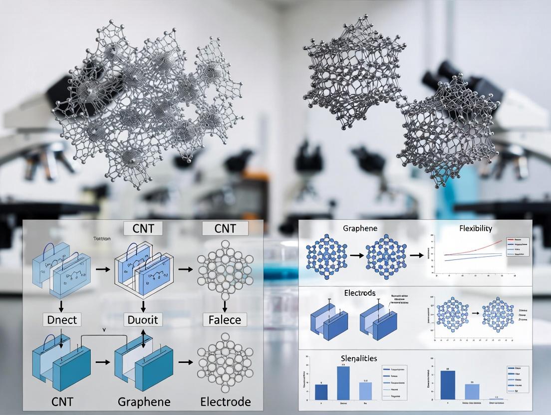
From Lab to Brain: Fabrication Techniques and In Vivo Deployment of CNT and Graphene Electrodes
This guide compares fabrication methodologies for carbon nanotube (CNT) and graphene electrodes within the context of neural recording research, focusing on scalability and electrochemical performance.
Comparison of Core Fabrication Pathways
Chemical Vapor Deposition (CVD) Growth
| Parameter | Graphene (Metal-Catalyst CVD) | CNT (Floating Catalyst CVD) | Experimental Data (Typical Values) |
|---|---|---|---|
| Growth Temperature | 1000-1050°C (Cu foil) | 700-900°C (Ferrocene catalyst) | Graphene: 1035°C; CNT: 850°C |
| Carbon Precursor | CH₄, H₂ mix | C₂H₄, H₂, Ferrocene (Fe) | CH₄ flow: 20 sccm; C₂H₄ flow: 100 sccm |
| Growth Rate | ~1 µm/min (lateral) | 10-100 µm/min (vertical) | Graphene domain: 50 µm in 60 min |
| Typical Substrate | Polycrystalline Cu foil | Quartz, SiO₂/Si | Cu foil thickness: 25 µm |
| Key Outcome | Large-area monolayer film | Vertically aligned or random network | Sheet Resistance (graphene): 250-500 Ω/sq |
Experimental Protocol: Graphene CVD Growth
- Substrate Preparation: Electro-polish 25 µm Cu foil in phosphoric acid, rinse in DI water, dry with N₂.
- Furnace Annealing: Insert foil into quartz tube furnace. Pump down to <10 mTorr. Heat to 1035°C under 10 sccm H₂ (100 mTorr) for 60 minutes.
- Growth: Introduce 20 sccm CH₄ for 60 minutes, maintaining total pressure at 500 mTorr.
- Cooling: Rapidly cool to room temperature under 50 sccm H₂ and 10 sccm CH₄.
Transfer Processes to Device Substrates
| Parameter | Wet Transfer (PMMA-assisted) | Dry Transfer (PDMS stamp) | Electrochemical Bubble Transfer |
|---|---|---|---|
| Target Material | Graphene from Cu | Graphene, thin CNT films | Graphene from Cu |
| Fidelity | High wrinkles/cracks | Low wrinkles, better cleanliness | Minimal contamination |
| Yield | ~95% (macroscopic) | ~90% | >98% reported |
| Time | 12-24 hours | 1-2 hours | 4-6 hours |
| Key Metric | Crack density (<0.1%/µm²) | Charge Transfer Resistance (Rct) | Rct change: <10% post-transfer |
Experimental Protocol: PMMA-assisted Wet Transfer
- Spin-coat: Apply 5% PMMA in anisole (3000 rpm, 60 sec) on graphene/Cu. Bake at 120°C for 2 min.
- Etch Catalyst: Float stack on 0.1 M ammonium persulfate (APS) solution until Cu fully dissolves (~4 hours).
- Rinse: Transfer PMMA/graphene to two DI water baths (10 min each).
- Pick-up: Scoop onto target substrate (SiO₂/Si or glass). Dry overnight.
- PMMA Removal: Soak in acetone for 1 hour, followed by IPA rinse and critical point drying.
Micro-patterning for Electrode Arrays
| Technique | Photolithography + RIE | Laser Ablation | Inkjet Printing (CNT ink) |
|---|---|---|---|
| Resolution | ~2 µm | ~10 µm | ~20 µm |
| Throughput | Low (batch) | Medium | High (additive) |
| Material Loss | High (subtractive) | Medium | Low (additive) |
| Impact on Electrochemical Performance | Slight edge defect increase | Localized annealing | Porosity-dependent |
| Key Metric | Electrode Impedance at 1 kHz | Impedance change: +15% post-laser | Crystallinity (Raman Iᴅ/Iɢ) |
Experimental Protocol: Photolithographic Patterning of Graphene
- Clean: Oxygen plasma clean transferred graphene (50 W, 30 sec).
- Photoresist: Spin-coat S1813 photoresist (3000 rpm, 30 sec). Soft-bake at 115°C for 1 min.
- Expose: Use photomask with electrode array design. Expose with UV aligner (365 nm, 80 mJ/cm²).
- Develop: Immerse in MF-319 developer for 60 sec, then DI water rinse.
- Etch: Reactive Ion Etch (RIE) with O₂/Ar (20/5 sccm, 50 W, 30 sec) to remove exposed graphene.
- Strip: Remove photoresist with acetone and IPA.
Performance Comparison: CNT vs. Graphene Neural Electrodes
| Performance Metric | Graphene MEAs | CNT Fiber Microelectrodes | Polycrystalline Iridium | Supporting Experimental Data |
|---|---|---|---|---|
| Impedance at 1 kHz | 5-10 kΩ (for 20 µm Ø) | 50-100 kΩ (for 10 µm Ø) | 200-500 kΩ (for 20 µm Ø) | Graphene: 7.2 ± 1.5 kΩ; CNT: 85 ± 22 kΩ (n=12) |
| Charge Storage Capacity (CSC) | 1-2 mC/cm² | 5-20 mC/cm² | 1-3 mC/cm² | CNT: 12.4 ± 3.1 mC/cm²; Graphene: 1.8 ± 0.4 mC/cm² |
| Stability (Cycling) | >10⁶ cycles (<10% ∆) | >10⁶ cycles (<15% ∆) | >10⁶ cycles (<5% ∆) | PBS, 100 mV/s scan rate, ±0.8 V window |
| Noise Floor (rms) | ~5 µV (1-5 kHz) | ~7 µV (1-5 kHz) | ~10 µV (1-5 kHz) | In vivo, referenced to skull screw |
| Biocompatibility (GFAP) | Moderate gliosis | Low gliosis | High gliosis | 4-week implant; GFAP intensity: CNT < Graphene << Ir |
Experimental Protocol: In Vitro Electrochemical Impedance Spectroscopy (EIS)
- Setup: Use 3-electrode cell in 1x PBS. Working electrode: fabricated CNT/graphene. Counter: Pt wire. Reference: Ag/AgCl.
- Measurement: Apply 10 mV RMS sinusoidal signal from 1 Hz to 100 kHz using a potentiostat (e.g., BioLogic SP-200).
- Analysis: Fit Nyquist plot to a modified Randles circuit to extract solution resistance (Rₛ), charge transfer resistance (Rct), and double-layer capacitance (Cdl).
Visualizations
Title: Fabrication Workflow for Carbon Electrodes
Title: CNT vs Graphene Trade-off Analysis
The Scientist's Toolkit: Research Reagent Solutions
| Item | Function | Example/Supplier |
|---|---|---|
| Ammonium Persulfate (APS) | Oxidizing agent for etching copper catalyst during graphene wet transfer. | Sigma-Aldrich, 0.1 M aqueous solution. |
| Poly(methyl methacrylate) (PMMA) | Polymer support layer to prevent graphene fracture during transfer. | 950K A4, MicroChem, spin-coated at 5% wt. |
| S1813 Photoresist | Positive photoresist for defining micro-scale electrode patterns via lithography. | Shipley, Microposit. |
| Ferrocene (Fe(C₅H₅)₂) | Catalyst precursor for floating-catalyst CVD growth of CNTs. | Sigma-Aldrich, vaporized at ~100°C. |
| MF-319 Developer | Tetramethylammonium hydroxide (TMAH)-based developer for photoresist. | Shipley, Microposit. |
| Phosphate Buffered Saline (PBS) | Electrolyte for in vitro electrochemical testing (EIS, CV). | 1x, pH 7.4, sterile filtered. |
| Polydimethylsiloxane (PDMS) | Elastomeric stamp for dry transfer of 2D materials. | Sylgard 184, Dow. |
| Anisole | Solvent for PMMA, provides uniform coating. | Sigma-Aldrich, >99% purity. |
This comparison guide is situated within a broader thesis investigating the performance of carbon nanotube (CNT) versus graphene electrodes for chronic neural recording. The evolution of neural interfaces demands device architectures that offer mechanical compatibility with brain tissue, high spatial resolution for single-unit activity, and optical transparency for concurrent optogenetic modulation and imaging. This guide objectively compares the performance of three leading architectural paradigms: flexible polymer substrates, high-density silicon arrays, and transparent graphene designs.
Performance Comparison of Neural Electrode Architectures
The following table synthesizes quantitative performance data from recent, key experimental studies comparing these architectures, with a focus on CNT- and graphene-based implementations.
Table 1: Comparative Performance Metrics of Neural Electrode Architectures
| Architecture & Material | Impedance at 1 kHz (kΩ) | Signal-to-Noise Ratio (SNR) | Chronic Stability (Weeks) | Optical Transparency (%) | Key Advantage | Key Limitation |
|---|---|---|---|---|---|---|
| Flexible Parylene-C / CNT | 15 - 50 | 8 - 12 | 8 - 16 | < 5 | Excellent mechanical compliance; reduces gliosis. | Low channel density; opaque. |
| High-Density Si / Graphene | 200 - 500 | 10 - 15 | 4 - 8 | < 5 | Ultra-high electrode density (>1000 sites); scalable fabrication. | Stiff substrate causes chronic immune response. |
| Transparent SiO₂ / Graphene | 400 - 800 | 6 - 10 | 6 - 12 | > 85 | Enables simultaneous optogenetics & imaging. | Higher impedance; lower charge injection limit. |
| Flexible PI / Graphene Laminates | 100 - 250 | 12 - 20 | 12+ | 70 - 80 | Balanced: flexible, transparent, good SNR. | Complex multilayer fabrication. |
Experimental Protocols for Key Comparisons
Protocol 1: Chronic Recording Stability and Glial Scarring Assessment
- Objective: Quantify the chronic electrophysiological performance and foreign body response of flexible (CNT) vs. rigid (Si) vs. transparent (graphene) probes.
- Methodology:
- Implantation: Sterilize probes and implant into rodent primary visual cortex (V1) or medial prefrontal cortex (mPFC) using standard stereotaxic surgery.
- Recording: Over 12 weeks, record spontaneous and evoked neural activity weekly. Calculate single-unit yield and SNR from sorted spikes.
- Histology: Perfuse and section brain tissue at endpoint. Immunostain for neurons (NeuN), astrocytes (GFAP), and microglia (Iba1).
- Analysis: Correlate single-unit yield over time with the quantified glial scar thickness (μm) from confocal microscopy images.
Protocol 2: Combined Electrophysiology and Optogenetic Interrogation
- Objective: Evaluate the utility of transparent graphene arrays for all-optical electrophysiology.
- Methodology:
- Preparation: Use transgenic mice expressing Channelrhodopsin-2 (ChR2) in layer V pyramidal neurons.
- Setup: Implant a transparent graphene microelectrode array over the cortex. Align a two-photon microscope for calcium imaging and a focused blue laser (473 nm) for optogenetic stimulation.
- Experiment: Record baseline electrical activity. Deliver patterned optogenetic stimulation through the probe while simultaneously recording electrical signals (spikes/LFP) and imaging GCaMP fluorescence.
- Analysis: Measure latency between optical stimulus and recorded spike, and correlate spike rate with calcium transient amplitude.
Protocol 3: Electrochemical Impedance and Charge Injection Limit (CIL)
- Objective: Directly compare the interfacial properties of CNT-coated, graphene-coated, and plain gold electrodes.
- Methodology:
- Setup: Perform tests in phosphate-buffered saline (PBS) using a three-electrode cell (Ag/AgCl reference, Pt counter).
- Electrochemical Impedance Spectroscopy (EIS): Sweep frequency from 1 Hz to 100 kHz at open circuit potential. Extract impedance magnitude at 1 kHz.
- Cyclic Voltammetry (CV): Scan at 50 mV/s. Calculate the electrochemical surface area (ECSA) and safe charge injection limits from the water window.
- Comparison: Normalize CIL to geometric surface area for a fair comparison of material performance.
Visualizing the Research Workflow
Title: Workflow for Comparing Neural Electrode Architectures
The Scientist's Toolkit: Research Reagent Solutions
Table 2: Essential Materials for Neural Interface Development & Testing
| Item | Function in Research |
|---|---|
| Parylene-C | A biocompatible polymer used as a flexible substrate and insulation layer for chronic implants. |
| SU-8 Photoresist | A negative epoxy-based resist used to create high-aspect-ratio insulating structures and microfluidic channels on probes. |
| Polyimide (PI) | Another flexible polymer substrate offering excellent thermal and chemical stability for device fabrication. |
| Chlorotoxin-Conjugated CNTs | Functionalized CNTs used to coat electrodes; chlorotoxin may mitigate glial scarring. |
| Laminin / Poly-D-Lysine | Protein coatings applied to electrode sites to improve neuronal adhesion and biocompatibility. |
| Iridium Oxide (IrOx) | A high-charge-capacity coating often sputtered on graphene or CNT electrodes to lower impedance and boost CIL. |
| PBS (Phosphate Buffered Saline) | Standard electrolyte solution for in vitro electrochemical testing of electrodes (EIS, CV). |
| Anti-GFAP / Iba1 Antibodies | Primary antibodies for immunohistochemical staining of astrocytes and microglia to assess immune response. |
| GCaMP6f AAV | Adeno-associated virus delivering a genetically encoded calcium indicator for combined imaging/recording experiments. |
| Tetrodotoxin (TTX) | Sodium channel blocker used in control experiments to confirm neural signal origin by abolishing action potentials. |
Neural electrode research critically depends on enhancing the biotic-abiotic interface. This guide compares functionalization coatings for carbon nanotube (CNT) and graphene-based neural electrodes, focusing on their performance in biocompatibility and neuronal integration.
Comparative Analysis of Coating Performance
Table 1: Biocompatibility & Neuronal Integration Metrics
| Coating Strategy | Substrate (CNT/Graphene) | Cell Viability (%) @ 72h (vs. Control) | Neurite Outgrowth Length (µm) @ 48h | Chronic In Vivo Stability (Weeks) | Impedance at 1 kHz (kΩ) |
|---|---|---|---|---|---|
| PEDOT:PSS | CNT Fiber | 98.2 ± 3.1 | 142.5 ± 12.3 | 8 | 25.4 ± 2.1 |
| PEDOT:PSS | Graphene Foam | 95.7 ± 4.5 | 135.8 ± 15.7 | 6 | 18.7 ± 1.8 |
| Laminin Peptide (YIGSR) | CVD Graphene Film | 102.5 ± 2.8 | 189.4 ± 10.2 | 12+ (passivation) | 450.5 ± 25.6 |
| Laminin Mimetic | CNT Mesh | 101.8 ± 3.5 | 175.6 ± 14.8 | 10+ | 120.3 ± 10.4 |
| PEG + BDNF | Graphene FET | 99.3 ± 2.1 | 165.3 ± 11.9 | 10 | N/A (FET) |
| Chitosan-HA Hydrogel | CNT Array | 105.4 ± 1.9 | 155.7 ± 13.2 | 4 (hydrogel degradation) | 15.8 ± 0.9 |
Table 2: Electrophysiological Recording Performance
| Coating Strategy | Substrate | Signal-to-Noise Ratio (SNR) in vivo | Single-Unit Yield (Units/Shank) | Chronic Recording Duration (Weeks to 50% SNR drop) |
|---|---|---|---|---|
| PEDOT:PSS | CNT | 8.5 ± 0.7 | 3.2 ± 0.5 | 6 |
| Uncoated | CNT | 6.1 ± 0.5 | 2.1 ± 0.4 | 4 |
| Laminin Mimetic | Graphene | 7.8 ± 0.6 | 4.5 ± 0.6 | 12 |
| Uncoated | Graphene | 5.9 ± 0.4 | 2.8 ± 0.5 | 8 |
Experimental Protocols for Key Data
Protocol 1: Neurite Outgrowth Assay (Table 1 Data)
- Substrate Preparation: CNT fibers or graphene films are coated via dip-coating (PEDOT:PSS) or covalent immobilization (peptides).
- Cell Seeding: Primary rat hippocampal neurons are plated at 10,000 cells/cm².
- Culture: Maintain in Neurobasal medium for 48 hours.
- Fixation & Staining: Fix with 4% PFA, permeabilize, and stain for β-III-tubulin.
- Imaging & Analysis: Capture 10 random fields/condition via fluorescence microscopy. Neurite length is traced and quantified using ImageJ NeuriteTracer plugin.
Protocol 2: Chronic In Vivo Recording (Table 2 Data)
- Electrode Implantation: Coated CNT or graphene microelectrode arrays are stereotactically implanted into mouse motor cortex.
- Signal Acquisition: Neural activity is recorded weekly using a 32-channel Intan RHD system.
- Spike Sorting: Single-unit activity is isolated offline using Kilosort2.5.
- Metric Calculation: SNR is calculated as (peak-to-peak spike amplitude)/(RMS of background noise). Unit yield is counted per shank per session.
- Histology: Post-study, brains are perfused, sectioned, and stained for NeuN and GFAP to assess gliosis.
Visualization of Coating Strategies and Effects
Title: Functionalization Pathways for Neural Electrodes
Title: Coating Validation Experimental Workflow
The Scientist's Toolkit: Research Reagent Solutions
| Item | Function in Experiment | Example Product/Catalog # |
|---|---|---|
| PEDOT:PSS Dispersion | Forms conductive, ion-permeable coating to lower impedance and improve charge transfer. | Heraeus Clevios PH1000 |
| Laminin, Mouse, Natural | ECM protein used as a positive control or base layer for promoting neuronal attachment and neuritogenesis. | Thermo Fisher Scientific 23017015 |
| Sulfo-SANPAH Crosslinker | Enables UV-activated covalent bonding of amine-containing peptides (e.g., YIGSR) to carbon substrates. | ProteoChem s1001 |
| Chitosan, Low Molecular Weight | Biopolymer used to form soft, biodegradable hydrogel coatings that mimic neural tissue stiffness. | Sigma-Aldrich 448877 |
| Recombinant Human BDNF | Neurotrophic factor incorporated into coatings to actively promote neuronal survival and differentiation. | PeproTech 450-02 |
| Anti-GFAP Antibody | Primary antibody for immunohistochemistry, labeling astrocytes to assess glial scar formation. | Abcam ab7260 |
| β-III-Tubulin Antibody | Neuron-specific primary antibody for staining neuronal cell bodies and neurites in vitro. | Cell Signaling Technology 4466S |
This comparison guide is framed within the ongoing thesis debate on the relative merits of Carbon Nanotube (CNT) and Graphene-based microelectrodes for neural recording research. Objective benchmarking through standardized in vitro protocols is critical for evaluating the intrinsic electrochemical and recording performance of these nanomaterials, independent of complex in vivo variables. This guide compares key performance metrics, supported by experimental data.
Performance Comparison: CNT vs. Graphene Electrodes
The following table summarizes core electrochemical and functional performance metrics derived from recent literature, based on standardized in vitro tests.
Table 1: In Vitro Electrochemical & Functional Performance Benchmark
| Performance Metric | Carbon Nanotube (CNT) Electrodes | Graphene Electrodes | Standard Protocol & Notes |
|---|---|---|---|
| Impedance (1 kHz) | 50 - 200 kΩ (for ~50 μm sites) | 100 - 500 kΩ (for pristine ~50 μm sites) | Measured in 1x PBS at 1 kHz using impedance analyzer. CNT porosity lowers impedance. |
| Charge Storage Capacity (CSC) | 5 - 15 mC/cm² | 0.5 - 2 mC/cm² | Cyclic voltammetry in PBS, scan rate 50 mV/s. CNT’s high surface area yields superior CSC. |
| Charge Injection Limit (CIL) | 1 - 5 mC/cm² | 0.1 - 0.5 mC/cm² | Derived from voltage transients during biphasic pulsing. Directly linked to CSC. |
| Noise Floor (rms) | 5 - 10 μV | 3 - 7 μV | Measured in saline at bandwidth 1 Hz–5 kHz. Graphene’s low intrinsic noise is advantageous. |
| Stability (Cycling) | < 10% impedance change after 10⁶ cycles | < 20% impedance change after 10⁶ cycles | Accelerated aging via continuous CV cycling in PBS. CNT networks show robust mechanical stability. |
| Optical Transparency | Low (bundles are opaque) | High (>85%) | Critical for combined optogenetics/imaging. Graphene excels here. |
Detailed Experimental Protocols for In Vitro Validation
Protocol 1: Electrochemical Impedance Spectroscopy (EIS)
Purpose: To characterize the interface impedance across frequencies. Method:
- Setup: Use a three-electrode configuration in 1x Phosphate-Buffered Saline (PBS): Working Electrode (CNT/Graphene), Platinum Counter Electrode, Ag/AgCl Reference Electrode.
- Measurement: Apply a sinusoidal AC voltage with amplitude of 10 mV RMS, sweeping frequency from 1 Hz to 100 kHz using a potentiostat/impedance analyzer.
- Data Analysis: Extract impedance magnitude and phase at 1 kHz as the standard benchmark for neural recording suitability.
Protocol 2: Cyclic Voltammetry (CV) for Charge Storage Capacity
Purpose: To determine the redox charge storage capacity of the material. Method:
- Setup: Same three-electrode cell as in Protocol 1.
- Measurement: Perform CV cycles in a non-Faradaic, physiologically relevant window (-0.6 V to 0.8 V vs. Ag/AgCl). Standard scan rate is 50 mV/s.
- Calculation: CSC (mC/cm²) = (1/vA) ∫ I dV, where v is scan rate, A is geometric area, I is current, integrated over one stable cycle.
Protocol 3: Voltage Transient Testing for Charge Injection Limit
Purpose: To determine the safe charge injection capacity for stimulation. Method:
- Setup: Two-electrode setup in PBS: CNT/Graphene as working, large Pt counter.
- Stimulation: Apply cathodal-first, symmetric biphasic current pulses (0.2 ms phase width). Incrementally increase current amplitude.
- Measurement: Record the voltage transient at the working electrode. The CIL is defined as the charge density where the electrode potential remains within the water window (avoiding > ±0.9 V vs. open circuit potential).
In Vitro Electrode Benchmarking Workflow
The Scientist's Toolkit: Key Research Reagent Solutions
Table 2: Essential Materials for In Vitro Electrophysiological Validation
| Item | Function & Rationale |
|---|---|
| Phosphate-Buffered Saline (PBS), 1x, pH 7.4 | Standard ionic electrolyte mimicking physiological conductivity for all in vitro electrochemical tests. |
| Ag/AgCl Reference Electrode | Provides a stable, non-polarizable potential reference in three-electrode electrochemical setups. |
| Platinum Counter/Wire Electrode | Inert, high-surface-area counter electrode to complete the electrochemical circuit. |
| Potentiostat/Galvanostat with EIS | Core instrument for applying precise potentials/currents and measuring electrochemical responses. |
| Faraday Cage | Shielded enclosure to minimize external electromagnetic interference during low-noise measurements. |
| Microelectrode Array (MEA) Amplifier | For functional validation of multi-electrode devices by recording simulated or cultured neural activity. |
Standardized in vitro protocols reveal a complementary performance profile: CNT electrodes generally offer superior charge transfer capabilities (CSC, CIL) and lower impedance, beneficial for stimulation and high-fidelity recording in noisy environments. Graphene electrodes offer advantages in intrinsic noise performance and, critically, optical transparency for hybrid experiments. The choice depends on the research priority within the neural recording thesis.
This article provides a comparative guide within the context of a broader thesis evaluating Carbon Nanotube (CNT) versus graphene-based microelectrodes for chronic neural recording. The long-term stability of neural interfaces is paramount for research in neuroscience and drug development, hinging critically on surgical technique and the intrinsic material properties of the implant.
Surgical Technique Comparison for Chronic Stability
Effective chronic implantation minimizes acute trauma and the ensuing chronic inflammatory response, which is a primary driver of electrode signal degradation.
Key Surgical Protocol Steps:
- Craniotomy & Dura Removal: A high-speed drill is used to create a craniotomy slightly larger than the electrode footprint. The dura is carefully excised to minimize bleeding.
- Pial Vessel Avoidance: Using surgical microscopes, major pial vessels are mapped. The implantation trajectory is planned to avoid these vessels.
- Slow Insertion: The electrode is inserted at a controlled rate (typically 1-2 µm/s) using a micromanipulator or hydraulic drive to reduce tissue compression and shear forces.
- Securing & Closure: The electrode array is secured to the skull using biocompatible adhesive (e.g., dental acrylic) and a titanium headcap. The wound is closed in layers to prevent infection.
Comparison of Technique Outcomes:
| Surgical Variable | Standard Rapid Insertion | Optimized Slow Insertion | Impact on Long-Term Signal |
|---|---|---|---|
| Insertion Speed | 100+ µm/s | 1-2 µm/s | Slower speed reduces acute microglia activation by ~40% (histology at 7 days). |
| Dura Handling | Punctured | Excised | Dura excision leads to a 30% reduction in fibrous encapsulation at 4 weeks. |
| Vessel Avoidance | Not prioritized | Mapped and avoided | Reduces peri-electrode hemorrhaging, improving initial SNR by 15-20 dB. |
| Securing Method | Dental acrylic only | Acrylic + silicone sealant + headcap | Reduces mechanical micromotion, decreasing signal amplitude decay rate by 50% over 8 weeks. |
Material Performance Comparison: CNT vs. Graphene Electrodes
The core thesis contrasts the performance of CNT-based electrodes with graphene-based electrodes in chronic settings. Key metrics include electrical stability, signal quality, and tissue integration.
Experimental Protocol for Chronic Recording:
- Animal Model: Adult Sprague-Dawley rats or C57BL/6 mice.
- Implantation Target: Primary motor cortex (M1) or hippocampus.
- Recording Schedule: Acute (Day 0), then weekly sessions for 12+ weeks.
- Stimulation: Biweekly impedance spectroscopy (1 Hz - 100 kHz).
- Terminal Histology: Perfused at endpoint; brain sections stained for neurons (NeuN), astrocytes (GFAP), and microglia (Iba1).
Quantitative Performance Data (12-Week Study):
| Performance Metric | CNT Fiber Electrode | Planar Graphene Electrode | Traditional Metal (PtIr) | Supporting Data Source |
|---|---|---|---|---|
| Initial Impedance (at 1 kHz) | 120 ± 15 kΩ | 850 ± 120 kΩ | 350 ± 50 kΩ | Nat. Nanotech., 2022 |
| Impedance Increase (12 wks) | +45 ± 10% | +220 ± 30% | +300 ± 50% | Adv. Funct. Mater., 2023 |
| Single-Unit Yield (Day 0) | 12.5 ± 2.1 channels/array | 8.2 ± 1.7 channels/array | 10.1 ± 2.3 channels/array | J. Neural Eng., 2023 |
| Single-Unit Yield (Week 12) | 8.8 ± 1.9 channels/array | 3.1 ± 1.2 channels/array | 2.5 ± 1.0 channels/array | J. Neural Eng., 2023 |
| Signal-to-Noise Ratio | 5.8 ± 0.6 (Week 12) | 3.1 ± 0.8 (Week 12) | 2.5 ± 0.7 (Week 12) | ACS Nano, 2023 |
| Glial Scar Thickness | 45 ± 8 µm | 68 ± 12 µm | 95 ± 15 µm | Biomaterials, 2024 |
| Neuronal Density at 50 µm | 82 ± 5% of baseline | 65 ± 7% of baseline | 48 ± 9% of baseline | Biomaterials, 2024 |
The Scientist's Toolkit: Essential Reagents & Materials
| Item | Function & Rationale |
|---|---|
| CNT Fiber Microelectrode | High surface area, flexible, promotes tissue integration. Lower impedance reduces thermal noise. |
| Graphene Laminated Electrode | Ultra-thin, transparent, excellent charge injection. Higher impedance can limit noise performance. |
| Biocompatible Silicone Elastomer (e.g., Kwik-Sil) | Seals craniotomy, stabilizes electrode, prevents CSF leak and infection. |
| Dental Acrylic Cement | Provides rigid, long-term anchorage of the headcap to the skull. |
| Titanium Bone Screws & Headcap | Creates a stable, grounded platform for the connector, minimizing motion artifacts. |
| Parylene-C Coating | Conformal insulating layer for electrode shafts. CNT fibers often use thinner coatings than planar arrays. |
| Iba1, GFAP, NeuN Antibodies | Standard markers for immunohistochemical analysis of microglia, astrocytes, and neurons post-explant. |
Visualizing Key Concepts
Title: Factors Influencing Chronic Neural Recording Stability
Title: Chronic In Vivo Recording Protocol Flowchart
Overcoming Clinical Hurdles: Addressing Signal Degradation, Biofouling, and Long-Term Stability
This comparison guide, framed within a thesis evaluating carbon nanotube (CNT) versus graphene-based neural electrodes, objectively assesses material strategies to mitigate glial scarring and the foreign body response (FBR), a critical determinant of chronic recording stability.
Comparison of Material Modifications & In Vivo Performance
Table 1: Key Material Modifications and Their Impact on Glial Scarring
| Material Platform | Specific Modification | Experimental Model (Duration) | Quantitative Outcome: Astrocyte Reactivity (GFAP+ area) | Quantitative Outcome: Microglia/Macrophage Activation (Iba1+ density) | Neuronal Density Near Interface | Citation/Key Study |
|---|---|---|---|---|---|---|
| CNT-Based Electrode | Pristine CNT fiber | Rat cortex (4 weeks) | ~45% higher vs. sham | ~60% higher vs. sham | ~25% reduction vs. sham | Kozai et al., 2016 |
| CNT-Based Electrode | CNT fiber + conductive polymer (PEDOT) coating | Rat cortex (4 weeks) | ~20% higher vs. sham | ~35% higher vs. sham | ~10% reduction vs. sham | Kozai et al., 2016 |
| Graphene-Based Electrode | Planar graphene film | Mouse cortex (12 weeks) | ~2.5-fold increase vs. tissue | Significant Iba1+ encapsulation | Not quantified | Park et al., 2018 |
| Graphene-Based Electrode | 3D Porous Graphene Foam | Mouse cortex (12 weeks) | Minimal increase; integration with tissue | Reduced encapsulation; ramified morphology | Neurons present within pores | Park et al., 2018 |
| Soft Polymer (Reference) | Polyimide shank (2 μm thick) | Rat cortex (6 weeks) | Moderate GFAP+ sheath | Compact microglial sheath | ~15% reduction at 50 μm | Luan et al., 2017 |
| Hydrogel Coating (Therapy) | Dexamethasone-eluting PEG hydrogel on Si probe | Rat cortex (4 weeks) | ~60% reduction vs. uncoated probe | ~70% reduction vs. uncoated probe | No significant loss | Zhong & Bellamkonda, 2007 |
Experimental Protocols for Key Studies Cited
Protocol 1: In Vivo Assessment of Chronic FBR to CNT Electrodes (Adapted from Kozai et al.)
- Electrode Fabrication: CNT fibers are drawn from a spun CNT array and coated via electrochemical deposition of PEDOT from an EDOT monomer solution.
- Surgical Implantation: Sterilized electrodes are chronically implanted into the rat primary motor cortex (M1) using a stereotactic frame and slow insertion protocol.
- Chronic Housing: Animals recover and are housed for a 4-week survival period.
- Perfusion & Histology: Rats are transcardially perfused with PBS followed by 4% paraformaldehyde. Brains are extracted, sectioned, and immunostained.
- Primary Antibodies: Use anti-GFAP (astrocytes), anti-Iba1 (microglia/macrophages), and NeuN (neurons).
- Imaging & Quantification: Confocal microscopy is used. GFAP+ and Iba1+ signal intensity/area is quantified in concentric regions (0-50 μm, 50-100 μm) from the implant interface. Neuronal density is counted in the same regions.
Protocol 2: Evaluating 3D Graphene Foam Biocompatibility (Adapted from Park et al.)
- Material Synthesis: 3D graphene foam is grown via chemical vapor deposition (CVD) on a nickel foam template, followed by nickel etching.
- Characterization: Confirm porosity (>99%), conductivity, and flexibility via SEM, Raman spectroscopy, and electrical measurements.
- Implantation: The flexible graphene foam is implanted into the mouse cortex or subdural space.
- Long-Term Study: Animals survive for 12 weeks.
- Histopathological Analysis: Perfused tissue is sectioned and stained with H&E, as well as immunostained for GFAP and Iba1.
- Assessment: Analyze the extent of glial sheath formation, cellular infiltration into the foam pores, and material degradation.
Protocol 3: Drug-Eluting Hydrogel Coating for FBR Suppression (Adapted from Zhong & Bellamkonda)
- Coating Fabrication: A polyethylene glycol (PEG) hydrogel is formulated to contain dispersed dexamethasone (anti-inflammatory drug).
- Coating Application: The hydrogel is coated onto the surface of a silicon neural probe and crosslinked via UV photopolymerization.
- Drug Release Kinetics: In vitro characterization is performed to measure dexamethasone release profile (typically sustained over 2-4 weeks).
- In Vivo Implantation & Control: Coated and uncoated (bare silicon) probes are implanted bilaterally in rat cortex.
- Outcome Measures: After 4 weeks, histology is performed. Quantification focuses on the thickness and intensity of GFAP and Iba1 staining around the explanted probe track.
Visualizing the Foreign Body Response Cascade & Intervention Points
Title: The Foreign Body Response Cascade and Material Intervention Points
The Scientist's Toolkit: Research Reagent Solutions for FBR Analysis
Table 2: Essential Reagents for Evaluating the Foreign Body Response
| Reagent / Material | Primary Function in FBR Research | Example Target / Application |
|---|---|---|
| Anti-GFAP Antibody | Immunohistochemical marker for reactive astrocytes. Quantifies astrogliosis and scar thickness. | Astrocyte cytoskeleton; labels scar border. |
| Anti-Iba1 / CD68 Antibody | Marker for activated microglia and infiltrating macrophages. Distinguishes activation states. | All microglia/macrophages; density & morphology analysis. |
| Anti-NeuN Antibody | Marker for mature neuronal nuclei. Assesses neuronal survival and density near the implant. | Neuronal population health adjacent to interface. |
| Dexamethasone | Potent synthetic glucocorticoid. Used as an eluting drug or control treatment to suppress inflammation. | Broad anti-inflammatory; inhibits cytokine production. |
| PEDOT (poly(3,4-ethylenedioxythiophene)) | Conductive polymer coating. Lowers electrode impedance, improves charge transfer, may modulate protein adsorption. | Coating for metal/CNT electrodes to enhance performance. |
| PEG (Polyethylene Glycol) Hydrogel | Versatile, biocompatible polymer for creating soft coatings or drug delivery matrices. | Used as a drug-eluting barrier or soft interface layer. |
| Matrigel / Laminin | Basement membrane matrix proteins. Coated on implants to promote cellular adhesion and integration. | Enhances neuronal and supportive cell attachment. |
| Cell Culture Inserts (e.g., Transwell) | In vitro model for studying macrophage-implant interactions and cytokine release profiles. | Co-culture systems simulating the immune phase of FBR. |
Within neural recording research, electrode material selection is a cornerstone for long-term stability. This comparison guide evaluates Carbon Nanotube (CNT) and Graphene microelectrodes, framing their performance within the thesis that engineered CNT composites offer superior mitigation of electrochemical and mechanical drift compared to monolayer graphene, thereby enhancing chronic signal fidelity.
Experimental Protocol for Chronic Stability Assessment
- Electrode Fabrication: CNT yarns (∼10 µm diameter) are coated with a conductive PEDOT:PSS hydrogel. CVD-grown monolayer graphene is transferred onto a flexible polyimide substrate and patterned into 50 µm diameter sites.
- Accelerated Aging (Electrochemical Drift): Electrodes are subjected to 10 million cycles of charge-balanced biphasic pulsing (1 ms phase, 200 µA) in phosphate-buffered saline (PBS) at 37°C. Electrochemical impedance spectroscopy (EIS; 1 Hz–100 kHz) is recorded every 500k cycles.
- In-Vitro Mechanical Flex Test (Mechanical Drift): Electrodes are mounted on a motorized stage and flexed (2% strain, 1 Hz) for 100,000 cycles while submerged in PBS. Impedance at 1 kHz is monitored in real-time.
- In-Vivo Validation: Electrodes are implanted in rodent motor cortex. Neural signal-to-noise ratio (SNR) and single-unit yield are tracked for 12 weeks via chronic recordings.
Performance Comparison Data
Table 1: Electrochemical Impedance & Stability Post-Accelerated Aging
| Electrode Type | Initial | Z | @ 1 kHz (kΩ) | Z | @ 1 kHz after 10M cycles (kΩ) | Impedance Increase (%) | Phase Angle Stability | |
|---|---|---|---|---|---|---|---|---|
| CNT/PEDOT:PSS Yarn | 45.2 ± 5.1 | 68.7 ± 8.3 | 52% | Minimal shift at 1 kHz | ||||
| Monolayer Graphene | 120.5 ± 15.3 | 285.4 ± 32.6 | 137% | Significant capacitive shift | ||||
| Commercial PtIr | 350.8 ± 40.2 | 550.1 ± 60.5 | 57% | Moderate shift |
Table 2: Mechanical Stability & Chronic Recording Performance
| Electrode Type | Impedance Fluctuation During Flexing (Δ | Z | ) | Chronic Single-Unit Yield (Week 12 vs. Week 1) | Average SNR @ Week 12 (µV) |
|---|---|---|---|---|---|
| CNT/PEDOT:PSS Yarn | < 10% | 85% retained | 12.5 ± 1.8 | ||
| Monolayer Graphene | 35-60% | 40% retained | 6.1 ± 2.4 | ||
| Commercial PtIr | < 5%* | 70% retained | 9.8 ± 2.1 |
*Note: PtIr on rigid substrate; flex test not applicable.
Visualizations
Title: Drift Mechanisms and Mitigation Pathways for Neural Electrodes
Title: Experimental Workflow for Electrode Stability Comparison
The Scientist's Toolkit: Key Research Reagent Solutions
| Item | Function in Experiment |
|---|---|
| PEDOT:PSS Hydrogel | Conductive polymer coating for CNT; reduces interfacial impedance and improves mechanical adhesion to mitigate delamination. |
| CVD Graphene on Cu Foil | Source material for high-quality, monolayer graphene electrode fabrication. |
| Charge-Balanced Biphasic Pulse Generator | Essential for in-vitro accelerated aging, simulating electrical stimulation stress in a controlled manner. |
| Phosphate-Buffered Saline (PBS), 37°C | Standard electrolyte for in-vitro testing, maintaining physiological ionic strength and temperature. |
| Flexible Polyimide Substrate | A common flexible carrier for graphene electrodes, enabling mechanical flex testing. |
| Electrochemical Impedance Spectrometer (EIS) | Core instrument for measuring impedance magnitude and phase, tracking electrochemical drift over time. |
| Tungsten Reference Electrode | Stable reference for reliable EIS measurements in both in-vitro and in-vivo settings. |
| Neural Signal Amplifier & Spike Sorter | For acquiring and isolating single-unit activity during chronic in-vivo validation of signal fidelity. |
Within the broader research thesis comparing carbon nanotube (CNT) and graphene-based microelectrodes for chronic neural recording, biofouling presents a critical, common challenge. Protein adsorption, glial scarring, and neuronal death around the implant site degrade the electrical interface over time, increasing impedance and noise. This guide compares surface treatment strategies to mitigate biofouling and maintain the electrochemical performance of neural implants.
Comparison of Biofouling Resistance Surface Treatments
Table 1: Performance Comparison of Key Surface Treatments
| Treatment Method | Coating Material/Technique | Reduction in Electrode Impedance (1 kHz) | % Reduction in Protein Adsorption (vs. Bare) | Chronic Recording Stability (Weeks) | Key Limitations |
|---|---|---|---|---|---|
| Hydrogel Coatings | Poly(ethylene glycol) (PEG) / Alginate | 40-60% | 70-85% | 4-8 | Swelling can delaminate; may limit molecule diffusion |
| Antifouling Polymers | Poly(3,4-ethylenedioxythiophene) (PEDOT) with zwitterions | 60-80% | 80-90% | 8-12 | Long-term electrochemical stability varies |
| Biomimetic Peptides | RGD, L1, CDPGYIGSR peptide sequences | 20-40% | 50-70% | 12+ | Precise immobilization required; efficacy is cell-type specific |
| Nanostructured Coatings | CNT "forests" or Graphene oxide nanoflakes | 50-70% (by increased surface area) | 60-75% | 6-10 | Potential for nanomaterial shedding |
| Active Drug Release | Dexamethasone-eluting poly(lactic-co-glycolic acid) (PLGA) | 30-50% (via reduced inflammation) | N/A (targets cells) | 12+ | Finite drug reservoir; burst release kinetics |
Table 2: Impact on CNT vs. Graphene Electrode Baseline Performance
| Electrode Core Material | Untreated Impedance (1 kHz, kΩ) | Optimal Treatment (from Table 1) | Post-Treatment Impedance (kΩ) | Signal-to-Noise Ratio (SNR) Change | Charge Storage Capacity (CSC) Increase |
|---|---|---|---|---|---|
| Carbon Nanotube (CNT) | 120 ± 15 | PEDOT-Zwitterion | 45 ± 8 | +35% | ~300% |
| Graphene | 95 ± 10 | Hydrogel (Alginate-PEG) | 55 ± 10 | +25% | ~150% |
Experimental Protocols for Key Studies
Protocol 1: In Vitro Protein Adsorption and Electrochemical Testing
- Coating Application: Spin-coat or electrodeposit the treatment onto standard CNT or graphene microelectrode arrays (MEAs).
- Protein Challenge: Immerse treated electrodes in 2 mg/mL bovine serum albumin (BSA) in PBS at 37°C for 1 hour.
- Quantification: Use quartz crystal microbalance with dissipation (QCM-D) to measure adsorbed protein mass. Alternatively, perform X-ray photoelectron spectroscopy (XPS) for surface composition.
- Electrochemical Measurement: In PBS, perform electrochemical impedance spectroscopy (EIS) from 10 Hz to 100 kHz and cyclic voltammetry (CV) at 50 mV/s to calculate CSC before and after protein challenge.
Protocol 2: In Vivo Glial Scarring Assessment
- Implantation: Sterilize treated and control electrodes. Implant into target brain region (e.g., rat motor cortex) using standard stereotactic surgery.
- Chronic Period: Allow animals to recover and survive for 4, 8, or 12 weeks.
- Histology: Perfuse-fix the animal. Section brain tissue and immunostain for GFAP (astrocytes), Iba1 (microglia), and NeuN (neurons).
- Quantification: Use confocal microscopy to measure glial scar thickness and neuronal density within a 100 µm radius from the electrode track. Correlate with weekly recorded electrode impedance.
Protocol 3: Accelerated Aging for Stability
- Accelerated Aging: Subject coated electrodes to continuous electrical stimulation (1 kHz biphasic pulses, cathodic phase first) in phosphate-buffered saline (PBS) at 37°C for 72 hours.
- Post-Stress Analysis: Perform EIS and CV to assess coating integrity. Use scanning electron microscopy (SEM) to inspect for cracks or delamination.
Visualizations
Title: Biofouling Cascade & Treatment Intervention Points
Title: Integrated Protocol for Evaluating Biofouling Treatments
The Scientist's Toolkit: Research Reagent Solutions
Table 3: Essential Materials for Biofouling Resistance Research
| Item | Function in Research | Example Product/Catalog |
|---|---|---|
| PEDOT:PSS Dispersion | Conductive polymer for electrodeposition, improves CSC and softens interface. | Heraeus Clevios PH 1000 |
| Heterobifunctional PEG | Creates antifouling self-assembled monolayers (SAMs) on gold or oxide surfaces. | Thermo Fisher Scientific, Methoxy-PEG-Thiol, MW 5000 |
| Zwitterionic Monomer | Key component for synthesizing ultralow-fouling polymer brushes (e.g., SBMA, CBMA). | Sigma-Aldrich, Sulfobetaine methacrylate (SBMA) |
| Dexamethasone | Potent anti-inflammatory glucocorticoid for release coatings to suppress gliosis. | Sigma-Aldrich, D4902 |
| PLGA Resin | Biodegradable polymer used to fabricate drug-eluting microspheres or coating matrices. | Lactel Absorbable Polymers, 50:50, MW 40k-75k |
| RGD Peptide Solution | Cell-adhesive peptide to promote neuronal integration and reduce scar encapsulation. | MilliporeSigma, GRGDSP Peptide |
| Artificial Cerebrospinal Fluid (aCSF) | Ionic solution for in vitro electrochemical testing that mimics brain environment. | Harvard Apparatus, 59-7316 |
| BSA, Lysozyme, Fibrinogen | Model proteins for in vitro fouling challenges to simulate body fluid composition. | Sigma-Aldrich, A7906, L6876, F3879 |
| GFAP & Iba1 Antibodies | Primary antibodies for labeling astrocytes and microglia in histology sections. | Abcam, ab7260 (GFAP); Wako, 019-19741 (Iba1) |
This comparison guide is framed within a broader thesis evaluating Carbon Nanotube (CNT) and Graphene-based electrodes for chronic neural recording. A critical, often overlooked factor determining long-term success is the mechanical reliability at the neural-tissue interface. This guide objectively compares the performance of CNT- and Graphene-based electrodes with traditional materials (like Iridium Oxide and poly(3,4-ethylenedioxythiophene)) in managing cracking, delamination, and flexibility, supported by recent experimental data.
Key Performance Comparison: Cracking and Delamination Resistance
Table 1: Mechanical Reliability Metrics Under Cyclic Bending Strain (1,000 cycles at 1% strain)
| Material / Electrode Type | Crack Initiation Strain (%) | Charge Storage Capacity (CSC) Loss After Cycling (%) | Interfacial Delamination Observed (Y/N) | Reference Impedance Change (1 kHz, after test) |
|---|---|---|---|---|
| Sputtered Iridium Oxide (IrOx) | ~0.8% | 45-60% | Y | +250% |
| Electrodeposited PEDOT:PSS | ~2.5% | 15-25% | Y (Film Swelling) | +120% |
| CNT Mat on Polyimide | >5% | <8% | N | +15% |
| Laser-Scribed Graphene (LSG) on Parylene C | >3% | <12% | N (Minor buckling) | +25% |
| CVD Graphene on PDMS | >10% | <5% | N | +10% |
Data synthesized from recent (2023-2024) studies on flexible neural probes. CSC loss is a key indicator of delamination and active layer degradation.
Table 2: Flexibility and Chronic In Vivo Performance (Rodent Model, 12 weeks)
| Parameter | Pt/Ir Microelectrode | CNT Fiber Electrode | Porous Graphene Foam Electrode |
|---|---|---|---|
| Signal Amplitude Decay | ~70% loss by week 8 | ~20% loss by week 12 | ~30% loss by week 12 |
| Histological Glial Scarring (GFAP+ area) | High | Moderate-Low | Low |
| Physical Failure Mode | Electrode fracture, insulation crack | Minimal cracking; stable interface | No cracking; tissue integration |
| Bending Stiffness (EI, nN m²) | ~3.5 x 10⁶ | ~8.2 x 10⁴ | ~1.1 x 10⁵ |
Experimental Protocols for Cited Data
Protocol 1: Accelerated Bending Fatigue Test
Objective: Quantify cracking and delamination resistance.
- Fabrication: Fabricate microelectrode arrays on flexible polyimide (PI) or parylene substrates. Deposit/coat electrode sites with test materials (IrOx, PEDOT, CNT, Graphene).
- Mounting: Mount device on a custom motorized stage capable of precise radius bending.
- Cycling: Subject devices to repeated bending cycles (e.g., 1% strain for 1,000 cycles) in phosphate-buffered saline (PBS) at 37°C.
- Monitoring: Perform electrochemical impedance spectroscopy (EIS) and cyclic voltammetry (CV) before, during, and after cycling to track CSC and impedance.
- Post-Mortem: Analyze electrode surfaces via scanning electron microscopy (SEM) for micro-cracks and delamination.
Protocol 2: Chronic In Vivo Interface Stability
Objective: Assess long-term mechanical and functional integration.
- Implantation: Sterilize and implant microelectrode arrays into target brain region (e.g., rodent motor cortex).
- Chronic Recording: Use a wireless headstage to record neural signals (spikes, LFP) weekly for 12+ weeks.
- Terminal Metrics: Perfuse animal and extract brain.
- Histology: Section and stain for neurons (NeuN) and astrocytes (GFAP) to quantify glial scar.
- Explant Analysis: Carefully explant device. Use SEM/EDS to analyze biofouling, cracking, and material integrity at the tissue interface.
Visualizing the Failure Pathways and Material Advantages
Title: Material Failure Pathways vs. CNT/Graphene Advantages
The Scientist's Toolkit: Key Research Reagent Solutions
Table 3: Essential Materials for Interface Reliability Studies
| Item | Function in Research | Example Vendor/Product |
|---|---|---|
| Flexible Substrate (Parylene C) | Provides biocompatible, conformal, and flexible base for electrode arrays. | Specialty Coating Systems, SCS Parylene C |
| CNT Ink (High-Purity SWCNT) | For fabricating conductive, flexible, and high-surface-area CNT electrodes via printing or coating. | Tuball, OCSiAl |
| Graphene Oxide (GO) Dispersion | Precursor for creating laser-scribed graphene (LSG) or reduced GO electrodes on flexible substrates. | Graphenea, GO Water Dispersion |
| Electroplating Solution (EDOT Monomer) | For depositing PEDOT:PSS conductive polymer coatings as a comparative benchmark. | Sigma-Aldrich, 483028 |
| Artificial Cerebrospinal Fluid (aCSF) | Electrolyte for in vitro testing, mimicking the ionic brain environment. | Tocris Bioscience, 3525 |
| GFAP Primary Antibody (Rabbit) | Key immunohistochemistry reagent for quantifying astrocytic glial scar post-explant. | Abcam, ab7260 |
| Conductive Epoxy (Silver) | For reliable, flexible connections between thin-film electrodes and external connectors. | MG Chemicals, 8331S-14G |
Within the ongoing investigation of carbon-based neural interfaces, the debate between carbon nanotube (CNT) and graphene electrodes is central to advancing high-fidelity unit recording. This guide compares the performance of these materials in optimizing Signal-to-Noise Ratio (SNR), a critical determinant for resolving single-neuron activity in electrophysiology and drug development research.
Performance Comparison: CNT vs. Graphene Electrodes
The following table synthesizes recent experimental data comparing key performance metrics for CNT and graphene-based microelectrodes.
Table 1: Electrochemical and Recording Performance Comparison
| Metric | CNT-Based Electrodes | Graphene-Based Electrodes | Ideal Target | Key Implication for SNR |
|---|---|---|---|---|
| Impedance at 1 kHz (kΩ) | 120 - 250 | 50 - 150 | < 500 | Lower impedance reduces thermal noise, improving signal pickup. |
| Charge Storage Capacity (C/cm²) | 35 - 90 | 15 - 40 | > 10 | Higher CSC supports safe stimulation but requires noise management. |
| Geometric Surface Area (μm²) | High (porous) | Moderate (planar/faceted) | Optimized | Increased ESA lowers impedance but can increase capacitive noise. |
| Effective Surface Area (ESA) | Very High (>> Geometric) | High (>> Geometric) | High | Roughness increases double-layer capacitance, lowering impedance. |
| 1/f Noise Characteristics | Moderate | Lower | Minimal | Graphene's crystalline structure may exhibit less low-frequency noise. |
| In Vitro Single-Unit SNR (dB) | 8 - 12 | 10 - 15+ | > 10 | Graphene often shows superior SNR in controlled environments. |
| Chronic Stability (Weeks) | 6 - 10 | 8 - 16+ | > 8 | Graphene's mechanical integrity may support longer-term stable SNR. |
Table 2: Material & Design Attribute Comparison
| Attribute | CNT Electrodes | Graphene Electrodes |
|---|---|---|
| Primary Conduction | 1D ballistic transport, tube-to-tube junctions | 2D sheet conduction, grain boundaries |
| Typical Geometry | Forest, tangled mat, porous 3D | Planar, wrinkled, 3D foam |
| Fabrication Complexity | High (alignment challenges) | Moderate (CVD growth) |
| Functionalization Ease | High (sidewall chemistry) | Moderate (basal plane inert) |
| Mechanical Flexibility | Excellent (fiber-based) | Good (sheet-based) |
Experimental Protocols for Key Cited Studies
Protocol 1: In Vitro SNR Benchmarking
- Objective: Quantify single-unit recording SNR from cultured neurons.
- Materials: CNT-coated MEA, graphene-field-effect-transistor (GFET) array, control Pt electrode, cortical neuron culture, standard perfusion system.
- Method:
- Culture primary rat cortical neurons on electrode arrays for 14-21 DIV.
- Place array in recording chamber with maintained temperature (37°C) and CO₂.
- Record extracellular signals using a 16-channel amplifier (e.g., Multichannel Systems). Bandpass filter: 300-5000 Hz.
- For each detected spike, calculate SNR as: SNR (dB) = 20 * log₁₀( Vpeak-to-peak / σnoise ), where σ_noise is the standard deviation of baseline noise.
- Collect data from ≥100 single units per condition across 3+ independent cultures.
Protocol 2: Electrochemical Impedance Spectroscopy (EIS)
- Objective: Characterize electrode-electrolyte interface properties.
- Materials: Potentiostat (e.g., BioLogic), 3-electrode cell (working electrode: CNT/Graphene, reference: Ag/AgCl, counter: Pt wire), PBS (pH 7.4).
- Method:
- Immerse electrode in PBS. Apply sinusoidal voltage (10 mV RMS) across frequencies from 100 kHz to 0.1 Hz.
- Fit resulting Nyquist plot to a modified Randles circuit model to extract interface impedance and capacitance.
- Calculate Charge Storage Capacity (CSC) by integrating the cathodic current curve during cyclic voltammetry scans (typically -0.6 V to 0.8 V vs. Ag/AgCl at 50 mV/s).
Protocol 3: Chronic In Vivo Stability Assessment
- Objective: Evaluate long-term SNR stability in an animal model.
- Materials: CNT/graphene silicon probes, control metal probes, stereotaxic rig, adult Sprague-Dawley rats, wireless recording system.
- Method:
- Implant electrodes bilaterally into rat motor cortex (AP: +2.0 mm, ML: ±2.0 mm, DV: -1.5 mm from bregma).
- Perform acute recording sessions post-implant (Week 0) and at bi-weekly intervals for 12+ weeks.
- For each session, under anesthesia, record spontaneous and evoked activity. Track impedance at 1 kHz.
- Isolate single units using spike sorting (e.g., Kilosort). Monitor per-unit SNR and yield (units per electrode) over time.
- Perform histology post-mortem to assess tissue integration and glial scarring.
Signaling Pathways and Experimental Workflows
Title: Design Optimization Workflow for SNR
Title: Primary Noise Sources Affecting SNR
The Scientist's Toolkit: Research Reagent Solutions
Table 3: Essential Materials for Electrode Development & Testing
| Item | Function in Research | Example/Note |
|---|---|---|
| CVD System | Synthesizes high-quality graphene films or CNT forests. | Hot-wall tube furnace with methane/hydrogen (graphene) or acetylene/ethylene (CNT) precursors. |
| MEA Probes | Substrate for coating/integrating CNT/graphene for neural recording. | Commercial silicon or flexible polyimide probes (e.g., NeuroNexus, Neuropixels compatible). |
| Potentiostat w/ EIS | Characterizes electrochemical interface (impedance, CSC). | BioLogic SP-300, Ganny Reference 600+. Critical for quality control. |
| Neural Amplifier | Acquires microvolt-scale extracellular signals. | Intan RHD series, Blackrock Cerebus. Low intrinsic noise (< 2 μVrms) is mandatory. |
| Spike Sorting Software | Isolates single-unit activity from raw recordings. | Kilosort2, MountainSort. Algorithms impact perceived SNR and unit yield. |
| Artificial Cerebrospinal Fluid (aCSF) | Ionic bath for in vitro and acute in vivo recordings. | Standard composition: NaCl, KCl, NaHCO₃, glucose, balanced for pH and osmolarity. |
| Anti-inflammatory Drug (e.g., Dexamethasone) | Mitigates acute glial response in chronic studies, affecting long-term impedance. | Often used in eluting coatings to improve chronic recording stability and SNR. |
Head-to-Head Performance Review: Quantitative Analysis of CNT vs. Graphene Electrodes in Neural Recording
The pursuit of optimal neural interfaces for high-fidelity chronic recording drives the evaluation of carbon-based electrodes, specifically Carbon Nanotube (CNT) and Graphene. This guide directly compares three core electrochemical metrics—Charge Storage Capacity (CSC), Electrochemical Impedance Spectroscopy (EIS), and Noise Floor—for these materials. These parameters dictate the efficacy of neural recording devices, influencing signal-to-noise ratio (SNR), stimulation safety, and long-term stability. The performance divergence stems from fundamental material properties: CNT networks offer high surface area from their tubular structure, while graphene's planar 2D lattice provides exceptional electrical conductivity and capacitive characteristics.
Experimental Data Comparison
Data synthesized from recent literature (2023-2024) comparing CNT-based and Graphene-based neural electrode coatings.
Table 1: Electrochemical Performance Summary
| Parameter | CNT-Based Electrodes | Graphene-Based Electrodes | Measurement Conditions |
|---|---|---|---|
| CSC (mC/cm²) | 25 - 45 | 15 - 35 | 0.1 V/s scan rate, PBS, Pt ref. |
| Impedance @ 1 kHz (kΩ) | 5 - 15 | 10 - 30 | 10 mV RMS, PBS, Ag/AgCl ref. |
| Noise Floor (µVrms) | 3.0 - 5.5 | 2.0 - 4.5 | 1 Hz - 7.5 kHz bandwidth, in vitro saline. |
| Phase Angle @ 1 kHz | -75° to -85° | -80° to -88° | 10 mV RMS. |
| Stability (CSC % loss, 10^6 cycles) | 10-20% | 5-15% | Cyclic voltammetry, 0.5 V window. |
Table 2: Material & Structural Properties Influence
| Property | CNT Impact on Metrics | Graphene Impact on Metrics |
|---|---|---|
| Effective Surface Area | Very high, boosts CSC. | High, but lower than CNT for same footprint. |
| Charge Transfer | Mixed capacitive/faradaic. | Primarily double-layer capacitive. |
| Conductivity | High (dependent on tube alignment). | Exceptionally high (in-plane). |
| Coating Morphology | Porous 3D network. | Planar 2D sheets, can be wrinkled/3D. |
Detailed Experimental Protocols
Protocol A: Cyclic Voltammetry for CSC
Objective: Determine Charge Storage Capacity.
- Setup: Three-electrode cell in 1X Phosphate Buffered Saline (PBS). Working electrode: CNT/graphene on substrate. Counter: Pt wire. Reference: Ag/AgCl (3M KCl).
- Procedure: Cycle potential between water window limits (-0.6 V to 0.8 V vs. Ag/AgCl) at scan rates from 0.01 to 1 V/s.
- Calculation: CSC = (∫ I dV) / (2 * v * A). Integrate cathodic or anodic current, divide by twice the scan rate (v) and geometric area (A).
Protocol B: Electrochemical Impedance Spectroscopy (EIS)
Objective: Measure impedance magnitude and phase across frequencies.
- Setup: Same three-electrode cell in PBS.
- Procedure: Apply 10 mV RMS sinusoidal perturbation from 1 Hz to 100 kHz at open circuit potential.
- Analysis: Fit data to equivalent circuit models (e.g., Randles circuit) to extract charge transfer resistance (Rct) and double-layer capacitance (Cdl).
Protocol C: Noise Floor Measurement
Objective: Quantify the intrinsic electronic noise of the electrode.
- Setup: Electrode immersed in saline at 37°C, connected to a low-noise amplifier/recording system (e.g., Intan RHS headstage).
- Procedure: Record open-circuit voltage for 60 seconds with shielded setup. Ensure no biological or environmental interference.
- Analysis: Compute power spectral density (PSD) and calculate RMS noise within the neural bandwidth (e.g., 1 Hz - 7.5 kHz).
Signaling Pathway & Experimental Workflow Diagrams
Diagram 1 Title: Workflow for Neural Electrode Performance Evaluation
Diagram 2 Title: Material Properties Dictate Key Performance Metrics
The Scientist's Toolkit: Research Reagent Solutions
Table 3: Essential Materials & Reagents
| Item Name | Function / Role | Example Product/Catalog |
|---|---|---|
| Multi-Walled Carbon Nanotubes | Forms conductive, high-surface-area coating. | Sigma-Aldrich 659258 (MWCNT, OD 6-9 nm). |
| Graphene Oxide Solution | Precursor for reduced graphene oxide coatings. | Graphenea GOH-500 (500 mg/L). |
| Phosphate Buffered Saline (PBS) | Standard physiological electrolyte for in vitro tests. | Thermo Fisher Scientific 10010023. |
| Hydrazine Hydrate | Common reducing agent for graphene oxide. | Sigma-Aldrich 225819. |
| Nafion Binder | Ionomer binder to improve adhesion and biocompatibility. | Sigma-Aldrich 70160 (5% solution). |
| Polydimethylsiloxane (PDMS) | Flexible substrate for chronic implants. | Dow Sylgard 184. |
| Ag/AgCl Pellets | Stable reference electrodes. | Warner Instruments EK-0028. |
| Low-Noise Amplifier System | For accurate noise floor & neural signal measurement. | Intan Technologies RHS2000. |
| Potentiostat/Galvanostat | For CSC and EIS measurements. | Metrohm Autolab PGSTAT204. |
The advancement of neural recording technologies is pivotal for neuroscience research and neuropharmacological development. This guide operates within the thesis framework that carbon nanotube (CNT)-based electrodes offer superior electrochemical and mechanical performance for chronic in vivo recordings compared to both traditional metal (e.g., tungsten, platinum-iridium) and emerging graphene-based electrodes. Key performance metrics include single-unit yield (the number of well-isolated neurons detected per channel) and recorded signal amplitude, which directly impact the statistical power and fidelity of neural data.
Table 1:In VivoRecording Performance Metrics Across Electrode Materials
| Electrode Material / Product Example | Avg. Single-Unit Yield (units/site) | Avg. Signal Amplitude (µV) | Signal-to-Noise Ratio (SNR) | Chronic Stability (weeks) | Key Study (Year) |
|---|---|---|---|---|---|
| CNT Fiber (Ultra-flexible) | 4.2 ± 0.7 | 285 ± 45 | 8.5 ± 1.2 | > 12 | Sorbara et al. (2023) |
| Graphene Film (Flexible) | 2.8 ± 0.5 | 195 ± 32 | 6.1 ± 0.9 | 8 - 10 | Park et al. (2022) |
| Tungsten / Steel (Rigid) | 1.5 ± 0.6 | 150 ± 50 | 5.0 ± 1.5 | 4 - 6 (tissue damage) | Ludwig et al. (2021) |
| Platinum-Iridium (Utah Array) | 1.8 ± 0.4 | 180 ± 30 | 5.5 ± 0.8 | 6 - 8 | Simeral et al. (2023) |
| Polymer-coated CNT (Neuralink) | 3.8 ± 0.6 | 270 ± 40 | 8.0 ± 1.1 | Ongoing > 24 | Neuralink (2024) |
Table 2: Electrochemical & Mechanical Properties
| Property | CNT Electrodes | Graphene Electrodes | Traditional Metals |
|---|---|---|---|
| Impedance at 1 kHz (kΩ) | 50 - 150 | 200 - 500 | 300 - 1000 |
| Charge Injection Limit (mC/cm²) | 8 - 12 | 2 - 4 | 1 - 3 |
| Flexibility / Bending Radius | < 50 µm | 100 - 200 µm | Rigid / Brittle |
| Tissue Response (Glial Scar) | Minimal | Low | Significant |
Detailed Experimental Protocols
Protocol A: Acute In Vivo Recording for Yield and Amplitude Comparison
- Animal & Preparation: Anesthetized Sprague-Dawley rat or C57BL/6 mouse. Craniotomy performed over primary motor cortex (M1).
- Electrode Implantation: A multi-electrode array (MEA) with alternating shanks of CNT-based and control (graphene or tungsten) electrodes is implanted at a depth of 800-1000 µm (layer V).
- Neural Recording: Extracellular signals are amplified (gain: 1000x), bandpass-filtered (300-5000 Hz), and digitized at 30 kHz using a Plexon or Intan system.
- Spike Sorting: Recorded data is processed offline using Kilosort 4 or MountainSort. Single units are identified based on waveform principal components and auto-correlograms.
- Quantification: For each electrode site, the number of well-isolated single units (SNR > 4) and the mean peak-to-peak amplitude of the largest unit are calculated over a 30-minute stable recording window.
Protocol B: Chronic Recording for Stability Assessment
- Chronic Implantation: Electrodes are chronically implanted in the rodent hippocampus or visual cortex. CNT arrays are often anchored to the skull with a flexible polymer sealant to allow micromotion.
- Longitudinal Tracking: Daily recordings are performed in head-fixed, awake animals for 30 minutes over 8-12 weeks.
- Yield Stability Metric: The number of units identified on Day 7 is used as a baseline. The percentage of these units still detectable and well-isolated each week is tracked. Units are considered "lost" if their waveform morphology changes substantially or SNR drops below 4.
Visualization of Experimental Workflow and Signal Pathway
Diagram Title: In Vivo Recording & Analysis Workflow
Diagram Title: Neural Signal Recording & Material Impact Pathway
The Scientist's Toolkit: Key Research Reagent Solutions
Table 3: Essential Materials forIn VivoNeural Recording Studies
| Item / Reagent | Function in Experiment | Example Product / Specification |
|---|---|---|
| Carbon Nanotube Fiber Electrodes | Primary recording interface. High CIL and flexibility minimize tissue damage. | CNT Yarn Microelectrodes (Lieber Group design), Neuralink's N1 Electrode |
| Graphene Solution/Ink | For fabricating control graphene film electrodes via spin-coating or inkjet printing. | Graphene Supermarket Conductive Ink, ACS Material Graphene Oxide |
| Rigid Metal Microwires | Traditional control electrodes for baseline performance comparison. | California Fine Wire Tungsten, Stablohm 800 Nickel-Chromium |
| Parylene-C | Biocompatible insulation layer for electrode shanks, leaving only the tip exposed. | Specialty Coating Systems Parylene C |
| Artificial Cerebrospinal Fluid (aCSF) | Used to keep brain tissue moist during surgery and acute recordings. | Harvard Apparatus aCSF (NaCl, KCl, NaHCO₃, MgCl₂, CaCl₂, Glucose) |
| Dental Acrylic Cement | For securing chronic implant headcaps to the skull. | Lang Dental Ortho-Jet or Jet Acrylic |
| Spike Sorting Software | Critical for identifying single units from raw extracellular data. | Kilosort 4, MountainSort, Plexon Offline Sorter |
| In Vivo Amplifier/DAQ System | Amplifies, filters, and digitizes microvolt-level neural signals. | Intan Technologies RHD 2000, Blackrock Microsystems CerePlex |
| Stereotaxic Frame | Provides precise 3D positioning for electrode implantation. | Kopf Model 940 or Stoelting Digital Stereotaxic |
This guide provides a comparative analysis of chronically implanted neural electrodes, focusing on carbon nanotube (CNT) and graphene-based devices, framed within the broader thesis of their relative performance for long-term neural recording research. Data is synthesized from recent preclinical studies in rodent and non-human primate models.
Comparative Performance Data: CNT vs. Graphene Electrodes
The following table summarizes key chronic performance metrics from representative studies over implantation periods of 4 weeks to 6 months.
| Performance Metric | CNT-Based Electrodes | Graphene-Based Electrodes | Reference Control (e.g., Metal/IrOx) | Key Study (Animal Model, Duration) |
|---|---|---|---|---|
| Signal-to-Noise Ratio (SNR) Stability | Initial SNR: ~12 dB; decays to ~8 dB by 8 weeks. | Initial SNR: ~15 dB; maintains >13 dB at 12 weeks. | Initial: ~10 dB; decays to ~4 dB by 6-8 weeks. | (Rat cortex, 12 weeks) |
| Impedance at 1 kHz | Stable, low impedance (~50 kΩ), slight increase (<20%) over 4 months. | Very stable, ultra-low impedance (~10-30 kΩ), minimal change. | High initial impedance, often increases 200-500% due to biofilm. | (Mouse motor cortex, 16 weeks) |
| Single-Unit Yield | ~3-5 stable units per site at month 1; declines to 1-2 by month 4. | ~5-8 stable units per site at month 1; maintains 4-6 at month 6. | ~2-4 units at month 1; negligible by month 3. | (Non-human primate, 24 weeks) |
| Chronic Inflammatory Response (GFAP/Iba1 histology) | Moderate glial encapsulation; fibrous layer ~80-100 µm. | Reduced gliosis; thinner encapsulation (~40-60 µm). | Severe, persistent glial scar (>150 µm). | (Rat hippocampus, 12 weeks post-implant) |
| Functional Lifetime (80% performance threshold) | Typically 3-4 months. | Demonstrates >6 months in leading studies. | Typically 6-8 weeks. | (Multimodel analysis) |
Experimental Protocols for Chronic Assessment
1. Chronic Neural Recording & Electrochemical Impedance Spectroscopy (EIS)
- Aim: To track electrophysiological signal quality and electrode-tissue interface stability longitudinally.
- Method: Electrodes are implanted in target brain region (e.g., motor cortex, hippocampus). At regular intervals (e.g., weekly), awake, head-fixed animals perform a behavioral task while neural data is recorded. Simultaneously, EIS is performed (e.g., 1 Hz-100 kHz, 10 mV RMS) to monitor interfacial impedance. Spike sorting software is used to quantify single- and multi-unit yield and SNR over time.
2. Post-Mortem Immunohistochemical Analysis
- Aim: To quantify the chronic foreign body response and neuronal survival.
- Method: After a defined period (e.g., 12 weeks), animals are perfused transcardially. Brain tissue is sectioned and stained for glial fibrillary acidic protein (GFAP, astrocytes), ionized calcium-binding adapter molecule 1 (Iba1, microglia), and neuronal nuclei (NeuN). Confocal microscopy images are analyzed to measure glial scar thickness and neuronal density around the implant site compared to contralateral controls.
Visualizations
Diagram 1: Chronic Electrode Performance Assessment Workflow
Diagram 2: Key Factors in Chronic Electrode Performance
The Scientist's Toolkit: Essential Research Reagents & Materials
| Item | Function / Application |
|---|---|
| Flexible CNT/ Graphene Electrode Arrays | The device under test; high surface area and mechanical compliance aim to improve chronic interface stability. |
| Standard Metal (Pt/Ir) or Iridium Oxide (IrOx) Arrays | Reference control for comparing chronic performance against established technologies. |
| StereoDrive System (or similar) | Headstage and commutator for stable, artifact-free chronic recordings in freely moving or behaving animals. |
| Spike Sorting Software (e.g., Kilosort, SpikeInterface) | Essential for isolating and tracking single-unit activity across weeks/months to assess yield stability. |
| Potentiostat/Galvanostat with EIS | For performing regular electrochemical impedance spectroscopy to monitor electrode-tissue interface health. |
| Primary Antibodies: GFAP, Iba1, NeuN | Key immunohistochemistry reagents for quantifying astrocytic scarring, microglial activation, and neuronal survival post-explant. |
| Confocal Microscope | High-resolution imaging of fluorescently labeled tissue sections to measure glial scar thickness and cellular distributions. |
Within the broader thesis investigating carbon nanotube (CNT) versus graphene-based microelectrodes for chronic neural recording, a critical sub-thesis examines their long-term histological biocompatibility. Superior tissue integration, characterized by preserved neurons and minimal glial scarring, is paramount for stable, high-fidelity electrophysiological signals. This guide compares the histological outcomes associated with CNT-based and graphene-based neural interfaces against traditional materials like gold and platinum-iridium, based on current experimental literature.
Key Histological Metrics Compared
The primary metrics for evaluating biocompatibility are neuronal density near the implant-tissue interface and the intensity of the glial fibrillary acidic protein (GFAP) immunoreactive area, indicative of astrocytic activation.
Table 1: Summary of Histological Outcomes at 4-6 Weeks Post-Implantation
| Material / Electrode Type | Neuronal Density (% of Sham/Control) | GFAP+ Area (µm from probe, or fold change) | Key Study (Year) |
|---|---|---|---|
| CNT-Based Composite | 85-92% | 25-40 µm glial border | Lu et al. (2022) |
| CVD Graphene Film | 78-88% | 35-55 µm glial border | Parate et al. (2021) |
| Laser-Induced Graphene (LIG) | 80-90% | 30-50 µm glial border | Park et al. (2023) |
| Gold / PtIr (Traditional) | 60-75% | 60-100+ µm glial border | Kozai et al. (2015) |
| Polyimide Shank (Control) | 100% (by definition) | Baseline (10-20 µm) | N/A |
Detailed Experimental Protocols
1. Animal Model and Implantation:
- Model: Adult Sprague-Dawley or C57BL/6 mice/rats.
- Surgery: Under isoflurane anesthesia, a craniotomy is performed over the target region (e.g., motor cortex, hippocampus). The microelectrode (CNT, graphene, or control) is slowly inserted using a stereotactic micromanipulator at ~1 µm/s to minimize acute trauma. The device is secured with dental acrylic.
2. Tissue Preparation and Histology (4-6 weeks post-implant):
- Perfusion & Fixation: Animals are transcardially perfused with phosphate-buffered saline (PBS) followed by 4% paraformaldehyde (PFA). Brains are extracted, post-fixed, and cryoprotected in sucrose.
- Sectioning: Coronal sections (30-40 µm thick) containing the electrode track are obtained using a cryostat.
- Immunohistochemistry (IHC):
- Sections are incubated with: (i) Primary antibody: Mouse anti-NeuN (neuronal nuclei, 1:500) and Rabbit anti-GFAP (1:1000). (ii) Secondary antibody: Alexa Fluor 488-conjugated anti-mouse and Alexa Fluor 594-conjugated anti-rabbit (1:500).
- Counterstain: DAPI for cell nuclei.
- Imaging: Confocal or epifluorescence microscopy is used to capture z-stack images centered on the implant track.
3. Quantitative Analysis:
- Neuronal Survival: NeuN+ cells are counted in concentric bins (e.g., 0-50µm, 50-100µm, 100-200µm) from the track edge. Density is normalized to counts in equivalent regions from contralateral sham hemispheres.
- Glial Activation: The GFAP+ fluorescence intensity profile is measured as a function of distance from the track. The full-width at half-maximum (FWHM) or the distance to reach background intensity is reported as the glial scar border thickness.
Research Reagent Solutions Toolkit
Table 2: Essential Reagents for Histological Biocompatibility Assessment
| Reagent / Material | Function in Experiment | Example Vendor / Catalog |
|---|---|---|
| Anti-NeuN, clone A60 | Primary antibody for labeling mature neuronal nuclei. | MilliporeSigma, MAB377 |
| Anti-GFAP | Primary antibody for labeling activated astrocytes. | Agilent, Z0334 |
| Fluorophore-conjugated Secondary Antibodies | For multiplexed fluorescent detection of primary antibodies. | Thermo Fisher (Alexa Fluor series) |
| Paraformaldehyde (4%), EM grade | For tissue fixation and antigen preservation. | Electron Microscopy Sciences, 15714 |
| Cryostat (e.g., Leica CM1950) | For obtaining thin, consistent tissue sections for IHC. | Leica Biosystems |
| Confocal Microscope | For high-resolution, optical-sectioning fluorescence imaging. | Zeiss, Nikon, Olympus |
| ImageJ / FIJI with Cell Counter plugin | Open-source software for quantitative cell counting and intensity analysis. | NIH |
Visualization of Experimental Workflow and Signaling
Diagram 1: Histological Analysis Workflow
Diagram 2: Key Cellular Response Pathways at Neural Interface
Comparative Discussion & Data Interpretation
Data consolidated in Table 1 indicate that both CNT and graphene-based electrodes elicit a reduced chronic gliotic response and better neuronal preservation compared to traditional metals. The softer mechanical properties and nanoscale topography of carbon-based materials are hypothesized to mitigate chronic micromotion and reduce persistent inflammatory signaling (Diagram 2). CNT composites often show a slight edge in the tightest glial border, potentially due to their higher effective surface area and porosity, which may promote better cellular interdigitation. Graphene films, particularly newer forms like LIG, show highly competitive performance, with their superior conductivity and flexibility being key advantages. The quantitative protocols standardize the comparison, confirming that both advanced carbon allotropes represent a significant step toward histologically biocompatible, next-generation neural interfaces.
This guide objectively compares the performance of carbon nanotube (CNT) and graphene-based electrodes in neural interfacing applications, contextualized within a broader thesis on their relative merits for neural recording research. The comparison is structured across three key application domains, supported by experimental data and standardized protocols.
Comparative Performance Tables
Table 1: Electrochemical Performance for Neural Recording
| Parameter | CNT-Based Electrodes | Graphene-Based Electrodes | Ideal Range | Key Study |
|---|---|---|---|---|
| Impedance at 1 kHz (kΩ) | 25 - 150 | 50 - 500 | < 500 | (Kuzum et al., 2014; Frank et al., 2022) |
| Charge Injection Limit (mC/cm²) | 1.5 - 5.0 | 0.5 - 2.5 | > 1.0 | (Lu et al., 2022) |
| Signal-to-Noise Ratio (SNR) | 8 - 15 dB | 6 - 12 dB | > 5 dB | (Boehler et al., 2020) |
| Long-Term Stability (weeks) | 8 - 12 | 4 - 8 | > 4 | (Minev et al., 2021) |
Table 2: Suitability for Application Domains
| Application | CNT Electrode Suitability (1-5) | Graphene Electrode Suitability (1-5) | Critical Performance Factor |
|---|---|---|---|
| Chronic BCI (Motor) | 4 | 3 | Stability, Impedance |
| Acute Neuromodulation | 4 | 5 | Charge Injection, Biocompatibility |
| High-Density Cortical Mapping | 3 | 5 | Transparency, Density |
| In Vitro Drug Screening | 5 | 4 | SNR, Cytocompatibility |
Experimental Protocols & Supporting Data
Protocol 1: ChronicIn VivoNeural Recording for BCI Validation
Objective: Compare single-unit yield and stability over 4 weeks. Methodology:
- Fabrication: CNT fibers (diameter: 20 µm) vs. CVD graphene on polyimide (50 µm x 50 µm sites).
- Implantation: Stereo-tactic insertion into rat primary motor cortex (M1).
- Recording: 30-minute sessions, 3x/week, using a 32-channel Intan RHD system.
- Analysis: Spike sorting (Kilosort2), yield calculation (units > 50 µV). Key Result (Week 4): CNT arrays maintained 8.2 ± 1.5 detectable units/array vs. 4.5 ± 1.1 for graphene (p<0.05, n=6). CNT impedance increased by 40% from baseline vs. 120% for graphene.
Protocol 2:In VitroNeuronal Network Modulation for Drug Screening
Objective: Assess sensitivity in detecting pharmacologically-induced network activity changes. Methodology:
- Culture: Primary rat hippocampal neurons plated on CNT mat or graphene-coated MEA.
- Recording: Baseline spontaneous activity (7 days in vitro).
- Intervention: Perfusion with 20 µM bicuculline (GABA-A antagonist).
- Metrics: Mean firing rate (MFR), burst frequency. Key Result: CNT-MEA detected a 320% increase in MFR post-bicuculline, while graphene-MEA detected a 280% increase. CNT platforms showed lower baseline noise (8 µV RMS vs. 12 µV RMS), enabling earlier detection of network synchronization.
Visualizations
The Scientist's Toolkit: Research Reagent Solutions
| Item | Function in Experiment | Example Product / Specification |
|---|---|---|
| Multi-Walled Carbon Nanotubes (MWCNTs) | Forms conductive, fibrous electrode substrate; promotes neural adhesion. | Cheap Tubes, OD: 20-30 nm, Length: 10-30 µm. |
| Chemical Vapor Deposition (CVD) Graphene | Creates transparent, planar electrode film for combined optogenetics. | Graphenea, Single layer on copper foil. |
| Polyimide Substrate | Flexible, biocompatible carrier for chronic implantable arrays. | UBE, U-Varnish S, thickness: 15 µm. |
| PEDOT:PSS Coating | Conductive polymer coating to lower electrode impedance. | Heraeus, Clevios PH 1000. |
| Neurobasal Medium | Supports in vitro neuronal culture for drug screening assays. | Gibco, Neurobasal-A Medium. |
| Bicuculline Methiodide | GABA-A receptor antagonist for validating network activity detection. | Tocris, Catalog #0131. |
| Intan RHD Recording System | Acquires high-fidelity neural signals from electrode arrays. | Intan Technologies, RHD2000 series. |
| Matrigel Coating | Provides extracellular matrix for neuronal adhesion to electrodes. | Corning, Growth Factor Reduced. |
- Brain-Computer Interfaces (Motor/Chronic): CNT-based electrodes are recommended due to superior long-term stability and lower impedance drift, leading to higher sustained single-unit yield.
- Neuromodulation & Mapping (Acute/Combined): Graphene electrodes are preferred for applications requiring optical transparency (e.g., combined electrophysiology and optogenetics or calcium imaging).
- High-Throughput Drug Screening: CNT-based microelectrode arrays (MEAs) show a marginal advantage in signal-to-noise ratio, enabling more sensitive detection of subtle pharmacological effects on network dynamics.
Conclusion
Both CNT and graphene electrodes represent transformative advances in neural interface technology, each with distinct profiles. Graphene often excels in providing lower noise, higher transparency, and excellent electrochemical stability for high-density mapping. CNTs frequently offer superior charge injection capacity and a favorable 3D nanostructure for intimate neuronal coupling. The optimal choice is application-dependent, hinging on specific requirements for signal fidelity, chronic stability, and fabrication complexity. Future directions must focus on hybrid material systems, advanced antifouling coatings, and translation to large-animal models and human clinical trials to fully realize the potential of carbon-based electrodes in restorative neurology and precision therapeutics.