Implementing ISO/TS 22082:2020: A Step-by-Step Guide to the Dechorionated Zebrafish Embryo Nanotoxicity Protocol
This comprehensive guide details the ISO/TS 22082:2020 protocol for assessing nanomaterial toxicity using dechorionated zebrafish embryos.
Implementing ISO/TS 22082:2020: A Step-by-Step Guide to the Dechorionated Zebrafish Embryo Nanotoxicity Protocol
Abstract
This comprehensive guide details the ISO/TS 22082:2020 protocol for assessing nanomaterial toxicity using dechorionated zebrafish embryos. Tailored for researchers and drug development professionals, it explores the scientific foundation, provides a detailed methodological walkthrough, offers troubleshooting for common issues, and validates the protocol against other models. The article synthesizes best practices to ensure robust, reproducible, and ethically-sound nanotoxicity screening in early-stage research and regulatory submissions.
Understanding ISO/TS 22082:2020: Why the Zebrafish Embryo is a Gold Standard for Nanotoxicity Screening
ISO/TS 22082:2020, "Nanotechnologies — Assessment of nanomaterial toxicity using dechorionated zebrafish embryo," provides a standardized test method for assessing the toxicological effects of nanomaterials using an in vivo vertebrate model at an early developmental stage. This technical specification establishes a protocol to generate reliable, reproducible, and comparable data on embryonic mortality, sublethal malformations, and hatching rates, which are critical endpoints for safety assessment.
Scope and Purpose
The scope of ISO/TS 22082 is strictly defined for testing water-dispersible nanomaterials using dechorionated wild-type zebrafish embryos from 4 to 6 hours post-fertilization (hpf) until 96 hpf. Its primary purpose is to support the hazard identification and risk assessment of engineered nanomaterials across regulatory, industrial, and research sectors. It fills a critical gap between in vitro assays and mammalian in vivo studies, offering a cost-effective, ethically favorable, and biologically complex model system.
Regulatory Context
The protocol is designed to inform several regulatory frameworks globally, including the OECD Guidelines for the Testing of Chemicals, REACH (EC 1907/2006) in the EU, and guidance from the US EPA and FDA. Data generated using this standardized method can contribute to dossier submissions for novel materials, chemicals, and nano-enabled products, promoting regulatory acceptance through methodological consistency.
Application Notes and Protocols
Key Application Notes
- Model Relevance: The zebrafish embryo offers high genetic and physiological homology to humans, particularly in early organogenesis, making it predictive for developmental toxicity.
- Dechorionation Rationale: Removal of the chorion barrier ensures direct and consistent exposure of the embryo to nanomaterials, eliminating variability in nanomaterial-chorion interactions that can impede dose-response accuracy.
- Nanomaterial Characterization: The protocol mandates pre-test characterization of the nanomaterial suspension (e.g., size distribution, agglomeration state, zeta potential) in the exposure medium, as these properties critically influence toxicity outcomes.
- Endpoint Sensitivity: Sublethal morphological assessments (e.g., pericardial edema, yolk sac edema, spinal curvature) are often more sensitive indicators of nanotoxicity than lethality alone.
Title: Acute Toxicity Test in Dechorionated Zebrafish Embryos
1. Principle: Healthy, dechorionated embryos are exposed to a range of concentrations of a nanomaterial dispersion. Embryonic mortality and sublethal malformations are recorded at 24, 48, 72, and 96 hpf. Hatching success is assessed from 48 to 72 hpf.
2. Materials & Reagents: (See "The Scientist's Toolkit" below).
3. Procedure:
- Embryo Collection & Selection: Collect embryos from group-spawned adult zebrafish. Under a stereomicroscope, select fertilized, normally developing embryos at the 4-6 cell stage (approx. 2 hpf).
- Dechorionation: At 4-6 hpf, manually remove the chorion using fine forceps or enzymatically digest using pronase (1 mg/mL for ~5-10 minutes). Rinse embryos thoroughly in embryo medium.
- Exposure Setup: Randomly distribute groups of 20 dechorionated embryos into individual wells of a 24-well plate, each containing 2 mL of test solution. Prepare a minimum of five concentrations of the nanomaterial dispersion in embryo medium, plus a negative (medium only) and a positive control (e.g., 3,4-dichloroaniline at 4 mg/L).
- Incubation & Observation: Incubate plates at 28 ± 1°C under a 14h/10h light/dark cycle. At 24, 48, 72, and 96 hpf, observe each embryo under a microscope. Record:
- Lethality: Coagulation, lack of somite formation, lack of detachment of the tail-bud from the yolk sac, or absence of heartbeat.
- Sublethal Malformations: Pericardial/yolk sac edema, spinal curvature, finfold malformations, and eye/snout/jaw abnormalities.
- Hatching Rate: Record the number of hatched embryos at 48, 72, and 96 hpf.
- Medium Renewal: Renew the test solutions every 24 hours to maintain exposure concentration and water quality.
- Data Analysis: Calculate the percentage of lethal and sublethal responses per concentration. Determine the LC50 (median lethal concentration) and EC50 (median effect concentration for a given malformation) using appropriate statistical methods (e.g., probit analysis).
Data Presentation
Table 1: Core Test Endpoints and Observation Criteria (ISO/TS 22082:2020)
| Time Point (hpf) | Endpoint Category | Specific Criteria | Quantitative Measure |
|---|---|---|---|
| 24, 48, 72, 96 | Lethality | Coagulation, no somites, no heartbeat. | Cumulative mortality (%) |
| 24, 48, 72, 96 | Developmalformation | Pericardial edema, yolk sac edema, spinal curvature. | Incidence (%) per malformation type |
| 48, 72, 96 | Hatching Inhibition | Embryo remains within chorion (if not dechorionated) or fails to hatch naturally. | Hatching rate (%) |
| 96 | Overall Toxicity | Combined analysis of lethal and sublethal effects. | LC50, EC50 (mg/L) |
Table 2: Example Experimental Design for Nanomaterial "X"
| Group | Concentration (mg/L) | Number of Embryos (n) | Medium Volume (mL) | Renewal Interval (h) |
|---|---|---|---|---|
| Negative Control | 0 (Embryo Medium) | 20 (x4 replicates) | 2.0 | 24 |
| Positive Control | 4 (3,4-Dichloroaniline) | 20 | 2.0 | 24 |
| Nanomaterial X | 1 | 20 (x4 replicates) | 2.0 | 24 |
| Nanomaterial X | 10 | 20 (x4 replicates) | 2.0 | 24 |
| Nanomaterial X | 50 | 20 (x4 replicates) | 2.0 | 24 |
| Nanomaterial X | 100 | 20 (x4 replicates) | 2.0 | 24 |
| Nanomaterial X | 200 | 20 (x4 replicates) | 2.0 | 24 |
Visualizations
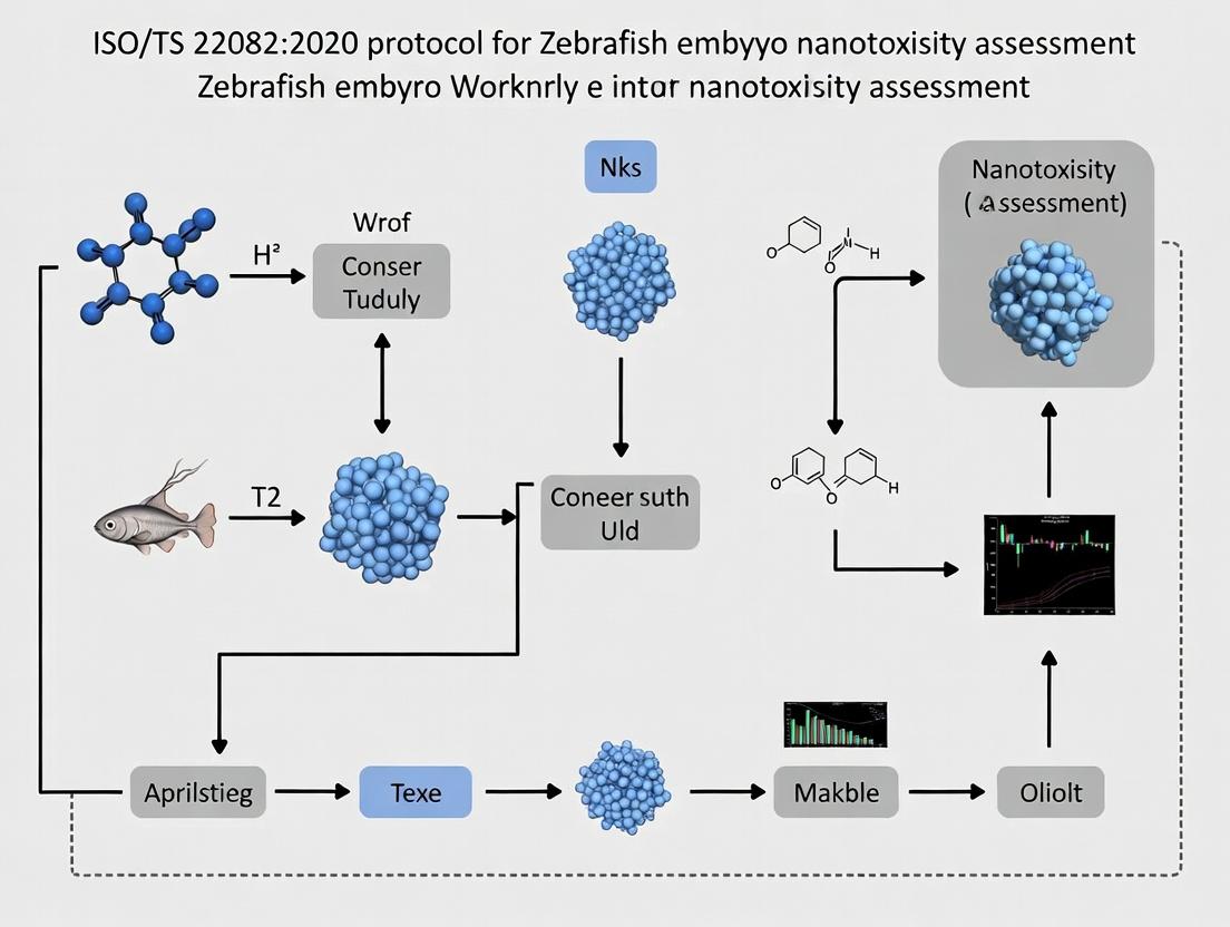
Title: ISO/TS 22082 Zebrafish Embryo Nanotoxicity Test Workflow
Title: Key Signaling Pathways in Nanomaterial-Induced Zebrafish Embryo Toxicity
The Scientist's Toolkit
Table 3: Essential Research Reagent Solutions for ISO/TS 22082 Protocol
| Item | Function / Purpose | Key Specifications / Notes |
|---|---|---|
| Wild-type Zebrafish (Danio rerio) | Source of embryos for testing. | Use healthy, well-maintained stocks (e.g., AB or TU strains). Spawning conditions must be standardized. |
| Embryo Medium (E3 Medium) | Standard medium for embryo rearing and exposure. | Contains: 5 mM NaCl, 0.17 mM KCl, 0.33 mM CaCl₂, 0.33 mM MgSO₄, pH ~7.2. |
| Pronase Solution | Enzyme for chorion digestion. | Used at ~1 mg/mL in embryo medium for batch dechorionation. Must be rinsed thoroughly post-treatment. |
| Positive Control | Validates test system sensitivity. | 3,4-Dichloroaniline (3,4-DCA) at 4 mg/L is recommended; must elicit consistent sublethal/lethal effects. |
| 24-well Cell Culture Plate | Vessel for embryo exposure. | Flat-bottom, sterile. One embryo per well in 2 mL solution to prevent cross-contamination. |
| Nanomaterial Stock Dispersion | The test substance. | Must be characterized (size, PDI, zeta potential) in embryo medium prior to testing. Sonication may be required. |
| Fine Forceps | Tool for manual dechorionation. | #5 or #55 Dumont forceps for precise manipulation of embryos under a stereomicroscope. |
| Stereomicroscope | For embryo selection, dechorionation, and observation. | Requires 10x-40x magnification with good depth of field for assessing malformations. |
The zebrafish (Danio rerio) embryo is a premier in vivo model for nanomaterial (NM) interaction studies, offering unique biological advantages aligned with the principles of ISO/TS 22082:2020, which provides a standardized framework for nanotoxicity testing using dechorionated embryos.
Key Biological Advantages:
- High Genetic & Physiological Homology: ~70% of human genes have a zebrafish orthologue, and conserved organ systems (cardiovascular, nervous, hepatic) allow for relevant human toxicity extrapolation.
- Optical Transparency: Embryos are externally fertilized and develop transparently, enabling real-time, high-resolution visualization of NM biodistribution, accumulation, and sub-lethal effects in vivo without invasive procedures.
- Rapid Ex Utero Development: Complete organogenesis occurs within 96 hours post-fertilization (hpf), facilitating high-throughput screening.
- High Fecundity: A single pair can produce hundreds of embryos weekly, providing robust statistical power and cost-effectiveness.
- Amenability to Genetic Manipulation: Ease of creating transgenic lines (e.g., with fluorescently tagged cell types) for mechanistic studies.
- Low Compound Requirement: Assays typically require microliter volumes of NM dispersion, crucial for evaluating precious novel materials.
- Regulatory Acceptance: The zebrafish embryo test (ZFET) is recognized in OECD Test Guideline 236 and is the basis for ISO/TS 22082:2020 for NM testing.
Application Notes: Key Parameters for Nanomaterial Studies
When employing the zebrafish embryo model under the ISO/TS 22082:2020 framework, critical parameters must be controlled to ensure reproducible and interpretable data on NM interactions.
Table 1: Critical Experimental Parameters for Zebrafish Embryo Nanomaterial Studies
| Parameter | ISO/TS 22082:2020 Considerations | Rationale & Impact |
|---|---|---|
| Chorion Status | Mandatory dechorionation (3-4 hpf) for NM exposure. | The chorion is a significant barrier, limiting NM bioavailability and leading to underestimation of toxicity. |
| Exposure Window | Initiate exposure by 4-6 hpf (post-dechorionation). | Ensures exposure during critical early developmental stages for consistent results. |
| Exposure Medium | Use standardized, defined media (e.g., E3 or ISO water). | Media composition (ions, pH, organic matter) drastically influences NM agglomeration/aggregation state and stability. |
| Nanomaterial Dispersion | Requires careful preparation (sonication, use of dispersants) and characterization (DLS, PDI, ζ-potential) immediately before exposure. | Aggregation state is the primary determinant of bioavailability, uptake, and toxicity. Must be reported. |
| Endpoint Analysis | Lethal (coagulation, lack of heartbeat) and sub-lethal (hatching rate, malformations, locomotion) at 24, 48, 72, 96 hpf. | Sub-lethal endpoints are often more sensitive and informative for NM interaction mechanisms. |
| Positive Control | Recommends use of a reference NM (e.g., 20 nm silver NPs) or chemical (e.g., 3,4-dichloroaniline). | Essential for intra- and inter-laboratory validation and protocol performance confirmation. |
Table 2: Quantitative Endpoint Sensitivity in Zebrafish Embryo Nanotoxicity Studies
| Endpoint | Typical Measurement Time (hpf) | Quantitative Readout | Relevance to Nanomaterial Interaction |
|---|---|---|---|
| Mortality/Coagulation | 24, 48, 72, 96 | LC50 (µg/mL or mg/L) | Acute toxicity, often related to NM mass dose or particle number. |
| Hatching Rate | 48, 60, 72, 96 | % Hatched Embryos | Indicator of developmental delay; chorion can trap NMs. |
| Malformation Score | 48, 72, 96 | % Affected; Severity Index | Specific teratogenicity (pericardial edema, yolk sac edema, spinal curvature). |
| Locomotor Activity | 96, 120 | Distance moved (pixels/unit time); Thigmotaxis | Neurodevelopmental toxicity, often a highly sensitive endpoint. |
| Cardiovascular Function | 48, 72, 96 | Heartbeat rate (bpm); Pericardial area (µm²) | Cardiotoxicity, often linked to oxidative stress or ion channel disruption. |
Detailed Protocols
Protocol 1: ISO/TS 22082:2020-Aligned Dechorionation and Nanomaterial Exposure
Aim: To prepare dechorionated zebrafish embryos for standardized nanomaterial toxicity assessment.
Materials: See "The Scientist's Toolkit" below. Procedure:
- Embryo Collection & Selection: Collect embryos from wild-type or transgenic zebrafish lines within 1 hour post-fertilization (hpf). At 3-4 hpf, under a stereo microscope, select fertilized embryos (cleavage visible) and place in a Petri dish with E3 medium.
- Dechorionation: a. Carefully remove E3 medium. b. Add 1-2 mL of pronase solution (1.5 mg/mL in E3) to the dish. c. Incubate at 28.5°C for 5-9 minutes, gently swirling occasionally. d. Monitor until chorions begin to rupture and detach. Immediately wash embryos 5-6 times with copious volumes of fresh E3 medium to completely remove chorions and residual pronase. e. Using a wide-bore pipette, transfer intact, dechorionated embryos to a new dish with fresh E3.
- Nanomaterial Exposure Setup (By 6 hpf): a. Prepare NM dispersions in E3 medium at 5-10X the desired final concentration. Sonicate (e.g., bath sonicator, 30-60 min) immediately prior to dilution. b. In a 24- or 96-well plate, add the appropriate volume of NM stock to E3 to achieve the final exposure volume (e.g., 2 mL/well for 24-well, 500 µL/well for 96-well). c. Randomly transfer 1 embryo per well (96-well) or 5-10 embryos per well (24-well) into the exposure solutions. Include a negative control (E3 only) and a positive control. d. Incubate the plate at 28.5°C on a 14h:10h light:dark cycle.
- Medium Refreshment (Optional for long exposures): At 24 hpf, carefully remove ~50% of the exposure medium without disturbing embryos and replace with freshly prepared NM solution of identical concentration to maintain dispersion stability.
Protocol 2: Assessment of Sub-Lethal Endpoints: Malformation and Locomotion
Aim: To quantify teratogenic and neurobehavioral effects of nanomaterial exposure.
Part A: Malformation Scoring (at 72 hpf)
- Fixation: Anesthetize embryos with tricaine (0.4 mg/mL). Transfer to 4% paraformaldehyde (PFA) in PBS and fix overnight at 4°C.
- Imaging: Wash 3x with PBS. Mount embryos laterally or dorsally in 3% methylcellulose on a depression slide.
- Scoring: Image using a bright-field stereomicroscope. Score each embryo for specific malformations:
- Pericardial Edema (PE): Present/Absent; Severity (1-mild, 2-severe).
- Yolk Sac Edema (YSE): Present/Absent.
- Spinal Curvature (SC): Present/Absent.
- Craniofacial Malformation (CF): Present/Absent.
- Analysis: Calculate the % of embryos with any malformation and the Malformation Severity Index (average score per embryo).
Part B: Locomotor Activity Assay (at 96 hpf)
- Preparation: Transfer live larvae (96 hpf) individually into the wells of a 96-well plate containing fresh E3 medium (300 µL).
- Acclimatization: Place the plate in a locomotor activity tracking system (e.g., ZebraBox, ViewPoint). Allow larvae to acclimate for 10-15 minutes in the dark.
- Testing Protocol: Program a standard light-dark cycle test (e.g., 10 min dark, 10 min light, 10 min dark). Record movement via video tracking.
- Data Analysis: Use software (e.g., ZebraLab) to quantify total distance moved (pixels/cm) during each light/dark phase. Hyper- or hypo-activity in dark phases is a sensitive indicator of neurotoxicity.
Visualizations
Diagram 1 Title: NM-Zebrafish Interaction Pathway from Exposure to Phenotype
Diagram 2 Title: ISO-Aligned Zebrafish Embryo Nanotoxicity Test Workflow
The Scientist's Toolkit
Table 3: Essential Research Reagent Solutions for Zebrafish Embryo Nanomaterial Studies
| Item | Function/Benefit | Key Consideration for NMs |
|---|---|---|
| E3 Embryo Medium (5 mM NaCl, 0.17 mM KCl, 0.33 mM CaCl₂, 0.33 mM MgSO₄) | Standardized, buffered medium for embryo rearing and exposure. | Low ionic strength can promote NM aggregation. Consistency is critical for comparability. |
| Pronase (from Streptomyces griseus) | Enzyme for rapid and gentle chemical dechorionation. | Must be thoroughly washed off to prevent proteolytic damage to embryo integument, which affects NM uptake. |
| Tricaine Methanesulfonate (MS-222) | Reversible anesthetic for embryo immobilization during imaging and sorting. | Does not interfere with NM stability at working concentrations (0.1-0.4 mg/mL). |
| Low-Melting Point Agarose or Methylcellulose | For immobilizing embryos/larvae during high-resolution imaging. | Must be prepared in NM-free medium to avoid unintended exposure during imaging. |
| Polyethylene Glycol (PEG) 400 or PVP Dispersants | Used to prepare stable, agglomerate-free NM stock dispersions in water. | Choice of dispersant can influence toxicity; must be controlled and reported. Use at minimal effective concentration. |
| Reference Nanomaterial (e.g., 20 nm PVP-coated Ag NPs) | Positive control material for protocol validation as per ISO/TS 22082:2020. | Provides a benchmark for inter-laboratory comparison and assay performance. |
| PTFE or UHMWPE Micropipette Tips & Vials | Low-adhesion labware for handling NM dispersions. | Minimizes loss of NMs due to adsorption to container walls, improving dosing accuracy. |
The ISO/TS 22082:2020 technical specification provides a standardized method for the use of zebrafish (Danio rerio) embryos in toxicity testing. Within this framework, the protocol for nanotoxicity assessment introduces a critical preparatory step: the mechanical or enzymatic removal of the chorion, the acellular protective membrane surrounding the embryo. This application note details the scientific rationale for dechorionation, establishing it as a key principle for generating reliable, reproducible, and biologically relevant data on engineered nanomaterial (ENM) toxicity.
Core Rationale: Overcoming Artifacts and Enhancing Bioavailability
The primary justification for dechorionation stems from the chorion's role as a significant barrier that can confound nanotoxicity assessments. Its pore size (approximately 0.5-0.7 μm) acts as a sieve, physically excluding larger aggregates and agglomerates of nanomaterials, while allowing only primary nanoparticles or very small clusters to penetrate. This creates an artificial size-selection process, preventing the testing of the true particle size distribution present in a suspension. Dechorionation eliminates this barrier, allowing direct and uniform exposure of the embryo to the ENM suspension as intended, thereby increasing the bioavailability of the test material and providing a more accurate representation of potential toxicity.
Table 1: Comparative Impact of Chorion Presence on Nanotoxicity Testing Parameters
| Parameter | Chorion-Intact Embryo | Dechorionated Embryo | Rationale for Dechorionation |
|---|---|---|---|
| Effective Exposure | Filtered, size-limited | Direct, full spectrum | Prevents false negatives from large/aggregated ENMs. |
| Dosimetry Accuracy | Low; chorion adsorption reduces delivered dose. | High; direct embryo contact. | Enables accurate correlation between nominal concentration and biological effect. |
| Uptake Kinetics | Delayed and attenuated. | Immediate and direct via skin, gills, GI tract. | Models realistic environmental or therapeutic exposure routes. |
| Oxidative Stress Onset | Slower, due to barrier effect. | Faster and more measurable. | Allows detection of a key nanotoxicity mechanism. |
| Hatching Rate | Endpoint can be affected by ENM-chorion interaction. | Not applicable; endpoint removed. | Eliminates confounding variable; focus on pure toxicity. |
| Protocol Reproducibility | Variable due to chorion batch/age differences. | High, standardizing the exposure interface. | Aligns with ISO/TS 22082:2020 goal of inter-laboratory reproducibility. |
Detailed Protocols for Dechorionation and Subsequent Exposure
Protocol 3.1: Mechanical Dechorionation (Pronase-Pretreatment Method)
This method is preferred under ISO/TS 22082:2020 as it minimizes mechanical stress on the embryo.
Materials:
- Healthy zebrafish embryos (4-6 hours post-fertilization, hpf).
- Pronase E solution (≥3 U/mL in embryo medium).
- Standard embryo medium (E3 or equivalent).
- Fine plastic transfer pipettes.
- Stericup filter units (0.22 μm) for sterilizing ENM suspensions.
- Watchmaker's forceps (Dumont #55).
- Stereo microscope.
Procedure:
- Pronase Treatment: Transfer approximately 50 embryos into a 60 mm Petri dish containing 5 mL of Pronase E solution. Incubate at 28.5°C for 8-10 minutes.
- Chorion Weakening: Periodically observe under microscope. The chorion will become soft and expand.
- Rinsing & Removal: Gently pour off Pronase solution and wash embryos 3x with 10 mL of fresh embryo medium. Using forceps or a wide-bore pipette, gently swirl or agitate the embryos. The weakened chorions will rupture and can be carefully separated from the embryos.
- Selection: Using a fire-polished glass or plastic pipette, transfer only successfully dechorionated, undamaged embryos to a new dish with fresh medium. Incubate until exposure.
Protocol 3.2: Direct Nanomaterial Exposure on Dechorionated Embryos
Following dechorionation at 4-6 hpf, expose embryos from 6-8 hpf onwards.
Procedure:
- ENM Dispersion: Prepare nanoparticle stock suspension in ultrapure water or appropriate vehicle. Sonicate (e.g., bath sonicator, 30 min) immediately prior to use. For in situ characterization, assess hydrodynamic size and zeta potential.
- Exposure Setup: At 6-8 hpf, array healthy dechorionated embryos into 24-well plates (1 embryo/mL/well). Prepare test concentrations by diluting the sonicated stock into embryo medium. Include a vehicle control (0.1% v/v max).
- Exposure: Carefully remove standard medium from each well and replace with 1 mL of the respective ENM test solution. Incubate plates at 28.5°C in the dark.
- Endpoint Assessment (24-96 hpf): Monitor lethal (coagulation, lack of somite formation, no heartbeat) and sublethal (malformations, reduced motility, pericardial edema, yolk sac absorption delay) endpoints according to ISO/TS 22082:2020. Perform imaging and molecular analyses as required.
Visualizing Key Pathways and Workflows
Title: Chorion Barrier Effect on Nanoparticle Exposure
Title: Dechorionation & Nanotoxicity Testing Workflow
Title: Key Nanotoxicity Signaling Pathways in Embryos
The Scientist's Toolkit: Essential Research Reagents & Materials
Table 2: Key Reagent Solutions for Dechorionation & Nanotoxicity Testing
| Item | Function/Benefit | Specification/Note |
|---|---|---|
| Pronase E (from S. griseus) | Enzymatically weakens the chorionic glycoprotein matrix for gentle mechanical removal. | Use ≥3 U/mL in embryo medium. Aliquot and store at -20°C. |
| Standard Embryo Medium (E3) | Isotonic, buffered solution for embryo maintenance and as a vehicle for test solutions. | 5 mM NaCl, 0.17 mM KCl, 0.33 mM CaCl₂, 0.33 mM MgSO₄, pH 7.2-7.4. |
| Dumont #55 Forceps | Fine-tipped tools for manual handling and dechorionation assistance. | Essential for precision work under a microscope. |
| Fire-Polished Glass Pipettes | For gentle transfer of dechorionated embryos without causing damage. | Smooth bore prevents physical shear stress. |
| Stericup Filter Units (0.22 µm) | For sterilizing nanoparticle stock solutions and biological reagents. | Use low-protein-binding PVDF membranes for accurate dosing. |
| Bath Sonicator | Essential for de-agglomerating nanoparticles in suspension immediately prior to exposure. | Standardizes the initial particle size distribution. |
| Methylene Blue | Antifungal agent for long-term embryo culture (>24h). | Use at very low concentration (0.0001%) to avoid interference. |
| Tricaine (MS-222) | Anesthetic for immobilizing embryos during imaging or precise staging. | Standard stock: 400 mg/mL, pH 7.0. Use at 160 mg/L in medium. |
| PBS (Ca²⁺/Mg²⁺-free) | For rinsing and preparation steps prior to molecular fixation or analysis. | Prevents premature hardening or precipitation during processing. |
Application Notes: Definitions and Context within ISO/TS 22082:2020
Within the ISO technical specification (TS) 22082:2020, which details a nanotoxicity test method using dechorionated zebrafish embryos, precise terminology is paramount. This framework aligns with broader ISO and OECD definitions for nanomaterials and nanoparticles, providing a standardized context for assessing biological endpoints.
Nanomaterial (ISO/TS 80004-1:2015): A material with any external dimension in the nanoscale (approximately 1–100 nm) or having internal structure or surface structure in the nanoscale. In the context of ISO/TS 22082, this is the parent substance being tested (e.g., a metal oxide powder, a polymer matrix).
Nanoparticle (ISO/TS 80004-2:2015): A nano-object with all three external dimensions in the nanoscale. For testing, the nanomaterial is often dispersed to create a suspension of nanoparticles (the exposure agent). Key characteristics include size, size distribution, shape, surface charge (zeta potential), and agglomeration/aggregation state in the exposure medium.
Endpoint (ISO/TS 22082:2020): A measurable biological parameter assessed to determine toxic effects. This protocol standardizes core endpoints for embryo viability, development, and morphology.
Table 1: Core Definitions and Quantitative Criteria in the ISO Framework
| Term | ISO Standard Source | Key Quantitative Dimension | Relevance to ISO/TS 22082:2020 |
|---|---|---|---|
| Nanoscale | ISO/TS 80004-1 | ~1 nm to 100 nm | Defines the size range of the material/particles of interest. |
| Nanomaterial | ISO/TS 80004-1 | Size, specific surface area | The test substance as manufactured. Requires characterization prior to dispersion. |
| Nanoparticle | ISO/TS 80004-2 | All three dimensions 1-100 nm | The primary unit in the exposure medium. Dispersion protocol critical. |
| Agglomeration | ISO/TS 80004-4 | Cluster strength (weak) | Reversible clustering affecting exposure dynamics in embryo medium. |
| Aggregation | ISO/TS 80004-4 | Cluster strength (strong) | Irreversible fusion affecting particle size and bioavailability. |
| Endpoint | ISO/TS 22082 | Lethal (LC50) & Sublethal (EC50) | Standardized measures of toxicity (e.g., coagulation, lack of somites, tail detachment). |
Experimental Protocol: Standard Dispersion and Exposure per ISO/TS 22082
This protocol details the preparation of nanoparticle suspensions and exposure of dechorionated zebrafish embryos for nanotoxicity assessment.
Materials & Reagents:
- Test nanomaterial (dry powder).
- Embryo medium (E3 or equivalent, 5 mM NaCl, 0.17 mM KCl, 0.33 mM CaCl₂, 0.33 mM MgSO₄).
- Stock dispersion solvent (e.g., ultrapure water with 0.05-0.1% w/v biocompatible stabilizer like bovine serum albumin (BSA) or sodium dodecyl sulfate (SDS) if justified).
- Dechorionated zebrafish embryos (wild-type AB strain, 4-6 hours post-fertilization (hpf)).
- 24-well or 96-well tissue culture plates.
- Sonication bath (bath sonicator) and/or probe sonicator.
Procedure:
Part A: Nanoparticle Stock Dispersion Preparation
- Weigh the appropriate amount of nanomaterial to achieve a high-concentration stock (e.g., 1000 mg/L).
- Add the nanomaterial to the stock dispersion solvent in a sterile vial.
- Pre-dispersion: Vortex the mixture vigorously for 1-2 minutes.
- Dispersion: Sonicate the suspension using a calibrated bath sonicator for 30 minutes at room temperature. For refractory materials, controlled probe sonication may be used (e.g., 1 min pulse on/off at 50 W). Note: Energy input must be reported.
- The stock suspension is used immediately to prepare serial dilutions in embryo medium.
Part B: Embryo Exposure and Endpoint Assessment
- Dechorionation: At 4-6 hpf, manually or enzymatically remove the chorion from viable embryos. Rinse twice in embryo medium.
- Plateing: Transfer 1 embryo per well into a multiwell plate containing 1-2 mL (24-well) or 100-200 µL (96-well) of exposure solution (nanoparticle dilution or control).
- Exposure Conditions: Incubate plates at 28 ± 1°C under a 14:10 hour light:dark cycle for the test duration (typically 96 hpf).
- Endpoint Recording: Assess embryos at 24, 48, 72, and 96 hpf using a stereomicroscope.
- Lethal Endpoints: Coagulation, lack of somite formation, non-detachment of the tail bud.
- Sublethal Endpoints: Malformations (pericardial edema, yolk sac edema, spinal curvature), reduced heartbeat, hatching success, motility.
Table 2: Key Endpoints and Assessment Criteria in Zebrafish Embryo Test
| Endpoint Category | Specific Measurement | Time of Assessment (hpf) | Typical Quantitative Output |
|---|---|---|---|
| Lethality | Embryo coagulation | 24, 48, 72, 96 | LC50 (concentration lethal to 50%) |
| Teratogenicity | Presence of malformations | 48, 72, 96 | EC50 (concentration causing effect in 50%) |
| Developmental Delay | Somite formation, tail detachment | 24 | Incidence rate (%) |
| Hatching | Hatching rate | 48, 60, 72, 96 | Hatching rate (%) at each timepoint |
| Cardiotoxicity | Heartbeat rate | 48, 72 | Beats per minute (bpm) |
Visualization: Pathways and Workflows
Nanotoxicity Pathway from Exposure to Endpoint
ZFET Experimental Workflow
The Scientist's Toolkit: Research Reagent Solutions
Table 3: Essential Materials for ISO/TS 22082-Compliant Nanotoxicity Testing
| Item / Reagent Solution | Function / Purpose in Protocol | Critical Notes |
|---|---|---|
| Standardized Zebrafish Embryo Medium (E3) | Provides isotonic, buffered environment for embryo development and nanoparticle exposure. | Must be particle-free; pH and conductivity should be consistent to control nanoparticle stability. |
| Biocompatible Dispersion Aid (e.g., BSA, Humic Acid) | Aids in creating stable, monodisperse nanoparticle suspensions in aqueous medium; can mimic environmental or biological matrices. | Concentration must be minimal and justified; it may influence bioavailability and toxicity. |
| Protease (e.g., Pronase) | For enzymatic dechorionation of embryos, ensuring uniform chemical exposure and high-throughput processing. | Must be thoroughly rinsed to avoid enzymatic interference with the test. |
| Morphological Staining Dyes (e.g., Alcian Blue, Alizarin Red) | Used for detailed assessment of skeletal or cartilage malformations (sublethal endpoint). | Applied post-fixation; not part of the core ISO protocol but a common extension. |
| Reactive Oxygen Species (ROS) Probe (e.g., DCFH-DA) | Fluorescent dye to quantify oxidative stress, a key molecular initiating event in nanotoxicity. | Used in supplementary mechanistic studies to link endpoints to pathways. |
| Nanoparticle Tracking Analysis (NTA) / DLS System | For characterizing hydrodynamic size distribution and concentration of nanoparticles in the exposure medium. | Critical pre-exposure step to confirm dispersion quality and dose metric. |
| High-Sensitivity Microbalance | For accurate weighing of small quantities of nanomaterial for stock preparation. | Requires calibration and an anti-static system due to nanomaterial electrostatic properties. |
Ethical and 3R (Replacement, Reduction, Refinement) Benefits Over Traditional Models.
This application note details the implementation of a dechorionated zebrafish embryo model for nanotoxicity assessment, aligned with ISO/TS 22082:2020. It explicitly outlines the ethical and 3R advantages of this approach over traditional mammalian and larval fish models, providing researchers with validated protocols for high-throughput, predictive toxicology.
Quantitative Comparison of Model Systems
The following tables summarize the key ethical, practical, and data-quality benefits of the dechorionated zebrafish embryo model.
Table 1: Ethical & 3R Benefits Analysis
| Aspect | Traditional Rodent Models | Traditional Larval Fish Models | Dechorionated Zebrafish Embryo (ISO/TS 22082) |
|---|---|---|---|
| Regulatory Status | Considered protected sentient beings; full animal use protocols required. | Considered protected at free-feeding stage (post-120 hpf in zebrafish). | Not considered protected procedures until independent feeding stage (before 120 hpf). |
| Replacement | Not applicable (in vivo mammal). | Partial replacement for adult fish tests. | Direct replacement for acute fish toxicity tests (OECD TG 203) and some mammalian screens. |
| Reduction | Low-throughput; high animal numbers per data point. | Moderate throughput. | High-throughput; one embryo yields multi-organ endpoint data; drastic reduction in animal use. |
| Refinement | Invasive procedures (e.g., gavage, injection) cause distress. | Potential distress from exposure in a contained environment. | Elimination of distress; chorion removal standardizes exposure; endpoints are largely non-invasive. |
| Sample Size | Typically n=5-10 per group. | Typically n=10-20 per group. | Typically n=24-32 per group; statistically robust with fewer total organisms. |
Table 2: Experimental Efficiency & Data Output
| Parameter | Traditional Mammalian Acute Tox | Dechorionated Zebrafish Embryo Assay |
|---|---|---|
| Test Duration | 14-28 days | 96-120 hours post-fertilization (hpf) |
| Compound Required | Milligrams to grams | Micrograms to milligrams |
| Cost per Compound | ~$15,000 - $30,000 | ~$1,000 - $3,000 |
| Endpoints Available | Mortality, histopathology, clinical chemistry | Mortality, malformation, motility, cardiotoxicity, neurotoxicity, hepatotoxicity, genotoxicity (via probes) |
| Mechanistic Insight | Terminal, requires many animals for tissues. | Real-time, in vivo, multi-parameter imaging on live organism. |
Core Protocol: Dechorionation and Nanomaterial Exposure (ISO/TS 22082:2020 Framework)
Protocol 2.1: Manual Dechorionation of Zebrafish Embryos
Purpose: To remove the chorionic barrier for direct and standardized nanomaterial exposure. Reagents/Materials: Wild-type (e.g., AB strain) zebrafish embryos (3-4 hpf), Pronase solution (1.5 mg/mL in embryo medium), sterile embryo medium (E3), plastic petri dishes (90 mm), fine forceps (Dumont #5), stereomicroscope. Procedure:
- Collect embryos and incubate at 28.5°C until ~4 hpf.
- Enzymatic Weakening: Transfer up to 100 embryos to a petri dish with 20 mL Pronase solution. Incubate for 8-10 minutes at 28.5°C until chorions appear slightly softened.
- Rinsing: Carefully decant Pronase solution. Gently rinse embryos 3x with 30 mL sterile embryo medium.
- Mechanical Removal: Under stereomicroscope, use fine forceps to gently tear the chorion. Apply minimal pressure to avoid embryo damage. Alternatively, gently roll embryos with forceps to expel them from the chorion.
- Post-Dechorionation Care: Transfer dechorionated embryos to a fresh dish with sterile embryo medium. Incubate at 28.5°C and inspect for normal development (shield stage at 6 hpf). Use only normally developing embryos for exposure within 2 hours.
Protocol 2.2: Nanomaterial Dispersion & Exposure
Purpose: To prepare stable nanomaterial dispersions and expose dechorionated embryos for toxicity assessment. Reagents/Materials: Test nanomaterial, embryo medium (E3) possibly with 0.1% Pluronic F-68 (stabilizer), sonicator (bath or probe), 24-well cell culture plates, dechorionated embryos (6 hpf). Procedure:
- Dispersion: Weigh nanomaterial. Prepare a concentrated stock dispersion (e.g., 1 mg/mL) in embryo medium (± stabilizer). Sonicate using a bath sonicator for 15 min or a probe sonicator (on ice, 30% amplitude, 2 min pulsed) immediately before dilution.
- Exposure Setup: In a 24-well plate, add 2 mL of each test concentration (e.g., 0, 1, 10, 50, 100 mg/L) per well. Include a negative (medium only) and a positive control (e.g., 4 mg/L 3,4-dichloroaniline).
- Embryo Transfer: Transfer one dechorionated embryo per well (n=24 per concentration). Incubate plate at 28.5°C in the dark.
- Monitoring & Renewal: For tests >24h, renew exposure solutions daily by carefully transferring embryos to a temporary plate, replacing solutions, and returning embryos.
Key Endpoint Assessment Protocols
Protocol 3.1: Sublethal Morphological Scoring (at 24, 48, 72, 96 hpf)
Purpose: To quantify teratogenic effects using a standardized scoring system. Procedure: Image each embryo under a brightfield stereomicroscope. Score the presence/severity of malformations: pericardial edema (0-3), yolk sac edema (0-2), tail malformation (0-2), spinal curvature (0-2), pigmentation defects (0-1). Calculate a Teratogenic Index (TI) = LC50 / EC50(malformation). A TI > 1 indicates teratogenic hazard.
Protocol 3.2: Behavioral Endpoint - Locomotor Activity (at 96 hpf)
Purpose: To assess neurodevelopmental toxicity via larval motility. Materials: 96-well plate, zebrafish larvae, tracking system (e.g., DanioVision, ViewPoint). Procedure:
- At 96 hpf, transfer one larva per well of a 96-well plate in fresh medium.
- Acclimate for 15 min in the tracking system.
- Record activity under alternating 10 min light / 10 min dark cycles for 60 min.
- Analyze total distance moved, particularly during dark phases (larval photomotor response). A ≥20% decrease vs. controls is considered significant.
Visualizing Pathways and Workflows
Title: Dechorionated Zebrafish Embryo Nanotoxicity Testing Workflow
Title: Key Nanotoxicity Pathways in Zebrafish Embryos
The Scientist's Toolkit: Research Reagent Solutions
| Item | Function & 3R Benefit |
|---|---|
| Wild-type Zebrafish (AB/TL strain) | Robust, genetically stable embryos. High fecundity enables massive reduction vs. mammalian models. |
| Pronase (from Streptomyces griseus) | Enzyme for chorion softening. Enables gentle, high-yield dechorionation for standardized exposure (Refinement). |
| Embryo Medium (E3) | Simple salt solution for embryo maintenance. Avoids use of complex animal sera. |
| Pluronic F-68 Non-ionic Surfactant | Stabilizes nanomaterial dispersions in aqueous media. Reduces aggregation for consistent, reproducible dosing (Refinement of exposure). |
| Morpholino Oligonucleotides | Enable gene knockdown without permanent genetic modification. Replaces some mammalian knockout models for mechanistic studies. |
| Fluorescent Molecular Probes (e.g., Acridine Orange, DCFH-DA) | In vivo staining for apoptosis and ROS. Provide rich mechanistic data from a single live embryo, reducing need for separate cohorts for histology. |
| 96-well Microplate with Lid | Format for high-throughput behavioral and viability screening. Enables reduction via miniaturization and parallel processing. |
| Automated Imaging & Tracking System | Enables objective, high-content phenotyping. Maximizes data per embryo (Reduction) and minimizes handling stress (Refinement). |
A Practical Walkthrough of the ISO/TS 22082 Protocol: From Embryo Preparation to Data Collection
This document provides detailed application notes and protocols for sourcing, preparing, and controlling the quality of materials and reagents essential for nanotoxicity research using dechorionated zebrafish embryos under the framework of ISO/TS 22082:2020. The standard mandates rigorous control of all experimental inputs to ensure the reliability, reproducibility, and validity of test results for hazard assessment of nanomaterials (NMs). This protocol is a critical component of a broader thesis establishing a standardized, internationally recognized testing methodology.
Sourcing Specifications for Key Reagents
All materials must be procured from qualified suppliers with appropriate Certificates of Analysis (CoA). Preference is given to reagents with purity grades specified for molecular biology or trace metal analysis.
Table 1: Primary Reagent Sourcing Specifications
| Reagent/Material | Specification / Grade | Key Quality Attribute | Recommended Supplier Type |
|---|---|---|---|
| Zebrafish Embryos (AB/TL strain) | Specific Pathogen Free (SPF) | <2% background deformity at 24 hpf | Accredited aquatic facility |
| Nanomaterial Test Substance | Characterization dossier per ISO/TS 22082 | Purity, size distribution (TEM), ζ-potential | Research or industrial NM producer |
| E3 Embryo Medium (without Methylene Blue) | Prepared in-house from component salts | Osmolarity: 290-310 mOsm/kg, pH 7.2-7.6 | USP/ACS grade salts |
| Pronase (for dechorionation) | Protease from Streptomyces griseus, ≥3,500 U/mg | Lyophilized powder, DNase/RNase-free | Molecular biology grade |
| Low-Melting Point Agarose | Gelling temp ~36°C | No additives (e.g., dyes, surfactants) | Electrophoresis grade |
| Polystyrene Microplates (for exposure) | 96-well, flat-bottom, tissue culture treated | Non-pyrogenic, sterile, embryo-tested | Specialized labware supplier |
| Paraquat (Positive Control) | Dichloride salt, ≥98% purity | Weighed and aliquoted under inert atmosphere | Analytical standard supplier |
| MS-222 (Tricaine) | Pharmaceutical standard (Finquel) | Buffered to pH 7.0 with Tris | Veterinary pharmaceutical grade |
Preparation Protocols
Preparation of E3 Embryo Medium (ISO/TS 22082 Compliant)
Materials: NaCl (ACS grade), KCl (ACS grade), CaCl₂·2H₂O (ACS grade), MgSO₄·7H₂O (ACS grade), Ultrapure Water (Type I, 18.2 MΩ·cm). Protocol:
- In a cleaned Class A volumetric flask, add 800 mL of Type I water.
- Weigh and dissolve: 0.294 g NaCl, 0.013 g KCl, 0.044 g CaCl₂·2H₂O, and 0.081 g MgSO₄·7H₂O.
- Stir until fully dissolved. Q.S. to 1 L with Type I water.
- Filter sterilize using a 0.22 µm PES membrane filter into an autoclaved bottle.
- Verify osmolarity (290-310 mOsm/kg) and pH (7.2-7.6). Store at 4°C for up to 2 weeks.
Preparation of Nanomaterial Stock Dispersions
Materials: Nanomaterial dry powder, Ultrapure Water (Type I), 0.05% (w/v) Bovine Serum Albumin (BSA, fatty acid-free) in E3 medium as a dispersant (if justified). Protocol:
- Pre-wet all contact surfaces (vials, pipette tips) with the selected dispersion vehicle to minimize adhesion.
- Weigh NM using a microbalance (sensitivity ± 1 µg) in a controlled environment (e.g., fume hood for powders).
- Add vehicle to achieve a high-concentration stock (e.g., 1000 µg/mL). Do not vortex if prone to aggregation.
- Sonicate the dispersion using a probe sonicator with a titanium microtip under controlled conditions (e.g., 20% amplitude, 30 sec pulse, 30 sec rest, 2 min total on ice).
- Characterize the hydrodynamic size and ζ-potential of the stock dispersion immediately using dynamic light scattering (DLS). Record data.
Quality Control Checklist
A daily log must be maintained for each experimental run.
Table 2: Pre-Experimental Quality Control Checklist
| Item | Acceptance Criterion | Check (✓/✗) | Corrective Action |
|---|---|---|---|
| Embryo Medium Osmolarity | 290 - 310 mOsm/kg | Prepare fresh batch | |
| Embryo Medium pH | 7.2 - 7.6 | Adjust with dilute HCl/NaOH | |
| Incubator Temperature | 28.0 ± 0.5 °C | Calibrate sensor | |
| NM Stock Dispersion Size (by DLS) | PDI < 0.4 (for monodisperse) | Re-sonicate or prepare fresh | |
| Pronase Activity | Complete chorion digestion in < 5 min | Prepare new aliquot | |
| Positive Control (Paraquat) | LC₅₀ within historical control limits (e.g., 15-35 µM) | Re-constitute from new stock | |
| Negative Control (E3 medium) | ≥ 90% embryo viability at 24 hpf | Discard affected clutch | |
| Agarose Gelling | Firm gel at 28°C in < 5 min | Adjust concentration | |
| Microplate Sterility | No microbial contamination after 24h incubation | Use new, sterile plate |
Experimental Protocol: Dechorionation and Exposure Setup
Title: Protocol for 96-Well Static Nanomaterial Exposure of Dechorionated Zebrafish Embryos
Materials:
- 24-48 hours post-fertilization (hpf) zebrafish embryos.
- Pronase solution (3 mg/mL in E3 medium, freshly prepared).
- E3 medium (as prepared in 2.1).
- Low-melting point agarose (1.2% in E3, kept at 40°C).
- Test solutions: NM dispersions, negative control (E3), positive control (e.g., 25 µM Paraquat).
- 96-well plates, stereomicroscope, precision pipettes.
Protocol:
- Bulk Dechorionation: Transfer ~50 embryos to a clean Petri dish. Remove all E3. Add 5 mL of Pronase solution. Swirl gently for 2-4 minutes until chorions visibly degrade.
- Washing: Carefully remove Pronase. Rinse embryos 5 times with 10 mL of fresh E3 medium to halt enzymatic activity.
- Selection: Under a stereomicroscope, select embryos at the same developmental stage (e.g., 24 hpf) with no obvious malformations.
- Embedding (for immobility): Using a cut pipette tip, transfer one embryo per well into a 96-well plate. Remove excess E3. Add 100 µL of warm (40°C) low-melting point agarose to immobilize the embryo. Allow to solidify for 5 minutes.
- Exposure: Gently overlay 100 µL of the appropriate test solution (NM dispersion, control) onto the agarose in each well. Ensure no air bubbles trap the embryo.
- Incubation & Assessment: Seal plate with gas-permeable membrane. Incubate at 28 ± 0.5°C. Assess endpoints (e.g., mortality, malformation, heartbeat) at 24h and 48h of exposure.
Visualizations
Diagram Title: Zebrafish Embryo Nanotoxicity Testing Workflow
Diagram Title: Quality Control Hierarchy for Nanotoxicity Assay
The Scientist's Toolkit
Table 3: Essential Research Reagent Solutions for Dechorionated Zebrafish Embryo Assay
| Item | Function/Justification | Critical Parameters |
|---|---|---|
| E3 Medium (w/o Methylene Blue) | Isotonic medium for embryo rearing and exposure. Methylene Blue is omitted to prevent antioxidant interference with NM toxicity. | Osmolarity, pH, sterile filtration. |
| Pronase Solution | Enzyme for efficient, gentle removal of the chorion, eliminating a potential diffusion barrier for NMs. | Activity concentration (3 mg/mL), fresh preparation to avoid loss of activity. |
| Low-Melting Point Agarose | Immobilizes embryo for consistent imaging and prevents ingestion of NMs, isolating trophic from waterborne exposure. | Gelling temperature (~36°C), purity (no additives). |
| BSA (Fatty Acid-Free) Stock | A justifiable dispersant agent to improve NM stability in aqueous media without undue toxicity. | Concentration (e.g., 0.05%), fatty acid-free to avoid metabolic confounding. |
| Nanomaterial Characterization Buffer | A standardized aqueous matrix (e.g., 5 mM NaCl) for DLS/ζ-potential measurements of NM stock. | Low ionic strength to prevent aggregation during measurement. |
| MS-222 (Tricaine) Stock | Anesthetic for humane euthanasia of embryos at endpoint or for prolonged imaging. | Buffered to pH 7.0 to avoid acid stress. |
| Paraquat Positive Control | Reference toxicant generating reactive oxygen species (ROS), a common NM toxicity pathway, to validate assay sensitivity. | Purity, accurate molar concentration. |
This protocol details the initial, critical steps for generating high-quality, synchronous zebrafish embryos intended for nanotoxicity testing under ISO/TS 22082:2020. Standardized breeding, spawning, and embryo collection are foundational for ensuring reproducibility in subsequent dechorionation, chemical exposure, and teratogenicity assessment.
Application Notes
- Standardization for Nanotoxicity Research: Consistent embryo quality is paramount for ISO/TS 22082:2020 compliance. Variability in parental health, water quality, and spawning conditions directly impacts embryo resilience and confounds nanomaterial toxicity endpoints.
- Temporal Precision: The 0-4 hours post-fertilization (hpf) window is selected to collect embryos prior to significant organogenesis, allowing for the observation of nanomaterial-induced effects throughout development. Collection within this timeframe ensures developmental synchrony.
- Water Quality as a Variable: Ionic composition and pH of system water can influence nanomaterial aggregation and bioavailability. Characterizing and maintaining husbandry water parameters is thus a critical pre-conditioning variable.
Detailed Protocol
Zebrafish Husbandry for Breeding Cohorts
Objective: Maintain optimal health and fecundity in broodstock.
- Housing: Keep zebrafish in a recirculating aquaculture system (RAS) with the following parameters:
- Temperature: 28.5 ± 1.0°C
- pH: 7.0 - 7.5
- Conductivity: 500 - 1500 µS/cm
- Photoperiod: 14h light:10h dark cycle.
- Nutrition: Feed adults a varied diet at least twice daily. A typical regimen includes:
- Morning: High-quality dry diet (e.g., 42-55% protein).
- Evening: Live or frozen Artemia nauplii and/or rotifers.
- Health Monitoring: Routinely screen for common pathogens (e.g., Pseudoloma neurophilia). Quarantine new stock for a minimum of 4 weeks.
Spawning Setup and Embryo Production
Objective: Generate a synchronous batch of fertilized embryos.
- Setup (Day before): In the afternoon, place breeding groups (typically at a 1:2 or 2:2 male-to-female ratio) into dedicated spawning tanks with a removable divider. Ensure the tank bottom has a mesh screen to separate adults from eggs post-spawning.
- Spawning Trigger: Remove the divider at the onset of the light cycle ("lights on"). Spawning typically occurs within 30-60 minutes.
- Egg Collection: Within 1 hour of spawning, carefully remove adults. Rinse eggs from the spawning tank's mesh or bottom with system water into a clean mesh sieve.
Embryo Collection and Selection (0-4 hpf)
Objective: Collect and select viable, fertilized embryos for experimentation.
- Rinsing: Gently rinse the collected eggs in system water.
- Debris Removal: Using a sterile transfer pipette, remove damaged eggs, feces, and other debris.
- Fertilization Check & Selection: At approximately 1-4 hpf, examine embryos under a stereomicroscope. Select only embryos that are fertilized (showing cell cleavage) and are developmentally normal. Discard unfertilized (clear, single-cell) or irregularly cleaving embryos.
- Disinfection (Optional, per ISO/TS 22082): Immerse selected embryos in a fresh solution of 0.003% (w/v) phenylthiourea (PTU) in embryo medium to inhibit pigment formation, if required for endpoint analysis. Alternatively, for surface disinfection, a brief rinse in 0.1% (v/v) bleach solution in embryo medium may be used, followed by multiple rinses in clean embryo medium.
- Incubation: Transfer selected embryos to a Petri dish containing fresh, pre-warmed (28.5°C) embryo medium (e.g., E3 medium). Place dish in a 28.5°C incubator until the dechorionation step.
Table 1: Optimal Zebrafish Broodstock Husbandry Parameters
| Parameter | Target Value | Acceptable Range | Measurement Frequency |
|---|---|---|---|
| Temperature | 28.5°C | 27.5 - 29.5°C | Continuous (Daily Log) |
| pH | 7.2 | 7.0 - 7.5 | Daily |
| Conductivity | 750 µS/cm | 500 - 1500 µS/cm | Daily |
| Ammonia (NH₃/NH₄⁺) | 0 mg/L | < 0.25 mg/L | Weekly |
| Nitrite (NO₂⁻) | 0 mg/L | < 0.25 mg/L | Weekly |
| Nitrate (NO₃⁻) | < 50 mg/L | < 200 mg/L | Weekly |
| Photoperiod | 14L:10D | N/A | Controlled |
Table 2: Embryo Collection Metrics & Expected Yield
| Metric | Target/Expected Outcome | Criteria for Rejection |
|---|---|---|
| Spawning Success Rate | > 70% per tank | If < 50%, review broodstock health/age. |
| Fertilization Rate (at 1-2 hpf) | > 90% | Batches with < 80% are not used. |
| Embryo Viability (at 4 hpf) | > 95% normally cleaving | Coagulated, asymmetric, or uncleaved embryos discarded. |
| Collection Window | 0 - 4 hours post-fertilization | Embryos older than 4 hpf introduce asynchrony. |
The Scientist's Toolkit
Table 3: Key Research Reagent Solutions
| Item | Function in Protocol | Example/Composition Notes |
|---|---|---|
| E3 Embryo Medium | Standard medium for embryo incubation and rinsing. | 5 mM NaCl, 0.17 mM KCl, 0.33 mM CaCl₂, 0.33 mM MgSO₄, pH ~7.2. |
| Phenylthiourea (PTU) Stock | Prevents melanin pigment formation for clear visualization of internal structures. | 0.003% (w/v) in E3 medium. Prepare fresh weekly. |
| Sodium Hypochlorite (Bleach) Solution | Used for surface disinfection of embryos or equipment. | Dilute stock to 0.1% (v/v) in E3 medium for brief rinses. |
| System Water | Water used in the main housing (RAS). Must be characterized for nanotoxicity studies. | Conditioned, reverse-osmosis (RO) water with added salts; parameters in Table 1. |
| Instant Ocean / Marine Salts | For reconstituting RO water to desired conductivity for system or embryo water. | Standardized salt mix ensures consistent ionic composition. |
| Methylene Blue | Antifungal agent; sometimes added to embryo medium for long-term holding. | Typical concentration: 0.0001% (w/v). Not used if conducting oxidative stress assays. |
Visualized Workflows
Title: Zebrafish Embryo Production and Collection Workflow
Title: Protocol Step 1 Role in Overall Nanotoxicity Thesis
Within the framework of ISO/TS 22082:2020, which standardizes nanotoxicity testing using zebrafish embryos, dechorionation is a critical preparatory step. The chorion is a protective acellular envelope that can act as a barrier, adsorbing test materials and potentially confounding toxicity results by limiting nanoparticle-embryo interaction. This application note details enzymatic and manual dechorionation methods, providing protocols and comparative analysis to ensure consistency and embryo viability for nanotoxicity assays.
Comparison of Dechorionation Methods
The choice of method balances efficiency, throughput, and embryo integrity. The following table summarizes key quantitative data from recent studies.
Table 1: Comparative Analysis of Dechorionation Methods
| Parameter | Enzymatic (Pronase) | Manual (Forceps) | Manual (Rolling) |
|---|---|---|---|
| Time per Embryo | ~5-10 min (batch processing) | ~0.5-1 min (skilled) | ~1-2 min (skilled) |
| Efficacy Rate | >95% (complete chorion removal) | ~90-98% (highly operator-dependent) | ~85-95% (operator-dependent) |
| Embryo Viability (24hpf) | 90-95% (with optimized protocol) | 85-92% (risk of physical damage) | 88-94% (lower direct impact risk) |
| Throughput | High (suitable for 50-100+ embryos) | Low to Medium (limited by operator skill/speed) | Medium (requires practice) |
| Skill Requirement | Low (standardized incubation) | High (fine motor control essential) | Moderate (consistent technique needed) |
| ISO/TS 22082:2020 Suitability | Excellent (high standardization, batch consistency) | Good (requires stringent operator training records) | Good (requires protocol uniformity) |
Detailed Experimental Protocols
Protocol 1: Enzymatic Dechorionation Using Pronase
Research Reagent Solutions & Materials:
- Pronase, Type XIV (from Streptomyces griseus): A broad-spectrum protease mixture that digests the proteinaceous chorion.
- E3 Embryo Medium (standard): 5 mM NaCl, 0.17 mM KCl, 0.33 mM CaCl₂, 0.33 mM MgSO₄, pH 7.2-7.6. Used for embryo rearing and dilution.
- Sterile Petri Dishes (60mm & 100mm): For incubation and washing steps.
- Fine Transfer Pipettes: For gentle handling of embryos.
- Incubator at 28.5°C: For maintaining optimal embryonic development.
- Stereomicroscope: For monitoring dechorionation progress.
Methodology:
- Preparation: Prepare a 2 mg/mL Pronase solution in E3 medium. Filter-sterilize (0.22 µm) and pre-warm to 28.5°C.
- Embryo Collection: Transfer embryos (at 4-6 hours post-fertilization, hpf) into a 60mm Petri dish.
- Enzyme Incubation: Remove existing E3 medium and add 5-10 mL of the prepared Pronase solution. Swirl gently.
- Incubation: Place dish in a 28.5°C incubator. Monitor every 2-3 minutes under a stereomicroscope. The chorion will thin and begin to rupture.
- Chorion Removal: Once ~80% of chorions are ruptured (typically 5-10 minutes), gently swirl the dish and use a fine pipette to create a gentle flow, aiding in the detachment of embryos from chorions.
- Washing: Immediately and carefully remove the Pronase solution using a pipette. Rinse embryos thoroughly with 3 x 10 mL volumes of fresh, pre-warmed E3 medium to ensure complete enzyme removal.
- Transfer & Validation: Transfer dechorionated embryos to a new dish with fresh E3. Inspect to ensure no chorion fragments remain attached.
Protocol 2: Manual Dechorionation Using Fine Forceps
Research Reagent Solutions & Materials:
- Dumont #5 or #55 Fine Forceps: Sharp, precision forceps for mechanical tearing of the chorion.
- 1-2% Agarose-Coated Petri Dishes: A soft substrate to cradle and stabilize the embryo during manipulation, preventing rolling and damage.
- E3 Embryo Medium.
- Stereomicroscope with Good Magnification (8x-50x): Essential for precise visualization.
Methodology:
- Preparation: Coat the bottom of a 60mm Petri dish with 1-2% agarose in E3, creating a grooved or flat, non-slip surface.
- Embryo Positioning: Under the stereomicroscope, transfer an embryo within its chorion to the agarose plate using a transfer pipette. Use minimal medium to prevent floating.
- Grasping: With one forceps tip, gently press down on the agarose near the embryo to stabilize the chorion. With the other forceps, grasp the chorion at a point away from the embryo proper.
- Tearing: Make a small, sharp tear in the chorion membrane. The internal pressure will often expel the embryo. Alternatively, carefully enlarge the tear and gently coax the embryo out.
- Transfer: Immediately use a fine pipette to transfer the free embryo to a fresh dish containing E3 medium.
- Repeat: Process embryos individually. Operator skill is critical to avoid puncturing or shearing the yolk or blastoderm.
Best Practices for Nanotoxicity Testing (ISO/TS 22082:2020 Context)
- Timing: Perform dechorionation post 4 hpf (post-epiboly initiation) to ensure developmental robustness. For nanotoxicity, expose embryos immediately after dechorionation to ensure maximal, unhindered compound interaction.
- Viability Controls: Always maintain a control group of non-dechorionated embryos from the same clutch to account for baseline viability.
- Blinding: When possible, dechorionation and subsequent assessments should be performed by different personnel to reduce observation bias.
- Documentation: Record the exact method, batch of enzyme (if used), incubation time, operator, and post-procedure viability. This aligns with ISO/TS 22082's requirement for traceability.
Visualization of Experimental Workflow
Dechorionation Protocol Decision Workflow
The Scientist's Toolkit: Essential Materials
Table 2: Key Research Reagent Solutions for Dechorionation
| Item | Function / Role in Protocol |
|---|---|
| Pronase, Type XIV | Proteolytic enzyme for digesting the proteinaceous chorion in the enzymatic method. |
| E3 Embryo Medium | Isotonic, buffered solution for maintaining embryo health during and after the procedure. |
| Fine Forceps (Dumont #5/55) | Precision tool for mechanically tearing the chorion in the manual method. |
| Agarose (Low Melt) | For creating a stable, non-slip substrate in Petri dishes to immobilize embryos for manual work. |
| Sterile Cell Culture Dishes | Provide a clean, controlled environment for embryo incubation and washing. |
| Fine-Bore Transfer Pipettes | Enable gentle aspiration and movement of embryos without causing mechanical damage. |
| Stereomicroscope | Provides the necessary magnification for visualizing embryos and chorions during manipulation. |
This application note details the critical procedures for nanomaterial dispersion, characterization, and exposure medium preparation, as mandated by ISO/TS 22082:2020 for nanotoxicity assessment using dechorionated zebrafish embryos. This step is foundational for ensuring reproducible, dose-relevant, and physiologically accurate exposure conditions within the broader testing protocol.
Nanomaterial Dispersion Protocol
A consistent dispersion protocol is vital to prevent aggregation and ensure stable, homogenous exposure media.
Protocol 2.1: Aqueous Dispersion for Stock Solution
- Weighing: Precisely weigh the nanomaterial using a microbalance in a controlled environment (e.g., glove box) to minimize static and contamination.
- Primary Dispersant: Transfer the powder to a clean glass vial containing the appropriate volume of ultrapure water (e.g., 18.2 MΩ·cm).
- Sonication: Subject the suspension to probe ultrasonication. Critical Parameters:
- Equipment: High-intensity ultrasonic probe sonicator (e.g., 100-400 W).
- Settings: Amplitude: 40-70%; Pulse cycle: 5 sec ON, 2 sec OFF.
- Duration & Cooling: 10-20 minutes total energy dose, with the sample vial immersed in an ice-water bath to prevent thermal degradation.
- Validation: Confirm dispersion stability (size by DLS) immediately after sonication and at 1-hour intervals.
Nanomaterial Characterization
Characterization of the dispersion prior to biological exposure is non-negotiable for dose confirmation and data interpretation.
Protocol 3.1: Dynamic Light Scattering (DLS) & Zeta Potential
- Sample Preparation: Dilute the sonicated stock dispersion 1:100 in the same medium used for dispersion (e.g., ultrapure water or simple salt solution). Filter diluent through a 0.1 µm syringe filter.
- Measurement: Load sample into a clean, disposable DLS cuvette or zeta cell. Avoid bubbles.
- Execution:
- Hydrodynamic Diameter (DH): Perform a minimum of 3 measurements at 25°C. Report intensity-weighted mean (Z-Average) and Polydispersity Index (PdI).
- Zeta Potential (ζ): Perform a minimum of 5 runs in an appropriate folded capillary cell. Report the mean and standard deviation.
- Data Interpretation: A PdI < 0.3 indicates a monodisperse suspension. |ζ| > 30 mV suggests good electrostatic stability.
Protocol 3.2: Concentration Verification via Inductively Coupled Plasma Mass Spectrometry (ICP-MS) For metallic/metal-oxide nanomaterials.
- Digestion: Mix 100 µL of the nanomaterial stock dispersion with 900 µL of trace metal-grade concentrated nitric acid (HNO3). Digest at 95°C for 2 hours or until clear.
- Dilution: Cool and dilute to a final acid concentration of 2% (v/v) with ultrapure water. Filter if necessary.
- Analysis: Analyze against a standard curve of the relevant element(s) using ICP-MS. Include appropriate blanks and quality control standards.
Table 1: Critical Characterization Parameters & Target Values (Representative Data)
| Parameter | Measurement Technique | Target/Expected Range (for stable dispersion) | Example Data for 50 nm Au NPs | ||
|---|---|---|---|---|---|
| Hydrodynamic Diameter | DLS | Z-Avg: < 2x primary particle size; PdI < 0.3 | 68 nm (PdI: 0.22) | ||
| Zeta Potential | Electrophoretic Light Scattering | ζ | > 30 mV (in low ionic strength) | -41.5 ± 3.2 mV | |
| Core Size | TEM (pre-study) | As per manufacturer certificate | 52.3 ± 5.1 nm | ||
| Elemental Concentration | ICP-MS | Within 90-110% of nominal gravimetric concentration | 48.7 µg/mL (Nominal: 50 µg/mL) |
Preparation of Exposure Solutions for Zebrafish Embryos
Exposure media must be isotonic, support embryo development, and maintain nanomaterial dispersion.
Protocol 4.1: Preparation of ISO Embryo Medium (E3)
- Recipe (1L): 5 mM NaCl, 0.17 mM KCl, 0.33 mM CaCl2, 0.33 mM MgSO4.
- Procedure: Dissolve reagents in ultrapure water. Adjust pH to 7.2 - 7.4. Sterilize by autoclaving or 0.22 µm filtration. Store at 4°C for up to one month.
Protocol 4.2: Spiking Protocol for Exposure Wells
- Working Solution: Prepare a 10x concentrated working dispersion of the nanomaterial in sterile E3 medium by diluting the characterized stock. Vortex thoroughly.
- Final Exposure Medium: In each well of a 24-well plate, add 450 µL of sterile E3 medium. Add 50 µL of the 10x nanomaterial working dispersion to achieve the final 1x exposure concentration. Gently swirl the plate to mix. Do not pipette up and down.
- Controls: Include a Vehicle Control (E3 + dispersant only) and a Negative Control (E3 only). A Positive Control (e.g., 3,4-dichloroaniline) is recommended for assay validation.
The Scientist's Toolkit
Table 2: Essential Research Reagent Solutions & Materials
| Item | Function in Protocol | Critical Notes |
|---|---|---|
| Ultrapure Water (18.2 MΩ·cm) | Primary dispersant for stock solutions; medium preparation. | Minimizes ionic interference during initial dispersion and characterization. |
| ISO Embryo Medium (E3) | Physiological exposure medium for dechorionated embryos. | Provides necessary ions for osmoregulation and development; low ionic strength may affect nanomaterial stability. |
| High-Intensity Probe Sonicator | Energy input to break up aggregates and create stable nanomaterial dispersions. | Calibration of delivered energy dose and consistent cooling are critical for reproducibility. |
| Sterile 0.22 µm Syringe Filters | Sterilization of embryo media and filtration of nanomaterial diluents for DLS. | Do not filter nanomaterial suspensions, as this will remove aggregates and alter the administered dose. |
| Trace Metal Grade Acids (e.g., HNO3) | Digestion of nanomaterials for ICP-MS analysis to verify concentration. | Essential for accurate quantification and avoiding contamination from lower-grade reagents. |
| Polystyrene 24-Well Plates | Vessel for embryo exposure. | Pre-rinse plates with E3 to remove potential surfactants that may affect nanomaterial behavior. |
Visualization of Workflow
Title: Workflow for Nanomaterial Dispersion and Exposure Prep
Title: Key Physicochemical Fate Pathways in Exposure Medium
Within the rigorous framework of ISO/TS 22082:2020 for dechorionated zebrafish embryo nanotoxicity testing, Step 4 is the critical translational phase where test substances meet the biological system under controlled, reproducible conditions. This section details the application notes and protocols for exposure setup, environmental incubation parameters, and statistical replication design, ensuring data robustness for hazard assessment.
Key Research Reagent Solutions & Materials
| Item | Function in ISO/TS 22082:2020 Context |
|---|---|
| Holmfeldt Stock Solution | Standardized reconstitution medium for nanomaterials (NMs), containing salts and dissolved organic carbon to simulate environmental or physiological conditions and stabilize NM dispersions. |
| Embryo Medium (E3) | Standard isotonic incubation medium (5 mM NaCl, 0.17 mM KCl, 0.33 mM CaCl₂, 0.33 mM MgSO₄). Used for control groups and as a dilution matrix for test solutions. |
| Methylcellulose (3%) | Used for temporary immobilization of embryos during precise microinjection or detailed morphological scoring, minimizing mechanical stress. |
| Polystyrene 24-Well Plates | Preferred exposure vessel. Provides sufficient volume (e.g., 2 mL per well) for static exposure, allows for individual embryo observation, and minimizes NM adhesion compared to some polymers. |
| PTFE (Teflon) Vial Caps | Used for storing NM stock dispersions to prevent adsorption of test substance to container walls, ensuring accurate exposure concentrations. |
| Paraquat (Positive Control) | Standardized chemical positive control for validation of assay responsiveness, inducing reproducible lethality and malformations. |
| MS-222 (Tricaine) | Anesthetic used for humane termination of experiments at defined endpoints. |
Exposure Setup Protocol
3.1. Dispersion of Nanomaterials
- Reconstitution: Prepare a 10x concentrated stock dispersion of the NM in Holmfeldt solution or appropriate vehicle. Sonicate using a probe sonicator (e.g., 40% amplitude, 30 sec pulse, 30 sec rest, 2 min total on ice) to achieve a homogenous dispersion.
- Serial Dilution: Perform serial dilutions in E3 embryo medium to create the final exposure concentrations (e.g., 1, 10, 50, 100 mg/L) directly in the 24-well plates. Prepare in triplicate for each concentration.
3.2. Embryo Transfer and Exposure Initiation
- At 4-6 hours post-fertilization (hpf), manually dechorionate healthy, normally developed embryos using fine forceps under a stereomicroscope.
- Randomly allocate one embryo per well into the pre-filled exposure plates. This eliminates cross-contamination and enables individual tracking.
- Exposure Volume: Use 2 mL of test solution per well per embryo.
- Controls: Include a Negative Control (E3 medium only) and a Vehicle Control (if applicable) in each plate. A Positive Control (e.g., 4 mg/L Paraquat) should be run with each independent experiment.
Incubation Conditions
Strict environmental control is mandated to isolate the toxicological signal from confounding variables.
| Parameter | ISO/TS 22082:2020 Recommended Setting | Rationale & Notes |
|---|---|---|
| Temperature | 28.0°C ± 0.5°C | Optimal for zebrafish embryogenesis. Variation can alter development rate and toxicity. |
| Photoperiod | 14h Light / 10h Dark | Maintains normal circadian rhythms and development. |
| Light Intensity | 100-300 lux at incubator shelf level | Sufficient for development; avoids phototoxicity. |
| Humidity | >60% RH within incubator | Prevents evaporation of exposure medium, which would artificially concentrate NMs. |
| Static Renewal | Full renewal at 24h intervals | For static exposure, renew solution to maintain water quality and stable NM concentration. Embryos are gently pipetted into a temporary holder during renewal. |
Replication Design and Statistical Power
A balanced design is crucial for meaningful statistical analysis.
5.1. Experimental Unit and Replication
- The individual embryo is the experimental unit.
- Intra-Experiment Replication: Minimum of 24 embryos per concentration per experiment, distributed across at least 3 independent wells/plates (e.g., 8 embryos per well, 3 wells).
- Inter-Experiment Replication: The entire assay must be performed on three separate occasions (biological replicates) using embryos from different parent spawns to account for biological variability.
5.2. Summary of Replication Design
| Tier | Replication Type | Minimum Requirement | Primary Purpose |
|---|---|---|---|
| Technical | Embryos per well | 1 | Avoids cross-contamination, enables tracking. |
| Intra-Exp. | Wells per concentration | 3 | Accounts for plate/position effects. |
| Intra-Exp. | Total embryos per concentration | 24 | Provides statistical power for dose-response analysis. |
| Inter-Exp. | Independent experiments | 3 | Ensures reproducibility across biological variability. |
Signaling Pathways in Nanotoxicity Endpoints
Exposure to NMs can perturb key developmental signaling pathways, leading to observed adverse outcomes.
Title: Key Signaling Pathways in Zebrafish Embryo Nanotoxicity
Experimental Workflow for Step 4
Title: Step 4 Exposure and Incubation Workflow
Within the framework of ISO/TS 22082:2020, the dechorionated zebrafish embryo model provides a robust, high-throughput platform for assessing nanomaterial toxicity. This protocol details the critical final phase: systematic monitoring and scoring of the three definitive apical endpoints—mortality, malformation, and hatching. Consistent and precise evaluation at these checkpoints is essential for generating reliable, reproducible data integral to hazard identification and risk assessment in nanotoxicology and early-stage drug development.
Quantitative Endpoint Definitions & Scoring Criteria
Adherence to standardized scoring criteria is paramount. The following tables define the key endpoints and their quantitative assessment windows.
Table 1: Key Toxicity Endpoints and Scoring Timepoints
| Endpoint | Definition (ISO/TS 22082:2020 Context) | Primary Observation Window (hours post-exposure, hpe) | Threshold for Positive Toxicity Signal |
|---|---|---|---|
| Mortality | Irreversible cessation of heartbeat and/or coagulation of the embryo. | 24, 48, 72, 96 hpe | ≥ 30% mortality in a treatment group triggers significant concern. |
| Malformation | Any persistent, abnormal morphological development compared to control. | 24, 48, 72, 96 hpe | Significant increase in incidence or severity vs. controls. |
| Hatching | Successful emergence of the larva from the chorion (for non-dechorionated assays) or developmental readiness to hatch. | 48 - 72 hpe (natural hatching window) | Delayed rate or significant reduction in % hatched. |
Table 2: Common Malformation Subtypes and Scoring Severity
| Malformation Category | Specific Defects | Severity Score (0-3) |
|---|---|---|
| Axis/Body | Shortened body axis, spinal curvature (scoliosis/lordosis). | 0: Absent; 1: Mild; 2: Moderate; 3: Severe |
| Craniofacial | Microcephaly, jaw malformation (agnathia/micrognathia), edema in pericardium or yolk sac. | 0: Absent; 1: Mild edema; 2: Severe edema; 3: Gross distortion |
| Fin/Tail | Malformed, underdeveloped, or absent fin folds; tail necrosis. | 0: Normal; 1: Slight shortening; 2: Severe truncation; 3: Absent |
Detailed Experimental Protocols
Protocol 5.1: Daily Monitoring and Mortality Assessment
Objective: To systematically assess embryo/larval viability and record mortality.
- Preparation: Pre-warm observation plates. Ensure stereomicroscope with calibrated brightfield illumination is ready.
- Observation Schedule: At each timepoint (24, 48, 72, 96 hpe), gently transfer the multi-well plate to the microscope stage.
- Scoring: For each embryo/larva:
- Check for a rhythmic heartbeat (atrial and ventricular) under 40-50x magnification.
- Look for signs of coagulation (opaque, milky white tissue) or somite degradation.
- Record: An embryo is scored as deceased if no heartbeat is detected and coagulation is evident. Remove deceased individuals immediately to prevent water quality deterioration.
- Data Recording: Log numbers per well in a dedicated datasheet. Calculate cumulative mortality percentage per treatment group.
Protocol 5.2: Malformation Phenotyping and Scoring
Objective: To identify and quantify morphological abnormalities.
- Standardized Imaging: At each observation timepoint, capture high-resolution, consistent images of each live embryo/larva (lateral and dorsal views) using a camera-mounted stereomicroscope.
- Systematic Checklist: Evaluate each individual against the following morphological criteria:
- Yolk Sac & Pericardium: Measure pericardial area and yolk sac dilation using image analysis software (e.g., ImageJ). Note any edema.
- Body Axis: Assess for normal straightening. Measure body length from the olfactory placode to the tip of the notochord.
- Somite Formation: Check for symmetrical, well-defined somites.
- Craniofacial Structures: Assess eye size, shape, and pigmentation (anophthalmia/microphthalmia). Evaluate jaw development.
- Tail & Fin Buds: Check for normal extension and integrity.
- Severity Index: Assign a severity score (0-3, see Table 2) for each malformation category. An overall "malformation score" per treatment group can be calculated as the mean of all individual scores.
Protocol 5.3: Hatching Rate Assessment
Objective: To determine the impact of nanomaterials on embryonic development and hatchability. * Note: This endpoint is primarily for non-dechorionated, chorionated embryo tests run in parallel for comparison. For dechorionated assays per ISO/TS 22082:2020, "developmental readiness" is noted. 1. Monitoring: Starting at 48 hpe, observe the integrity of the chorion and any tearing indicative of hatching initiation. 2. Counting: At 72 hpe, count all fully hatched larvae. A larva is considered hatched when it is completely free of the chorion. 3. Calculation: Hatching rate = (Number of hatched larvae / Initial number of live embryos) * 100%. Record delayed hatching (partial emergence) as a qualitative observation.
Visualization of Endpoint Relationships and Workflow
Diagram Title: Toxicity Endpoint Monitoring Workflow
Diagram Title: Molecular Pathways to Apical Endpoints
The Scientist's Toolkit: Research Reagent Solutions
Table 3: Essential Materials for Endpoint Monitoring
| Item/Reagent | Function & Rationale | Example/Supplier Note |
|---|---|---|
| Stereomicroscope with Cold Light Source | High-resolution, real-time observation of live embryos without heat-induced stress. Requires 8x-50x magnification range. | Leica M80, Nikon SMZ18, or equivalent with a gooseneck LED illuminator. |
| High-Speed Camera for Microscopy | Captures clear, still images and time-lapse videos for detailed morphological analysis and documentation. | Cameras with ≥5 MP resolution and software triggering (e.g., Zeiss Axiocam). |
| Embryo Medium (E3 or ISO Water) | Standardized, buffered medium for maintaining embryos. Essential for consistent background in toxicity tests. | 5 mM NaCl, 0.17 mM KCl, 0.33 mM CaCl₂, 0.33 mM MgSO₄, pH 7.2. |
| PTU (1-Phenyl-2-thiourea) | A tyrosinase inhibitor used to suppress melanin pigmentation for clearer visualization of internal structures. | Typical working concentration: 0.003% (w/v). Add post-6 hpf. |
| MS-222 (Tricaine) | Anesthetic for immobilizing larvae during detailed imaging or prolonged observation, ensuring welfare. | Stock: 4 g/L in embryo medium. Use ~160 mg/L for immobilization. |
| Image Analysis Software | Quantifies morphological parameters (body length, pericardial area, eye distance) from digital images. | Fiji/ImageJ with appropriate plugins (e.g., ROI manager, Z-project). |
| Multi-Well Plate with Lid | Platform for housing embryos during exposure and observation. Polystyrene plates are standard. | 24- or 96-well plates, depending on throughput needs. |
| Automated Liquid Handling System | For high-throughput, precise dispensing of nanomaterials, media, and fixatives. Reduces operator variability. | Systems from Tecan, Hamilton, or Beckman Coulter. |
Troubleshooting ISO/TS 22082: Solving Common Problems and Optimizing Your Nanotoxicity Assay
1.0 Introduction and Thesis Context Within the framework of ISO/TS 22082:2020, which standardizes the zebrafish embryotoxicity test using dechorionated embryos, the pre-exposure health of the embryo is the critical, non-negotiable foundation for valid nanotoxicity research. Poor initial embryo viability directly invalidates the protocol's core requirement for a consistent biological substrate, leading to irreproducible data, false-positive toxicity signals, or an inability to discern specific nanoparticle effects from background morbidity. This application note details protocols and metrics to ensure robust embryo health prior to nanomaterial exposure, thereby upholding the integrity of ISO/TS 22082:2020-compliant research.
2.0 Quantitative Data on Embryo Health Metrics Key parameters for assessing embryo viability at the point of selection for dechorionation and exposure are summarized below.
Table 1: Quantitative Benchmarks for Wild-Type (e.g., AB strain) Zebrafish Embryo Health Pre-Exposure (0-4 hpf)
| Parameter | Optimal Range / Score | Acceptable Threshold | Indicators of Poor Viability |
|---|---|---|---|
| Fertilization Rate | > 90% | ≥ 80% | < 80% |
| Developmental Staging | Synchronous (±1 stage) | Synchronous (±2 stages) | Severe asynchrony (>3 stages) |
| Morphological Score | 10 (see Protocol 3.1) | ≥ 9 | Score ≤ 8 |
| Coatable/Uncoated Egg Quality | < 5% irregular/dead | < 10% irregular/dead | > 15% irregular/dead |
| Perivitelline Space | Uniform, clear | Slightly variable | Collapsed or absent |
| Yolk | Spherical, translucent | Slight granulation | Severe granulation, opaque |
| Cleavage | Symmetrical, well-defined | Minor asymmetry | Gross asymmetry, fragmentation |
3.0 Experimental Protocols for Ensuring Pre-Exposure Viability
Protocol 3.1: Embryo Source and Husbandry for Optimal Gamete Quality Objective: To generate embryos with inherently high viability.
- Breeding Colony Maintenance: Maintain broodstock at low density (≤ 5 fish/L) in dedicated, pathogen-free recirculating systems. Water quality: pH 6.8-7.5, conductivity 500-1500 µS/cm, temperature 28.0 ± 0.5°C, nitrite <0.2 mg/L.
- Nutrition: Feed adults a varied diet including high-quality live (e.g., Artemia), frozen, and formulated feeds ≥2 times daily.
- Spawning Triggers: Use timed, automated light cycles (14h light:10h dark). For controlled breeding, set up male and female pairs (1:1 or 2:1) in spawning tanks with dividers the evening before collection. Remove dividers at light onset.
- Egg Collection: Collect embryos within 1 hour post-fertilization (hpf) using fine mesh sieves. Rinse with system water.
Protocol 3.2: Morphological Scoring System for Embryo Selection Objective: To objectively select only embryos meeting strict health criteria before dechorionation.
- Stage Embryos: At 2-4 hpf, align embryos under a stereomicroscope. Discard any that are unfertilized (clear, no cell division).
- Score Each Embryo (Scale: 0-2 per criterion, total 10):
- Symmetry of Cleavage (2): Blastomeres of equal size and shape.
- Perivitelline Space (2): Uniform and clearly visible around the entire embryo.
- Yolk Appearance (2): Smooth, spherical, and translucent.
- Coat Integrity (2): No visible cracks, debris, or fungal growth.
- General Appearance (2): No cytoplasmic streaming, granulation, or blebbing.
- Selection: Only embryos with a perfect score of 10 proceed to dechorionation. Document the percentage of embryos meeting this criterion for each spawn.
Protocol 3.3: Dechorionation Pre-Screening and Health Verification Objective: To confirm viability withstands the dechorionation process mandated by ISO/TS 22082:2020.
- Pre-Dechorionation Hold: Post-selection, hold scored embryos in a Petri dish with E3 medium (without methylene blue) at 28°C for 1 hour.
- Viability Re-check: Observe embryos. Discard any showing developmental arrest, coagulation, or morphological degradation.
- Controlled Dechorionation: Perform enzymatic (e.g., pronase) or manual dechorionation per ISO/TS 22082:2020. Critical Step: Time the procedure to minimize embryo handling stress (< 5 minutes).
- Post-Dechorionation Check: Immediately after chorion removal, transfer embryos to fresh E3 medium. Under microscope, confirm the integrity of the yolk cell membrane and blastoderm. Any embryos with torn membranes or lysis are discarded. Only these verified, dechorionated embryos are suitable for nanotoxicity exposure.
4.0 Visualization of Workflow and Critical Pathways
Title: Workflow for Ensuring Pre-Exposure Embryo Viability
Title: Stress Pathways from Poor Embryo Health Confound Nanotoxicity
5.0 The Scientist's Toolkit: Research Reagent Solutions
Table 2: Essential Materials for Pre-Exposure Embryo Health Management
| Item / Reagent | Function / Purpose | Critical Specification |
|---|---|---|
| Recirculating Aquaculture System | Maintains optimal, stable water quality for broodstock. | Temperature control (±0.5°C), mechanical & biological filtration, UV sterilization. |
| High-Protein Live Feed (e.g., Artemia) | Provides essential nutrients for gametogenesis and embryo yolk quality. | Newly hatched, decapsulated to reduce pathogen risk. |
| E3 Embryo Medium | Isotonic medium for embryo holding post-collection and post-dechorionation. | 5 mM NaCl, 0.17 mM KCl, 0.33 mM CaCl₂, 0.33 mM MgSO₄; prepared with ultrapure water. |
| Pronase (from Streptomyces griseus) | Protease for enzymatic dechorionation per ISO/TS 22082:2020. | High-purity, suitable for cell culture. Working conc. ~1.5 mg/mL. |
| Fine Forceps (Dumont #5) | For manual dechorionation or removal of non-viable embryos. | Biology grade, fine tip for precise manipulation. |
| Stereo Microscope with Cold Light Source | For embryo scoring, selection, and dechorionation verification. | Magnification 6x-50x, LED illumination to prevent heating. |
| PTFE-Coated Mesh Sieves | For gentle collection and rinsing of embryos. | Mesh size ~1 mm, non-adhesive coating prevents damage. |
| Low-Binding Petri Dishes | For holding embryos during scoring and post-dechorionation. | Prevents adhesion and physical stress to delicate embryos. |
This application note addresses the critical challenge of preparing stable, well-characterized nanomaterial (NM) dispersions in aqueous media. Within the thesis research context of implementing the ISO/TS 22082:2020 nanotoxicity protocol using dechorionated zebrafish embryos, the quality and consistency of NM dispersions are paramount. Unstable agglomeration or sedimentation can lead to irreproducible exposure concentrations, confounding toxicity results and invalidating the high-throughput screening potential of this vertebrate model. This document provides detailed protocols for dispersion preparation, characterization, and verification, ensuring alignment with ISO/TS 22082:2020 requirements for reliable and comparable nanotoxicological data.
Key Dispersion Parameters & Characterization Data
The stability of a nanomaterial dispersion is quantified through multiple complementary techniques. The target values for a suitable dispersion, as per recent literature and ISO guidance, are summarized below.
Table 1: Target Characterization Parameters for Stable Nanomaterial Dispersions in ISO/TS 22082 Assays
| Parameter | Measurement Technique | Target Range for Stable Dispersion | Relevance to Zebrafish Embryo Assay | |||
|---|---|---|---|---|---|---|
| Hydrodynamic Diameter (Dh) | Dynamic Light Scattering (DLS) | < 200 nm (monomodal distribution) | Ensures bioavailability and consistent exposure. | |||
| Polydispersity Index (PdI) | Dynamic Light Scattering (DLS) | < 0.2 | Indicates a monodisperse sample; high PdI suggests aggregation. | |||
| Zeta Potential (ζ) | Electrophoretic Light Scattering | > | ±30 | mV (excellent stability) | Predicts colloidal stability; prevents rapid agglomeration in exposure medium. | |
| UV-Vis Absorption Profile | UV-Vis Spectroscopy | Characteristic peak unchanged over 24h | Monitors dissolution or agglomeration via plasmon shift (for metals). | |||
| Isoelectric Point (IEP) | Zeta Potential vs. pH titration | Should be far from assay pH (e.g., 7.4) | Avoids instability near the point of zero charge. | |||
| Critical Coagulation Concentration (CCC) | Stability in varied ionic strength | High CCC value (> 50 mM NaCl) | Indicates stability in physiological salt conditions. |
Detailed Protocols
Protocol 1: Standardized Dispersion of Powder Nanomaterials for Aqueous Exposure
Adapted from NIST and ISO/TS 22082 guidelines.
Objective: To produce a reproducible, stable stock dispersion (e.g., 1000 µg/mL) of a powder nanomaterial in ultrapure water.
Materials:
- Nanomaterial powder (e.g., TiO2, SiO2, Ag NPs).
- Ultrapure water (18.2 MΩ·cm, 0.22 µm filtered).
- Probe sonicator (with titanium tip, e.g., 3 mm diameter).
- Weighing balance (microgram precision).
- Glass vial or borosilicate tube.
- Ice-water bath.
- Dispersant (if justified; e.g., 0.05% w/v Bovine Serum Albumin).
Procedure:
- Pre-wetting: Weigh 10 mg of nanomaterial into a clean glass vial. Add 1 mL of dispersant solution (or water) and allow to sit for 1 hour to pre-wet.
- Primary Dispersion: Add 9 mL of ultrapure water to achieve a nominal 1000 µg/mL concentration. Cap the vial and vortex vigorously for 30 seconds.
- Probe Sonication: Immerse the probe tip ~1 cm into the liquid center. Place the vial in an ice-water bath to dissipate heat.
- Settings: 40% amplitude, 10-minute total duration, pulsed cycle (10 sec ON / 5 sec OFF).
- Post-Sonication: Allow the dispersion to equilibrate at room temperature for 5 minutes. Vortex briefly before use or dilution.
- Note: Sonication energy (J/mL) must be documented and kept constant across all experiments.
Protocol 2: Characterization of Dispersion Stability Prior to Assay
Objective: To verify the quality and stability of the prepared NM dispersion immediately before dilution into embryo exposure medium.
Materials:
- Prepared NM dispersion.
- Zeta potential/DLS instrument (e.g., Malvern Zetasizer Nano ZS).
- Disposable folded capillary zeta cells & polystyrene cuvettes.
- UV-Vis spectrophotometer.
- Exposure medium (e.g., E3 medium for zebrafish embryos).
Procedure: Part A: DLS & Zeta Potential
- Dilute the stock dispersion to 50 µg/mL using ultrapure water. Vortex briefly.
- Load 1 mL into a DTS1070 zeta cell. Insert into instrument.
- DLS Run: Measure at 25°C, with an equilibration time of 120 sec. Perform minimum 3 runs. Record Z-average (Dh) and PdI.
- Zeta Potential Run: Set to Smoluchowski model. Perform minimum 12 sub-runs. Record mean zeta potential and conductivity.
- Stability Check: Repeat measurement on the same sample after 2 hours at room temperature. A change in Dh > 20% indicates instability.
Part B: Stability in Exposure Medium
- Dilute the stock dispersion to the intended highest test concentration (e.g., 100 µg/mL) in E3 embryo medium.
- Immediately measure Dh and PdI (as in Part A).
- Incubate the diluted exposure dispersion at 28°C (zebrafish standard) for 24 hours.
- Measure Dh and PdI again after 24h. Compare to time-zero measurements.
- Acceptance Criteria: For the assay to proceed, the 24h Dh in E3 medium should be < 250 nm and the PdI < 0.25.
Visual Workflows & Pathways
Diagram Title: Workflow for Preparing & QC-ing Nanomaterial Dispersions for Zebrafish Assay
The Scientist's Toolkit: Essential Research Reagent Solutions
Table 2: Key Materials for Nanomaterial Dispersion in Aquatic Toxicity Studies
| Item/Reagent | Function & Rationale | Example Product/Catalog |
|---|---|---|
| Ultrapure Water | Dispersion medium to avoid interference from ions/contaminants during initial sonication. | Millipore Milli-Q (18.2 MΩ·cm) |
| Bovine Serum Albumin (BSA) | Biomolecular dispersant; provides steric stabilization, mimics protein corona formation, improves biocompatibility. | Sigma-Aldrich A7906 |
| Sodium Dodecyl Sulfate (SDS) | Anionic surfactant for electrostatic stabilization of hydrophobic NMs. Use with caution (biological effects). | Fisher Scientific BP166 |
| E3 Embryo Medium | Standard zebrafish exposure medium; final dispersion matrix for toxicity testing. | 5 mM NaCl, 0.17 mM KCl, 0.33 mM CaCl₂, 0.33 mM MgSO₄ |
| Disposable Zeta Cells | For zeta potential measurement; prevents cross-contamination between samples. | Malvern DTS1070 |
| Disposable Syringe Filters | For sterile filtration of media; NOT for filtering NM dispersions (alters dose). | PES, 0.22 µm, Fisher Scientific 09-720-004 |
| Probe Sonicator | Applies high shear energy to break apart agglomerates and produce primary particles. | QSonica Q125 (with microtip) |
| Dynamic Light Scattering (DLS) Instrument | Measures hydrodynamic size (Dh) and polydispersity (PdI) of particles in suspension. | Malvern Panalytical Zetasizer Ultra |
| pH/Ion Meter | Critical for monitoring exposure medium pH, which affects NM surface charge and stability. | Mettler Toledo SevenExcellence |
Application Notes: Standardizing Dechorionation for Nanotoxicity Testing Under ISO/TS 22082:2020
ISO/TS 22082:2020 provides a standardized framework for nanotoxicity testing using dechorionated zebrafish embryos. A critical technical hurdle identified in inter-laboratory comparisons is inconsistent manual dechorionation, which directly compromises embryo integrity and leads to variable, non-reproducible toxicity data. This document outlines the sources of inconsistency, quantifies their impact, and provides a validated, detailed protocol to ensure embryo health and data reliability.
Quantified Impact of Inconsistent Dechorionation: Table 1: Common Dechorionation Pitfalls and Their Measured Impact on Embryo Viability
| Pitfall | Typical Error Rate | Resultant Embryo Damage (%) | Effect on LC50 Data Variability (95% CI Width Increase) |
|---|---|---|---|
| Over-digestion with Pronase (>3 min) | 15-25% of batches | 40-60% mortality at 24 hpf | Up to 300% |
| Incomplete Chorion Removal | 20-30% of embryos | Altered nanoparticle uptake; 35% decrease in assay sensitivity | ~150% |
| Physical Shear Force (pipetting) | 10-20% of embryos | 25-35% morphological defects (yolk sac rupture) | ~200% |
| Inadequate Post-Digestion Washing | N/A | 50-70% residual pronase toxicity in controls | Makes data non-interpretable |
Core Principle: The chorion is a protective barrier that must be removed uniformly and gently to allow direct, consistent chemical exposure while preserving the embryo's structural and developmental integrity.
Detailed Protocol: Consistent Enzymatic Dechorionation for ISO/TS 22082:2020 Compliance
I. Materials & Reagent Preparation
Table 2: Research Reagent Solutions for Dechorionation
| Reagent/Material | Specification/Function | Critical Notes |
|---|---|---|
| Pronase, Type XIV | Lyophilized powder from S. griseus. Enzymatically digests the proteinaceous chorion. | Must be prepared fresh for each use. Stock concentration: 10 mg/mL in E3 medium. Filter sterilize (0.22 µm). |
| E3 Embryo Medium | Standard medium for zebrafish embryo maintenance (5 mM NaCl, 0.17 mM KCl, 0.33 mM CaCl₂, 0.33 mM MgSO₄). | pH must be stabilized at 7.2 ± 0.2. |
| Low-Melting Point Agarose | For immobilizing embryos post-dechorionation for imaging. | 1.0-1.2% solution in E3. Maintain at 42°C to prevent heat shock. |
| Sterile Plastic Petri Dishes | For pronase digestion and washing. | Pre-rinse with E3 to reduce static adhesion. |
| Fire-polished Glass Pasteur Pipettes | For gentle embryo transfer. | Tip opening ~1.5x embryo diameter. Essential to prevent shear damage. |
| Sterile Cell Strainer | 70 µm mesh nylon. For rapid bulk washing and pronase removal. | Pre-wet with E3. |
II. Step-by-Step Workflow
- Embryo Collection: Collect embryos at shield stage (6 hpf). Visually screen under stereomicroscope, discarding unfertilized or irregular eggs.
- Pronase Digestion:
- Transfer up to 100 embryos to a Petri dish.
- Remove all E3 medium.
- Critical Step: Add 10 mL of pre-warmed (28.5°C) E3 medium containing 0.3 mg/mL pronase (30x dilution of stock). Do not exceed this concentration.
- Gently swirl and incubate at 28.5°C for exactly 2.5 minutes.
- Chorion Removal & Washing:
- Immediately pour the embryo/pronase solution over the 70 µm sterile cell strainer to catch embryos.
- Rinse vigorously with ≥ 500 mL of fresh, pre-warmed E3 medium to completely remove pronase.
- Back-wash embryos from the strainer into a clean Petri dish with E3 using a gentle stream from a fire-polished pipette.
- Dechorionation Verification & Transfer:
- Observe embryos. Chorions will appear as transparent, loose sacs. Gently agitate water flow with pipette tip to fully liberate embryos. Do NOT mechanically squeeze or pinch embryos.
- Using a fire-polished pipette, transfer healthy, dechorionated embryos to a fresh dish with E3.
- Post-Procedural Incubation: Incubate dechorionated embryos for 1 hour at 28.5°C in fresh E3 before proceeding to nanomaterial exposure. This recovery period is critical for embryo stabilization.
III. Quality Control Checkpoint Before nanomaterial exposure, assess batch viability. A valid batch for ISO/TS 22082 testing must show ≥ 95% survival and ≤ 5% morphological abnormality at the 1-hour post-dechorionation checkpoint.
Visualization: Experimental Workflow and Critical Control Points
Diagram Title: Zebrafish Embryo Dechorionation & QC Workflow
Visualization: Factors Leading to Embryo Damage
Diagram Title: Root Causes of Embryo Damage from Inconsistent Dechorionation
ISO/TS 22082:2020 provides a standardized framework for nanomaterial toxicity testing using dechorionated zebrafish embryos. A primary challenge in adhering to this protocol, and in nanotoxicity assessment generally, is the frequent occurrence of high background toxicity or unclear dose-response relationships. These issues can stem from nanomaterial aggregation, ion leaching, interactions with test media components, or adsorption to exposure vessels, confounding the true biological effect. This application note details protocols and strategies to identify, mitigate, and interpret such confounding factors to ensure robust, ISO/TS 22082:2020-compliant data.
Table 1: Common Sources and Solutions for High Background Toxicity
| Source of Background Effect | Proposed Mechanism | Experimental Mitigation Strategy | ISO/TS 22082:2020 Relevance |
|---|---|---|---|
| Nanomaterial Aggregation | Increased effective particle size, settling, altered bioavailability. | Sonication pre-dispersion; use of dispersants (e.g., BSA, NOM); dynamic exposure systems. | Clause 7.3.2 highlights the need for material characterization in the exposure medium. |
| Ion Leaching (e.g., Zn²⁺, Ag⁺) | Dissolved ions cause acute toxicity independent of particles. | Centrifugal filtration; dialysis of stock suspensions; measurement of ion concentration. | Aligns with the requirement to identify the true test substance (Clause 4). |
| Reactive Oxygen Species (ROS) Generation | High surface reactivity leads to oxidative stress in embryos. | Include ROS scavengers (e.g., N-acetylcysteine) in control experiments; measure ROS in situ. | Supports endpoint analysis (Clause 8) for mechanistic understanding. |
| Adsorption to Exposure Vessel | Reduced exposure concentration over time. | Use of coated plates (e.g., polyethylene); concentration verification via ICP-MS. | Ensures accurate dosing (Clause 7.4). |
| Media Component Interaction | Precipitation or complexation with salts (e.g., Ca²⁺, Mg²⁺). | Pre-screen nanomaterials in media; adjust media hardness/buffering. | Part of preliminary test design (Clause 6). |
Experimental Protocols for Clarifying Dose-Response
Protocol 3.1: Separation of Particulate vs. Ionic Toxicity
Objective: To differentiate toxicity originating from nanomaterial particles versus leached ions. Materials: Test nanomaterial, zebrafish embryo medium (E3), centrifugal filter devices (3 kDa MWCO), ICP-MS/OES. Procedure:
- Prepare a concentrated nanomaterial suspension in E3 medium per ISO/TS 22082:2020 guidelines.
- Split the suspension into two aliquots.
- Aliquot 1 (Total): Sonicate and expose directly.
- Aliquot 2 (Filtered): Centrifuge at 5,000 x g for 10 min. Pass supernatant through a 3 kDa centrifugal filter at 4,000 x g for 30 min. Retain the filtrate (ionic fraction).
- Analyze both aliquots for total metal/element concentration via ICP-MS.
- Conduct parallel embryo exposures (24-96 hpf) with the Total suspension, the Filtered fraction, and a control of reconstituted ions at concentrations matching the filtrate.
- Compare LC50/EC50 values for mortality and sublethal endpoints (e.g., pericardial edema, yolk sac absorption).
Protocol 3.2: Assessment of Nanomaterial Stability and Exposure Verification
Objective: To monitor and account for particle aggregation and sedimentation during exposure. Materials: Dynamic Light Scattering (DLS) instrument, UV-Vis spectrophotometer, multi-well plates. Procedure:
- Time-zero Characterization: Characterize nanomaterial hydrodynamic diameter (DLS) and absorbance in exposure medium immediately after preparation.
- Temporal Monitoring: Plate the exposure solutions in the same well type used for embryo tests. Measure DLS and absorbance from the top 2 mm of the well at 0, 6, 24, and 48 hours.
- Concentration Gradient Mapping: At experiment termination (e.g., 96 hpf), carefully sample 50 µL from the top, middle, and bottom of exposure wells without disturbing settled material. Digest samples and quantify nanomaterial content via ICP-MS.
- Data Integration: Corrogate the measured concentration gradient with observed biological effects. Normalize the nominal concentration to the measured exposure concentration in the embryo's vicinity.
Diagram: Experimental Workflow for Toxicity Source Identification
Title: Workflow to Decouple Nanomaterial Toxicity Sources
The Scientist's Toolkit: Essential Research Reagent Solutions
Table 2: Key Reagents and Materials for Robust Nanotoxicity Testing
| Item | Function & Rationale | Example Product/Catalog |
|---|---|---|
| BSA (Bovine Serum Albumin) | A biocompatible dispersant; coats nanoparticles to reduce aggregation in biological media without high inherent toxicity. | Sigma-Aldrich A7906 |
| NOM (Natural Organic Matter) | Standardized humic/fulvic acids; simulates environmental dispersion conditions and provides a more realistic exposure scenario. | International Humic Substances Society Standard Suwannee River NOM |
| 3 kDa MWCO Centrifugal Filters | For rapid separation of free ions (< 1-2 nm) from nanoparticulate fractions in suspension. | Amicon Ultra-0.5 mL Centrifugal Filters (UFC5003) |
| Polyethylene-Coated Multi-Well Plates | Minimizes nanoparticle adhesion to well walls, preserving exposure concentration in solution. | Thermo Scientific 12-Well Plate (144687) |
| N-Acetylcysteine (NAC) | A potent ROS scavenger; used in control experiments to determine if observed toxicity is mediated by oxidative stress. | Sigma-Aldrich A9165 |
| Zebrafish Embryo Medium (E3) | Standardized, low-ion background medium for consistent embryo rearing and nanomaterial testing per ISO. | In-house per recipe: 5 mM NaCl, 0.17 mM KCl, 0.33 mM CaCl₂, 0.33 mM MgSO₄ |
| Sonicator with Microtip | Provides consistent, high-energy dispersion of nanomaterial stock suspensions to break up aggregates. | QSonica Q125 (with 1/8" microtip) |
| Certified Reference Nanoparticles | Positive control materials with known toxicity profiles (e.g., PVP-Ag NPs, ZnO NPs) for assay calibration. | Joint Research Centre Nanomaterials Repository (e.g., JRCNM62100a) |
Diagram: Signaling Pathways in Nanomaterial-Induced Embryo Toxicity
Title: Key Toxicity Pathways in Zebrafish Embryos Post-NM Exposure
ISO/TS 22082:2020 provides a foundational protocol for nanotoxicity testing using dechorionated zebrafish embryos. This framework standardizes core procedures but explicitly acknowledges the need for protocol optimization based on specific nanoparticle (NP) properties and research objectives. The broader thesis context posits that rigid adherence to fixed exposure durations, medium composition, and scoring criteria can lead to inaccurate toxicity profiles. This document details application notes and protocols for optimizing these three critical parameters to improve assay sensitivity, reproducibility, and biological relevance within the ISO/TS 22082:2020 framework.
Table 1: Optimization of Exposure Duration (Post-Fertilization)
| Exposure Window (hpf) | Biological Stage | Advantages | Reported Impact on LC50 (Example: Ag NPs) |
|---|---|---|---|
| 4 - 24 hpf | Early organogenesis | Captures early developmental defects; high sensitivity. | LC50: 2.1 mg/L (narrow CI) |
| 24 - 48 hpf | Organogenesis & growth | Assesses effects on formed organs; lower natural mortality. | LC50: 3.8 mg/L (broader CI) |
| 4 - 48/96 hpf | Full larval development | Comprehensive profile; aligns with OECD TG 236. | LC50: 1.7 mg/L (most conservative) |
Note: hpf = hours post-fertilization. CI = Confidence Interval. Data synthesized from recent studies (2023-2024).
Table 2: Optimization of Medium Composition
| Medium Component | Standard (ISO/TS 22082) | Optimization Variant | Rationale & Impact on NP Behavior |
|---|---|---|---|
| Ionic Strength | Standard Danieau's/E3 medium | Reduced salt (1/4x Danieau's) | Reduces NP aggregation, increasing bioavailability and often observed toxicity. |
| OM Content | Minimal (≤ 5 mg C/L) | Added NOM (e.g., Suwannee River FA, 10 mg C/L) | Mimics environmental conditions; can stabilize or aggregate NPs via coating, altering uptake. |
| Protein Supplement | None (for pristine NP testing) | Added BSA (0.1 - 1%) or embryo water extract | Models protein corona formation; can decrease acute toxicity by reducing reactive surface area. |
Table 3: Optimization of Scoring Criteria: Sublethal Endpoints
| Endpoint Category | Standard Assessment (ISO) | Enhanced Scoring System (Optimized) | Quantification Method |
|---|---|---|---|
| Morphological | Coagulation, somite formation, tail detachment | Pericardial edema area (px²), Yolk sac edema index, Body length (µm) | Image analysis (e.g., ImageJ) |
| Functional | Spontaneous movement, heartbeat (presence/absence) | Heart rate (bpm), Touch response score (0-3), Swim bladder inflation (binary) | High-speed video, visual scoring |
| Neurological | Not specified | Locomotor activity (distance moved/20 min), Response to light/dark transition | Automated tracking (ZebraBox, ViewPoint) |
Experimental Protocols
Protocol 3.1: Determining Optimal Exposure Duration
Aim: To identify the exposure window that maximizes sensitivity for a specific NP class. Materials: Synchronized wild-type (AB strain) zebrafish embryos, dechorionated at 4 hpf. Test NP suspension in standard Danieau's medium. Procedure:
- Cohort Setup: At 4 hpf, distribute viable embryos into 24-well plates (1 embryo/mL/well, n=24 per group).
- Staggered Exposure: Prepare a master NP solution. Expose cohorts to the same NP concentration (e.g., 10 mg/L) but initiate exposure at different start times: Group A (4 hpf), Group B (24 hpf), Group C (48 hpf).
- Termination & Analysis: Terminate all exposures simultaneously at 96 hpf. Immediately perform lethality (coagulation, lack of heartbeat) and sublethal scoring (Table 3).
- Data Interpretation: The window showing the steepest concentration-response curve and highest sublethal endpoint incidence is the most sensitive for that NP.
Protocol 3.2: Testing Medium Composition Variants
Aim: To evaluate how ionic strength and organic matter affect NP toxicity. Materials: NPs, Danieau's medium (standard and 1/4x dilution), Suwannee River Fulvic Acid (SRFA) stock, BSA. Procedure:
- NP Dispersion: Prepare a 100x concentrated NP stock in ultrapure water with sonication (30 s, 40% amplitude).
- Medium Preparation: Create four exposure media for the same NP: a. Standard Danieau's + NP. b. 1/4x Danieau's (diluted with ultrapure water) + NP. c. Standard Danieau's + 10 mg C/L SRFA + NP. d. Standard Danieau's + 0.5% BSA + NP.
- Characterization: Measure hydrodynamic diameter and zeta potential of NPs in each medium using DLS (immediately and after 24h).
- Exposure: Expose embryos (4-96 hpf) to each medium with a range of NP concentrations. Score at 24, 48, 72, 96 hpf.
Protocol 3.3: Implementing Quantitative Scoring Criteria
Aim: To quantitatively assess pericardial edema and locomotor activity. Materials: Stereomicroscope with camera, ImageJ software, automated zebrafish tracking system (e.g., ViewPoint). Procedure for Pericardial Edema:
- Image Capture: At 96 hpf, anesthetize larvae with tricaine and capture lateral view images under 4x magnification.
- Image Analysis (ImageJ):
- Open image, set scale (pixels/µm).
- Use the polygon selection tool to trace the pericardial sac area.
- Record area in pixels². Convert to µm².
- Calculate Edema Index = (Mean AreaTreated / Mean AreaControl).
- Threshold: An Edema Index > 1.5 is typically considered a significant positive response.
Procedure for Locomotor Activity:
- Setup: At 120 hpf, transfer individual larvae to 96-well plates in fresh medium.
- Acclimation: Place plate in tracking device in dark for 15 min.
- Testing: Program a 20-minute test: 10 min dark, 5 min light, 5 min dark.
- Analysis: Export total distance moved (cm) per larval during each light/dark phase. Compare treated groups to vehicle control.
Visualization: Diagrams & Workflows
Diagram 1: Three-Pillar Optimization Workflow (75 chars)
Diagram 2: Key Nanotoxicity Signaling Pathway (65 chars)
The Scientist's Toolkit: Research Reagent Solutions
Table 4: Essential Materials for Protocol Optimization
| Item Name | Supplier Example | Function in Optimization |
|---|---|---|
| Danieau's Medium (30x Stock) | MilliporeSigma or in-house prep | Standardized embryo rearing and exposure medium. Base for ionic strength modifications. |
| Suwannee River Fulvic Acid (SRFA) | International Humic Substances Society | Standard natural organic matter (NOM) to model environmental conditions and study NP-NOM interactions. |
| Low-Melting Point Agarose | Thermo Fisher Scientific | For embedding larvae during high-resolution imaging for morphometric scoring. |
| Tricaine Methanesulfonate (MS-222) | Western Chemical Inc. | Reversible anesthetic for immobilizing embryos/larvae during precise imaging and scoring. |
| Bovine Serum Albumin (BSA), Fatty-Acid Free | MilliporeSigma | Used to model protein corona formation in biological fluids. |
| Polyethylene Glycol (PEG) Coated Plates | ViewPoint Life Sciences | Essential for automated behavioral tracking to prevent larval adhesion. |
| Pronase (Protease from S. griseus) | Roche Diagnostics | For rapid, standardized, and gentle enzymatic dechorionation of embryos. |
| N-Phenylthiourea (PTU) | Alfa Aesar | Inhibits melanogenesis (pigmentation) to improve visualization of internal organs for scoring. |
Validating Your Data: How the ISO/TS 22082 Protocol Compares to Other Toxicity Models
This document provides detailed application notes and protocols for benchmarking the ISO/TS 22082:2020 dechorionated zebrafish embryo toxicity assay against established in vitro cell-based assays. Within the broader thesis on validating ISO/TS 22082:2020 for nanotoxicity screening, this comparative analysis is critical for defining the assay's applicability domain, understanding its predictive value for human biology, and interpreting data where model systems diverge.
Benchmarking studies typically compare endpoints such as lethality (e.g., LC50), sublethal morphological impacts, and mechanistic markers (e.g., oxidative stress, apoptosis) across platforms. The following table synthesizes common findings from recent comparative nanotoxicity studies.
Table 1: Comparative Endpoints Between Zebrafish Embryo and In Vitro Cell Assays
| Toxicity Endpoint | In Vitro Cell Assay (Example) | Zebrafish Embryo Assay (ISO/TS 22082) | Typical Correlation | Noted Discrepancies & Potential Causes |
|---|---|---|---|---|
| Acute Cytotoxicity / Lethality | MTT/WST-1 (IC50), Neutral Red Uptake | 96-hpf Lethality (LC50) | Strong for many soluble toxins. | Often seen with nanoparticles (NPs); embryos may be more sensitive due to multi-organ exposure and chorion barrier dynamics. |
| Oxidative Stress | DCFH-DA assay (ROS), GSH/GSSG ratio | Whole-embryo ROS staining (e.g., H2DCFDA), GPx/SOD activity | Good correlation for pro-oxidant chemicals. | Discrepancy may arise from differential antioxidant capacity, NP biodistribution, and metabolic activation in whole organism. |
| Apoptosis | Caspase-3/7 activity, Annexin V staining | Acridine Orange staining, TUNEL assay | Moderate correlation in exposed tissues. | Apoptosis patterns are tissue-specific in embryos, while cell assays provide a homogenized signal. |
| Developal Toxicity | Not directly measured. | Morphological scoring (pericardial edema, yolk sac absorption, spine malformation) | N/A – unique to whole organism. | Key advantage of zebrafish embryo, capturing complex teratogenic effects absent in monolayer cultures. |
| Neurotoxicity | Neuronal cell viability, microelectrode array (MEA) | Tail coiling assay, spontaneous movement, locomotor response. | Functional endpoints show complementary data. | Cell assays target specific neuronal pathways; embryos integrate systemic & developmental neurotoxicity. |
| Biodistribution & Uptake | Cellular uptake (flow cytometry, microscopy) | Visual tracking of fluorescent NPs, ICP-MS on whole homogenate. | Qualitative agreement on uptake potential. | Quantitative differences significant; embryonic intake routes (skin, gills, GI) differ from cellular endocytosis. |
Detailed Experimental Protocols
Protocol 3.1: Benchmarking Acute Toxicity: MTT vs. Embryo Lethality
Aim: To derive and compare concentration-response curves for a test nanomaterial using in vitro cytotoxicity and zebrafish embryo lethality.
- Materials: Test nanomaterial suspension, HepG2 cells, cell culture medium, MTT reagent, DMSO, dechorionated zebrafish embryos (6-hpf), E3 embryo medium, 24-well & 96-well plates.
- In Vitro MTT Protocol (ISO 10993-5):
- Seed HepG2 cells in 96-well plate at 10,000 cells/well. Incubate for 24h.
- Prepare serial dilutions of nanomaterial in culture medium. Replace medium with treatments (n=6 wells/concentration).
- Incubate for 24h. Add MTT solution (0.5 mg/mL final). Incubate 4h.
- Carefully aspirate medium, add DMSO to dissolve formazan crystals.
- Measure absorbance at 570 nm with 630 nm reference. Calculate IC50 via non-linear regression.
- Zebrafish Embryo LC50 Protocol (ISO/TS 22082:2020 Adapted):
- Distribute 10 healthy dechorionated embryos per well into 24-well plates containing 2 mL E3 medium.
- Add nanomaterial to achieve desired concentration range (e.g., 1-100 mg/L). Include vehicle control.
- Incubate at 28°C. Record lethal endpoints (coagulation, lack of somite formation, no heartbeat) at 24, 48, 72, and 96 hpf.
- Calculate LC50 at each timepoint using probit analysis or Spearman-Karber method.
Protocol 3.2: Comparative Oxidative Stress Analysis
Aim: To assess and correlate ROS generation in cells and whole embryos.
- Materials: DCFH-DA probe, H2DCFDA probe, fluorescent plate reader, confocal microscope.
- Cellular ROS (DCFH-DA):
- Treat cells in black-walled 96-well plates as in Protocol 3.1.
- After exposure, load cells with 10 µM DCFH-DA in PBS for 30 min at 37°C.
- Wash twice with PBS. Measure fluorescence (Ex/Em: 485/535 nm).
- Embryonic ROS (H2DCFDA Staining):
- Expose embryos (n=20/concentration) to nanomaterial in 12-well plates from 6-48 hpf.
- Wash embryos and incubate in 10 µg/mL H2DCFDA in E3 for 1h in dark.
- Anesthetize, image live embryos under standardized confocal settings. Quantify fluorescence intensity in regions of interest (e.g., yolk sac, liver rudiment).
Visualizing Signaling Pathways and Workflows
Diagram 1: Comparative toxicity pathways in vitro vs. zebrafish embryo.
Diagram 2: Benchmarking workflow for nanotoxicity.
The Scientist's Toolkit: Research Reagent Solutions
Table 2: Essential Materials for Comparative Nanotoxicity Studies
| Item/Category | Function & Rationale | Example Product/Specification |
|---|---|---|
| Standardized Nanomaterials | Benchmarking requires well-characterized materials to compare across labs. | NIST gold nanoparticles (AuNPs), ZnO NM-110 (JRC Repository). |
| Dechorionation Tools | Essential for consistent exposure in zebrafish assay per ISO/TS 22082. | Pronase enzyme solution (1-2 mg/mL), fine forceps. |
| Viability/Lethality Stains | Distinguish live/dead cells and embryonic tissues. | Trypan Blue (cell exclusion), Acridine Orange (embryo apoptosis). |
| ROS Detection Probes | Quantify oxidative stress in both systems. | Cell-permeable DCFH-DA (cells), H2DCFDA (embryos). |
| Zebrafish Embryo Medium | Maintain embryo health during exposure; affects nanomaterial behavior. | E3 medium (5 mM NaCl, 0.17 mM KCl, 0.33 mM CaCl2, 0.33 mM MgSO4). |
| Metabolic Activity Assay Kits | Measure cell viability as a key in vitro benchmark. | MTT, WST-1, or AlamarBlue assay kits. |
| High-Content Imaging System | Quantify morphological endpoints in embryos and cell cultures. | Automated microscope with analysis software (e.g., ImageXpress). |
| Inductively Coupled Plasma Mass Spectrometry (ICP-MS) | Quantify nanomaterial uptake/biodistribution quantitatively. | Required for mass-based dose comparison across models. |
Within the framework of ISO/TS 22082:2020 dechorionated zebrafish embryo nanotoxicity research, the selection of an appropriate in vivo model is critical. This document compares the zebrafish embryo model with traditional rodent models, evaluating their predictive value for human outcomes and translational relevance in toxicology and drug discovery.
Quantitative Comparison of Model Attributes
Table 1: Core Comparative Metrics of Zebrafish Embryo vs. Rodent Models
| Attribute | Zebrafish Embryo (Dechorionated) | Rodent (Mouse/Rat) | Implication for Predictive Value |
|---|---|---|---|
| Development Time | Key organogenesis within 24-48 hpf | Key organogenesis over days/weeks | Rapid screening possible with zebrafish. |
| Throughput | High (10s-100s per condition) | Low (typically n=3-10) | Zebrafish enables higher statistical power and dose-range testing. |
| Genetic Tractability | High (transgenics, CRISPR) | Moderate (complex, costly) | Zebrafish allows rapid pathway interrogation. |
| Systemic Complexity | Intermediate (vertebrate systems, innate immune) | High (full adaptive immunity, complex CNS) | Rodents better for late-stage, integrated physiology. |
| Compound Requirement | Micrograms | Milligrams to grams | Zebrafish conserves scarce or expensive compounds. |
| Cost per Model | Very low (< $1 per embryo) | High (housing, care, ethics) | Zebrafish reduces early-phase R&D costs. |
| 3R's Alignment | Replacement & Reduction (pre-rodent filter) | Often the regulatory "gold standard" | Zebrafish minimizes rodent use in early screening. |
| Translational Concordance | ~70-85% for developmental toxicity | ~75-90% for chronic/carcinogenicity | Both have significant but context-dependent value. |
Table 2: Predictive Value Analysis in Specific Domains
| Toxicity/Disease Domain | Zebrafish Embryo Predictive Concordance* | Rodent Model Predictive Concordance* | Key Translational Gaps Addressed by Zebrafish |
|---|---|---|---|
| Developmental Toxicity | 80-87% | 75-85% | Rapid, visual assessment of teratogenesis. |
| Cardiotoxicity | 78-82% (QT, function) | 80-88% (hemodynamics) | High-throughput functional cardiac screening. |
| Hepatotoxicity | 70-75% (steatosis, necrosis) | 75-85% (complex DILI) | Early detection of hepatotoxic phenotypes. |
| Neurotoxicity | 75-80% (behavior, neural death) | 80-90% (complex behavior) | Rapid behavioral and morphological CNS analysis. |
| Nanomaterial Toxicity | Primary ISO/TS 22082 focus | Limited, variable protocols | Standardized biodistribution & uptake imaging. |
*Concordance estimates refer to alignment with known human outcomes based on meta-analyses of published validation studies.
Detailed Experimental Protocols
Protocol 1: ISO/TS 22082:2020 - Standardized Zebrafish Embryo Nanotoxicity Screen
Purpose: To assess the toxicity of nanomaterials (NMs) using dechorionated zebrafish embryos as a pre-rodent screening tool. Materials: See "Scientist's Toolkit" below. Procedure:
- Embryo Collection & Dechorionation: Collect wild-type (e.g., AB strain) embryos. At 4-6 hours post-fertilization (hpf), manually or enzymatically remove the chorion using pronase (1-2 mg/mL) for 5-10 minutes. Rinse thoroughly in embryo medium (E3).
- Nanomaterial Dispersion: Prepare NM stock suspensions per ISO/TS 22082 guidelines. Sonicate (e.g., bath sonicator, 30 min) to minimize aggregation. Characterize size (DLS) and concentration.
- Exposure Setup: At 6 hpf, array 30 dechorionated embryos per condition in 24-well plates. Add 2 mL of NM suspension per well. Include vehicle control (e.g., 0.1% DMSO in E3) and positive control (e.g., 4 mM 3,4-dichloroaniline).
- Exposure Regimen: Incubate at 28°C for 24-120 hpf. Do not feed. Replace exposure solution daily for long-term assays.
- Endpoint Assessment (at 24, 48, 72, 96, 120 hpf):
- Lethality: Count coagulated embryos/larvae.
- Morphological Malformations: Score under stereomicroscope for yolk sac edema, pericardial edema, spinal curvature, craniofacial defects, and pigmentation.
- Sublethal Endpoints: Measure heartbeat (bpm) at 48 hpf, assess spontaneous movement (24 hpf), and touch-evoked response (48 hpf).
- Data Analysis: Calculate LC50/EC50 using probit analysis. Statistical significance vs. control determined via one-way ANOVA with Dunnett's post-hoc test (p<0.05).
Protocol 2: Complementary Rodent Acute Systemic Toxicity Study (OECD 420)
Purpose: To provide in vivo systemic toxicity data for regulatory submission, following zebrafish prioritization. Materials: Rats (e.g., Sprague-Dawley, 8-12 weeks), dosing equipment (gavage needles, syringes), clinical pathology analyzers, histology supplies. Procedure:
- Dose Selection: Based on zebrafish NOAEL/LOAEL, apply allometric scaling (body surface area) to estimate a rodent starting dose.
- Animal Allocation: House rats under standard conditions. After acclimation, randomly assign to groups (n=5/sex/group): Vehicle control, three dose levels (scaled from zebrafish), and a satellite group for recovery.
- Dosing: Administer test article (or vehicle) via oral gavage in a single bolus. Observe meticulously for the first 4 hours, then daily.
- Clinical Observations: Record body weight, food consumption, and detailed clinical signs (lethargy, piloerection, neurological symptoms) daily for 14 days.
- Termination & Necropsy: At day 15, euthanize via CO2. Perform gross necropsy. Collect and weigh key organs (liver, kidneys, spleen, heart, lungs, brain). Preserve tissues in 10% NBF for histopathology.
- Clinical Pathology: Collect terminal blood for hematology and serum chemistry (ALT, AST, BUN, Creatinine).
- Analysis: Determine MTD (Maximum Tolerated Dose). Compare organ weight changes and pathology findings to zebrafish endpoints (e.g., liver size vs. zebrafish hepatotoxicity).
Signaling Pathway & Workflow Visualizations
Title: Conserved Nano-Toxicity Pathways Across Models
Title: Integrated Screening Workflow: Zebrafish to Rodent
The Scientist's Toolkit
Table 3: Key Research Reagent Solutions for Zebrafish Embryo Nanotoxicity (ISO/TS 22082)
| Item | Function/Description | Example Vendor/Product |
|---|---|---|
| Wild-type Zebrafish Strains (AB, TU) | Genetically stable, standard background for toxicology. | ZIRC (Zebrafish International Resource Center), EZRC. |
| Pronase E | Protease for enzymatic dechorionation of embryos. | Sigma-Aldrich, P8811. |
| E3 Embryo Medium | Standard isotonic buffer for embryo housing and exposure. | 5 mM NaCl, 0.17 mM KCl, 0.33 mM CaCl2, 0.33 mM MgSO4. |
| Low-Melt Agarose | For immobilizing larvae during imaging (e.g., heartbeat). | Thermo Fisher Scientific, 16520100. |
| Reference Nanomaterials | Positive/negative controls for protocol validation (e.g., PSNPs, Ag NPs). | Joint Research Centre (JRC) nanomaterials repository. |
| 4 mM 3,4-Dichloroaniline | Standard positive control for aquatic toxicity (induces malformations). | Sigma-Aldrich, D65801. |
| PTU (1-Phenyl-2-thiourea) | Suppresses pigment formation for improved imaging clarity. | Sigma-Aldrich, P7629. |
| High-Throughput Imaging System | Automated imaging and analysis of embryo plates (mortality, malformations). | PerkinElmer Phenotypic ArrayScan, Union Biometrica BioSorter. |
Within a thesis on ISO/TS 22082:2020 nanotoxicity protocol research, this document provides application notes and protocols for validating dechorionated zebrafish embryo assay results against published literature for specific nanomaterials (NMs). ISO/TS 22082 offers a standardized 96-hour post-fertilization (hpf) toxicity test. This validation is critical for confirming the protocol's reliability, identifying NM-specific response patterns, and building a robust, comparative database for regulatory and research purposes.
Case Study 1: Silver Nanoparticles (AgNPs)
ISO/TS 22082 Parameter in Focus: Embryo mortality and sublethal morphological malformations at 24, 48, 72, and 96 hpf.
Literature Comparison & Validation: Recent studies consistently report AgNP toxicity mediated by Ag⁺ ion release, oxidative stress, and notochord distortion. Validation involves comparing LC₅₀ values and malformation types.
Quantitative Data Comparison: Table 1: Validation of AgNP Toxicity Data (Citrate-coated, ~20 nm)
Toxicity Endpoint ISO/TS 22082 Test Result (This Thesis) Published Literature Range (2020-2024) Validation Outcome 96-h LC₅₀ 1.8 mg/L 0.5 - 3.2 mg/L Within Range Key Malformation (Frequency) Notochord distortion (85% at 2.0 mg/L) Notochord/axis curvature (60-95%) Consistent Onset of Mortality >48 hpf Typically >24-48 hpf Consistent Detailed Experimental Protocol for AgNP Validation:
- NM Preparation: Prepare a 10 mg/L stock of characterized AgNPs (citrate-coated, 20±5 nm) in embryo medium. Sonicate (bath sonicator, 15 min, 25°C) before serial dilution.
- Embryo Exposure: Dechorionate wild-type (AB strain) embryos at 4-6 hpf. Distribute 20 embryos per well (24-well plate) in 2 mL of test solution (0.1, 0.5, 1.0, 2.0, 5.0 mg/L AgNPs and controls). Use four replicates per concentration.
- Incubation & Observation: Incubate at 28±1°C. Record mortality and photograph embryos at 24, 48, 72, and 96 hpf under a stereomicroscope.
- Malformation Scoring: Use a standardized malformation index scoring card for notochord distortion, pericardial edema, and eye/jaw development.
- Ion Release Control: Include a AgNO₃ (ionic silver) treatment group (e.g., 0.05 mg/L) to differentiate particle vs. ion effects.
- Data Analysis: Calculate LC₅₀ using probit analysis. Compare malformation profiles and LC₅₀ with literature values via statistical t-test (p<0.05 for significance).
Case Study 2: Titanium Dioxide Nanoparticles (TiO₂ NPs)
ISO/TS 22082 Parameter in Focus: Sublethal endpoints, particularly oxidative stress biomarkers and behavioral changes (spontaneous movement at 24 hpf).
Literature Comparison & Validation: Literature indicates TiO₂ NPs (anatase) are potent induces of reactive oxygen species (ROS) leading to behavioral and neurodevelopmental effects, even at low mortality.
Quantitative Data Comparison: Table 2: Validation of TiO₂ NP Sublethal Effects (Anatase, ~30 nm)
Toxicity Endpoint ISO/TS 22082 Test Result (This Thesis) Published Literature Range (2020-2024) Validation Outcome 96-h Mortality <10% at 100 mg/L Typically <20% at ≤100 mg/L Consistent ROS Increase (DCF assay) 3.5-fold at 50 mg/L 2-5 fold increase at 10-100 mg/L Within Range Spontaneous Movement (24 hpf) Decreased by 40% at 50 mg/L 30-60% decrease Consistent Photomotor Response (96 hpf) Hyperactivity at 10 mg/L Altered locomotor activity common Consistent Detailed Experimental Protocol for TiO₂ NP Oxidative Stress & Behavior:
- NM Preparation: Disperse TiO₂ NPs (anatase, 30 nm) in embryo medium via vortexing (1 min) and sonication (probe, 2 min, 40% amplitude, ice bath).
- Embryo Exposure: Expose dechorionated embryos (4-6 hpf) as in Case Study 1. Concentrations: 1, 10, 50, 100 mg/L.
- Behavioral Assay (24 hpf): At 24 hpf, record 5-minute videos of embryos (n=20 per group) in a temperature-controlled chamber. Quantify spontaneous tail coiling frequency using automated tracking software (e.g., ZebraLab).
- Oxidative Stress Measurement (96 hpf): Pool 10 embryos per group. Homogenize in cold buffer. Centrifuge at 10,000 x g for 15 min at 4°C. Use supernatant for:
- ROS: Dichloro-dihydro-fluorescein diacetate (DCFH-DA) assay, measure fluorescence (Ex/Em 485/535 nm).
- Antioxidant Enzyme: Measure catalase (CAT) activity via the decomposition of H₂O₂ at 240 nm.
- Validation: Compare dose-response curves for behavior and ROS with published studies using correlation analysis.
Pathway and Workflow Visualizations
AgNP Toxicity Signaling Pathway (79 chars)
Literature Validation Workflow for ISO/TS 22082 (78 chars)
The Scientist's Toolkit: Research Reagent Solutions
Table 3: Essential Materials for ISO/TS 22082 Validation Studies
| Item | Function / Relevance | Example / Specification |
|---|---|---|
| Characterized Nanomaterials | Test articles; crucial for reproducibility. Must be well-defined. | AgNPs (citrate, 20 nm), TiO₂ (anatase, 30 nm). Report size, PDI, Z-pot, coating. |
| Embryo Medium (E3) | Standardized exposure medium for zebrafish embryos. | 5 mM NaCl, 0.17 mM KCl, 0.33 mM CaCl₂, 0.33 mM MgSO₄, pH 7.2. |
| Dechorionation Tools | To remove chorion, ensuring direct NM-embryo contact per ISO/TS 22082. | Fine forceps (#5 Dumont) or pronase enzyme treatment (1 mg/mL). |
| ROS Detection Kit | Quantifies oxidative stress, a key NM toxicity mechanism. | DCFH-DA or CellROX Green Reagent. |
| Behavioral Analysis Software | Quantifies sublethal neurotoxicity (spontaneous movement, photomotor). | ZebraLab (ViewPoint), DanioVision (Noldus), or EthoVision. |
| Catalase Activity Assay Kit | Measures antioxidant enzyme response to NM-induced oxidative stress. | Colorimetric kit based on H₂O₂ consumption at 240 nm. |
| Automated Liquid Handler | For high-precision, reproducible dosing of NM dispersions in 96-well format. | Essential for screening multiple concentrations and replicates. |
| Statistical Analysis Software | For LC₅₀ calculation and comparison with literature data. | GraphPad Prism, R, with PROBIT analysis module. |
The drive to reduce, refine, and replace (3Rs) animal testing in chemical and pharmaceutical safety assessment has led to the development of Integrated Testing Strategies (ITS). ITS combine data from various non-animal methods—including in silico, in vitro, and alternative in vivo models—to produce a robust, weight-of-evidence safety decision. The regulatory acceptance of ITS conclusions hinges critically on the standardization, transparency, and reliability of each component's experimental protocol. This article examines this pivotal role of the protocol within the specific context of nanomaterial toxicity testing using the dechorionated zebrafish embryo, guided by the ISO/TS 22082:2020 standard, and its integration into broader ITS frameworks.
ISO/TS 22082:2020: A Protocol for Standardization
ISO/TS 22082:2020, "Nanotechnologies — Assessment of the toxic potential of engineered nanomaterials by testing the embryonic zebrafish (Danio rerio)," provides a critical protocol foundation. It standardizes the dechorionation, exposure, and endpoint assessment of zebrafish embryos (from 4 to 6 hours post-fertilization up to 96 hpf) to nanomaterials (NMs).
Core Protocol Workflow:
- Embryo Collection: Spawning and collection of healthy embryos.
- Dechorionation: Manual or enzymatic removal of the chorion before 8 hpf to ensure direct NM-embryo contact.
- NM Dispersion: Preparation of NM stock suspensions using a standardized dispersion protocol (e.g., with selected dispersants, sonication energy).
- Exposure: Placement of embryos (typically n=20-30 per concentration) into multi-well plates containing serial dilutions of NM suspensions. A negative control (standard dilution water) and a positive control (e.g., 3,4-dichloroaniline) are mandatory.
- Incubation & Assessment: Incubation at 26 ± 1°C with a defined light-dark cycle. Lethal and sublethal endpoints are assessed at 24, 48, 72, and 96 hpf.
- Data Analysis: Calculation of LC50, EC50, and NOEC/LOEC values for endpoints like coagulation, lack of somite formation, non-detachment of tail, and lack of heartbeat.
Table 1: Key Endpoints Mandated by ISO/TS 22082:2020
| Endpoint Category | Specific Measurement | Timepoint (hpf) | Typical Quantitative Output |
|---|---|---|---|
| Lethality | Coagulation of embryo | 24, 48, 72, 96 | LC50 (mg/L) |
| Teratogenicity | Malformation score (e.g., pericardial edema, yolk sac edema, spinal curvature) | 24, 48, 72, 96 | EC50 (mg/L), NOEC |
| Hatchability | Rate of successful hatch | 48, 72, 96 | % Hatch, EC50 |
| Sub-lethal | Spontaneous movement (early neurotoxicity) | 24 | Frequency count |
| Sub-lethal | Heart rate (cardiotoxicity) | 48, 72 | Beats per minute |
The Protocol as the Bridge to ITS
A standardized protocol like ISO/TS 22082:2020 enables reliable data generation that can be integrated with other information sources within an ITS. The diagram below illustrates how the protocol anchors the zebrafish embryo model within a modular ITS for nanomaterial risk assessment.
Diagram Title: Protocol as the Core Module in a Nanomaterial ITS
Detailed Experimental Protocol: Integrating Transcriptomics
To move beyond apical endpoints, the base protocol can be extended with mechanistic investigations. Below is a detailed protocol for RNA extraction and sequencing from exposed embryos for integration into an ITS.
Protocol: RNA-seq from Dechorionated Zebrafish Embryos Exposed to NMs (Post-ISO/TS 22082:2020 exposure).
A. Sample Preparation (Post-96 hpf assessment)
- Pooling: Pool surviving embryos from each treatment group (e.g., control, NOEC, EC50) into 1.5 mL microtubes (n=15-20 embryos per pool, minimum 3 pools per group).
- Washing: Rinse embryos 3x with cold PBS to remove adherent NMs.
- Homogenization: Add 500 µL of Qiazol lysis reagent to each tube. Homogenize immediately using a motorized pestle for 30 seconds on ice.
- Storage: Store homogenates at -80°C or proceed to RNA extraction.
B. Total RNA Extraction (Using miRNeasy Kit)
- Thaw samples on ice. Add 100 µL chloroform, vortex vigorously for 15 sec, incubate 2-3 min at RT.
- Centrifuge at 12,000 x g for 15 min at 4°C. Transfer the upper aqueous phase to a new tube.
- Add 1.5 volumes of 100% ethanol, mix by pipetting.
- Transfer up to 700 µL to an RNeasy Mini column. Centrifuge at ≥8000 x g for 15 sec. Discard flow-through.
- Perform on-column DNase I digestion (15 min RT) as per kit instructions.
- Wash with RW1 and RPE buffers.
- Elute RNA in 30-50 µL RNase-free water. Assess purity (A260/A280 ~2.0) and integrity (RIN > 8.5 via Bioanalyzer).
C. Library Prep & Sequencing
- Use 500 ng total RNA for ribosomal RNA depletion.
- Construct cDNA libraries using a stranded kit (e.g., NEBNext Ultra II).
- Validate libraries with a Bioanalyzer, quantify by qPCR.
- Sequence on an Illumina platform for 30-40 million 150bp paired-end reads per sample.
Table 2: Example RNA-seq Data Output for ITS Integration
| Sample Group | DEGs vs Control | Key Enriched Pathways (KEGG) | Proposed Mechanism | ITS Weight |
|---|---|---|---|---|
| NM-A (NOEC) | 45 | Oxidative phosphorylation, Ribosome | Early metabolic disruption | Supportive |
| NM-A (EC50) | 510 | p53 signaling, Apoptosis, Chemical carcinogenesis | Genotoxicity & cell death | Strong |
| NM-B (EC50) | 320 | PPAR signaling, Fatty acid metabolism | Endocrine & metabolic dysfunction | Strong |
Visualizing Mechanistic Pathways from Protocol-Derived Data
Gene expression data from the extended protocol can elucidate toxicity pathways. The diagram below maps a hypothetical signaling pathway activated by NM-induced oxidative stress, a common finding.
Diagram Title: Signaling Pathway from Zebrafish Embryo Exposure
The Scientist's Toolkit: Research Reagent Solutions
Table 3: Essential Materials for Dechorionated Zebrafish Embryo Nanotoxicity Testing
| Item/Category | Example Product/Description | Function in Protocol |
|---|---|---|
| Zebrafish Embryos | Wild-type (AB/TL) or transgenic lines (e.g., Tg(fli1a:EGFP)) | The test system for assessing toxicity and biodistribution. |
| Nanomaterial Dispersant | 0.05% (w/v) Sodium Pyrophosphate (PP) or 2% (v/v) Fetal Bovine Serum (FBS) in standard dilution water. | Aids in creating stable, monodisperse NM suspensions for reproducible exposure. |
| Dechorionation Enzyme | Pronase from Streptomyces griseus (≥3.5 units/mg solid) | Enzymatically removes the chorion for direct NM-embryo contact. |
| Exposure Plate | 24-well cell culture plate (non-treated, flat-bottom) | Holds individual embryos and exposure medium for easy observation and scoring. |
| Positive Control | 3,4-Dichloroaniline (DCA) stock solution (e.g., 100 mg/L in acetone) | Validates embryo sensitivity and assay performance in each run. |
| Lysis Buffer for OMICs | Qiazol Lysis Reagent or equivalent (phenol/guanidine-based) | Simultaneously lyses embryos and stabilizes RNA for downstream transcriptomics. |
| RNA Extraction Kit | miRNeasy Mini Kit (or similar with DNase step) | Provides high-purity, high-integrity total RNA including small RNAs. |
| Fixative for Imaging | 4% Paraformaldehyde (PFA) in PBS | Fixes embryos for subsequent histopathology or confocal microscopy of NM uptake. |
The strict adherence to a detailed, standardized protocol like ISO/TS 22082:2020 is non-negotiable for generating reliable data from complex alternative models like the dechorionated zebrafish embryo. It ensures intra- and inter-laboratory reproducibility, defines the biological relevance and limitations of the model, and creates the data quality necessary for computational integration within an ITS. Ultimately, it is this rigorous protocolization that builds the confidence required for regulatory bodies to accept ITS conclusions, thereby accelerating the paradigm shift towards next-generation, animal-free safety assessment.
This application note, framed within a broader thesis on ISO/TS 22082:2020 research, details the specific capabilities and inherent constraints of the dechorionated zebrafish embryo model for nanotoxicity assessment. This standardized protocol is a pivotal tool in early-stage toxicological screening but has defined predictive boundaries that researchers must acknowledge.
Table 1: Predictive Accuracy of the Zebrafish Embryo Protocol for Mammalian Toxicity Outcomes
| Endpoint in Zebrafish Embryo (ISO/TS 22082) | Correlation with Rodent Acute Systemic Toxicity (LD50) | Correlation with Human Organ-Specific Toxicity (e.g., Hepatotoxicity) | Key Supporting Study / Meta-Analysis Reference |
|---|---|---|---|
| Lethality (LC50) | High (~80% concordance) | Moderate (~65% concordance) | [1] Selderslaghs et al., 2012; [2] Comparative analysis by the EU Framework Programme 7 Project ZF-HEALTH. |
| Teratogenicity (Malformations) | High for developmental toxicity | Limited for adult-onset chronic disease | [3] Tal et al., 2020 review in Nature Protocols. |
| Cardiovascular Function (Heart Rate, Pericardial Edema) | Good for cardiotoxicity screening | Low for predicting electrophysiological arrhythmias | [4] Garcia et al., 2016; [5] ISO/TS 22082:2020 validation data. |
| Neurotoxicity (Spontaneous Movement, Axon Pathfinding) | Moderate for acute neurotoxicants | Poor for complex neurobehavioral or neurodegenerative effects | [6] d’Amora et al., 2019 in International Journal of Molecular Sciences. |
| Hepatotoxicity (Liver Morphology, Yolk Sac Utilization) | Emerging, moderate correlation | Low for predicting idiosyncratic drug-induced liver injury (DILI) | [7] He et al., 2014 model for nanoparticle-induced steatosis. |
Experimental Protocols for Key Predictive Assays
Protocol 1: Standardized Lethality (LC50) and Teratogenicity Assessment (ISO/TS 22082 Core)
Objective: To determine the concentration causing 50% embryo lethality (LC50) and identify morphological malformations within 96 hours post-fertilization (hpf). Materials: Synchronized wild-type (e.g., AB strain) zebrafish embryos, dechorionation pronase solution, 24-well plates, test substance/nanomaterial suspension in embryo medium. Method:
- Dechorionate embryos at 4-6 hpf using 1 mg/mL pronase for 10 minutes, followed by three washes in embryo medium.
- At 6 hpf, distribute 5 embryos per well into 24-well plates containing 2 mL of serially diluted test substance per well. Include a vehicle control.
- Incubate at 28.5°C. Record lethal and sublethal endpoints at 24, 48, 72, and 96 hpf.
- Lethality: Score an embryo as dead if no heartbeat is observed. Calculate LC50 using probit analysis or nonlinear regression (e.g., GraphPad Prism).
- Teratogenicity: Score surviving embryos for malformations: pericardial edema, yolk sac edema, axial curvature, craniofacial defects, and fin malformations. Express as incidence (%) per concentration.
Protocol 2: Functional Cardiovascular Toxicity Assessment
Objective: To quantify sublethal cardiotoxic effects via heart rate and rhythm. Materials: Dechorionated embryos (72 hpf), stereomicroscope with high-speed camera or automated zebrafish heart rate analysis system (e.g., DanioScope). Method:
- Expose embryos to sub-lethal concentrations of test material from 6-72 hpf as per Protocol 1.
- At 72 hpf, anesthetize embryos lightly with 0.02% tricaine.
- Place embryo laterally in a depression slide. Under the microscope, record a 30-second video of the ventricular region.
- Manually count heartbeats over 15 seconds (multiply by 4 for beats per minute, bpm) or use automated software.
- Analyze for bradycardia (<120 bpm) or tachycardia (>180 bpm) and visually assess for arrhythmia or atrial-ventricular dissociation.
Protocol 3: Neurobehavioral Toxicity (Touch-Evoked Response)
Objective: To assess sensorimotor integration deficits. Materials: Dechorionated embryos (48 hpf), fine tactile stimulus (e.g., nylon monofilament). Method:
- Expose embryos to test material from 6-48 hpf.
- At 48 hpf, transfer individual embryo to a fresh dish with embryo medium.
- Using a fine filament, gently touch the tail tip.
- Score the response: 0 = no response; 1 = slow, incomplete coil; 2 = normal, rapid coil away from stimulus. Minimum n=20 embryos per group.
Visualizing Protocol Workflow and Predictive Scope
Diagram Title: Zebrafish embryo nanotoxicity protocol workflow and predictive scope.
The Scientist's Toolkit: Research Reagent Solutions
Table 2: Essential Materials for ISO/TS 22082:2020-Compliant Nanotoxicity Screening
| Item | Function in Protocol | Key Consideration for Nanotoxicity |
|---|---|---|
| Wild-type Zebrafish Embryos (AB/TL strain) | Standardized biological model for all assays. | Use synchronized spawns (<1h window) to ensure developmental consistency. |
| Pronase (from Streptomyces griseus) | Enzymatic dechorionation to ensure direct compound/embryo contact. | Critical for nanoparticle testing; chorion can adsorb materials, skewing dose. |
| Embryo Medium (E3 or ISO-standard) | Maintenance medium for embryo incubation. | Must be characterized for ion content/pH; can influence nanomaterial aggregation and stability. |
| Methylcellulose (3%) | Immobilization medium for high-resolution imaging. | Inert hydrogel; preferable to anesthetic for static imaging to avoid pharmacological interference. |
| Tricaine Methanesulfonate (MS-222) | Reversible anesthetic for procedures. | Standardize exposure time (<5 min) to avoid confounding toxicity in neurobehavioral assays. |
| Nanomaterial Dispersion Vehicle | To uniformly suspend test nanomaterials. | Requires characterization (DLS, zeta potential) in the embryo medium to confirm stable dispersion. |
| Automative Imaging & Analysis System (e.g., DanioScope, ZebraLab) | High-throughput phenotyping of viability, morphology, movement. | Essential for objective, quantitative scoring and reducing observer bias in large-scale nano-screening. |
The ISO/TS 22082:2020 protocol is a robust, reproducible tool for predicting acute and developmental toxicity, offering high concordance with mammalian acute systemic toxicity data. Its scope is deliberately bounded to early life stages and apical endpoints. It cannot model adaptive immune responses, multi-organ chronic toxicity, or human-specific pharmacokinetics. Proper application requires understanding these limits, using the protocol as a high-throughput filter within a integrated testing strategy.
Conclusion
The ISO/TS 22082:2020 protocol provides a standardized, ethical, and scientifically robust framework for early-stage nanotoxicity assessment. By mastering its foundational principles, meticulous methodology, and optimization strategies, researchers can generate high-quality, reproducible data that complements in vitro studies and informs further in vivo testing. Its adoption strengthens the reliability of safety screening, accelerates the development of safer nanomaterials, and supports regulatory decision-making. Future directions will involve integrating omics technologies (transcriptomics, proteomics) with phenotypic endpoints, expanding the protocol to assess complex nanomaterials (e.g., coated, degradable), and fostering global data-sharing initiatives to build predictive toxicological models, ultimately bridging the gap between nanomaterial innovation and clinical safety.