Mastering Colloidal Stability: A Scientific Guide to Balancing Electrostatic and Steric Stabilization in Nanoparticle Formulations
This comprehensive guide explores the critical principles and practical strategies for achieving long-term colloidal stability in nanoparticle suspensions essential for drug delivery, diagnostics, and advanced materials.
Mastering Colloidal Stability: A Scientific Guide to Balancing Electrostatic and Steric Stabilization in Nanoparticle Formulations
Abstract
This comprehensive guide explores the critical principles and practical strategies for achieving long-term colloidal stability in nanoparticle suspensions essential for drug delivery, diagnostics, and advanced materials. We dissect the foundational science of DLVO theory and steric hindrance, detail methodologies for implementing combined stabilization approaches, provide troubleshooting frameworks for common aggregation scenarios, and present validation techniques for comparative analysis. Tailored for researchers and formulation scientists, this article synthesizes current literature and best practices to enable the rational design of stable, functional nanoscale systems.
The Science of Stability: Understanding DLVO Theory, Steric Forces, and the Colloidal Balance
Technical Support Center
Troubleshooting Guide: Identifying and Mitigating Aggregation
Common Problem: Opalescence or Haziness in Formulation Buffer
- Q: My therapeutic protein solution has become visibly opalescent or hazy after buffer exchange or storage. What should I do?
- A: Visible opalescence is a strong indicator of sub-visible or nascent visible aggregate formation. Immediate steps:
- Stop planned administration or filling.
- Characterize: Perform dynamic light scattering (DLS) to measure hydrodynamic radius increase. Use micro-flow imaging (MFI) or light obscuration to count and size particles >1 µm.
- Diagnose: Check recent process history (e.g., shear from pumping, temperature shift, interfacial exposure). Review buffer components (pH, ionic strength).
- Mitigate: Consider filtration (0.22 µm may clog; pre-filter). Re-evaluate stabilizers (e.g., increase polysorbate concentration, add a steric stabilizer like PEG).
- A: Visible opalescence is a strong indicator of sub-visible or nascent visible aggregate formation. Immediate steps:
Common Problem: Loss of Potency in Bioassay
- Q: My biological activity assay shows reduced potency, but SEC-HPLC shows only a minor decrease in monomeric peak. Why?
- A: Aggregates can be bioactive but with altered pharmacokinetics or can sequester active monomer. Steps:
- Analyze aggregates: Use analytical ultracentrifugation (AUC) or field-flow fractionation (FFF) coupled to MALS to determine if aggregates are covalent or non-covalent.
- Test the aggregate fraction isolated via SEC for receptor binding or enzyme inhibition—it may be antagonistic.
- Investigate sub-visible particles that may not elute in standard SEC. This highlights a functional cost where traditional analytics underestimate the problem.
- A: Aggregates can be bioactive but with altered pharmacokinetics or can sequester active monomer. Steps:
Common Problem: Increased Backpressure in Filtration or Chromatography
- Q: I'm experiencing abnormally high system pressure during sterile filtration or column purification. Is this aggregation-related?
- A: Yes, this is a classic economic and process cost of aggregation. Clogged filters and fouled columns are direct consequences.
- Immediate Action: Replace the filter pre-emptively. Do not force filtration, as it may shear the protein and worsen aggregation.
- Root Cause Analysis: Test the feed solution via DLS and nanoparticle tracking analysis (NTA). Determine if aggregates are pre-existing or formed during the process step (e.g., from air bubbles, contact with pump seals).
- Prevention: Implement in-line DLS for process monitoring. Optimize hold times and tank geometries to minimize air-liquid interface exposure.
- A: Yes, this is a classic economic and process cost of aggregation. Clogged filters and fouled columns are direct consequences.
FAQs on Stabilization Strategies
Q: How do I choose between electrostatic (charge-based) and steric (polymer-based) stabilization for my biologic? A: The choice is central to the thesis of balancing these forces. Use this diagnostic table:
| Formulation Characteristic | Favor Electrostatic Stabilization | Favor Steric Stabilization |
|---|---|---|
| Ionic Strength | Low to medium (< 150 mM) | High (e.g., physiological saline) |
| pH Sensitivity | Stable far from pI | Effective across a wider pH range |
| Mechanism | Increases repulsive energy barrier via surface charge. | Provides a physical, hydrated barrier that prevents particle close approach. |
| Common Excipients | Histidine, citrate, phosphate buffers; adjust pH. | Polysorbate 20/80, Poloxamer 188, PEGylated lipids. |
| Risk | Sensitive to salt-induced screening; may not prevent aggregation at pI. | Potential for free radical generation (polysorbates); micelle formation. |
Q: What are the most critical experiments to perform when developing a stabilization strategy? A: A tiered approach is recommended, framed within the core research thesis:
- Forced Degradation Studies: Stress the molecule (thermal, freeze-thaw, shear) and analyze aggregates via multiple orthogonal methods (SEC, DLS, MFI).
- Zeta Potential Mapping: Measure net surface charge (zeta potential) across a pH range (e.g., pH 3-9) to identify the isoelectric point (pI) and regions of high electrostatic stability.
- Steric Stabilizer Screening: Test various surfactants/polymers across a concentration range under stressed conditions to find the minimum effective concentration.
- Synergy Testing: Combine optimal pH (electrostatic) with optimal steric stabilizer and challenge the formulation. The goal is a robust, synergistic stabilization.
Experimental Protocols
Protocol 1: Orthogonal Aggregation Analysis Post-Stress
Title: Comprehensive Particle Characterization After Thermal Stress Objective: To quantify and size protein aggregates using SEC, DLS, and NTA after a controlled heat stress.
- Sample Preparation: Aliquot 500 µL of protein formulation (2 mg/mL) into low-protein-binding microcentrifuge tubes.
- Stress Condition: Incubate samples in a thermal block at 40°C for 7 days. Include a control stored at 2-8°C.
- Analysis:
- SEC-HPLC: Inject 50 µL onto a suitable size-exclusion column (e.g., TSKgel UP-SW3000). Use mobile phase matched to formulation buffer. Integrate monomer, fragment, and aggregate peaks.
- DLS: Dilute stressed sample 1:10 in formulation buffer to avoid multiple scattering. Measure in a quartz cuvette. Report Z-average size, PDI, and intensity size distribution.
- NTA: Dilute sample 1:1000 to achieve 20-100 particles/frame. Inject into NanoSight cell. Record five 60-second videos. Report particle concentration (particles/mL) and mean/mode size for >100 nm particles.
Protocol 2: Zeta Potential vs. pH Profiling
Title: Determining Isoelectric Point and Electrostatic Stability Window Objective: To map the net surface charge of the therapeutic nanoparticle as a function of pH.
- Buffer Series: Prepare 20 mM buffers across pH 3-9 (e.g., citrate, phosphate, Tris, histidine). Include 1 mM KCl as background electrolyte.
- Sample Dialysis: Dialyze 1 mL of protein/nanoparticle solution against each buffer overnight at 4°C.
- Measurement: Load dialyzed sample into a folded capillary cell for a zeta potential instrument (e.g., Malvern Zetasizer). Set instrument parameters (viscosity, dielectric constant) for water at 25°C.
- Data Analysis: Perform at least 3 runs per sample. Plot mean zeta potential (mV) vs. pH. The pI is where zeta potential = 0. Identify pH zones where magnitude is > |±15| mV, indicating likely electrostatic stabilization.
Mandatory Visualizations
Title: Stabilization Balance Thesis Core Concept
Title: Aggregation Troubleshooting Experimental Workflow
The Scientist's Toolkit: Research Reagent Solutions
| Reagent / Material | Function in Aggregation Research |
|---|---|
| Polysorbate 20 & 80 | Non-ionic surfactants providing steric stabilization by adsorbing at interfaces, preventing surface-induced denaturation and aggregation. |
| Sucrose / Trehalose | Cryoprotectants and stabilizers that act via the preferential exclusion mechanism, stabilizing the native protein conformation in solution. |
| L-Histidine HCl Buffer | A common buffer for biologics providing electrostatic stabilization at a pH (∼6.0) often far from the pI of many proteins, while also having low complexation risk. |
| Methionine / Sodium Thioctate | Antioxidants used to mitigate oxidation-induced aggregation, especially in formulations with polysorbates prone to peroxide formation. |
| PEGylated Lipids (e.g., DSPE-PEG2000) | Provides a dense, covalent steric barrier for lipid nanoparticles and liposomes, preventing aggregation via strong hydration and entropic repulsion. |
| Size-Exclusion Chromatography (SEC) Columns (e.g., TSKgel, Zenix) | High-resolution columns for separating monomeric protein from aggregates and fragments based on hydrodynamic size. |
| Zetasizer Nano ZSP (or equivalent) | Instrument for measuring hydrodynamic size (DLS), size distribution, and zeta potential (surface charge) of particles in solution. |
| Micro-Flow Imaging (MFI) System | Provides particle count, size distribution, and visual morphology of sub-visible and visible particles (1-70 µm) in a formulation. |
Technical Support Center
Troubleshooting Guide & FAQs
Q1: During zeta potential measurement, my sample shows erratic, fluctuating values. What could be the cause and how do I fix it? A: Erratic zeta potential readings are often due to low sample conductivity or high ionic strength, which can cause electrode polarization or sample heating.
- Troubleshooting Steps:
- Check Ionic Strength: Excess salt (>10 mM) can compress the double layer and mask the true zeta potential. Dilute the sample with its original dispersion medium (e.g., deionized water or a specific buffer).
- Verify Conductivity: For aqueous samples, ensure conductivity is between 0.1 and 20 mS/cm. Use a conductivity meter.
- Clean Electrodes: Follow the instrument manual to clean or equilibrate the measurement cell electrodes. Protein or polymer adsorption can foul electrodes.
- Control Temperature: Allow the sample to thermally equilibrate in the instrument for 2-5 minutes before measurement. Use the instrument's temperature controller.
- Relevant Protocol: "Zeta Potential Measurement for Sensitive Nanoparticle Dispersions (Malvern Zetasizer Nano Series)." Load sample, equilibrate at 25°C for 120 seconds, perform 3-5 runs with automatic attenuation selection, report mean and standard deviation.
Q2: My nanoparticle suspension is stable (high zeta potential) in pure water but aggregates immediately in biological buffers. How can I improve buffer compatibility? A: This indicates the ionic strength of the buffer is compressing the electrostatic double layer, reducing the energy barrier predicted by DLVO theory.
- Troubleshooting Steps:
- Modify Surface Charge: Increase the surface charge density. For citrate-capped gold nanoparticles, add more citrate and re-heat. For polymeric particles, introduce stronger ionic groups (sulfates over carboxylates).
- Introduce Steric Stabilization: Co-adsorb a non-ionic polymer (e.g., PEG or poloxamer) to provide a steric barrier. This works synergistically with electrostatic repulsion.
- Optimize Buffer: Reduce salt concentration if possible. Switch to a lower ionic strength buffer (e.g., 2 mM HEPES vs. 150 mM PBS). Ensure the pH keeps your surface groups fully ionized.
- Relevant Protocol: "PEG Grafting for Steric-Electrostatic Stabilization." Activate nanoparticle surface with EDC/NHS. Add methoxy-PEG-amine (5 kDa) at 100x molar excess. React for 4 hours at room temperature. Purify via centrifugal filtration. Characterize zeta potential and hydrodynamic size before/after.
Q3: According to DLVO theory, my calculated interaction energy barrier is >15 kT, yet my particles still aggregate over time. What am I missing? A: Classical DLVO only considers van der Waals attraction and electrostatic repulsion. Time-dependent aggregation often points to "non-DLVO" forces or secondary minima aggregation.
- Troubleshooting Steps:
- Check for Bridging: If your stabilizing polymer or surfactant is too low in concentration, it can bind to multiple particles, causing bridging flocculation. Increase stabilizer concentration.
- Secondary Minima: Particles can be trapped in a shallow secondary minimum (a few kT deep). Gentle agitation or a slight increase in surface charge can help particles escape.
- Hydrophobic Interactions: Uncoated hydrophobic patches can cause strong, irreversible aggregation. Consider surfactants or hydrophilic coatings.
- Polymer Depletion: The presence of free, non-adsorbing polymer in solution can create a depletion force causing aggregation. Remove unbound polymer via dialysis or filtration.
Q4: How do I accurately measure surface charge density for input into DLVO calculations? A: Surface charge density is best derived from a combination of titration and zeta potential.
- Experimental Protocol: "Potentiometric Titration for Surface Charge Density."
- Prepare a concentrated nanoparticle dispersion in a low-ionic-strength background electrolyte (e.g., 1 mM KCl).
- Titrate across a wide pH range (e.g., 3 to 11) using an autotitrator with 0.1 M HCl and KOH.
- Record pH and amount of titrant added. Run an identical titration on the supernatant (after ultracentrifugation) to account for background.
- The difference in titrant consumption between the dispersion and the supernatant at a given pH gives the surface charge. Divide by the total surface area (from BET or microscopy) to get charge density.
Table 1: Zeta Potential Stability Benchmarks
| Zeta Potential Range (mV) | Stability Prediction | Susceptibility to Ionic Strength |
|---|---|---|
| 0 to ±5 | Rapid aggregation or flocculation | Extreme |
| ±10 to ±30 | Incipient instability | High |
| ±30 to ±40 | Moderate stability | Moderate |
| ±40 to ±60 | Good stability | Low |
| > ±60 | Excellent stability | Very Low |
Table 2: DLVO Energy Barrier Guidelines for Stability
| Total Energy Maximum (Vmax) | Depth of Secondary Minimum | Predicted Stability Outcome |
|---|---|---|
| < 0 kT (No barrier) | Any | Fast, irreversible aggregation |
| 0 - 10 kT | Shallow (< 1-2 kT) | Slow aggregation, reversible flocculation |
| 10 - 15 kT | Moderate | Metastable, sensitive to shear |
| > 15 kT | Deep or Shallow | Stable dispersion |
The Scientist's Toolkit: Key Research Reagent Solutions
| Item | Function in Experiment |
|---|---|
| Zetasizer Nano ZSP (Malvern Panalytical) | Measures zeta potential, particle size, and molecular weight via Dynamic Light Scattering (DLS) and Electrophoretic Light Scattering (ELS). |
| Polyethylene Glycol (PEG), 2-20 kDa | Non-ionic polymer used to provide steric stabilization, shielding van der Waals attraction and reducing opsonization in biological media. |
| Sodium Citrate Tribasic Dihydrate | Common reducing agent and anionic stabilizer for noble metal nanoparticles (e.g., Au, Ag). Provides electrostatic repulsion via carboxylate groups. |
| (3-Aminopropyl)triethoxysilane (APTES) | Silane coupling agent used to introduce primary amine groups onto oxide surfaces (SiO2, TiO2), allowing for charge reversal and further functionalization. |
| Poloxamer 407 (Pluronic F127) | Triblock copolymer (PEO-PPO-PEO). Adsorbs onto hydrophobic surfaces via PPO block, providing steric stabilization via hydrophilic PEO chains. |
| MES or HEPES Buffer (Low Ionic Strength) | Biological buffers suitable for maintaining pH with minimal salt content, helping to preserve a strong electrostatic double layer. |
| Potassium Chloride (KCl), 1M Stock | Used to prepare background electrolyte for zeta potential measurements and DLVO calculations, allowing control of ionic strength. |
| Dispersion Technology Software (DT1200) | Software for modeling DLVO interaction potentials between particles based on input parameters like Hamaker constant, surface potential, and size. |
Experimental Workflow and Theory Diagrams
Title: Nanoparticle Stabilization Development Workflow
Title: Components of DLVO Interaction Energy
Troubleshooting Guide & FAQs
Q1: My PEGylated nanoparticles are still aggregating in high-ionic-strength buffers. What could be wrong? A: This likely indicates insufficient steric layer thickness or density. The electrostatic component of your stabilization is being screened by salt, placing full burden on the steric layer. Key issues and solutions:
- Insufficient Grafting Density: Your polymer brushes may be too sparse. Increase the molar ratio of functionalized PEG to nanoparticle surface sites during conjugation.
- PEG Chain Length Too Short: The steric barrier (L) must exceed the distance where van der Waals attractions become significant. For particles >50nm, use PEG chains >5 kDa.
- Conformational Collapse: Certain pH or temperature conditions can cause PEG or other polymers to transition from a "brush" to a "mushroom" or even "collapsed" conformation, drastically reducing the steric barrier. Check the buffer compatibility and cloud point of your polymer.
Q2: How do I experimentally determine if I have achieved a high enough grafting density for a true "brush" conformation? A: You need to characterize the hydrated layer. Use Dynamic Light Scattering (DLS) to measure the hydrodynamic radius (Rh) before and after polymer grafting. A significant increase indicates successful coating. More advanced techniques:
- X-ray Photoelectron Spectroscopy (XPS): Quantifies surface elemental composition, confirming polymer presence and providing a surface density estimate.
- Isothermal Titration Calorimetry (ITC): Can measure the heat change upon binding of polymers to nanoparticles, helping optimize grafting conditions.
Q3: I see "bridging flocculation" in my system with grafted copolymer stabilizers. How do I resolve this? A: Bridging occurs when a single polymer chain adsorbs to two or more particles. This is a common failure mode.
- Cause: Typically, using block copolymers where one block has strong affinity for the particle surface, but the stabilizing block is too short or sparse.
- Solution: Ensure the stabilizing block (e.g., PEG) is significantly longer and more abundant than the anchoring block. Preferential adsorption of the anchor block can be enhanced by adjusting solvent chemistry during coating. Switch to a graft (comb) polymer architecture where multiple stabilizing chains are attached to a single anchor point.
Q4: Does PEGylation always improve stability? What are its limitations? A: No. PEG's efficacy depends on its conformation, which is environment-dependent.
- Limitations: PEG can undergo oxidative degradation. In vivo, it can induce anti-PEG antibodies. At high temperatures or specific ionic conditions, PEG layers can become less effective.
- Alternative Polymers: Consider polysaccharides (dextran, chitosan), poly(vinyl pyrrolidone) (PVP), or poly(oxazoline)s (POx) for improved stability under specific conditions. The choice depends on your application's pH, temperature, and biological environment.
Experimental Protocol: Quantifying Steric Stabilization via Critical Flocculation Temperature (CFT)
Objective: To determine the stability of sterically stabilized nanoparticles by identifying the temperature at which aggregation begins.
Materials:
- Sterically stabilized nanoparticle dispersion (e.g., PNIPAM-grafted particles)
- A suitable non-adsorbing solvent (e.g., water, specific buffer)
- Dynamic Light Scattering (DLS) instrument with temperature control
- Vortex mixer
Methodology:
- Sample Preparation: Dilute the nanoparticle dispersion to an appropriate concentration for DLS (typically causing 100-500 kcps scattering intensity).
- Temperature Ramp: Place the sample in the DLS instrument. Set a temperature gradient (e.g., from 15°C to 50°C) with increments of 1-2°C.
- Equilibration: Allow the sample to equilibrate at each temperature for 3-5 minutes.
- Measurement: At each temperature, perform 3-5 DLS measurements to determine the Z-average hydrodynamic diameter and polydispersity index (PDI).
- Data Analysis: Plot the hydrodynamic diameter vs. temperature. The CFT is identified as the temperature at which a sharp, sustained increase in diameter is observed, indicating aggregation. For thermo-responsive polymers like PNIPAM, this relates directly to the conformational collapse of the stabilizing brush.
Table 1: Common Steric Stabilizers and Their Properties
| Stabilizer Polymer | Typical Mw (kDa) | Key Mechanism | Advantage | Limitation | CFT/CFPT* |
|---|---|---|---|---|---|
| Poly(ethylene glycol) (PEG) | 2 - 20 | Hydrated brush, steric repulsion | Biocompatible, FDA-approved | Can oxidize, anti-PEG immunity | N/A |
| Poly(N-isopropylacrylamide) (PNIPAM) | 10 - 100 | Thermo-responsive brush-to-globule | Tunable CFT ~32°C | Non-biodegradable, hysteresis | ~32°C |
| Poly(vinyl pyrrolidone) (PVP) | 10 - 40 | Steric repulsion, H-bonding | Good chemical stability | Strong binder, can cause bridging | N/A |
| Dextran | 10 - 100 | Hydrated brush, steric repulsion | Biodegradable, hydrophilic | Polydisperse, can be metabolized | N/A |
| Poly(2-oxazoline) (POx) | 5 - 50 | Tunable brush properties | High stability, low immunogenicity | Less established history | Tunable |
*CFT: Critical Flocculation Temperature; CFPT: Critical Flocculation Phase Transition Temperature.
Diagram: Decision Tree for Diagnosing Steric Stabilization Failure
The Scientist's Toolkit: Key Reagents for Steric Stabilization Experiments
| Item | Function | Key Consideration |
|---|---|---|
| mPEG-Thiol (e.g., HS-PEG-OCH₃) | Gold-standard for grafting to gold surfaces & other metals via strong Au-S bond. Provides monofunctional, non-crosslinking steric layer. | Use fresh or properly stored aliquots to avoid oxidation of thiol group. |
| mPEG-NHS Ester | Reacts with primary amine groups (-NH₂) on particle surfaces (e.g., amine-modified silica, proteins). Common for PEGylation. | Reaction pH must be ~8.5; avoid amine-containing buffers. |
| Heterobifunctional PEG (e.g., NH₂-PEG-COOH) | Allows for controlled, oriented conjugation and further functionalization of the nanoparticle surface after PEGylation. | The longer the PEG chain, the greater steric barrier but potentially lower final grafting density. |
| Block Copolymer (e.g., Pluronic F127) | Contains anchor (polypropylene oxide) and stabilizing (PEG) blocks. Self-assembles onto hydrophobic surfaces. | Prone to desorption under dilution; critical micelle concentration matters. |
| Dextran-Amine | Polysaccharide-based steric stabilizer. Biodegradable alternative to PEG for certain applications. | High polydispersity can affect reproducibility of layer thickness. |
| PNIPAM-NHS | Temperature-responsive polymer. Allows creation of "smart" dispersions that aggregate upon heating past its LCST. | The LCST can be tuned by copolymerization; precise molecular weight control is key. |
| Dynamic Light Scattering (DLS) Instrument | Essential tool for measuring hydrodynamic size, PDI, and monitoring aggregation in real-time. | Always filter samples (0.22 µm) and report intensity-weighted distributions. |
| Zeta Potential Analyzer | Measures surface charge (ζ-potential) to decouple electrostatic from steric stabilization contributions. | Not a direct measure of steric layer; use to confirm charge masking after polymer coating. |
Technical Support Center: Troubleshooting Guides & FAQs
FAQ 1: My nanoparticle suspension is aggregating despite using a steric stabilizer (e.g., PEG). What are the primary troubleshooting steps?
Answer: This is often due to insufficient electrostatic contribution. Electrosteric stabilization requires a balance. First, measure the zeta potential of your suspension. If the magnitude is below |20| mV, the electrostatic defense is weak. Troubleshoot as follows:
- Check Ionic Strength: High salt concentration screens surface charge. Dialyze against a low-ionic-strength buffer or dilute your sample.
- Verify pH: Ensure the pH is not at the isoelectric point (pI) of your particles or polymer. Adjust pH to move away from the pI to increase surface charge.
- Assess Polymer Grafting Density: Low grafting density of your steric stabilizer creates "bald patches" where van der Waals attraction dominates. Re-optimize your coating protocol to increase density.
FAQ 2: How do I experimentally determine if my stabilization mechanism is purely steric, purely electrostatic, or electrosteric?
Answer: Perform a series of stability assays under varying conditions. Follow this protocol:
- Prepare three aliquots of your stabilized nanoparticle dispersion.
- Aliquot A (Electrostatic Test): Add a concentrated salt solution (e.g., NaCl) to achieve a final concentration of 0.5 M. A purely electrostatically stabilized system will aggregate rapidly.
- Aliquot B (Steric Test): Adjust the pH to the known isoelectric point of your nanoparticle core. A purely sterically stabilized system should remain stable.
- Aliquot C (Electrosteric Test): Subject it to both high salt (0.5 M) and isoelectric pH. A robust electrosterically stabilized system may resist aggregation.
- Monitor aggregation via dynamic light scattering (DLS) for size increase or visually for sedimentation.
Table 1: Diagnostic Outcomes for Stabilization Mechanisms
| Condition | Electrostatic Stabilization | Steric Stabilization | Electrosteric Stabilization |
|---|---|---|---|
| High Ionic Strength | Rapid Aggregation | Stable | Often Stable |
| pH → Isoelectric Point | Rapid Aggregation | Stable | Stable |
| High Ionic + pI | Aggregation | Stable | May Aggregate Slowly |
FAQ 3: What is a reliable protocol for coating gold nanoparticles with a charged polymer (e.g., poly(acrylic acid)) to achieve electrosteric stabilization?
Answer: Here is a standard ligand exchange protocol: Materials: Citrate-stabilized AuNPs (10 nm), Poly(acrylic acid) (PAA, 2 kDa), 0.1M NaOH, 0.1M HCl, DI water, Centrifugal filters (10 kDa MWCO). Protocol:
- Adjust the pH of the PAA solution (10 mg/mL) to 8-9 using 0.1M NaOH to ensure carboxylate groups are deprotonated.
- Mix the AuNP solution with the PAA solution at a 1:5 volume ratio (optimize for full coverage).
- Sonicate the mixture for 30 minutes at 25°C.
- Allow ligand exchange to proceed for 12 hours under gentle stirring.
- Purify the PAA-coated AuNPs by three rounds of centrifugation/filtration using 10 kDa MWCO filters to remove free citrate and unbound PAA.
- Redisperse the final particles in DI water or desired buffer. Characterize by DLS (for hydrodynamic size) and zeta potential measurement (should be strongly negative).
FAQ 4: How can I quantify the synergy between electrostatic and steric components in my formulation?
Answer: The synergy can be quantified by measuring the critical coagulation concentration (CCC) under different polymer grafting densities. Protocol:
- Prepare a series of nanoparticle samples with systematically increasing grafting density (σ) of a charged polymer (e.g., polymethacrylate).
- For each sample, perform a salt titration. Gradually add aliquots of a concentrated NaCl solution and measure the hydrodynamic diameter (DLS) after each addition.
- Determine the CCC—the salt concentration at which the particle size increases sharply (coagulation).
- Plot CCC vs. Grafting Density (σ). A synergistic electrosteric mechanism will show a non-linear, sharp increase in CCC after a threshold σ, indicating a defense much stronger than the sum of its parts.
Table 2: Example Data: CCC vs. Grafting Density for Polymethacrylate-Coated Silica NPs
| Grafting Density, σ (chains/nm²) | CCC (mM NaCl) | Observed Mechanism |
|---|---|---|
| 0.1 | 25 | Weak electrostatic, rapid aggregation |
| 0.2 | 45 | Electrostatic dominance |
| 0.3 | 120 | Onset of synergy |
| 0.4 | >500 | Strong electrosteric synergy |
The Scientist's Toolkit: Research Reagent Solutions
| Reagent / Material | Primary Function in Electrosteric Stabilization Research |
|---|---|
| Poly(ethylene glycol) Thiol (HS-PEG-COOH) | Gold-standard steric stabilizer for metal NPs. Thiol group anchors to gold, PEG provides steric cloud, terminal COOH adds electrostatic repulsion. |
| Poly(acrylic acid) (PAA) | A pH-responsive polymer providing adjustable electrostatic (via COO⁻) and steric stabilization. Used for coating various nanomaterials. |
| Polystyrene-b-poly(acrylic acid) Block Copolymer | Provides a robust steric barrier (polystyrene block) and a dense charged corona (PAA block). Model system for studying electrosteric effects. |
| Zeta Potential Analyzer | Instrument to measure the effective surface charge (zeta potential) of particles in suspension, critical for diagnosing electrostatic contribution. |
| Dynamic Light Scattering (DLS) Instrument | Measures hydrodynamic diameter and size distribution to monitor aggregation in real-time under stress conditions. |
| Centrifugal Filters (various MWCO) | For purifying coated nanoparticles from excess ligands and unreacted precursors, crucial for controlling grafting density. |
Visualization: Experimental Workflow & Mechanism
Diagram 1: Workflow for Diagnosing Stabilization Mechanism
Diagram 2: Electrosteric vs. Single-Mechanism Defense
Troubleshooting Guides & FAQs
FAQ 1: Why do my nanoparticles rapidly aggregate upon dilution or buffer exchange?
Answer: This is a classic sign of insufficient electrostatic repulsion, often due to low ionic strength or an inappropriate pH. Dilution lowers the particle concentration, reducing collision frequency, but if the pH is near the particle's isoelectric point (pI) or the ionic strength is too low to support an effective double layer, the energy barrier to aggregation vanishes. Verify the pH is at least 2 units away from the pI of your particle and consider adding a low concentration of a monovalent salt (e.g., 1-10 mM NaCl) to establish a stable double layer.
FAQ 2: My sterically stabilized particles (using PEG) are clumping at elevated temperatures (e.g., 37°C). What's the cause?
Answer: This likely indicates the system is near the theta temperature (Θ) for your polymer-solvent pair. As temperature increases, the solvent quality for the stabilizing polymer (e.g., PEG) decreases. At or above the Θ temperature, the polymer chains collapse, drastically reducing the steric repulsion barrier. Check the Θ temperature for your specific PEG molecular weight in your buffer. Consider using a polymer with a higher Θ temperature or adjusting the formulation to improve solvent quality (e.g., modifying pH or adding a cosolvent).
FAQ 3: How can I systematically diagnose the primary cause of instability in my colloidal suspension?
Answer: Follow a structured diagnostic protocol:
- Measure Zeta Potential vs. pH: Identify the pI and ensure formulation pH provides a high surface charge (|ζ| > 30 mV for electrostatic stabilization).
- Perform a Critical Coagulation Concentration (CCC) Test: Determine the salt concentration at which rapid aggregation begins. A low CCC indicates weak electrostatic stabilization.
- Conduct Temperature Ramp DLS: Monitor hydrodynamic diameter from 4°C to 50°C. A sudden increase at a specific temperature points to solvent quality or steric layer failure.
FAQ 4: Increasing particle concentration for my assay leads to increased polydispersity. Is this expected?
Answer: Yes, this is a common challenge. Higher particle concentration increases collision frequency. If the stabilization energy barrier (electrostatic, steric, or electrosteric) is not sufficiently high, aggregation will occur more rapidly, leading to increased polydispersity. This is particularly critical when balancing electrostatic and steric forces; one mechanism may be adequate at low concentration but fail at high concentration. Re-optimize stabilizer density or solution conditions at your target working concentration.
Experimental Protocols
Protocol 1: Determining Zeta Potential vs. pH Profile Objective: To identify the isoelectric point and optimal pH for electrostatic stabilization.
- Prepare a diluted sample of the nanoparticle suspension in 1 mM KCl.
- Using a titrator, adjust the pH from 2 to 11 in incremental steps (0.5 pH units).
- At each pH, allow equilibration for 2 minutes, then measure the zeta potential via electrophoretic light scattering.
- Plot zeta potential vs. pH. The pI is where the curve crosses 0 mV. For stability, formulate at a pH where |ζ| > 30 mV.
Protocol 2: Critical Coagulation Concentration (CCC) Determination via Dynamic Light Scattering (DLS) Objective: To assess the robustness of electrostatic stabilization against ionic strength.
- Prepare a series of vials with a constant nanoparticle concentration.
- Add an aliquote of concentrated salt solution (e.g., NaCl, CaCl₂) to each vial to create a logarithmic concentration series (e.g., 1 mM, 10 mM, 100 mM, 500 mM).
- Immediately load each sample into a DLS instrument after gentle mixing.
- Monitor the hydrodynamic diameter every minute for 10 minutes.
- The CCC is identified as the lowest salt concentration at which a rapid, monotonic increase in diameter is observed.
Protocol 3: Assessing Steric Stabilization Integrity via Temperature Ramp Objective: To evaluate the thermal stability of a sterically stabilized nanoparticle system.
- Load a sample of sterically stabilized nanoparticles (e.g., PEGylated) into a DLS instrument with a temperature controller.
- Equilibrate at 4°C for 5 minutes.
- Program a temperature ramp from 4°C to 50°C at a rate of 0.5°C/min.
- Continuously measure the hydrodynamic diameter and scattering intensity.
- Plot diameter vs. temperature. A sharp inflection point indicates the failure temperature of the steric layer.
Data Tables
Table 1: Impact of pH on Zeta Potential and Hydrodynamic Diameter of Model Polystyrene Nanoparticles
| pH | Zeta Potential (mV) | Hydrodynamic Diameter (nm) | Polydispersity Index (PDI) | Observation |
|---|---|---|---|---|
| 3.0 | +42 ± 2 | 101 ± 1 | 0.05 ± 0.01 | Stable |
| 4.5 | +15 ± 3 | 105 ± 3 | 0.08 ± 0.02 | Marginal |
| 6.0 (pI) | 0 ± 1 | Aggregation | N/A | Unstable |
| 8.0 | -38 ± 2 | 102 ± 1 | 0.04 ± 0.01 | Stable |
| 10.0 | -52 ± 1 | 101 ± 1 | 0.03 ± 0.01 | Very Stable |
Table 2: Critical Coagulation Concentration (CCC) for Different Stabilization Mechanisms
| Stabilization Type | Stabilizing Agent | Counter-Ion | CCC (mM) | Key Inference |
|---|---|---|---|---|
| Electrostatic | Citrate | Na⁺ | 250 | Moderate |
| Electrostatic | Citrate | Ca²⁺ | 2.4 | Sensitive to divalent ions |
| Steric | PEG-5k Da | Na⁺ | > 1000 | Excellent ionic tolerance |
| Electrosteric | PEG-Carboxylate | Ca²⁺ | 45 | Enhanced over electrostatic alone |
Visualizations
Title: Diagnostic Workflow for Colloidal Instability
Title: Factors Influencing DLVO and Non-DLVO Interaction Energies
The Scientist's Toolkit: Research Reagent Solutions
| Item | Primary Function in Stability Studies |
|---|---|
| Zeta Potential Reference Standards | Calibration of electrophoretic mobility measurements (e.g., -50 mV ± 5 mV standards). |
| Monodisperse Silica or Polystyrene Nanospheres | Model particles for establishing baseline stability behavior under different conditions. |
| Polyethylene Glycol (PEG) Thiols/Carboxylates (various MW) | For introducing steric or electrosteric stabilization layers on gold or carboxylated particles. |
| Critical Micelle Concentration (CMC) Surfactants (e.g., SDS, Tween 80) | To study and impart electrostatic or steric stabilization via adsorption. |
| High-Purity Monovalent/Divalent Salts (NaCl, CaCl₂) | For precisely controlling ionic strength and performing CCC experiments. |
| pH Buffers (Citrate, Phosphate, Tris, Borate) | To systematically investigate pH effects without confounding ionic strength changes. |
| Temperature-Sensitive Polymers (e.g., PNIPAM) | For explicit study of thermal effects on steric stabilization and aggregation. |
| Size Exclusion Chromatography (SEC) Columns | For separating aggregated species from monodisperse populations post-stability challenge. |
Practical Formulation Strategies: Techniques for Implementing Dual Stabilization Mechanisms
Technical Support Center
Troubleshooting Guides & FAQs
Q1: My nanoparticles aggregate immediately upon mixing with a protein solution, despite using a charged polymer stabilizer. What went wrong?
A: This is likely due to charge neutralization or bridging flocculation. The protein's isoelectric point (pI) may be near the solution pH, causing it to adsorb and neutralize the particle's surface charge. Follow this guide:
- Measure Zeta Potential: Check the zeta potential of your particles in the exact buffer used, before protein addition. It should be |±30| mV for strong electrostatic stabilization.
- Check Protein pI: Determine the pI of your target protein. Adjust the solution pH to be at least 2 units away from the protein's pI to ensure it carries a strong net charge.
- Evaluate Charge Density: The charged polymer may have insufficient charge density. Consider switching from a weak polyelectrolyte (e.g., chitosan, pAA) to a strong one (e.g., poly(styrene sulfonate), poly(diallyldimethylammonium chloride)) and ensure full charge at your working pH.
- Protocol - Assessing Stability:
- Materials: Zeta potential analyzer, dynamic light scattering (DLS) instrument, pH meter.
- Method: Disperse particles in buffer. Measure hydrodynamic diameter (Dh) and zeta potential (ζ). Add incremental amounts of protein solution, mixing gently. Measure Dh and ζ after each addition. Aggregation is indicated by a rapid increase in Dh and a drop in |ζ| towards 0 mV.
- Solution: If aggregation occurs, pre-adjust the protein solution to a pH where its charge is opposite to that of the particle, promoting electrostatic repulsion, or introduce a non-ionic steric stabilizer (e.g., Pluronic F127) for combined stabilization.
Q2: I am using a PEG-based surfactant for steric stabilization, but particles still clump during freeze-thaw cycles. How can I improve cryostability?
A: PEG alone may not provide sufficient steric barrier against ice crystal-induced aggregation. The issue is likely inadequate surface coverage or missing a cryoprotectant.
- Verify Surface Coverage: Ensure the surfactant concentration is above the critical micelle concentration (CMC) and allows for complete monolayer adsorption. Use isothermal titration calorimetry or surface tension measurements to confirm.
- Introduce a Cryoprotectant: Add a small molecule cryoprotectant (e.g., 5-10% w/v sucrose or trehalose) to the formulation. These molecules form a glassy matrix, separating particles during freezing.
- Protocol - Freeze-Thaw Stability Test:
- Materials: DLS instrument, vial freezer (-80°C), water bath (25°C).
- Method: Prepare 1 mL aliquots of the nanoparticle dispersion with the proposed cryoprotectant. Measure initial Dh and PDI. Freeze at -80°C for 24 hours, then thaw in a 25°C water bath until completely liquid. Vortex gently. Repeat for 3-5 cycles. Measure Dh and PDI after the 1st, 3rd, and final cycle. >10% increase in Dh or PDI >0.1 indicates failure.
- Solution: Optimize the ratio of steric surfactant to cryoprotectant. Consider using a block copolymer with a longer hydrophobic anchor (e.g., switch from Pluronic F68 to F108) for stronger adsorption.
Q3: When switching from a sulfate (SO₄⁻) to a carboxylate (COO⁻) charged group, my particle suspension becomes unstable at low pH. Why?
A: This is due to the difference in pKa between strong acid (sulfate) and weak acid (carboxylate) groups.
- Sulfate groups (pKa <1) remain fully deprotonated and charged from pH 2 upwards.
- Carboxylate groups (pKa ~4-5) protonate and lose their charge at pH values below their pKa, eliminating electrostatic repulsion.
- Solution: For applications requiring stability at low pH (e.g., oral drug delivery), select strong acid groups (sulfate, sulfonate) or ensure the formulation pH is always above the pKa of carboxylate groups. Alternatively, combine with a pH-insensitive steric stabilizer.
Q4: How do I choose between anionic, cationic, or non-ionic surfactants for my biocompatible formulation?
A: The choice depends on the target application, particle surface, and toxicity profile. See the table below.
Table 1: Surfactant Selection Guide for Biocompatible Formulations
| Surfactant Type | Common Examples | Key Advantages | Key Disadvantages | Ideal For |
|---|---|---|---|---|
| Anionic | Sodium dodecyl sulfate (SDS), Dioctyl sulfosuccinate | Strong electrostatic stabilization, high CMC (easier removal) | Can be cytotoxic, sensitive to divalent cations, may denature proteins | Non-biological applications, templates, where wash-off is needed. |
| Cationic | Cetyltrimethylammonium bromide (CTAB), DDAB | Strong adsorption to negatively charged surfaces (e.g., silica, DNA) | Generally more cytotoxic than anionic/non-ionic, can cause oxidative stress | Gene delivery (binds nucleic acids), antibacterial coatings. |
| Non-Ionic | Polysorbates (Tween 80), Poloxamers (Pluronic F68), Span 80 | Low toxicity, excellent steric stabilization, less disruptive to proteins, insensitive to pH/ionic strength | Stabilization can be temperature-dependent (cloud point), may require higher concentrations | In vivo drug delivery, protein-stabilized emulsions/nanoparticles. |
Experimental Protocols
Protocol 1: Determining Optimal Polymer-to-Particle Ratio for Electrosteric Stabilization Objective: To find the minimum concentration of a charged polymer (e.g., polyacrylic acid, PAA) required to stabilize colloidal particles against aggregation in physiological saline. Materials:
- Nanoparticle dispersion (1 wt%, 100 nm, bare surface)
- Poly(acrylic acid) (PAA, Mw ~50,000) solution, 1 mg/mL in DI water
- Phosphate Buffered Saline (PBS), 10X concentrate
- Dynamic Light Scattering (DLS) instrument
- Zeta potential analyzer
- Vortex mixer Method:
- Prepare a master mix of nanoparticles in PBS to a final concentration of 0.1 wt% and 1X PBS. This is your "challenge medium."
- In a 96-well plate or small vials, prepare a series of PAA solutions in the challenge medium. The concentrations should range from 0 to 0.1 mg/mL (e.g., 0, 0.001, 0.005, 0.01, 0.05, 0.1 mg/mL).
- Add an equal volume of the 0.1 wt% nanoparticle dispersion to each PAA solution. Final conditions: 0.05 wt% particles, 1X PBS, variable PAA.
- Vortex each sample for 10 seconds.
- Incubate at 25°C for 30 minutes.
- Measure the hydrodynamic diameter (Dh) and polydispersity index (PDI) of each sample via DLS.
- Measure the zeta potential of selected samples across the concentration range. Analysis: Plot Dh and PDI vs. PAA concentration. The optimal ratio is the lowest concentration where Dh remains at the initial value and PDI < 0.2. This point should correlate with a zeta potential |ζ| > 25 mV, indicating electrosteric stabilization.
Protocol 2: Evaluating Combined Steric-Electrostatic Stabilization Against Salt-Induced Aggregation Objective: Compare the stability of particles stabilized with either electrostatic (charged surfactant) or electrosteric (charged polymer + non-ionic surfactant) mechanisms under increasing ionic strength. Materials:
- Sample A: Particles stabilized with 1 mM SDS (electrostatic).
- Sample B: Particles stabilized with 0.01 mg/mL PAA + 0.1% w/v Pluronic F68 (electrosteric).
- Sodium Chloride (NaCl) stock solution, 5 M.
- DLS instrument. Method:
- Prepare 2 mL aliquots of Sample A and Sample B.
- For each sample, prepare a series of 200 µL aliquots in separate vials.
- Add NaCl stock to each vial to achieve final concentrations of 0, 50, 100, 200, 500, and 1000 mM. Mix gently.
- Incubate for 1 hour at 25°C.
- Measure Dh and PDI for each vial. Analysis: Plot Dh vs. [NaCl] for both samples. The critical coagulation concentration (CCC) is identified by a sharp increase in Dh. Sample B (electrosteric) will demonstrate a significantly higher CCC than Sample A (purely electrostatic), showcasing the enhanced stability from the combined approach.
Diagrams
Diagram Title: Decision Workflow for Stabilization Mechanism Selection
Diagram Title: Mechanisms of Nanoparticle Stabilization
The Scientist's Toolkit: Research Reagent Solutions
Table 2: Essential Materials for Electrosteric Stabilization Research
| Reagent/Material | Function & Role in Stabilization | Example Use Case |
|---|---|---|
| Poly(Acrylic Acid) (PAA) | Weak polyanion providing electrostatic repulsion; carboxyl groups allow pH-responsive behavior and further conjugation. | Creating a charged particle surface for pH-dependent stability or as a foundation for layer-by-layer assembly. |
| Poloxamer 407 (Pluronic F127) | Triblock non-ionic surfactant (PEO-PPO-PEO) providing steric stabilization via dense PEO brushes; low toxicity. | Stabilizing drug-loaded polymeric nanoparticles for intravenous injection. |
| Chitosan | Weak polycation (amino groups) offering electrostatic stabilization and mucoadhesive properties. | Forming nanoparticles for oral or nasal drug delivery where interaction with mucous membranes is desired. |
| Citric Acid | Small molecule chelating agent and anionic stabilizer; can be used to functionalize particle surfaces with carboxylate groups. | Providing a simple negative charge to metal or metal oxide nanoparticles during synthesis. |
| DSPE-PEG(2000) | Phospholipid conjugated to polyethylene glycol; acts as a steric stabilizer and anchor in lipid-based systems. | PEGylating liposomes or lipid nanoparticles to impart "stealth" properties and prolong blood circulation time. |
| Sodium Dodecyl Sulfate (SDS) | Strong anionic surfactant providing high electrostatic surface charge; high CMC allows easy removal. | Temporarily stabilizing emulsion templates or nanoparticles during synthesis before exchanging to a biocompatible surfactant. |
| Zeta Potential Analyzer | Instrument to measure the effective surface charge (electrokinetic potential) of particles in dispersion. | Quantifying the success of surface modification and predicting colloidal stability via the DLVO theory. |
| Dynamic Light Scattering (DLS) | Instrument to measure hydrodynamic size distribution and polydispersity index (PDI) of particles in suspension. | Monitoring particle aggregation in real-time under different environmental stresses (pH, salt, serum). |
Troubleshooting Guides & FAQs
General Protocol Issues
Q1: My steric layer is unstable under physiological buffer conditions (e.g., PBS). The particles aggregate after a few hours. What went wrong? A: This is a classic sign of insufficient anchoring. For covalent grafting, ensure your particle surface has the necessary reactive groups (e.g., -OH, -COOH, -NH2). If using silane chemistry on oxides, check that the surface is thoroughly hydroxylated and anhydrous conditions were used during coupling. For physical adsorption of polymers like PEG-lipids or polypeptides, the chosen anchor block may have too low affinity for your particle surface. Increase the anchor block hydrophobicity or consider switching to a multi-anchor polymer. In the context of balancing steric and electrostatic stabilization, remember that a weak steric layer will fail when the electrostatic repulsion is screened by high ionic strength buffers.
Q2: How do I determine if my coating is covalently attached or just physically adsorbed? A: Perform a stability challenge test. Protocol: Take two identical aliquots of your coated nanoparticles. To the first, add a concentrated solution of a strong surfactant (e.g., 1% w/v SDS). To the second, add pure buffer. Incubate at 40-50°C for 1-2 hours with mild agitation. Analyze both aliquots via Dynamic Light Scattering (DLS) for size increase (indicative of aggregation) and via a colorimetric assay (e.g., BCA for proteins, iodine test for PEG) for released polymer in the supernatant after ultracentrifugation. Covalently grafted layers will show minimal change in size and low supernatant polymer.
Q3: My covalent grafting protocol yields inconsistent coating densities between batches. What are the critical parameters to control? A: The most common variables are:
- Surface Group Density: Quantify reactive groups on your bare nanoparticles titrimetrically or via spectroscopy before each batch.
- Water Content: For many coupling reactions (e.g., silanization, NHS-ester chemistry), trace water competes and hydrolyzes the active species. Use anhydrous solvents and ensure particles are fully dried (lyophilization recommended).
- Reaction Concentration & Time: Use a large excess of grafting molecule (10-100x relative to surface sites) and ensure the reaction proceeds for at least 12-24 hours with gentle mixing.
Covalent Grafting-Specific Issues
Q4: During PEG silane grafting onto silica nanoparticles, I get extensive particle cross-linking and gelation. How do I prevent this? A: Gelation occurs due to silane molecules bridging between particles. To mitigate:
- Use a monofunctional silane (e.g., mPEG-silane with a single -Si(OMe)3 group) instead of a tri-functional one.
- Perform the reaction under high dilution (low particle concentration, < 1 mg/mL).
- Add the silane reagent dropwise to a vigorously stirring particle suspension.
- Consider using a milder methoxy-silane instead of a more reactive ethoxy-silane.
Q5: The biofunctional ligand (e.g., an antibody) I coupled to my steric layer is inactive. How can I preserve activity? A: Orientation is key. Avoid random coupling via lysine amines. Use site-specific coupling strategies:
- For antibodies, periodate-oxidize carbohydrate chains on the Fc region and couple to hydrazide-functionalized PEG chains.
- Use click chemistry (e.g., azide-alkyne) between a specifically modified ligand and your coated particle.
- Always perform coupling in mild, non-denaturing buffers (avoid amines like Tris if using NHS chemistry) and at 4°C.
Physical Adsorption-Specific Issues
Q6: The physically adsorbed polymer (e.g., Pluronic F127) desorbs upon dilution, leading to aggregation in vivo. Any solutions? A: Yes. Physical adsorption is an equilibrium process. Solutions include:
- Use polymers with stronger anchoring blocks: Replace PEO-PPO-PEO triblocks (Pluronics) with polymers containing multiple alkyl chains or lipid anchors.
- Crosslink after adsorption: Use adsorbing polymers with functional groups (e.g., amine-terminated PEO-lipid) and gently crosslink them in situ on the particle surface using a mild homobifunctional crosslinker (e.g., BS3).
- Increase polymer concentration during formulation and perform a thorough purification to remove only the loosely bound excess, leaving a tightly packed, high-affinity layer.
Q7: How can I measure the thickness and density of my physically adsorbed steric layer? A: Use a combination of:
- DLS: Measure the hydrodynamic size increase before and after coating.
- Thermogravimetric Analysis (TGA): Quantify the organic weight loss of the coated particles to calculate grafted amount.
- X-ray Photoelectron Spectroscopy (XPS): Surface elemental composition confirms coating presence and can estimate thickness for thin layers.
- ζ-Potential: A significant change in surface charge indicates successful coating, especially for neutral polymers like PEG.
Quantitative Data Comparison
Table 1: Covalent vs. Physical Adsorption for Steric Layer Formation
| Parameter | Covalent Grafting | Physical Adsorption |
|---|---|---|
| Anchoring Strength | Irreversible, covalent bond. | Reversible, based on affinity (hydrophobic, electrostatic). |
| Layer Stability | Excellent; stable against dilution, surfactant, and extreme pH. | Variable; can desorb under dilution, competitive displacement, or changing conditions. |
| Typical Grafting Density | 0.2 - 1.5 chains/nm² (controlled by reaction). | 0.1 - 0.8 chains/nm² (controlled by equilibrium). |
| Protocol Complexity | High. Requires surface activation, anhydrous conditions often. | Low. Simple mixing/incubation steps. |
| Time to Coating | Long (hours to days). | Short (minutes to hours). |
| Impact on Core Material | Risk of surface modification or damage from harsh chemistry. | Minimal, uses mild conditions. |
| Best For | Long-circulating in vivo therapeutics, harsh application environments. | In vitro diagnostics, quick prototyping, sensitive core materials. |
| Key Challenge | Controlling uniformity and avoiding cross-linking. | Preventing desorption and ensuring reproducibility. |
Table 2: Common Coating Molecules and Their Properties
| Coating Material | Typical Anchor/ Chemistry | Common Substrate | Grafting Method | Notes for Steric Stabilization |
|---|---|---|---|---|
| mPEG-Silane | Siloxane bond (Si-O-Si) | SiO₂, metal oxides | Covalent | Gold standard for oxide nanoparticles. Provides dense, neutral brush. |
| PEG-Thiol | Thiol-Au bond | Gold nanoparticles, surfaces | Covalent | Strong, self-assembled monolayer. Density controlled by incubation time/conc. |
| PEG-Phospholipid (DSPE-PEG) | Hydrophobic insertion | Lipid bilayers, polymeric NPs | Physical Adsorption | Excellent for liposomes. Can migrate or exchange. |
| Poloxamer/Pluronic (PEO-PPO-PEO) | Hydrophobic PPO insertion | Hydrophobic surfaces (PS, PLGA) | Physical Adsorption | Economical. Moderate stability. PPO anchor strength is temperature-sensitive. |
| Poly(L-lysine)-g-PEG | Electrostatic (Lysine) + hydrophobic | Anionic surfaces (citrate-Au, ITO) | Physical Adsorption | "Brushes" with cationic anchor. Can be tuned for charge balance. |
Experimental Protocols
Protocol 1: Covalent Grafting of mPEG-Silane onto Iron Oxide Nanoparticles (for Steric Stabilization) Objective: Create a hydrolytically stable, sterically preventing clumping in high-ionic-strength media. Materials: Carboxylated Fe₃O₄ nanoparticles (10 nm, 1 mg/mL in water), (3-Aminopropyl)triethoxysilane (APTES), mPEG-Succinimidyl Carboxymethyl (mPEG-SCM, 5 kDa), anhydrous DMSO, phosphate buffer (0.1 M, pH 7.4), magnetic separation rack. Procedure:
- Surface Amination: Activate Fe₃O₄ NPs with APTES. Adjust particle suspension to pH ~5 with acetic acid. Add APTES (10 µL per mg NP) and sonicate for 2 hours at room temperature. Separate NPs magnetically and wash 3x with ethanol/water (1:1).
- PEGylation: Redisperse aminated NPs in pH 7.4 phosphate buffer. Dissolve mPEG-SCM in anhydrous DMSO to 100 mM. Add PEG solution to NP suspension at a 1000:1 molar excess over estimated surface amines. React for 12 hours at 4°C with gentle rotation.
- Purification: Separate NPs magnetically. Resuspend in PBS and place on a magnetic rack for 5 minutes. Carefully remove supernatant. Repeat this washing cycle 5 times.
- Validation: Characterize by DLS (size and PDI), ζ-potential (shift towards neutral), and FTIR (appearance of C-O-C ether stretch at ~1100 cm⁻¹).
Protocol 2: Physical Adsorption of Poloxamer 338 onto Polystyrene Nanoparticles Objective: Quickly establish a steric barrier to prevent aggregation in serum-containing media. Materials: Carboxylated polystyrene nanoparticles (100 nm, 1% solids), Poloxamer 338 (F108), PBS, centrifugal filters (100 kDa MWCO). Procedure:
- Solution Preparation: Dissolve Poloxamer 338 in PBS to create a 10% (w/v) stock solution. Dilute to 1% for working solution.
- Adsorption: Mix 100 µL of PS nanoparticle stock with 900 µL of 1% Poloxamer solution. Final NP concentration ~0.1% solids, Poloxamer in vast excess.
- Incubation: Incubate the mixture at room temperature for 1 hour with gentle end-over-end mixing.
- Purification: To remove unadsorbed polymer, transfer the mixture to a 100 kDa MWCO centrifugal filter. Centrifuge at 4000 RCF for 10 minutes. Retain the concentrate. Add 1 mL of fresh PBS to the filter and centrifuge again. Repeat for a total of 5 washes.
- Validation: Characterize by DLS (should see a moderate size increase of 5-15 nm) and measure the critical flocculation concentration (CFC) by adding NaCl to assess stability improvement.
The Scientist's Toolkit: Research Reagent Solutions
| Item | Function in Coating/Functionalization |
|---|---|
| mPEG-SCM (Succinimidyl Carboxymethyl PEG) | Heterobifunctional PEG for covalent "end-on" grafting to amine-functionalized surfaces. Creates a dense brush. |
| DSPE-PEG(2000) (1,2-distearoyl-sn-glycero-3-phosphoethanolamine-N-[methoxy(PEG)-2000]) | Lipid-PEG conjugate for physical insertion into hydrophobic membranes or surfaces of nanoparticles. |
| (3-Aminopropyl)triethoxysilane (APTES) | Common coupling agent to introduce primary amine groups onto hydroxylated oxide surfaces for further conjugation. |
| Sulfo-SMCC (Sulfosuccinimidyl 4-(N-maleimidomethyl)cyclohexane-1-carboxylate) | Heterobifunctional crosslinker for creating stable, oriented conjugates between surface amines and thiol-containing ligands. |
| Tween-80 (Polysorbate 80) | Nonionic surfactant used for physical adsorption coating to provide steric hindrance and improve colloidal stability. |
| EZ-Link NHS-Biotin | Small molecule for biotinylating surface amines, enabling subsequent high-affinity capture or labeling via streptavidin. |
| Click Chemistry Toolkit (e.g., DBCO-PEG-NHS, Azide-PEG-Lipid) | Enables modular, bioorthogonal, and efficient coupling under mild physiological conditions for precise functionalization. |
Diagrams
Coating Protocol Decision and Validation Flow
Balancing Steric and Electrostatic Forces for Stability
Technical Support Center
Troubleshooting Guide & FAQs
Q1: During polyelectrolyte layer-by-layer (LbL) deposition, my layer thickness is highly inconsistent. What could be the cause? A: Inconsistent thickness often stems from poor control of solution ionic strength or pH, inadequate rinsing, or variable adsorption times.
- Troubleshooting Steps:
- Verify Ionic Strength: Use a conductivity meter to ensure consistency of your salt (e.g., NaCl) concentration across all polyelectrolyte and rinse solutions. Even small deviations can significantly alter chain conformation and adsorption kinetics.
- Standardize Rinsing: Implement a fixed protocol: e.g., three rinse baths, each with 60 seconds of gentle agitation. Manually counting seconds can introduce error; use a timer.
- Control Adsorption Time: Ensure each adsorption step lasts a minimum time to reach saturation. Use a quartz crystal microbalance (QCM) in a test run to determine the saturation time for your specific polymer pair and conditions.
Q2: My nanoparticle dispersion, previously stabilized by a charged polymer layer, is aggregating upon storage. How can I diagnose if the issue is charge density loss or steric shield collapse? A: This core stability problem requires systematic diagnosis to balance electrostatic and steric forces.
- Diagnostic Protocol:
- Measure Zeta Potential: Use dynamic light scattering (DLS) to measure the zeta potential. A shift towards neutral charge (e.g., from ±30 mV to ±10 mV) indicates a loss of electrostatic stabilization, possibly due to polymer desorption or charge screening.
- Perform a Salt Challenge Test: Add incremental volumes of a concentrated NaCl solution to aliquots of your dispersion. Immediate aggregation at low ionic strength (<100 mM) suggests weak steric stabilization. Gradual aggregation as ionic strength increases points to electrostatic screening.
- Check for Desorption: Isolate the coated nanoparticles via centrifugation (gentle, to avoid compaction), re-disperse the pellet in pure solvent, and measure the zeta potential again. A large change suggests polymer desorption is occurring during storage.
Q3: When trying to increase charge density by adsorbing more polyelectrolyte, I observe particle clumping instead. Why? A: This is a classic sign of "overcharging" and bridging flocculation. Adding an excess of high-molecular-weight polymer can cause individual polymer chains to adsorb onto multiple particles, pulling them together.
- Solution:
- Reduce Polymer Concentration: Perform adsorption isotherm experiments to find the minimal concentration needed for monolayer saturation.
- Optimize Addition Method: Add the polyelectrolyte solution dropwise under vigorous stirring (e.g., using a syringe pump) to ensure rapid dilution and homogeneous mixing before it can bridge particles.
- Consider Polymer Architecture: Switch to a lower molecular weight polymer or a branched/brush architecture that provides a more defined, dense steric barrier once adsorbed.
Q4: How can I accurately measure the dry thickness of an adsorbed polymer layer on a flat substrate for calibration? A: Ellipsometry is the preferred technique for non-destructive, precise dry thickness measurement.
- Experimental Protocol:
- Substrate Preparation: Use clean, smooth silicon wafers (oxide layer characterized) as model substrates alongside your particle experiments.
- Layer Deposition: Follow the exact same LbL or adsorption protocol used for particles on the wafer.
- Ellipsometry Measurement: Use a spectroscopic ellipsometer to measure the change in polarization of reflected light. Model the substrate (Si/SiO₂) and then fit the data with an additive layer (e.g., Cauchy model) to determine the polymer layer thickness. Measure at multiple spots to ensure uniformity.
Table 1: Impact of Ionic Strength on LbL Film Properties
| NaCl Concentration (mM) | Average Layer Thickness per Bilayer (nm) | Film Roughness (RMS, nm) | Zeta Potential of Coated Particles (mV) |
|---|---|---|---|
| 0 | 1.2 ± 0.3 | 0.5 | +45 ± 3 |
| 100 | 3.5 ± 0.6 | 1.8 | +38 ± 4 |
| 500 | 6.1 ± 1.2 | 3.5 | +25 ± 5 |
Note: Data simulated for a model system of Poly(diallyldimethylammonium chloride) (PDADMAC) and Poly(sodium 4-styrenesulfonate) (PSS) on 200 nm polystyrene particles.
Table 2: Troubleshooting Matrix for Particle Aggregation
| Observed Problem | Potential Cause (Electrostatic) | Potential Cause (Steric) | Diagnostic Test |
|---|---|---|---|
| Aggregation on storage | Charge neutralization by impurities | Polymer desorption over time | Measure zeta potential over time; analyze supernatant after centrifugation. |
| Aggregation upon dilution | Reduced ionic strength below critical stabilization | N/A (steric is dilution stable) | Conduct stability test in serial dilutions of the same buffer. |
| Aggregation in serum | Protein adsorption & charge screening | Protein adsorption & competitive displacement | Incubate with serum; measure size (DLS) and zeta potential change. |
Experimental Protocols
Protocol: Determining the Minimal Stabilizing Polymer Concentration via Critical Flocculation Concentration (CFC) Objective: To find the minimum polymer concentration required to prevent aggregation at a given ionic strength, informing optimal dosing for charge/steric control. Materials: Nanoparticle dispersion, polyelectrolyte stock solution, salt solution (e.g., 1M NaCl), DLS instrument. Procedure:
- Prepare a series of 10 test tubes each containing 2 mL of your nanoparticle dispersion at a standard concentration (e.g., 0.1 mg/mL).
- To each tube, add a different volume of polyelectrolyte stock solution to create a concentration series (e.g., 0, 5, 10, 20, 40, 60, 80, 100, 150, 200 µg/mL). Mix thoroughly.
- Incubate for 30 minutes to allow adsorption equilibrium.
- Add a challenging dose of salt (e.g., 50 µL of 1M NaCl to each tube for a final [NaCl] = 25 mM).
- Vortex briefly and incubate for 15 minutes.
- Measure the hydrodynamic diameter of the particles in each tube using DLS.
- Analysis: Plot diameter vs. polymer concentration. The concentration where the diameter sharply increases indicates the CFC. The optimal stabilizing concentration is typically 1.5-2x the CFC.
Protocol: Quartz Crystal Microbalance with Dissipation (QCM-D) for In-Situ Layer Growth Monitoring Objective: To monitor the real-time adsorption mass, thickness (hydrated), and viscoelastic properties of polyelectrolyte layers. Materials: QCM-D instrument, gold or silica-coated sensor crystals, polyelectrolyte solutions, buffer solutions. Procedure:
- Mount the sensor in the flow module. Establish a stable baseline with your running buffer (e.g., 10 mM NaCl, pH 6.5) at a constant flow rate (e.g., 100 µL/min).
- Switch the inlet to the first polyelectrolyte solution (e.g., cationic polymer) for a fixed period (e.g., 10-15 minutes), monitoring the frequency (ΔF, related to mass) and dissipation (ΔD, related to film softness) shifts.
- Switch back to buffer to rinse away loosely adsorbed material until stable signals are achieved.
- Switch to the second, oppositely charged polyelectrolyte solution and repeat steps 2-3.
- Repeat for multiple bilayers.
- Analysis: Use the Sauerbrey or a viscoelastic model (if ΔD is large) to calculate adsorbed mass and hydrated thickness per layer. This provides direct feedback on layer density and growth regime (linear vs. exponential).
Visualizations
Title: Diagnostic Path for Particle Aggregation
Title: Standard Layer-by-Layer (LbL) Deposition Workflow
The Scientist's Toolkit: Research Reagent Solutions
Table 3: Essential Materials for Layer & Stability Control Experiments
| Item | Function & Relevance |
|---|---|
| Poly(diallyldimethylammonium chloride) (PDADMAC) | A common, strong cationic polyelectrolyte for building LbL films or imparting positive surface charge. |
| Poly(sodium 4-styrenesulfonate) (PSS) | A common, strong anionic polyelectrolyte, often paired with PDADMAC for model LbL studies. |
| Pluronic F-127 or Poloxamer 407 | A non-ionic triblock copolymer surfactant providing steric stabilization via PEO chains. |
| Poly(ethylene glycol) Thiol (HS-PEG-COOH) | A model grafting molecule for creating steric brushes on gold surfaces/particles; carboxyl end allows further functionalization. |
| Sodium Chloride (NaCl), High Purity | To control ionic strength, which directly modulates polyelectrolyte conformation, layer thickness, and electrostatic screening. |
| Phosphate Buffered Saline (PBS), 10x Concentrate | A standard physiological buffer for testing stability in biologically relevant conditions. |
| Quartz Crystal Microbalance with Dissipation (QCM-D) | Instrument for real-time, label-free measurement of adsorbed mass, hydration, and viscoelasticity of growing layers. |
| Zeta Potential Cell & DLS Cuvettes | Disposable cells for measuring particle size (DLS) and surface charge (zeta potential) in various media. |
Technical Support Center: Troubleshooting & FAQs
Context: This support center is designed within the context of ongoing research into optimizing nanoparticle colloidal stability. The focus is on balancing electrostatic (e.g., zeta potential) and steric (e.g., PEGylation, polymer brushes) stabilization mechanisms to prevent aggregation and ensure reproducible performance in drug delivery applications.
Troubleshooting Guide: Common Experimental Issues
Issue 1: Rapid Aggregation of LNPs Post-Formulation
- Possible Cause: Inadequate PEG-lipid content or degradation, leading to insufficient steric barrier. Incorrect buffer ionic strength screening surface charge.
- Diagnostic Steps: Measure zeta potential (electrostatic indicator) and hydrodynamic size via DLS over time in storage buffer. Check pH.
- Solution: Optimize molar percentage of PEG-lipid (typically 1.5-5%). Ensure buffer pH is away from the particle's isoelectric point. Consider adding a cryoprotectant (e.g., sucrose) for lyophilization.
Issue 2: Low Drug Loading Efficiency in PLGA NPs
- Possible Cause: Poor solubility of drug in polymer matrix or rapid drug diffusion into aqueous phase during emulsion.
- Diagnostic Steps: Measure encapsulation efficiency (EE%) via centrifugation/ultrafiltration and HPLC.
- Solution: Adjust organic solvent choice (e.g., dichloromethane vs. ethyl acetate). Use ion pairing for hydrophilic drugs. Optimize the aqueous-to-organic phase ratio.
Issue 3: Inconsistent Sizes in Gold Nanoparticle (AuNP) Synthesis
- Possible Cause: Variable reduction kinetics or citrate stabilizer degradation.
- Diagnostic Steps: Monitor reaction color and use UV-Vis spectroscopy to check plasmon peak consistency.
- Solution: Use fresh reagents, strictly control temperature and stirring speed. Consider alternative stabilizers (e.g., tannic acid) for tighter size control.
Issue 4: Premature Drug Release from Micelles
- Possible Cause: Critical micelle concentration (CMC) too high for dilution in vivo, or unstable core-forming block.
- Diagnostic Steps: Measure CMC using pyrene fluorescence assay. Perform in vitro release study in PBS with surfactants.
- Solution: Use polymers with lower CMC (e.g., longer hydrophobic blocks). Crosslink the micelle core or shell for enhanced stability.
Frequently Asked Questions (FAQs)
Q1: What zeta potential value is generally considered sufficient for electrostatic stabilization? A: A magnitude greater than |±30| mV typically indicates good stability in low-ionic-strength environments. However, for in vivo applications, a combination with steric stabilization (PEG) is necessary, and the zeta potential may be deliberately moderated to reduce non-specific uptake.
Q2: How do I choose between a PEG coating and a polymeric brush for steric stabilization? A: PEG (e.g., DSPE-PEG) is the gold standard for LNPs and provides a "stealth" effect. For polymeric NPs (e.g., PLA, PLGA), grafting or adsorbing thicker, denser polymer brushes (e.g., PEO, PVA) can offer superior steric hindrance against aggregation, especially in high-ionic-strength media.
Q3: My inorganic nanoparticles (e.g., silica, iron oxide) aggregate in physiological buffer. How can I stabilize them? A: This indicates that electrostatic stabilization alone is insufficient. You must introduce a steric component. Common strategies include coating with a silica shell and subsequently silanizing with PEG-silane, or direct ligand exchange with bifunctional molecules (e.g., catechol-PEG).
Q4: What is the most critical parameter to monitor for batch-to-batch reproducibility? A: The Polydispersity Index (PDI) from Dynamic Light Scattering (DLS) is a key indicator. A PDI < 0.1 is monodisperse, 0.1-0.2 is moderately polydisperse, and >0.2 indicates a very broad distribution, often linked to aggregation or inconsistent synthesis.
Table 1: Comparative Overview of Nanocarrier Properties & Stabilization Strategies
| Nanocarrier Type | Typical Size Range (nm) | Typical Zeta Potential (mV) | Primary Stabilization Mechanism(s) | Key Stability Challenge |
|---|---|---|---|---|
| Lipid Nanoparticles (LNPs) | 50-150 | Slightly negative to +10 (cationic) | Steric (PEG-lipids) + Moderate Electrostatic | PEG shedding; aggregation during lyophilization. |
| Polymeric (PLGA/PLA) | 100-300 | Negative (-20 to -40) | Electrostatic + Steric (Surfactants e.g., PVA) | Hydrolytic degradation alters surface properties. |
| Inorganic (Gold, SiO₂) | 10-100 | Highly negative (citrate-Au) or tunable | Electrostatic (requires added steric for bio-use) | Aggregation in saline; ligand exchange complexity. |
Table 2: Common Stabilizing Agents & Their Functions
| Reagent | Typical Use Case | Function in Stabilization |
|---|---|---|
| DSPE-PEG2000 | LNPs, Liposomes, Micelles | Steric: Provides a hydrophilic polymer brush that creates a hydration barrier, preventing opsonization and particle approach. |
| Polyvinyl Alcohol (PVA) | PLGA Nanoparticles | Steric/Electrostatic: Residual PVA on particle surface provides steric hindrance and can influence surface charge. |
| Citrate Ions | Gold Nanoparticle Synthesis | Electrostatic: Adsorbs to Au surface, providing a negative charge that repels adjacent particles. |
| Poloxamer 407 (F-127) | Iron Oxide NPs, Micelles | Steric: Amphiphilic block copolymer adsorbs/anchors, providing a thick PEO brush. |
Experimental Protocols
Protocol 1: Formulation of PEGylated Lipid Nanoparticles (LNPs) via Microfluidic Mixing Objective: To reproducibly prepare stable, siRNA-encapsulating LNPs. Materials: Cationic lipid (e.g., DLin-MC3-DMA), DSPC, Cholesterol, DMG-PEG2000, siRNA in citrate buffer (pH 4.0), ethanol. Procedure:
- Prepare Lipid Phase: Dissolve cationic lipid, DSPC, cholesterol, and DMG-PEG2000 (molar ratio 50:10:38.5:1.5) in ethanol.
- Prepare Aqueous Phase: Dilute siRNA in 10 mM citrate buffer (pH 4.0).
- Mixing: Use a microfluidic device (e.g., NanoAssemblr). Set flow rate ratio (aqueous:organic) to 3:1 and a total flow rate of 12 mL/min.
- Dialyze: Immediately dialyze the formed LNP suspension against PBS (pH 7.4) for 2 hours at 4°C to remove ethanol and raise pH.
- Characterize: Measure size (PDI) by DLS, zeta potential, and encapsulation efficiency (ribogreen assay).
Protocol 2: Assessing Colloidal Stability via Time-Based Dynamic Light Scattering (DLS) Objective: To monitor nanoparticle aggregation under simulated physiological conditions. Procedure:
- Dilute the purified nanoparticle suspension in three different buffers: (a) DI water, (b) PBS (pH 7.4), (c) Cell culture medium (+10% FBS).
- Incubate at 37°C with gentle shaking.
- At predetermined time points (0h, 2h, 6h, 24h, 48h), take aliquots.
- Measure the hydrodynamic diameter (Z-average) and PDI using a DLS instrument.
- Interpretation: A >20% increase in Z-average and/or a significant rise in PDI indicates aggregation and inadequate stabilization in that medium.
Visualizations
Title: LNP Formulation via Microfluidics Workflow
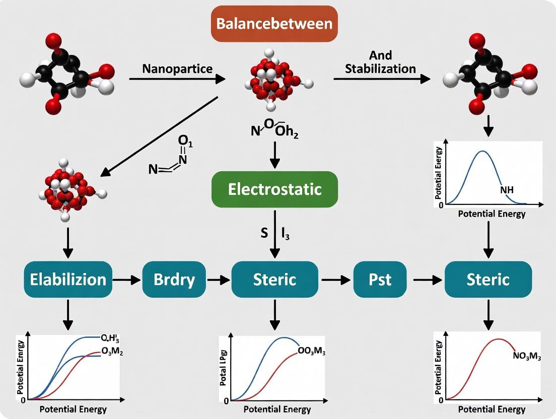
Title: Balancing Electrostatic and Steric Stabilization
The Scientist's Toolkit: Research Reagent Solutions
Table 3: Essential Materials for Nanoparticle Stabilization Research
| Item | Function/Application | Example Product/Brand |
|---|---|---|
| Zetasizer Nano | Measures hydrodynamic size (DLS), PDI, and zeta potential. | Malvern Panalytical |
| DSPE-PEG Variants | Provides steric stabilization and stealth properties for lipid-based systems. | Avanti Polar Lipids |
| PLGA Resorbable Polymers | Core-forming polymer for sustained-release nanoparticles. Requires stabilizer (PVA). | Lactel (Evonik) |
| Microfluidic Mixer | Enables reproducible, scalable nanoparticle formulation with tight size control. | NanoAssemblr (Precision NanoSystems) |
| Dialysis Membranes | Purifies nanoparticles by removing organic solvents, uncapsulated drugs, and free stabilizers. | Spectra/Por (MWCO 3.5-100 kDa) |
| Citrate-Stabilized AuNPs | Ready-made model inorganic nanoparticles for surface modification studies. | Cytodiagnostics, NanoComposix |
| RiboGreen Assay Kit | Quantifies siRNA/RNA encapsulation efficiency in LNPs. | Invitrogen (Thermo Fisher) |
Technical Support Center
Troubleshooting Guides & FAQs
Q1: During nanoprecipitation, my nanoparticles immediately form a visible, milky aggregate instead of a slightly opalescent suspension. What went wrong? A: This indicates catastrophic aggregation, typically due to insufficient stabilization force at the moment of particle formation. First, verify that your anti-solvent is being added rapidly with vigorous mixing (e.g., using a syringe pump and magnetic stirrer at 1000 rpm). Second, reassess your stabilizer concentration. If using a single polymer like PVA, you may be below the critical concentration for effective steric stabilization. Action: Perform a stabilizer screening experiment, incrementally increasing concentration from 0.1% to 5% (w/v). If the problem persists, consider switching to or adding a charged stabilizer (e.g., sodium cholate) to introduce electrostatic repulsion, creating a combined steric-electrostatic barrier.
Q2: My formulation is initially stable but aggregates over 24 hours during storage at 4°C. How can I improve shelf-life? A: Time-dependent aggregation suggests your formulation is in a metastable state. The balance of forces is precarious. Key parameters to check:
- Zeta Potential: Measure immediately after preparation and after 24 hours. A shift towards neutral (±10 mV) indicates loss of electrostatic stabilization. Consider adding a stabilizing agent like Poloxamer 188 or TPGS, which provide strong steric hindrance less sensitive to ionic strength changes.
- Osmotic Pressure: Differential osmotic pressure across the nanoparticle membrane can cause swelling and fusion. Action: Add a cryoprotectant like trehalose (5-10% w/v) or sucrose before storage. This forms a glassy matrix, immobilizing particles.
- pH: Small shifts can protonate/deprotonate surface groups. Ensure your buffer (e.g., 10 mM citrate for acidic, 10 mM PBS for neutral/alkaline) has adequate capacity.
Q3: My Dynamic Light Scattering (DLS) data shows multiple peaks or a very high Polydispersity Index (PDI > 0.3). How do I achieve a monodisperse population? A: High PDI indicates heterogeneous nucleation and growth during synthesis. To promote uniform particle formation:
- Refine Mixing: Transition from magnetic stirring to microfluidic or confined impingement jet mixing for reproducible, rapid solvent displacement.
- Purify: Use differential centrifugation or membrane filtration to remove oversized aggregates or debris. For example, centrifuge at a low relative centrifugal force (e.g., 2,000 x g for 5 min) to pellet aggregates, then collect the supernatant.
- Post-Formulation Processing: Implement a brief sonication step (e.g., probe sonication at 20% amplitude for 30 seconds on ice) to disrupt loose aggregates before measurement.
Q4: How do I choose between steric, electrostatic, or combined stabilization for my specific API? A: The choice depends on your Active Pharmaceutical Ingredient (API) properties and administration route. Use this decision framework:
| API/Route Consideration | Recommended Stabilization | Rationale & Example |
|---|---|---|
| Hydrophobic, for IV injection | Combined (Steric + Electrostatic) | Prevents opsonization and aggregation in high-ionic-strength blood. Example: PLGA nanoparticles with cationic coating (e.g., CTAB) + PEG. |
| pH-sensitive, for oral delivery | Primarily Steric | Electrostatic stabilization fails in varying GI pH. Steric polymers (e.g., PVP, HPMC) maintain integrity. |
| Targeting specific cells | Primarily Electrostatic | A controlled surface charge (slightly positive) can enhance cellular uptake. Must balance with cytotoxicity. |
Key Experimental Protocols
Protocol 1: Formulation Screening via Solvent Displacement (Nanoprecipitation) Objective: To produce and screen batches of nanoformulations with varying stabilizers.
- Prepare Organic Phase: Dissolve 10 mg of your hydrophobic API (e.g., curcumin) in 1 mL of acetone.
- Prepare Aqueous Phase: Prepare 5 mL of aqueous solutions containing different stabilizers (e.g., 1% PVA, 0.5% Poloxamer 188, 0.1% sodium cholate, or combinations).
- Mixing: Using a syringe pump, inject the organic phase into the vigorously stirred (1000 rpm) aqueous phase at a rate of 0.5 mL/min.
- Evaporation: Stir the resulting suspension uncovered for 2-3 hours to evaporate organic solvent.
- Analysis: Characterize each batch immediately for size, PDI, and zeta potential (see Protocol 2).
Protocol 2: Characterization of Size, PDI, and Zeta Potential Objective: To quantitatively assess nanoparticle stability and quality.
- Sample Preparation: Dilute 20 µL of raw nanoparticle suspension in 2 mL of appropriate buffer (e.g., 1 mM KCl for zeta potential). Ensure the count rate is within instrument specifications.
- Dynamic Light Scattering (DLS):
- Load sample into a disposable cuvette.
- Set temperature to 25°C, equilibrium time 120 sec.
- Run measurement with at least 12 sub-runs.
- Record the Z-Average Diameter (d.nm) and Polydispersity Index (PDI).
- Zeta Potential Measurement:
- Load diluted sample into a clear disposable zeta cell.
- Set parameters: dielectric constant 78.5, viscosity 0.8872 mPa.s.
- Perform at least 3 measurements with >15 runs each.
- Record the Zeta Potential (ζ, mV).
Protocol 3: Accelerated Stability Testing Objective: To predict physical stability under stress conditions.
- Sample Allocation: Aliquot 1 mL of purified nanoformulation into 2 mL cryovials (n=3 per condition).
- Stress Conditions:
- Thermal: Store vials at 4°C, 25°C, and 40°C for 7 days.
- Freeze-Thaw: Subject vials to 3 cycles of freezing at -20°C for 12h and thawing at 25°C for 12h.
- Analysis: After stress, vortex samples gently and analyze for changes in diameter, PDI, and zeta potential versus time-zero controls. An increase in diameter >10% or |ζ| change >5 mV indicates instability.
Data Presentation
Table 1: Stabilizer Screening for PLGA Nanoparticles (API: Docetaxel)
| Stabilizer System (Concentration) | Avg. Size (nm) | PDI | Zeta Potential (mV) | Observation after 1 wk @ 4°C |
|---|---|---|---|---|
| None (Control) | 450 ± 120 | 0.45 | -3.2 ± 1.5 | Heavy aggregation, precipitation |
| PVA (1% w/v) | 165 ± 5 | 0.12 | -10.5 ± 2.1 | No size change, slight opalescence |
| Poloxamer 188 (0.5% w/v) | 180 ± 8 | 0.08 | -5.8 ± 1.8 | No change, clear suspension |
| Sodium Cholate (0.1% w/v) | 155 ± 10 | 0.15 | -42.3 ± 3.5 | No size change |
| PVA (1%) + Sodium Cholate (0.05%) | 158 ± 4 | 0.05 | -35.1 ± 2.4 | No change, optimal stability |
Table 2: Critical Quality Attributes (CQA) Target Ranges
| Parameter | Ideal Target Range | Acceptable Range | Analytical Method | ||||||
|---|---|---|---|---|---|---|---|---|---|
| Particle Size | 80 - 150 nm | 70 - 200 nm | Dynamic Light Scattering | ||||||
| Polydispersity Index (PDI) | < 0.10 | ≤ 0.20 | DLS (Cumulants analysis) | ||||||
| Zeta Potential (Steric) | N/A | N/A | N/A | ||||||
| Zeta Potential (Electrostatic) | > | +30 | or <-30 | mV | > | +20 | or <-20 | mV | Electrophoretic Light Scattering |
| Encapsulation Efficiency | > 90% | > 80% | HPLC/UV after separation |
The Scientist's Toolkit: Research Reagent Solutions
| Item | Function in Nanoformulation |
|---|---|
| Poly(D,L-lactide-co-glycolide) (PLGA) | Biodegradable polymer matrix; forms the nanoparticle core. |
| Polyvinyl Alcohol (PVA) | Steric stabilizer; adsorbs to particle surface, preventing close approach. |
| D-α-Tocopheryl Polyethylene Glycol Succinate (TPGS) | Steric stabilizer & permeation enhancer; derived from Vitamin E. |
| Poloxamer 188 (Pluronic F68) | Non-ionic triblock copolymer steric stabilizer; reduces protein adsorption. |
| Sodium Cholate | Anionic surfactant; provides electrostatic & steric stabilization. |
| Cetyltrimethylammonium Bromide (CTAB) | Cationic surfactant; provides positive charge for electrostatic stabilization. |
| Trehalose Dihydrate | Cryoprotectant; protects nanoparticles during lyophilization/storage. |
| Dichloromethane (DCM) / Acetone | Organic solvents for dissolving polymer/API in emulsion/evaporation methods. |
Diagrams
Title: Forces Governing Nanoparticle Stability
Title: Nanoformulation Development Workflow
Diagnosing Instability: A Troubleshooting Guide for Aggregation, Flocculation, and Ostwald Ripening
Troubleshooting Guide & FAQ
Q1: How can I differentiate between a failure of electrostatic repulsion and a failure of steric stabilization when observing particle aggregation in my colloidal dispersion?
A: The kinetics and reversibility of aggregation are key. Electrostatic collapse (e.g., from salt screening or pH shift) often causes rapid, irreversible flocculation that is sensitive to ionic strength. Steric inadequacy (e.g., from poor surface coverage or solvent quality change) typically leads to slower, more reversible aggregation, often upon temperature change or freeze-thaw cycling. Conduct a critical zeta potential measurement; a value moving within ±10 mV suggests electrostatic collapse. For steric failure, perform a steric layer thickness measurement via dynamic light scattering (DLS) in a good vs. poor solvent.
Q2: What is the definitive experimental protocol to diagnose the primary cause of instability in a PEGylated liposome or nanoparticle formulation?
A: Follow this sequential diagnostic protocol:
- Measure Zeta Potential vs. Ionic Strength: Dilute the sample in a series of NaCl solutions (0-150 mM). Plot zeta potential. A steep decline indicates electrostatic stabilization was present and is being screened.
- Perform a Stability Test in Aqueous Two-Phase System: Mix sample with a dextran/PEG system. A sterically stabilized particle will partition based on the surface polymer chemistry, not charge.
- Conduct a Temperature-Ramp DLS Experiment: Heat the sample from 25°C to 60°C while monitoring hydrodynamic radius. A sharp increase near the theta temperature of the polymer indicates steric failure due to polymer collapse.
Q3: Our siRNA-loaded lipid nanoparticles (LNPs) aggregate upon storage at 4°C. Is this more likely a charge or steric issue?
A: For ionizable lipid-based LNPs, this is frequently a sign of steric inadequacy. At the acidic pH of internal buffers and 4°C, the PEG-lipid conjugate may undergo phase separation or the PEG corona may collapse, reducing steric repulsion. Diagnose by: (i) checking if aggregation reverses upon gentle warming and vortexing (suggests steric), and (ii) measuring the particle size before and after dialysis against a low-ionic-strength buffer. If dialysis doesn't prevent aggregation, steric coverage is likely insufficient.
Q4: What quantitative data should I compare to conclusively assign the failure mode?
A: Summarize key metrics in the following table:
| Diagnostic Metric | Electrostatic Collapse (Positive Sign) | Steric Inadequacy (Positive Sign) | Typical Measurement Method |
|---|---|---|---|
| Zeta Potential (mV) | Absolute value < ±15 mV at formulation pH & IS | Absolute value > ±20 mV but aggregation still occurs | Electrophoretic Light Scattering |
| Critical Flocculation Point | Low ionic strength (< 50 mM NaCl) causes aggregation | High ionic strength tolerated, but aggregation at specific temperature | DLS monitoring of size vs. [Salt] or Temp |
| Aggregation Reversibility | Irreversible upon dilution or pH adjustment | Partially reversible upon changing solvent quality or temperature | DLS/Turbidity before and after stress removal |
| Steric Layer Thickness (nm) | Layer thin or absent; small difference in Rh (good vs. poor solvent) | Layer > 5 nm, but may show collapse in poor solvent | DLS in good solvent (water) vs. poor solvent (e.g., high salt or alcohol) |
| Critical Aggregation Temperature | Not observed | Clearly observed near polymer theta temperature | DLS with temperature ramp |
Experimental Protocol: Determining Steric Layer Thickness and Solvent Quality
Objective: To measure the effective steric barrier thickness and assess its stability. Materials: Nanoparticle dispersion, deionized water, 2M ammonium sulfate ((NH₄)₂SO₄) solution, DLS instrument, temperature-controlled cuvette holder. Method:
- Filter all solvents through a 0.02 µm filter.
- Dilute the nanoparticle sample 100-fold in deionized water (good solvent for PEG). Equilibrate at 25°C for 5 minutes. Measure the hydrodynamic radius (Rh_water) via DLS (perform 5 runs, take median).
- Separately, dilute an identical aliquot of the nanoparticle sample 100-fold in 1.5M (NH₄)₂SO₄ solution (a poor solvent for PEG). Equilibrate at 25°C for 5 minutes. Measure Rh_salt.
- Calculate approximate steric layer thickness: δ ≈ (Rhsalt - Rhwater) / 2. A δ < 2 nm suggests negligible or collapsed steric stabilization.
- (Optional) Take the sample in poor solvent and perform a temperature scan from 25°C to 50°C, monitoring Rh. A sharp increase indicates polymer collapse and confirms steric failure mode.
The Scientist's Toolkit: Key Reagent Solutions
| Reagent / Material | Primary Function in Stability Diagnostics |
|---|---|
| NaCl or KCl Solutions (0-500 mM) | Titrant for screening electrostatic interactions and determining Critical Flocculation Concentration (CFC). |
| Ammonium Sulfate ((NH₄)₂SO₄) Solution | A "salting-out" agent used as a poor solvent for PEG and many common steric stabilizers to test layer collapse. |
| pH Adjustment Buffers (e.g., citrate, phosphate, Tris) | To probe the ionization state of surface charged groups and identify the isoelectric point (IEP). |
| PEG/Dextran Aqueous Two-Phase System | To confirm the dominance of steric stabilization by observing partitioning based on surface polymer chemistry. |
| Fluorescently-Labeled Polymer Analog (e.g., FITC-PEG-lipid) | To quantify surface coverage and confirm grafting density of steric stabilizers via fluorescence techniques. |
| Calorimetry Standard (e.g., Tris buffer) | For validating temperature measurements in DLS during temperature-ramp experiments for steric failure. |
Decision Tree for Initial Failure Mode Diagnosis
Mechanistic Comparison of Two Stability Failure Modes
Troubleshooting & FAQ Center
Q1: During colloidal stability tests, my nanoparticle formulation rapidly aggregates upon addition of a standard PBS buffer. What is the immediate cause and how can I diagnose it?
A: This is a classic symptom of electrostatic screening. The high ionic strength of PBS compresses the electrical double layer, reducing the repulsive force between particles. To diagnose:
- Check Zeta Potential: Measure the zeta potential of your formulation in both water and low-ionic-strength buffer (e.g., 1 mM KCl). If the value drops significantly (e.g., from ±30 mV to ±10 mV) in PBS, electrostatic screening is confirmed.
- Determine Critical Coagulation Concentration (CCC): Perform a serial dilution of salt (e.g., NaCl) and note the concentration at which aggregation occurs (via DLS size increase or visual inspection). Compare to theoretical CCC values (see Table 1).
Q2: My sterically stabilized (PEGylated) nanoparticles are still sensitive to pH changes near the formulation's pKa. Why does this happen, and how can I improve robustness?
A: PEG provides steric hindrance but may not fully shield underlying charge groups. pH shifts near the pKa of your core or coating material can alter ionization states, affecting hydration and conformation of the polymer layer.
- Solution: Introduce a mixed stabilization approach. Incorporate a small, controlled percentage of a permanently charged moiety (e.g., a sulfonate group) alongside PEG. This provides a residual electrostatic component that works synergistically with steric hindrance, creating a higher energy barrier against aggregation at varying pH levels.
Q3: What is the most reliable experimental protocol to map the stability landscape of a formulation against ionic strength and pH?
A: Use a High-Throughput Microplate Stability Assay.
- Materials: Nanoparticle suspension, phosphate or citrate buffers (pH range 4-9), NaCl stock solution (2M), 96-well plate, plate reader with dynamic light scattering (DLS) or static light scattering (turbidity) capability.
- Protocol: a. Prepare a matrix in the microplate: vary pH along rows and NaCl concentration (0-500 mM) along columns. b. Dilute a concentrated nanoparticle stock into each well at a final, consistent particle concentration. c. Seal the plate, incubate at desired temperature (e.g., 25°C) for 1-24 hours. d. Measure the hydrodynamic diameter (via DLS plate reader) or optical density (at 600 nm) for each well. e. Plot stability maps (size or OD vs. pH and ionic strength) to identify stable operating zones.
Q4: How do I differentiate between aggregation caused by ionic strength versus specific ion effects (Hofmeister series)?
A:
- Test with Different Salts: Perform CCC experiments using a series of sodium salts (NaCl, Na₂SO₄, NaSCN). Observe the order of effectiveness.
- Diagnostic Table:
| Observation | Likely Cause | Confirmation Experiment |
|---|---|---|
| Aggregation follows ionic strength (I) regardless of anion type. | Non-specific electrostatic screening. | CCC correlates with 1/(zeta potential)^4 as per DLVO theory. |
| Aggregation order is Cl⁻ > SO₄²⁻ (at same ionic strength). | Specific ion (Hofmeister) effect. CCC varies significantly for salts of same valence. | Further test with potassium salts to rule out cation-specific effects. |
Table 1: Critical Coagulation Concentration (CCC) for Common Valences
| Electrolyte Valence (z) | Theoretical CCC (mol/L) for ψ₀ ≈ 75 mV | Approximate CCC for Model Latex Spheres (mM) |
|---|---|---|
| 1:1 (e.g., NaCl) | 0.075 | 40 - 150 |
| 2:1 (e.g., MgCl₂) | 0.0009 | 2 - 5 |
| 3:1 (e.g., AlCl₃) | 0.0001 | 0.1 - 0.3 |
Note: Theoretical values derived from simplified DLVO. Experimental values depend on surface potential and Hamaker constant.
Table 2: Impact of pH on Stabilization Mechanisms
| Stabilization Type | Most Stable pH Region | Vulnerability | Mitigation Strategy |
|---|---|---|---|
| Purely Electrostatic (e.g., citrate-coated) | Far from isoelectric point (IEP) | High ionic strength, pH near IEP | Formulate in low-salt buffer, buffer pH away from IEP. |
| Purely Steric (e.g., thick PEG layer) | Broad, but can be pH-sensitive if polymer chargeable. | Poor solvent conditions, specific adsorption. | Use non-ionic polymers (e.g., Pluronics), ensure good solvent quality. |
| Electrosteric (e.g., charged polymer brush) | Broadest. Electrostatic component reinforces steric layer. | Extreme pH that neutralizes charge AND collapses polymer. | Optimize charge density to balance electrostatic robustness and stealth properties. |
Key Experimental Protocols
Protocol 1: Determining Zeta Potential vs. pH Titration Objective: Identify the isoelectric point and assess surface charge variation.
- Equipment: Zeta potential analyzer with titrator module.
- Procedure: a. Dilute nanoparticle sample in 1 mM KCl background electrolyte. b. Set titrator to add incremental amounts of 0.1M HCl or 0.1M KOH. c. After each pH adjustment, allow equilibration (2 min), then measure zeta potential (minimum 5 runs). d. Plot zeta potential vs. pH. The pH where zeta potential = 0 is the IEP.
Protocol 2: Assessing Steric Layer Robustness via Solvent Quality Objective: Test the integrity of the steric stabilizing layer.
- Materials: PEGylated nanoparticle suspension, polyethylene glycol (PEG) 6000, phosphate buffer.
- Procedure: a. Prepare a series of solutions with increasing concentration of PEG 6000 (0-30% w/v) in phosphate buffer. This creates a worsening solvent quality ("crowding" and dehydration effect). b. Mix equal volumes of nanoparticle suspension with each PEG solution. c. Incubate at room temperature for 1 hour. d. Measure hydrodynamic diameter by DLS. A sudden increase indicates collapse or insufficient steric layer thickness.
Visualizations
Diagram 1: Stabilization Mechanism Decision Logic
Diagram 2: Experimental Workflow for Stability Mapping
The Scientist's Toolkit: Research Reagent Solutions
| Item | Function & Rationale |
|---|---|
| Zeta Potential Reference Standards (e.g., DTAP latex) | Validate instrument performance for accurate surface charge measurement. |
| Low-Ionic-Strength Buffers (e.g., 2-5 mM HEPES, Bis-Tris) | Provide pH control without significant electrostatic screening during characterization. |
| PEG Polymers (varying MW & end-groups) | Model steric stabilizers. Thicker layers (higher MW) provide better steric hindrance. Functional end-groups (e.g., -COOH, -NH₂) allow electrosteric tuning. |
| Chaotropic & Kosmotropic Salts (e.g., NaSCN, (NH₄)₂SO₄) | Probe specific ion effects (Hofmeister series) on colloidal stability and polymer hydration. |
| Microplate for High-Throughput Screening | Enables efficient mapping of stability across multi-dimensional parameter space (pH, ionic strength, temperature). |
| Dynamic Light Scattering (DLS) with Titrator | Essential for automated measurement of particle size and zeta potential as a function of pH or salt concentration. |
Technical Support Center
Troubleshooting Guides & FAQs
Q1: During Layer-by-Layer (LbL) assembly, my particles show immediate, severe aggregation after the addition of the first polycation layer. What is the primary cause and solution?
A: This is typically a sign of excessive polymer charge density or insufficient steric bulk, leading to bridging flocculation.
- Primary Cause: The high charge density polycation is neutralizing the particle's surface charge too rapidly and completely, leaving no electrostatic or steric barrier. It may also be bridging between multiple particles.
- Troubleshooting Steps:
- Dilute Polymer Solution: Reduce the concentration of the adsorbing polymer by 50-80%.
- Increase Ionic Strength: Add a low concentration of salt (e.g., 10-50 mM NaCl) to the polymer solution. This screens electrostatic interactions, allowing the polymer to adsorb in a more loopy, sterically stabilizing conformation.
- Switch to a Copolymer: Use a polycation with a lower charge density block (e.g., a copolymer with PEG or hydrophobic segments) to introduce steric stabilization from the first layer.
- Verify pH: Ensure the solution pH is such that both the particle surface and the polymer are fully charged.
Q2: My multilayered particles are stable in storage but aggregate instantly upon introduction to cell culture media or blood serum. How can I improve colloidal stability in biological fluids?
A: This is a classic failure of electrostatic stabilization due to "charge screening" by the high ionic strength and competing polyelectrolytes in biological fluids.
- Primary Cause: Reliance solely on electrostatic stabilization, which is screened in high-ionic-strength environments.
- Troubleshooting Steps:
- Incorporate a Steric Stabilizer: The final outer layer must be a dense, hydrophilic, neutrally charged polymer. Poly(ethylene glycol) (PEG) is the gold standard. Use a PEG-grafted polyelectrolyte or adsorb a triblock copolymer (e.g., Pluronic F127) as the final layer.
- Optimize Architecture: Implement a "mixed layer" architecture where the final layer is a blend of a charged polymer and a non-ionic steric stabilizer.
- Increase Hydrophilicity: Choose polymers with hydrogen-bonding groups (e.g., polysaccharides like chitosan/hyaluronic acid) for outer layers, as they better retain a hydration shell.
Q3: How do I determine the optimal Molecular Weight (MW) of the polyelectrolyte for my system?
A: The optimal MW balances strong adsorption (higher MW) with the ability to form a dense, conformationally adaptable layer (lower MW can pack more tightly).
- Guidelines:
- For a Thin, Dense Layer (Primary Electrostatic Stabilization): Use lower MW polymers (10-50 kDa). They adsorb in a flatter conformation, leading to higher charge density per unit area.
- For a Thick, Steric Brush Layer: Use higher MW polymers (50-200 kDa) or grafted polymers. They extend further into solution, creating a physical barrier against van der Waals attraction.
- Experimental Protocol: Perform adsorption isotherms (via QCM-D or depletion assay) and measure zeta potential & hydrodynamic size for a series of MWs at fixed concentration. The optimal MW gives a plateau in adsorbed mass and a high, stable zeta potential.
Q4: The zeta potential of my particles does not reverse sign after the adsorption of an oppositely charged layer. Does this mean the layer failed to adsorb?
A: Not necessarily. Several factors can explain this.
- Possible Causes & Diagnostics:
- Charge Overcompensation is Incomplete: The polymer dose or adsorption time may be insufficient. Solution: Increase polymer concentration or incubation time.
- Polymer Charge is Patchy: The polymer adsorbs in patches, leaving some original surface charge exposed. Solution: Use a lower charge density polymer or add salt to encourage smoother adsorption.
- The Layer is Too Thin: The shear plane for zeta potential measurement may lie outside the new polymer layer. Confirm adsorption by measuring an increase in hydrodynamic diameter via Dynamic Light Scattering (DLS).
- Instrument Limitation: High conductivity samples can make zeta potential measurements unreliable.
Table 1: Effect of Polycation Molecular Weight on Layer Properties and Stability
| Polycation MW (kDa) | Hydrodynamic Thickness Increase (nm) | Zeta Potential after Layer (mV) | Stability in 150 mM NaCl (Time to Aggregation) |
|---|---|---|---|
| 15 | 2.1 ± 0.3 | +28 ± 3 | < 2 hours |
| 50 | 4.5 ± 0.6 | +35 ± 2 | 12 hours |
| 150 | 8.7 ± 1.1 | +31 ± 4 | > 48 hours |
| 150 (PEG-grafted) | 12.5 ± 1.5 | +12 ± 2 | > 1 week |
Table 2: Impact of Outer Layer Architecture on Stability in Biological Media
| Outer Layer Architecture | Zeta Potential in PBS (mV) | Hydrodynamic Diameter in 10% FBS (Change after 1h) | Visual Aggregation in Serum? |
|---|---|---|---|
| Pure PEI (High Charge Density) | +40 ± 3 | +285% | Severe |
| Pure PGA (High Charge Density) | -45 ± 2 | +190% | Severe |
| PGA / PEG-b-PLL Mixed Layer | -15 ± 5 | +15% | None |
| PGA / Pluronic F127 Adsorbed | -8 ± 3 | +5% | None |
Experimental Protocols
Protocol 1: Determining Optimal Polymer Concentration for LbL Assembly Objective: To find the minimum polymer concentration required for complete charge reversal without causing aggregation. Materials: Core nanoparticle dispersion, polyelectrolyte solutions (0.1-5 mg/mL in 10 mM NaCl, pH adjusted), zeta potential & DLS instrument. Steps:
- Dilute the core nanoparticle dispersion to a standard concentration (e.g., 0.1 mg/mL) in 10 mM NaCl buffer.
- Measure the initial zeta potential and diameter (DLS).
- Add a fixed volume of the lowest polyelectrolyte concentration (0.1 mg/mL) to the nanoparticle dispersion under rapid stirring. Stir for 15 minutes.
- Measure zeta potential and diameter. Centrifuge gently if needed to remove unbound polymer.
- Repeat steps 3-4 with incrementally higher polymer concentrations on fresh nanoparticle samples.
- Analysis: The optimal concentration is the lowest one that achieves a stable, reversed zeta potential (plateau value) with no significant increase in aggregate size (PdI < 0.2).
Protocol 2: Assessing Robustness via Salt Challenge Test Objective: To evaluate the electrostatic vs. steric stabilization contribution of a built multilayer. Materials: Multilayered nanoparticle dispersion, NaCl solutions (0.15 M, 0.5 M, 1.0 M), DLS instrument. Steps:
- Prepare a 1 mL aliquot of the final nanoparticle dispersion.
- Measure the baseline hydrodynamic diameter (Z-avg) and polydispersity index (PdI).
- Add the appropriate volume of concentrated NaCl stock to the aliquot to achieve the first target concentration (e.g., 150 mM). Mix immediately.
- Incubate for 30 minutes at room temperature.
- Measure Z-avg and PdI.
- Repeat steps 3-5 for higher salt concentrations on fresh aliquots.
- Analysis: A system relying purely on electrostatics will show a rapid, large increase in diameter (>50%) at 150 mM NaCl. A system with good steric stabilization will show minimal size change (<20%) even at 1.0 M NaCl.
Visualizations
Diagram Title: LbL Optimization Decision Workflow
Diagram Title: Balancing Electrostatic and Steric Stabilization
The Scientist's Toolkit: Research Reagent Solutions
Table 3: Essential Materials for LbL Stabilization Research
| Item | Function & Rationale |
|---|---|
| Poly(ethylene imine) (PEI), Branched & Linear | A model polycation with high charge density. Used to study the effects of MW and branching on initial layer adsorption and stability. |
| Poly(acrylic acid) (PAA) / Poly(sodium styrene sulfonate) (PSS) | Model polyanions. PAA's charge is pH-responsive, allowing control. PSS provides strong, permanent negative charge. |
| PEG-grafted Copolymers (e.g., PEG-b-PLL, PEG-b-PEI) | Critical for introducing steric stabilization. The charged block adsorbs, while the PEG block extends to form a hydration cloud. |
| Pluronic Surfactants (F68, F127) | Non-ionic triblock copolymers (PEO-PPO-PEO). Adsorb via the hydrophobic PPO block, providing a dense PEO brush for steric stabilization. |
| Chitosan & Hyaluronic Acid | Natural, biocompatible polyelectrolytes. Provide different charge densities and bio-interactive properties for specialized applications. |
| Quartz Crystal Microbalance with Dissipation (QCM-D) | Measures adsorbed polymer mass and viscoelastic properties in situ on a flat surface, informing layer growth and rigidity. |
| Zeta Potential Analyzer | The primary tool for tracking layer-by-layer charge reversal and predicting electrostatic stability. |
| Dynamic & Static Light Scattering (DLS/SLS) | Measures hydrodynamic diameter, polydispersity (PdI), and aggregation state in real-time under various conditions. |
Troubleshooting Guides & FAQs
Q1: Why do my nanoparticles aggregate rapidly when added to serum for cell culture experiments, and how can I stabilize them? A: Serum proteins can adsorb to nanoparticle surfaces, destabilizing the electrostatic and/or steric shielding you engineered. This is often due to insufficient steric stabilization density or an incorrect surface charge (zeta potential) in physiological ionic strength.
- Troubleshooting Steps:
- Measure Zeta Potential in Serum: Use Dynamic Light Scattering (DLS) to measure the zeta potential of your particles in 10-50% serum or PBS/physiological buffer (150 mM NaCl). A shift towards neutral charge (e.g., from ±30 mV to near ±10 mV) indicates significant protein adsorption and electrostatic shield failure.
- Increase Steric Layer Density: If using PEG or another polymer, increase the grafting density. A high-density brush conformation is more resistant to protein adsorption than a mushroom conformation.
- Optimize Surface Chemistry: Consider using block copolymers (e.g., Pluronic F127) or introducing serum-resistant ligands like high-molecular-weight PEG (≥ 5 kDa) or specific zwitterionic polymers.
Q2: My particle formulation appears stable initially but forms large clumps after a single freeze-thaw cycle. What is the cause and solution? A: Freezing concentrates particles and cryoprotectants in the remaining liquid, leading to ice crystal formation and physical forces that push particles together, overcoming repulsive barriers.
- Troubleshooting Steps:
- Introduce Cryoprotectants: Add non-reducing sugars (e.g., trehalose, sucrose) at 5-10% (w/v). They form a vitrified matrix that separates particles during freezing.
- Control Freeze/Thaw Rate: Flash-freeze in liquid nitrogen and thaw rapidly in a 37°C water bath to minimize time in the high-concentration phase. Avoid slow thawing at 4°C.
- Increase Pre-Freeze Stability: Ensure your particles have a strong steric barrier (optimal polymer coverage) and a high absolute zeta potential (> ±30 mV) in the storage buffer before freezing to maximize the combined stabilization forces.
Q3: How can I predict and ensure the long-term (6-24 month) stability of my colloidal dispersion for a drug product? A: Long-term stability requires a formulation that balances electrostatic repulsion (for long-range ordering) with steric hindrance (for short-range, ionic-strength-independent protection).
- Troubleshooting Steps:
- Perform Accelerated Stability Studies: Store samples at 4°C, 25°C, and 40°C. Monitor hydrodynamic size (by DLS) and PDI weekly/monthly. Aggregation at higher temperatures predicts faster degradation at recommended storage temperatures.
- Optimize Storage Buffer: Use a buffer with a slightly alkaline pH (e.g., 7.4-8.5) to maintain negative surface charge, and include 1-5% sucrose as a stabilizer. Avoid phosphate buffers with divalent cations (like Ca²⁺) which can bridge and precipitate particles.
- Conduct Forced Degradation Studies: Expose samples to oscillating pH, UV light, or oxidizers to identify the weakest point in your stabilization strategy.
Table 1: Impact of Surface Modification on Serum Stability
| Polymer Coating | Grafting Density | Initial Zeta Potential (mV) in Water | Zeta Potential (mV) in 50% Serum | Size Increase in Serum after 24h (%) | Key Finding |
|---|---|---|---|---|---|
| None (Plain Particle) | N/A | -35.2 ± 2.1 | -8.5 ± 1.3 | > 300% (Aggregation) | Electrostatic stabilization alone fails in serum. |
| PEG 2kDa (Low Density) | ~0.2 chains/nm² | -12.5 ± 1.8 | -6.2 ± 0.9 | ~150% | Low steric density is insufficient. |
| PEG 5kDa (High Density) | ~0.8 chains/nm² | -8.4 ± 0.7 | -9.1 ± 0.8 | < 10% | High-density brush provides excellent serum stability. |
Table 2: Efficacy of Cryoprotectants Against Freeze-Thaw-Induced Aggregation
| Cryoprotectant | Concentration | Size (nm) Before Freeze-Thaw | Size (nm) After 3 Freeze-Thaw Cycles | Polydispersity Index (PDI) After |
|---|---|---|---|---|
| None (PBS only) | N/A | 105.3 ± 1.5 | 2450 ± 320 | 0.52 |
| Sucrose | 5% (w/v) | 108.1 ± 2.0 | 115.6 ± 3.1 | 0.12 |
| Trehalose | 10% (w/v) | 107.8 ± 1.7 | 110.2 ± 2.5 | 0.09 |
| Bovine Serum Albumin | 1% (w/v) | 106.5 ± 1.9 | 185.4 ± 15.2 | 0.21 |
Detailed Experimental Protocols
Protocol 1: Assessing Serum Stability via DLS and Zeta Potential Objective: To quantify nanoparticle stability and protein adsorption upon exposure to biological media. Materials: Nanoparticle dispersion, fetal bovine serum (FBS), phosphate-buffered saline (PBS, 1x), DLS/Zetasizer instrument. Procedure:
- Prepare a 50% (v/v) serum solution by mixing equal parts FBS and 1x PBS.
- Dilute the nanoparticle stock to the recommended measurement concentration (e.g., 0.1-0.5 mg/mL) using (a) 1x PBS and (b) the 50% serum solution.
- Incubate both samples at 37°C for 30 minutes.
- Transfer to appropriate cuvettes/zeta cells.
- Measure the hydrodynamic diameter and PDI via DLS (3 measurements per sample).
- Measure the zeta potential via electrophoretic light scattering (5-12 runs per sample).
- Analysis: Compare the size and zeta potential in PBS vs. serum. A significant increase in size/PDI and a decrease in absolute zeta potential indicate serum protein adsorption and poor stability.
Protocol 2: Systematic Freeze-Thaw Stability Test Objective: To evaluate formulation robustness to temperature cycling. Materials: Nanoparticle dispersion, cryoprotectant solutions, cryovials, liquid nitrogen, 37°C water bath. Procedure:
- Aliquot identical volumes (e.g., 500 µL) of the nanoparticle formulation into 5 cryovials per condition (e.g., no protectant, with 5% sucrose, with 10% trehalose).
- For the "slow freeze" group, place vials at -80°C for 24 hours. For the "flash freeze" group, submerge vials in liquid nitrogen for 5 minutes, then transfer to -80°C.
- Thaw samples rapidly by placing in a 37°C water bath with gentle swirling until just ice-free.
- Allow samples to equilibrate to room temperature.
- Vortex gently to mix.
- Analyze particle size and PDI via DLS immediately.
- Repeat the freeze-thaw cycle (steps 2-6) for up to 5 cycles.
- Analysis: Plot size and PDI versus freeze-thaw cycle number. An effective cryoprotectant will maintain baseline values.
Visualizations
The Scientist's Toolkit: Research Reagent Solutions
| Item | Primary Function in Stabilization Studies |
|---|---|
| Dynamic Light Scattering (DLS) / Zetasizer | Measures hydrodynamic particle size (diameter), size distribution (PDI), and zeta potential (surface charge). Essential for quantifying stability. |
| Polyethylene Glycol (PEG), various MWs | The gold-standard steric stabilizer. High-MW, high-density PEG brushes create a hydration shell and physical barrier against aggregation and protein adsorption. |
| Trehalose / Sucrose | Cryoprotectants and lyoprotectants. Form amorphous glasses during freezing/drying, separating particles and preventing ice-crystal-induced damage. |
| Fetal Bovine Serum (FBS) | Complex protein mixture used to model the in vivo environment and test for opsonization and colloidal stability in biological media. |
| Size Exclusion Chromatography (SEC) Columns | Used to purify nanoparticles after surface modification, removing unbound polymers and reagents that can interfere with stability. |
| Zwitterionic Polymers (e.g., PMPC, PSBMA) | Alternative to PEG; form super-hydrophilic surfaces that strongly bind water, providing exceptional resistance to protein fouling and serum aggregation. |
Technical Support Center: Troubleshooting & FAQs
Q1: During formulation, my nanoparticles rapidly aggregate upon adding a therapeutic payload. What is the primary cause and correction strategy?
A: This is a classic sign of electrostatic destabilization. The payload likely neutralizes or reduces the surface charge (zeta potential) on the nanoparticles. First, measure the zeta potential before and after payload addition. If the magnitude drops below |±20| mV, electrostatic repulsion is insufficient.
- Correction: Introduce a small-molecule additive like sodium citrate (1-10 mM) to the aqueous phase. Citrate anions can adsorb onto the particle surface, restoring negative charge and increasing electrostatic repulsion. Alternatively, shift to a hybrid stabilizer cocktail. For example, supplement your existing ionic surfactant (e.g., SDS) with a non-ionic polymer like Poloxamer 188 (0.1% w/v), which provides steric hindrance to compensate for lost electrostatic force.
Q2: My hybrid (electrostatic + steric) stabilized formulation remains stable at 4°C but aggregates at physiological temperature (37°C). Why?
A: Temperature-induced aggregation often points to the failure of the steric component. Many polymers (e.g., certain PEG derivatives, polysorbates) have lower critical solution temperatures (LCST) or can undergo conformational changes.
- Correction:
- Characterize: Perform Dynamic Light Scattering (DLS) with a temperature ramp from 25°C to 40°C to monitor hydrodynamic size change.
- Optimize Cocktail: Replace the thermo-sensitive steric stabilizer with a more robust one like hydroxypropyl methylcellulose (HPMC) or polyethylene glycol (PEG) with higher molecular weight (e.g., PEG 5kDa vs. 2kDa). Ensure the electrostatic component (e.g., a charged lipid like DOTAP) remains intact at the higher temperature.
- Additive Use: Incorporate a small-molecule stabilizing additive like trehalose (5% w/v), which can form a protective hydrogen-bonding network at the particle surface, dampening the effect of thermal energy on particle collision.
Q3: How do I determine the optimal ratio between electrostatic and steric stabilizers in a hybrid cocktail?
A: This requires a systematic Design of Experiments (DoE) approach, with colloidal stability as the key response variable.
- Protocol: Prepare a matrix of formulations varying the concentration of your ionic stabilizer (e.g., Sodium Cholate: 0.1% to 0.5% w/v) and your steric stabilizer (e.g., Pluronic F127: 0.5% to 5% w/v). Use a sonication/high-pressure homogenization standard protocol for all. Characterize each batch immediately (T=0) and after stress (T=24h at 25°C) using the metrics in the table below.
Table 1: Optimization Data for Hybrid Stabilizer Cocktail (Model System: PLGA Nanoparticles)
| Stabilizer Cocktail Composition | Zeta Potential (mV) | PDI (T=0) | Hydrodynamic Size (nm, T=0) | Size Increase after 24h (%) | Visual Inspection |
|---|---|---|---|---|---|
| 0.2% Na. Cholate only | -35.2 ± 1.5 | 0.12 | 152 ± 3 | +45% | Slight precipitate |
| 2% F127 only | -1.5 ± 0.8 | 0.18 | 165 ± 5 | +8% | Opalescent |
| 0.25% Na. Cholate + 3% F127 | -28.7 ± 2.1 | 0.08 | 158 ± 2 | +<2% | Clear, no change |
| 0.1% Na. Cholate + 5% F127 | -15.4 ± 1.2 | 0.10 | 171 ± 4 | +5% | Clear |
| 0.5% Na. Cholate + 0.5% F127 | -40.1 ± 1.8 | 0.25 | 145 ± 8 | +60% | Rapid aggregation |
Q4: I am using a charged polymer (e.g., chitosan) for electrostatic stabilization and PEG for sterics, but I see precipitation. What's happening?
A: This is likely due to charge neutralization and bridging flocculation. The positively charged chitosan and negatively charged terminal groups of some PEGs (or impurities) can interact, causing large, insoluble complexes.
- Correction: Implement a sequential stabilization protocol. First, allow adsorption of the primary electrostatic stabilizer (chitosan) to reach equilibrium. Then, add the steric stabilizer (PEG) in a separate, controlled step at low concentration with vigorous mixing. Consider using a small-molecule additive like arginine hydrochloride (5-15 mM) in the buffer. Arginine can act as a "molecular lubricant," reducing non-specific polymer-polymer interactions and preventing bridging.
Experimental Protocol: Evaluating Hybrid Cocktail Efficacy Against Salt-Induced Aggregation
Objective: To test the resilience of hybrid vs. single-mechanism stabilizers against physiological salt stress.
Materials: (See "Scientist's Toolkit" below) Method:
- Formulation: Prepare three nanoparticle batches using high-pressure homogenization (20,000 psi, 5 cycles):
- Batch A (Electrostatic): 0.3% Sodium Dodecyl Sulfate (SDS).
- Batch B (Steric): 2% Polyvinyl alcohol (PVA, Mw 30-70 kDa).
- Batch C (Hybrid): 0.15% SDS + 1% PVA.
- Stress Test: Take 1 mL of each batch and add 1 mL of 150 mM NaCl phosphate buffer (pH 7.4) under gentle vortexing.
- Analysis: Immediately measure the zeta potential and hydrodynamic diameter (via DLS) of the stressed samples. Compare to unstressed controls (diluted with DI water).
- Incubation: Store stressed samples at 25°C for 2 hours, then measure size again and record any visible aggregation.
The Scientist's Toolkit: Key Research Reagent Solutions
| Reagent / Material | Primary Function in Stabilization |
|---|---|
| Sodium Citrate | Small-molecule additive; provides ionic strength & can adsorb to surfaces to enhance negative charge (electrostatic). |
| Trehalose / Sucrose | Small-molecule additive; forms a vitrified or hydration layer, reducing interfacial energy and preventing close approach (steric/thermodynamic). |
| Poloxamer 188 (Pluronic F68) | Non-ionic triblock copolymer; provides robust steric hindrance via hydrophilic PEG chains. |
| D-α-Tocopheryl Polyethylene Glycol Succinate (TPGS) | Hybrid stabilizer molecule; combines the steric bulk of PEG with the hydrophobic anchor of Vitamin E. |
| Chitosan (low MW) | Cationic biopolymer; provides electrostatic stabilization in acidic environments. |
| DOTAP (Lipid) | Cationic lipid; confers strong positive surface charge for electrostatic repulsion and cell interaction. |
| Hydroxypropyl Methylcellulose (HPMC) | Non-ionic cellulose polymer; provides temperature-insensitive steric stabilization. |
| ArgHCl / HisHCl | Small-molecule amino acid additives; mitigate aggregation by suppressing protein-particle or particle-particle interactions. |
Visualizations
Title: Troubleshooting Stabilization Failure Flowchart
Title: Sequential Hybrid Cocktail Formulation Workflow
Benchmarking Performance: Validation Methods and Comparative Analysis of Stabilization Approaches
Troubleshooting Guides & FAQs
Dynamic Light Scattering (DLS)
Q1: Why is my DLS measurement showing multiple size populations or a high polydispersity index (PDI) when I expect a monodisperse sample? A: This is often due to aggregation or contamination. Within the thesis context of balancing stabilization forces, a high PDI indicates insufficient electrostatic or steric stabilization, leading to clumping.
- Troubleshooting Steps:
- Filter samples through appropriate syringe filters (e.g., 0.22 µm) before measurement.
- Check solvent compatibility. Ensure the dispersant's ionic strength and pH are optimized for your stabilization strategy. Use a low ionic strength buffer (e.g., 1 mM KCl) for electrostatically stabilized particles.
- Verify concentration. Too high a concentration can cause multiple scattering. Dilute the sample until the count rate is within the instrument's optimal range.
- Clean the cuvette meticulously with filtered solvent to remove dust.
Q2: My intensity-based size distribution differs significantly from my volume or number distribution. Which one should I trust? A: The intensity distribution is the primary result. Large aggregates scatter light disproportionately more than small particles, skewing this distribution. For assessing clumping, the intensity peak is sensitive to aggregates. The volume/number distributions are mathematical models and can be unreliable for polydisperse or aggregating samples. Always report the intensity-based hydrodynamic diameter and PDI.
Zeta Potential
Q3: My zeta potential measurement has low reproducibility or shows extreme variability between runs. A: This typically stems from electrode issues, sample preparation, or instrument calibration.
- Troubleshooting Steps:
- Clean and condition electrodes. Follow manufacturer protocols. For platinum electrodes, rinse with distilled water and ethanol. Ensure no air bubbles are trapped.
- Standardize sample preparation. Use a consistent sample volume, equilibration time (temperature), and sonication protocol prior to loading.
- Calibrate with a standard zeta potential tracer (e.g., -50 mV ± 5 mV standard) before critical experiments.
- Control temperature precisely, as mobility is temperature-dependent.
Q4: How do I interpret zeta potential values in the context of steric-electrostatic stabilization? A: Zeta potential indicates electrostatic repulsion strength. For combined stabilization:
- |ζ| > |30| mV: Strong electrostatic stabilization dominates.
- |ζ| ~ |20| mV: Moderate electrostatic contribution; steric layers (e.g., PEG) are crucial to prevent clumping.
- |ζ| < |15| mV: Electrostatic repulsion is weak; steric stabilization must be robust to prevent aggregation. A near-zero ζ in a steric system may still be stable.
Electron Microscopy (TEM/SEM)
Q5: My TEM sample shows extensive particle aggregation on the grid, unlike my DLS results in solution. A: This is a common artifact due to sample drying and grid-surface interactions, critical for studying clumping.
- Troubleshooting Steps:
- Use negative staining (e.g., 1-2% uranyl acetate) to embed and isolate particles, preserving their solution state.
- Apply a hydrophilic coating. Glow-discharge carbon-coated grids to create a hydrophilic surface, improving dispersion.
- Optimize loading. Use a lower concentration and blot more gently to minimize capillary forces during drying.
- Consider cryo-TEM for the most accurate assessment of in-solution morphology and aggregation state.
Q6: I cannot get clear SEM images of my polymer-coated nanoparticles; the coating appears to "melt" under the beam. A: Many steric stabilizers (polymers) are beam-sensitive.
- Troubleshooting Steps:
- Reduce beam energy and current. Start with low kV (e.g., 5-10 kV) and use a minimal probe current.
- Use a conductive coating. Apply a thin, uniform layer of gold/palladium or carbon via sputter coater.
- Consider environmental SEM (ESEM) if available, which allows imaging of hydrated or non-conductive samples with less damage.
Spectroscopic Techniques (UV-Vis, FTIR, Raman)
Q7: My UV-Vis spectrum for gold nanoparticles shows broadening and a red-shift over time. What does this indicate? A: This is a classic sign of aggregation. The surface plasmon resonance (SPR) peak is sensitive to particle size and inter-particle distance. Broadening and red-shifting indicate increased electronic coupling due to clumping, signaling a failure of your stabilization strategy.
Q8: How can FTIR confirm the successful grafting of a steric stabilizer (e.g., PEG-thiol) onto nanoparticle surfaces? A: Use FTIR in ATR mode to detect characteristic polymer bands.
- Protocol: Dry a concentrated nanoparticle solution onto the ATR crystal. Compare spectra to pure ligand and bare nanoparticles. Look for the appearance of PEG's strong C-O-C stretch at ~1100 cm⁻¹ and the suppression of the bare nanoparticle's surface ligand peaks.
| Technique | Key Measured Parameter | Typical Target Value for Stability (Context-Specific) | Common Issue & Diagnostic Value for Clumping | ||||||
|---|---|---|---|---|---|---|---|---|---|
| DLS | Hydrodynamic Diameter (Z-avg), PDI | PDI < 0.1 (monodisperse); Stable size over time. | Increased size & PDI over time: Direct indicator of aggregation. | ||||||
| Zeta Potential | Zeta Potential (ζ) | ζ | > 30 mV (electrostatic); | ζ | shift upon coating. | ζ | decrease towards zero: Loss of electrostatic barrier, high aggregation risk. | ||
| UV-Vis Spectroscopy | SPR Peak Wavelength & FWHM | Sharp, stable peak. | Peak broadening/red-shift: Indicator of aggregation via plasmon coupling. | ||||||
| TEM | Primary Particle Size, Morphology | Uniform dispersion on grid. | Aggregates on grid: May indicate instability or drying artifacts. | ||||||
| FTIR/ Raman | Functional Group Presence/Shift | Clear signature of coating ligands. | Missing ligand peaks or shifts: Suggests incomplete or unstable steric layer. |
Experimental Protocol: Assessing Steric-Electrostatic Stabilization Against Aggregation
Title: Sequential Analysis of Nanoparticle Stability
Objective: To systematically evaluate the contribution of electrostatic and steric forces to colloidal stability under physiological-like conditions.
Materials:
- Synthesized nanoparticles with combined electrostatic/steric coatings.
- Dispersant: 1 mM KCl solution (low ionic strength), 1X PBS (high ionic strength).
- 0.22 µm nylon syringe filters.
- Disposable zeta potential cells & DLS cuvettes.
- pH meter.
- Ultrasonic water bath.
Procedure:
- Sample Preparation: Split the nanoparticle stock into two aliquots. Dialyze or dilute one into 1 mM KCl and the other into 1X PBS to final concentration of 0.1 mg/mL. Sonicate for 5 minutes.
- Baseline Measurement (Low Ionic Strength): Filter the 1 mM KCl sample. Measure hydrodynamic size (3 runs, 10 sub-runs each) and zeta potential (minimum 3 runs of 10-100 cycles) at 25°C.
- Stress Test (High Ionic Strength): Repeat Step 2 with the PBS sample. This screens the electrostatic component.
- Temporal Stability: Place both samples in a 37°C incubator. Measure size and PDI at 0, 1, 4, 8, 24, and 48 hours.
- Validation: After 48 hours, prepare TEM grids (via negative stain) from samples showing the least size change for morphological correlation.
The Scientist's Toolkit: Key Research Reagent Solutions
| Item | Function in Stabilization Studies |
|---|---|
| Polyethylene Glycol (PEG) Thiols/Alkovysilanes | Provides steric stabilization layer; grafting density is critical for preventing clumping. |
| Citrate/Tannic Acid | Common reducing agent & electrostatic stabilizer for metal NPs; provides negative surface charge. |
| Poloxamers (e.g., Pluronic F127) | Triblock copolymers offering robust steric stabilization through hydrophobic adsorption. |
| Potassium Chloride (KCl) Solution | Used to precisely adjust ionic strength and screen electrostatic interactions in stability assays. |
| Uranyl Acetate (2% aqueous) | Negative stain for TEM; embeds particles, allowing visualization of spacing and aggregation state. |
| Phosphate Buffered Saline (PBS), 10X | High ionic strength medium for stress-testing colloidal stability. |
| Dialysis Tubing (MWCO appropriate) | For purifying nanoparticles and exchanging dispersants to precisely control the medium. |
| 0.22 µm Nylon Syringe Filters | Essential for removing dust and large aggregates prior to DLS/zeta potential measurements. |
Diagrams
Title: Dual-Modal Stabilization Strategy for Nanoparticles
Title: Stability Analysis Experimental Workflow
Technical Support Center
Troubleshooting Guides & FAQs
Q1: During accelerated stability testing (40°C/75% RH), our nanoparticle suspension shows rapid increase in turbidity. What are the primary causes and corrective actions?
A: A rapid turbidity increase indicates aggregation. Primary causes and solutions are:
| Cause | Diagnostic Check | Corrective Action | ||
|---|---|---|---|---|
| Insufficient Steric Barrier | Measure hydrodynamic diameter (DLS) before/after stress. >20% increase confirms. | Increase concentration of steric stabilizer (e.g., Poloxamer 188) by 0.5-1.0% w/v. | ||
| Electrostatic Shield Collapse | Measure zeta potential. Shift to | ±10 mV | confirms. | Adjust pH away from isoelectric point or increase ionic stabilizer. |
| Critical Process Temperature Exceeded | Check if storage temp exceeds stabilizer's cloud point (for non-ionics). | Reformulate with stabilizer having a higher cloud point (e.g., Poloxamer 407). | ||
| Oxygen Degradation | Test under nitrogen atmosphere. If improved, oxidation is cause. | Add anti-oxidants (0.01-0.05% ascorbic acid) or use inert gas headspace. |
Q2: When using the CIELAB method for color stability, what does a ΔE*ab > 5 indicate, and how should we proceed?
A: ΔE*ab > 5 indicates a "major difference" perceptible to the human eye, signaling potential chemical degradation or physical instability.
| ΔE*ab Range | Interpretation | Recommended Investigator Actions |
|---|---|---|
| 0 - 1 | Imperceptible change. | No action required for appearance. |
| 1 - 2 | Slight difference. | Monitor closely. |
| 2 - 5 | Perceptible difference. | Check individual L, a, b* shifts (see table below). Initiate chemical assay. |
| > 5 | Major difference. | Priority Action: 1) Correlate with assay for active degradation. 2) Check packaging for light/oxygen ingress. 3) Review formulation for susceptible dyes or actives. |
Q3: Our turbidity (NTU) measurements are inconsistent between replicates. What are common methodological pitfalls?
A: Inconsistent NTU readings often stem from sample preparation or instrument handling.
| Problem | Root Cause | Solution |
|---|---|---|
| Bubbles in Cuvette | Vortexing or shaking prior to reading. | Let sample settle for 2 mins; gently tap cuvette to dislodge bubbles. |
| Particle Settling | Large particles settling during reading. | Use a small magnetic stirrer in cuvette holder if possible; otherwise, take rapid, timed readings. |
| Cuvette Imperfections | Scratches or fingerprints on optical surface. | Clean with lens tissue; use matched quartz cuvettes. |
| Incorrect Dilution | Aggregates break up or form during dilution. | Use a dispersion medium identical to the formulation buffer. Perform serial dilution. |
| High Concentration | Readings beyond instrument linear range (>1000 NTU). | Dilute sample until NTU reading is between 50-800 NTU for accuracy. |
Table 1: Accelerated Stability Testing Conditions & Acceptance Criteria
| Stress Condition | Typical Duration | Purpose | Stability Indicating Parameters (Acceptance Criteria) | ||
|---|---|---|---|---|---|
| 40°C ± 2°C / 75% RH ± 5% | 1, 3, 6 months | Predict long-term shelf-life. | Size (PDI <0.25), Zeta Potential ( | >±20 mV | ), Turbidity (<50% increase), Potency (>95%). |
| 25°C ± 2°C / 60% RH ± 5% | 0, 3, 6, 12, 24 months | Long-term real-time reference. | As above. | ||
| Cycling Study (4°C40°C, 24h cycles) | 1-2 weeks | Assess mechanical/thermal stress. | Visual inspection (no precipitate), Size (no irreversible aggregation). |
Table 2: Interpreting CIELAB Color Coordinate Shifts
| Coordinate | Direction of Change | Potential Physical/Chemical Implication |
|---|---|---|
| ΔL* (Lightness) | Positive (Brighter) | Formation of reflective precipitates or crystals. |
| Negative (Darker) | Oxidation, formation of colored degradants, aggregation. | |
| Δa* (Red-Green) | Positive (Redder) | Acidification, specific oxidative pathways. |
| Negative (Greener) | Often less common; possible complexation. | |
| Δb* (Yellow-Blue) | Positive (Yellower) | Most common. Maillard reaction, hydrolysis, oxidation. |
| Negative (Bluer) | Photodegradation of certain chromophores. |
Table 3: Turbidity (NTU) Ranges and Correlation to Physical State
| Sample Type | Expected NTU Range (at 650 nm) | Correlation to Particle Size (DLS) |
|---|---|---|
| Clear Solution | < 10 NTU | Diameter < 50 nm, no significant light scatter. |
| Opalescent Suspension (Stable) | 10 - 200 NTU | Diameter 50-500 nm, stable dispersion. |
| Hazy/Milky (Unstable) | 200 - 1000+ NTU | Diameter > 500 nm, onset of aggregation/creaming. |
Experimental Protocols
Protocol 1: Integrated Stability Assessment Workflow Objective: To concurrently monitor electrostatic, steric, and macroscopic stability under accelerated conditions.
- Sample Preparation: Prepare 10 mL of nanoparticle formulation. Split into 20 x 2mL sterile vials.
- Stress Conditions: Place vials in stability chambers: 4°C (control), 25°C/60% RH, 40°C/75% RH. Pull triplicates at t=0, 1, 2, 4, 8, 12 weeks.
- Analysis at Each Time Point: a. Visual & Turbidity: Invert vial gently 3x. Visually score for precipitation, color. Measure NTU in undiluted sample. b. Size & Zeta Potential: Dilute 20 µL sample in 2 mL of filtered (0.1 µm) deionized water or original buffer (for low ionic strength). Measure Z-average diameter, PDI, and zeta potential via DLS. c. Colorimetry: Place sample in standard glass vial. Measure CIELAB L, a, b* values against a white standard tile background using a calibrated colorimeter. Calculate ΔE*ab relative to t=0. d. Chemical Assay: Filter sample (0.22 µm) and analyze for active ingredient concentration via HPLC.
Protocol 2: CIELAB Color Difference Measurement for Formulations Objective: Quantify color change as a stability indicator.
- Instrument Calibration: Calibrate colorimeter per manufacturer's instructions using provided black and white tiles.
- Standardization: Set illuminant to D65 (daylight) and observer angle to 10° (standard for pharmaceuticals).
- Sample Presentation: Fill a clear, optical-quality glass vial (e.g., 20 mL scintillation vial) with 15 mL of sample. Use a consistent sample depth.
- Measurement: Place vial against the measurement port. Take 5 readings, rotating the vial 90° between each. Record average L, a, b* values.
- Calculation: Compute ΔEab = √[(ΔL)^2 + (Δa)^2 + (Δb)^2], where Δ values are the difference from the t=0 reference standard.
Diagrams
Integrated Stability Assessment Workflow
CIELAB Coordinate Shift Decision Logic
The Scientist's Toolkit: Key Research Reagent Solutions
Table 4: Essential Materials for Electrostatic/Steric Stability Research
| Item | Function in Stability Studies | Example(s) & Notes |
|---|---|---|
| Steric Stabilizers | Provide a physical barrier to prevent particle coalescence. | Poloxamer 188/407: Non-ionic triblock copolymers. Polysorbate 80: Often used in biologics. Polyethylene Glycol (PEG) lipids: For liposomes/nano-lipid particles. |
| Electrostatic Stabilizers | Impart surface charge for electrostatic repulsion. | Sodium Dodecyl Sulfate (SDS): Anionic. Cetyltrimethylammonium Bromide (CTAB): Cationic. Citric Acid/Phosphate Buffers: Control pH to maintain charge. |
| Model Challenging Electrolytes | Test robustness of electrostatic stabilization (Debye screening). | Sodium Chloride (NaCl) solutions: 0.1M to 1.0M for ionic strength challenge. |
| Antioxidants | Mitigate oxidative degradation pathways that can affect color & potency. | Ascorbic Acid, Alpha-tocopherol: For aqueous phases. Butylated Hydroxytoluene (BHT): For lipid phases. |
| Standardized Latex Beads | Calibrate and validate DLS, zeta potential, and turbidity instruments. | NIST-traceable polystyrene beads: e.g., 100 nm ± 5 nm diameter. |
| Stability Testing Chambers | Provide controlled temperature and humidity for accelerated studies. | ICH-compliant chambers capable of maintaining 40°C/75% RH, 25°C/60% RH. Use data loggers for verification. |
| Optical Quality Cuvettes/Vials | Ensure accurate, reproducible turbidity and colorimetry readings. | Quartz cuvettes for UV-vis turbidity; clear glass vials with uniform wall thickness for colorimetry. |
Technical Support Center: Troubleshooting & FAQs
FAQ 1: My electrostatically stabilized nanoparticle suspension is aggregating upon dilution with pure water. Why does this happen and how can I prevent it? Answer: This is a classic failure mode of purely electrostatic (e.g., citrate-capped) stabilization. Aggregation occurs because dilution lowers the ionic strength of the suspension, collapsing the electrical double layer and reducing the Debye length. This allows van der Waals forces to dominate, causing clumping. Prevention: Avoid drastic dilution with low-ionic-strength solvents. Instead, dialyze against or dilute with a buffer that maintains a consistent, low ionic strength (e.g., 1-10 mM). For long-term stability, consider transitioning to a steric or combined formulation.
FAQ 2: I am using a PEG-based steric stabilizer, but my particles still aggregate in high-ionic-strength media (e.g., PBS or biological fluids). What went wrong? Answer: Purely steric stabilization can fail under two common conditions: 1) Insufficient graft density of the polymer (PEG) on the particle surface, or 2) Use of a PEG chain length that is too short for the specific medium. In high-ionic-strength media, electrostatic repulsion is screened; if the polymer layer is too thin or sparse, particles can get close enough for attractive forces to cause aggregation. Troubleshooting: Increase the molecular weight of the PEG (e.g., from 2kDa to 5kDa) to increase the hydrodynamic thickness of the layer. Optimize your surface conjugation chemistry to maximize grafting density.
FAQ 3: How do I experimentally determine whether my formulation is primarily electrostatically or sterically stabilized? Answer: Perform a critical coagulation concentration (CCC) test.
- Protocol: Prepare a series of test tubes with identical nanoparticle concentrations but increasing concentrations of a salt (e.g., NaCl for electrostatic screening or MgCl₂ for stronger charge screening). After mixing and incubating for 15-30 minutes, measure the hydrodynamic diameter (Dh) via Dynamic Light Scattering (DLS) or monitor absorbance at the plasmon peak (for metals) via UV-Vis.
- Interpretation: A purely electrostatic system will show a sharp increase in Dh (or drop in absorbance) at a specific salt concentration (the CCC). A purely steric system will show little to no change in Dh across a wide range of salt concentrations. A combined (electrosteric) system will show remarkable resistance to aggregation even at high salt, only aggregating under extreme conditions.
Experimental Data Summary
Table 1: Stabilization Performance Under Stress Conditions
| Formulation Type | Model System | Critical Coagulation [NaCl] (mM) | Stability in PBS (48h, Dh change) | Freeze-Thaw (-20°C, 3 cycles) |
|---|---|---|---|---|
| Purely Electrostatic | Citrate-capped AuNPs | 25-50 | Severe Aggregation (>200% ΔDh) | Fails (Irreversible aggregation) |
| Purely Steric | PEG(5kDa)-capped AuNPs | >1000 | Stable (<10% ΔDh) | Moderately Stable (May need sonication) |
| Combined (Electrosteric) | PEG(5kDa)-COOH AuNPs | >1000 | Stable (<5% ΔDh) | Most Stable (Fully redispersible) |
Table 2: Key Characterization Metrics for Comparison
| Characterization Technique | Purpose | Expected Outcome for Electrostatic | Expected Outcome for Steric/Combined |
|---|---|---|---|
| Zeta Potential (ζ) | Measure surface charge | High magnitude (> ±30 mV) | Low to moderate (Can be near neutral for dense PEG) |
| DLS Hydrodynamic Size | Measure particle size & dispersity | May increase with time/ionic stress | Remains constant under various conditions |
| UV-Vis Spectroscopy | Monitor aggregation (plasmon shift) | Peak broadening & red-shift upon aggregation | Peak position & shape remain stable |
The Scientist's Toolkit: Essential Research Reagents
| Item | Function in Stabilization Research |
|---|---|
| Citrate (e.g., Trisodium Citrate) | Reducing agent & classic electrostatic stabilizer for noble metal nanoparticles. |
| mPEG-Thiol (e.g., mPEG-SH, 2kDa-10kDa) | Gold-standard steric stabilizer for gold and other surfaces; forms self-assembled monolayer. |
| Carboxylated PEG-Thiol (e.g., HS-PEG-COOH) | Provides combined electrosteric stabilization; COOH group adds charge and bio-conjugation handle. |
| Dynamic Light Scattering (DLS) / Zeta Potential Analyzer | Instrument for measuring hydrodynamic size, polydispersity index (PDI), and zeta potential. |
| UV-Vis-NIR Spectrophotometer | For tracking nanoparticle stability via plasmon band shifts and monitoring concentration. |
| Dialysis Membranes (MWCO 3.5k-14kDa) | For purifying nanoparticles and exchanging dispersion media without causing aggregation. |
Experimental Protocol: Formulation & Direct Comparison
Title: Synthesis and Comparative Stability Testing of AuNP Formulations. Workflow:
- Synthesis of Citrate-capped AuNPs (Electrostatic): Heat 100 mL of 1 mM HAuCl₄ to boiling. Rapidly add 10 mL of 38.8 mM sodium citrate under stirring. Continue heating for 15 min until color stabilizes (red). Cool.
- Ligand Exchange to mPEG-SH (Steric): Take 10 mL of citrate-AuNPs. Add mPEG-SH (5kDa) in 1000x molar excess over surface Au atoms. Stir for 4 hours. Purify via centrifugation (14k rpm, 20 min) and resuspend in water. Repeat 3x.
- Ligand Exchange to HS-PEG-COOH (Combined): Repeat Step 2 using HS-PEG-COOH (5kDa).
- Stability Test - Salt Challenge: For each formulation, prepare aliquots with final NaCl concentrations of 0, 10, 50, 100, 500, and 1000 mM. Measure Dh and ζ-potential after 30 min incubation.
- Stability Test - Serum Challenge: Incubate each formulation (v/v) with 10% FBS in PBS at 37°C. Measure Dh at 0, 1, 4, and 24 hours.
Visualizations
Title: Experimental Workflow for Stabilization Comparison
Title: Mechanisms of Nanoparticle Stabilization
Technical Support Center: Troubleshooting & FAQs
This support center addresses common experimental challenges in correlating colloidal stability with biological performance, framed within a thesis on balancing electrostatic and steric stabilization to prevent nanoparticle clumping.
FAQ & Troubleshooting Guide
Q1: My nanoparticles aggregate immediately upon dilution into biological media (e.g., PBS, cell culture medium). What are the primary causes and solutions?
A: This is a classic failure of colloidal stabilization in high ionic strength environments.
- Cause: Reliance on purely electrostatic stabilization (e.g., from citrate or charged polymers), which is screened by salts in the media, reducing the Debye length and eliminating repulsive forces.
- Troubleshooting Steps:
- Introduce Steric Components: Co-graft or adsorb steric stabilizers like polyethylene glycol (PEG), polysorbates (Tween 80), or block copolymers (Pluronic F127).
- Optimize Balance: Use a mixed-layer approach. For a gold nanoparticle with a citrate charge layer, add a thiolated PEG (SH-PEG) to create a combined electrostatic-steric shield.
- Pre-condition Media: Dilute nanoparticles first in a low-ionic-strength buffer (e.g., 1 mM HEPES) before gradual addition to full-strength media.
- Check Zeta Potential: Post-modification, measure zeta potential in both water and PBS. A shift towards neutral values in water but maintained stability in PBS suggests successful steric stabilization.
Q2: I observe a strong correlation between in vitro cellular uptake and colloidal stability, but the in vivo biodistribution does not follow the same trend. Why?
A: In vitro conditions cannot replicate the complex in vivo environment.
- Cause: The "protein corona" effect. Upon intravenous injection, proteins rapidly adsorb onto nanoparticles, forming a new biological identity that dictates biodistribution. A formulation stable in serum-free medium may be heavily opsonized in blood.
- Troubleshooting Steps:
- Perform Serum Incubation Studies: Pre-incubate your most stable formulations with 100% fetal bovine serum (FBS) or mouse/rat plasma for 1 hour at 37°C. Re-measure hydrodynamic size and zeta potential. The formulation with the smallest change in size (lowest corona-induced aggregation) often correlates better with predictable biodistribution.
- Use "Stealth" Stabilizers: High-density, long-chain PEGylation (e.g., PEG-5kDa) remains the gold standard to minimize opsonization and prolong circulation.
- Validate with DLS & NTA: Use Dynamic Light Scattering (DLS) and Nanoparticle Tracking Analysis (NTA) to quantify the percentage of aggregates formed post-serum incubation.
Q3: How can I quantitatively differentiate between aggregates formed due to instability and controlled clusters used for targeting?
A: This requires multi-modal characterization.
- Cause: Instability-driven aggregation is polydisperse and random, while engineered clustering is monodisperse and often ligand-directed.
- Troubleshooting Steps:
- Size Distribution Analysis: Use DLS polydispersity index (PDI) and NTA visual tracking. A PDI >0.3 with a broad, skewed NTA histogram suggests uncontrolled aggregation.
- Microscopy: Use Transmission Electron Microscopy (TEM) to visualize morphology. Random, irregular clumps indicate instability; uniform, spaced assemblies suggest design.
- Functional Assay: Perform a competitive inhibition assay. For targeted clusters, uptake should be specifically blocked by free ligand. Non-specific aggregation shows no inhibition.
Table 1: Impact of Stabilization Strategy on Colloidal and Biological Parameters
| Stabilization Formulation | Zeta Potential in Water (mV) | Zeta Potential in PBS (mV) | HD in PBS after 2h (nm) | % Size Increase post-50% FBS | In Vitro Uptake (RFU) | Liver Accumulation (%ID/g) |
|---|---|---|---|---|---|---|
| Citrate-only (Electrostatic) | -38.5 ± 2.1 | -5.2 ± 1.8 | >1000 (precipitate) | N/A | 15 ± 5 | 85 ± 6 |
| PEG-2kDa (Steric) | -3.5 ± 0.9 | -2.1 ± 0.5 | 52.3 ± 1.2 | 15 ± 3 | 42 ± 8 | 65 ± 7 |
| Citrate + PEG-2kDa (Mixed) | -31.7 ± 1.5 | -8.5 ± 1.2 | 48.7 ± 0.9 | 8 ± 2 | 55 ± 6 | 45 ± 5 |
| PEG-5kDa (Dense Stealth) | -1.8 ± 0.5 | -0.5 ± 0.3 | 55.8 ± 1.5 | 5 ± 1 | 38 ± 7 | 15 ± 4 |
HD: Hydrodynamic Diameter; %ID/g: Percent Injected Dose per gram of tissue; RFU: Relative Fluorescence Units. Data are representative means ± SD.
Experimental Protocols
Protocol 1: Serum Stability Assay for Predicting Opsonization
- Dilution: Dilute purified nanoparticles in 1x PBS to a standard concentration (e.g., 0.1 mg/mL).
- Incubation: Mix 100 µL of nanoparticle solution with 100 µL of 100% FBS. Vortex gently.
- Control: Prepare a matching control with 100 µL nanoparticles + 100 µL PBS.
- Condition: Incubate both samples at 37°C for 1 hour with slow rotation.
- Characterization: Dilute each sample 1:10 in PBS and measure hydrodynamic size and PDI via DLS. Calculate the percentage increase in diameter:
((D_{serum} - D_{control}) / D_{control}) * 100. - Interpretation: Formulations with <20% size increase are considered highly stable against corona-driven aggregation.
Protocol 2: In Vitro Cellular Uptake Correlation with Stability
- Cell Seeding: Seed relevant cells (e.g., HeLa, RAW 264.7) in 24-well plates at 2.5 x 10^5 cells/well. Culture for 24h.
- Nanoparticle Dosing: Prepare nanoparticle doses in complete cell culture medium using the pre-conditioning method (stepwise dilution). Apply to cells.
- Incubation: Incubate for a standardized time (e.g., 4h).
- Washing: Wash cells 3x with cold PBS to remove non-internalized particles.
- Quantification: Lyse cells with 1% Triton X-100. Measure nanoparticle signal (fluorescence, ICP-MS for metals) in the lysate. Normalize to total protein content (BCA assay).
- Correlation: Plot normalized uptake value against the nanoparticle's PDI in culture medium (measured separately).
Visualizations
Title: Balance of Electrostatic and Steric Stabilization for Nanoparticles
Title: Functional Validation Workflow from Synthesis to Biodistribution
The Scientist's Toolkit: Research Reagent Solutions
Table 2: Essential Materials for Colloidal Stability & Validation Studies
| Item | Function & Rationale |
|---|---|
| Thiolated Polyethylene Glycol (SH-PEG-COOH/NH2, various MWs) | Gold-standard for creating a steric barrier on metal nanoparticles. Thiol provides strong anchor; PEG chain reduces protein adsorption. |
| Pluronic F127 / Poloxamer 407 | Non-ionic triblock copolymer surfactant. Provides steric stabilization, often used for polymeric nanoparticles and to prevent non-specific binding. |
| DLS & Zeta Potential Analyzer | Instrument to measure hydrodynamic size distribution (PDI) and surface charge (zeta potential) in different buffers, the primary stability metrics. |
| Nanoparticle Tracking Analysis (NTA) System | Complements DLS by providing particle concentration and visualizing size distribution of polydisperse samples, crucial for detecting aggregates. |
| Sucrose or Glycerol Density Gradient Media | Used in ultracentrifugation to separate monodisperse nanoparticles from aggregates for purification and analysis. |
| Size-Exclusion Chromatography (SEC) Columns | HPLC/GPC columns to separate nanoparticles by hydrodynamic size, purifying main population from aggregates or free stabilizers. |
| Fluorescent Dye (e.g., Cy5, DiD) or Radioisotope (e.g., Zr-89, In-111) | Tags for reliable, quantitative tracking of nanoparticles in complex biological matrices (cells, serum, tissues). |
| Pre-formed Protein Corona Kits | Commercial kits containing isolated human serum proteins for controlled corona formation studies in simplified systems. |
Troubleshooting Guide & FAQs
Q1: During the formulation of a sterically stabilized nanoparticle suspension, we observe rapid particle clumping upon dilution with a low-ionic-strength buffer. What is the likely cause and how can it be resolved?
A: This is a classic sign of insufficient electrostatic repulsion in a primarily steric stabilization system. The steric barrier (e.g., PEG layer) may be compromised upon dilution, reducing surface coverage density. Simultaneously, the low ionic strength reduces electrostatic screening, but if the intrinsic surface charge (zeta potential) is too low, van der Waals forces dominate, causing aggregation.
- Troubleshooting Steps:
- Measure the zeta potential of the formulation pre- and post-dilution. A magnitude below |±10| mV indicates weak electrostatic stabilization.
- Characterize the hydrodynamic radius via Dynamic Light Scattering (DLS) pre- and post-dilution to confirm aggregation.
- Solution: Reformulate to introduce a co-stabilizing ionic surfactant (e.g., sodium dodecyl sulfate for anionic charge, cetrimide for cationic charge) or adjust the pH to move the particle surface further from its isoelectric point, thereby increasing zeta potential while maintaining the steric layer.
Q2: Our electrostatically stabilized protein colloidal system meets initial release specs but shows significant particle growth after 3 months of real-time stability at 2-8°C. How should we address this for regulatory batch release?
A: Regulatory agencies (FDA, EMA) require stability data to support the proposed shelf-life. Instability indicates the initial criteria were insufficient.
- Investigation Protocol:
- Perform a root cause analysis: Check for changes in zeta potential (indicating charge decay) and pH over time.
- Analyze for chemical degradation (e.g., deamidation, oxidation) that could alter surface charge groups.
- Implement Enhanced Stability Protocols: Beyond size and charge, use Orthogonal Analytical Methods like Nanoparticle Tracking Analysis (NTA) for sub-population detection, and assess morphology via TEM.
- Regulatory Path: Propose revised, tighter acceptance criteria for batch release (e.g., lower mean size limit, higher absolute zeta potential) based on the stability data trend. Implement these for subsequent clinical batches and include a stability commitment in the regulatory filing.
Q3: When scaling up from preclinical (lab) to clinical (GMP) batches of a dual electrosteric stabilized liposome, the polydispersity index (PDI) increases. What process parameters are critical?
A: Scale-up alters mixing dynamics, affecting the self-assembly and coating uniformity critical for dual stabilization.
- Critical Process Parameter (CPP) Checklist:
- Mixing Shear Rate: Low shear may yield incomplete lipid hydration or uneven polymer (e.g., PEG-lipid) incorporation. High shear can damage vesicles. Maintain consistent shear stress (e.g., via controlled impeller speed/Reynolds number) across scales.
- Temperature Control during Formulation: The phase transition temperature of lipids is crucial. Inconsistent temperature can lead to heterogeneous bilayer formation.
- Addition Rate of Steric Stabilizer: The rate of PEG-lipid or polymer addition must be controlled to ensure even surface grafting and optimal surface density for steric protection.
- Purification/Tangential Flow Filtration (TFF) Parameters: Scale-up of TFF must maintain consistent wall shear stress to prevent aggregation during concentration/diafiltration.
Experimental Protocols
Protocol 1: Assessing Electrosteric Stability via Salt Challenge
Objective: To evaluate the robustness of a colloidal system leveraging both electrostatic and steric stabilization against ionic strength-induced aggregation. Methodology:
- Prepare a standardized suspension of the nanoparticle (e.g., 1 mg/mL).
- Prepare a series of sodium chloride (NaCl) solutions in the same buffer (e.g., 0.1 M to 2.0 M).
- Mix equal volumes of nanoparticle suspension and NaCl solution in a quartz cuvette.
- Immediately measure the hydrodynamic diameter (Dh) and zeta potential (ζ) using Dynamic Light Scattering (DLS) at time zero (t=0).
- Monitor the Dh at 5-minute intervals for 60 minutes.
- Record the critical coagulation concentration (CCC), defined as the NaCl concentration at which a >20% increase in Dh is observed within 10 minutes. Interpretation: A high CCC indicates a robust stabilization mechanism. Primarily electrostatic systems will have a low CCC. Systems with effective steric components maintain stability at higher ionic strengths.
Protocol 2: Accelerated Stability Testing for Batch Release Specification Setting
Objective: To establish science-based stability criteria for preclinical/clinical batch release using stress conditions. Methodology:
- Sample Allocation: Aliquot the finished drug product batch (clinical) or formulation (preclinical) into sealed vials.
- Stress Conditions:
- Thermal: Incubate samples at 5°C, 25°C/60% RH, and 40°C/75% RH (per ICH Q1A(R2) guidelines).
- Mechanical Stress: Subject samples to agitation (e.g., 100 rpm orbital shaking) for 24-72 hours.
- Freeze-Thaw: Conduct 3-5 cycles between -20°C (or -80°C) and room temperature.
- Analysis Time Points: t=0, 1 week, 2 weeks, 1 month, 3 months.
- Key Analytical Tests:
- Particle Size & PDI: By DLS.
- Surface Charge: Zeta potential measurement.
- Visual Inspection: For color, opacity, precipitation.
- Assay/Potency: Drug content analysis (e.g., HPLC).
- Steric Layer Integrity: Using a competitive assay (e.g., fluorescence quenching of a labeled PEG terminus).
- Data Analysis: Define acceptance criteria (e.g., Dh change ≤ 10%, PDI ≤ 0.2, ζ potential change ≤ |±5| mV) based on the point where significant degradation or aggregation begins under stress.
Table 1: Stability Data Comparison: Electrostatic vs. Steric vs. Electrosteric Systems
| Stabilization Mechanism | Formulation Example | Initial Zeta Potential (mV) | Initial Size (nm) | Size after 1M NaCl (nm) | Size after 3 Freeze-Thaw Cycles (nm) |
|---|---|---|---|---|---|
| Electrostatic Only | Chitosan Nanoparticles (pH 5.0) | +35.2 ± 2.1 | 110 ± 5 | Aggregated | 150 ± 25 |
| Steric Only | PEGylated PLGA Nanoparticles | -3.5 ± 1.0 | 85 ± 3 | 88 ± 4 | Aggregated |
| Electrosteric (Dual) | PEGylated Liposomes with Anionic Lipid | -28.5 ± 1.8 | 95 ± 2 | 98 ± 3 | 102 ± 5 |
Table 2: Regulatory Stability Requirements for Batch Release (Example)
| Batch Type | Stability Data Required for Release | Typical Parameters Monitored | ICH Guideline Reference |
|---|---|---|---|
| Preclinical (Tox) | Accelerated data (e.g., 1-3 month) supporting duration of animal studies. | Size, PDI, Zeta Potential, Drug Content, Sterility/Endotoxin. | ICH Q1A(R2), ICH Q6A |
| Phase I/II Clinical | Real-time data at recommended storage condition covering clinical trial duration. | All preclinical parameters plus impurities, degradation products, pH, particulate matter. | ICH Q1A(R2), ICH Q5C (Biotech) |
| Phase III/Commercial | Full long-term & accelerated data to propose shelf-life. | Comprehensive quality attributes, including container closure integrity. | ICH Q1A(R2), Q1B, Q5C |
Diagrams
Diagram 1: Stability Factor Decision Map
Diagram 2: Batch Release Stability Protocol Workflow
The Scientist's Toolkit: Research Reagent Solutions
| Item | Function in Stabilization Research | Example/Note |
|---|---|---|
| Polyethylene Glycol (PEG) Derivatives | Provides steric stabilization by forming a hydrated, neutral polymer layer that prevents particle close approach. | PEG-DSPE: Commonly used for liposome and lipid nanoparticle coating. |
| Ionic Surfactants | Imparts electrostatic stabilization via adsorption to particle surface, generating high surface charge (zeta potential). | SDS (Anionic), CTAB (Cationic): Used in nanoparticle synthesis and stabilization. |
| Zeta Potential Analyzer | Measures the electrostatic potential at the slipping plane, critical for quantifying electrostatic stabilization. | Malvern Zetasizer Nano series is industry standard. |
| Dynamic Light Scattering (DLS) Instrument | Determines hydrodynamic particle size distribution and Polydispersity Index (PDI), key for detecting aggregation. | Also used for stability-indicating assays. |
| Tangential Flow Filtration (TFF) System | Purifies and concentrates nanoparticle suspensions while maintaining steric/electrostatic layer integrity. | Critical for scale-up and buffer exchange into final formulation buffer. |
| Controlled Environment Stability Chambers | Provides ICH-compliant storage conditions (temperature, humidity) for real-time and accelerated stability studies. | Espec, ThermoFisher Scientific. |
| Size Exclusion Chromatography (SEC) Columns | Separates nanoparticles from free polymer/unbound stabilizer or aggregates, assessing coating efficiency. | Sepharose CL-4B, TSKgel columns for analytical quantification. |
| Fluorescently-Labeled Polymers | Enables direct visualization and quantification of steric stabilizer adsorption/desorption kinetics. | e.g., FITC-PEG, for confocal microscopy or fluorescence quenching assays. |
Conclusion
Achieving robust colloidal stability is not a choice between electrostatic and steric mechanisms but a deliberate orchestration of both. As this guide illustrates, a foundational understanding of DLVO theory and polymer physics must inform pragmatic formulation strategies, which are then refined through systematic troubleshooting and validated with rigorous analytical benchmarking. For biomedical research, mastering this balance is paramount—it directly translates to enhanced drug loading, predictable pharmacokinetics, reproducible efficacy, and ultimately, successful clinical translation. Future directions point toward smart, stimuli-responsive stabilizers and machine-learning-driven formulation design, promising even greater control over nanoparticle fate in complex biological environments. The path to reliable nanomedicines is built on the stable foundation of well-balanced particle dispersions.