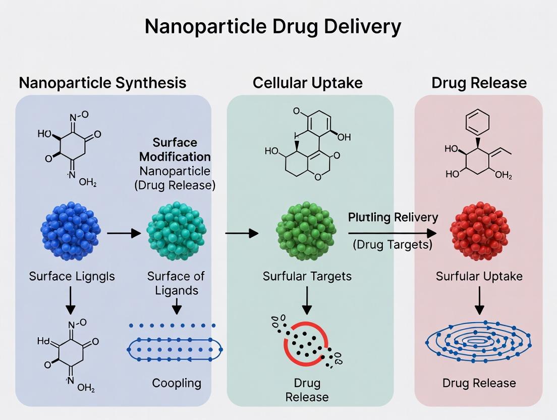
Nanoparticle Drug Delivery: Core Principles, Design Strategies, and Clinical Translation for Researchers
This comprehensive guide for researchers and drug development professionals explores the fundamental principles of nanoparticle drug delivery systems.
Nanoparticle Drug Delivery: Core Principles, Design Strategies, and Clinical Translation for Researchers
Abstract
This comprehensive guide for researchers and drug development professionals explores the fundamental principles of nanoparticle drug delivery systems. We detail the core concepts of nanomedicine, from rational material selection and synthesis methods to advanced targeting strategies and surface engineering. The article provides practical insights into formulation optimization, characterization techniques, and overcoming biological barriers. Furthermore, we examine rigorous validation protocols, comparative analyses of nanoplatforms, and the critical regulatory pathway to clinical translation, offering a holistic view essential for advancing next-generation therapeutics.
The Nano-Scale Frontier: Core Concepts and Rationale for Drug Delivery
This guide, framed within a broader thesis on the basic principles of nanoparticle drug delivery, details the defining characteristics of the nanoscale and the Enhanced Permeability and Retention (EPR) effect. The EPR effect is a cornerstone concept that exploits the unique pathophysiology of tumor vasculature to facilitate targeted drug accumulation.
Defining the Nano-Realm: Key Physicochemical Properties
Nanoparticles (NPs) for drug delivery are typically defined as constructs between 1-1000 nm, with the 10-200 nm range being optimal for systemic administration. Their efficacy is governed by interdependent core properties.
Table 1: Key Physicochemical Properties of Nanoparticles and Their Impact
| Property | Typical Optimal Range | Primary Biological Impact |
|---|---|---|
| Size | 10 - 200 nm | Determines renal clearance (<10 nm), vascular extravasation, and tissue penetration. |
| Surface Charge (Zeta Potential) | ±10 - ±30 mV | Influences stability, opsonization, and cellular uptake. Near-neutral or slightly negative reduces non-specific binding. |
| Hydrophobicity/Lipophilicity | Log P ~ 1-5 (varies) | Affects protein corona formation, circulation time, and membrane interactions. |
| Shape | Spherical, Rod-like, Disc-like | Alters flow dynamics, margination, and internalization kinetics. |
| Surface Functionalization | PEG density: 5-20% molar ratio | Modulates stealth (anti-opsonization), targeting (ligand conjugation), and biocompatibility. |
The Enhanced Permeability and Retention (EPR) Effect: Mechanism and Heterogeneity
The EPR effect describes the passive accumulation of macromolecules and nanoparticles in tumor tissue due to:
- Enhanced Permeability: Tumor vasculature exhibits wide fenestrations (100-2000 nm) due to malformed, discontinuous endothelial cells and poor pericyte coverage.
- Retention: Tumors lack functional lymphatic drainage, leading to the accumulated agents being trapped interstitially.
Diagram 1: Mechanism of the EPR Effect in Tumor vs. Normal Tissue
Important Consideration: Recent research highlights the significant heterogeneity of the EPR effect across tumor types, locations, and individual patients, which is a major challenge in clinical translation.
Experimental Protocols for Characterizing NPs and Evaluating the EPR Effect
Protocol 1: Dynamic Light Scattering (DLS) for Hydrodynamic Size & Zeta Potential
- Objective: Determine nanoparticle hydrodynamic diameter (size) and surface charge (zeta potential).
- Materials: Purified NP suspension, appropriate buffer (e.g., 1xPBS, 1mM KCl), DLS/Zetasizer instrument.
- Method:
- Dilute NP sample in clear, disposable cuvette to avoid signal saturation (typically 0.1-1 mg/mL).
- For size: Measure at a fixed angle (e.g., 173° backscatter) at 25°C. Perform minimum 3 runs, report Z-average diameter and polydispersity index (PDI).
- For zeta potential: Use folded capillary cell. Measure electrophoretic mobility and convert to zeta potential via Smoluchowski equation. Perform >10 measurements.
Protocol 2: In Vivo Fluorescence Imaging for EPR Evaluation
- Objective: Visualize and quantify passive tumor accumulation of NPs.
- Materials: Tumor-bearing mouse model (e.g., subcutaneous xenograft), fluorescently labeled NPs (e.g., Cy5.5, DyLight 800), in vivo fluorescence imaging system, anesthesia setup.
- Method:
- Randomize mice into control and treatment groups.
- Administer fluorescent NPs via tail vein injection at a standardized dose (e.g., 5 mg/kg).
- Anesthetize mice at predetermined time points (e.g., 1, 4, 24, 48h) and image using appropriate excitation/emission filters.
- Quantify mean fluorescence intensity (MFI) in the tumor region of interest (ROI) and normalize to a background tissue ROI. Calculate tumor-to-background ratio.
- At terminal time point, excise organs (tumor, liver, spleen, kidneys, heart, lungs) for ex vivo imaging to quantify biodistribution.
The Scientist's Toolkit: Research Reagent Solutions
Table 2: Essential Materials for NP Characterization and EPR Studies
| Reagent/Material | Function/Description | Key Provider Examples |
|---|---|---|
| PLGA (Poly(lactic-co-glycolic acid)) | Biodegradable, FDA-approved polymer for forming NP core; encapsulates both hydrophilic/hydrophobic drugs. | Sigma-Aldrich, LACTEL Absorbable Polymers |
| DSPE-mPEG (2000) | Phospholipid-PEG conjugate for creating "stealth" nanoparticles; reduces opsonization and extends circulation half-life. | Avanti Polar Lipids, Nanocs |
| Cy5.5 NHS Ester | Near-infrared fluorescent dye for labeling amine-containing NPs; enables in vivo tracking and EPR quantification. | Lumiprobe, GE Healthcare |
| Matrigel Basement Membrane Matrix | Used for establishing subcutaneous tumor xenografts; provides a scaffold for tumor cell growth. | Corning |
| Dylight 800 NHS Ester | Far-red/NIR dye for in vivo imaging; minimizes tissue autofluorescence for deeper tissue penetration. | Thermo Fisher Scientific |
| PD-10 Desalting Columns | For purifying and buffer-exchanging synthesized NPs via size-exclusion chromatography. | Cytiva |
Signaling Pathways Influencing Vascular Permeability in Tumors
Diagram 2: Key Pathways in Tumor Vascular Hyperpermeability
Thesis Context: Within the fundamental principles of nanoparticle (NP) drug delivery research, the core objective is to engineer carriers that overcome the intrinsic biopharmaceutical limitations of active pharmaceutical ingredients (APIs). This whitepaper details how nanoplatforms address the triad of solubility, stability, and pharmacokinetic (PK) challenges, thereby translating molecular efficacy into clinical therapeutic outcomes.
Addressing Poor Aqueous Solubility
Poor solubility remains a primary cause of drug candidate attrition. Nanocarriers enhance apparent solubility by molecular dispersion, surface adsorption, or encapsulation.
Mechanism: Nanoformulations increase the surface area-to-volume ratio, promoting interaction with aqueous media. For crystalline drugs, nanoparticles can create a metastable high-energy polymorph or an amorphous solid dispersion, enhancing dissolution rate and saturation solubility via the Ostwald-Freundlich equation.
Quantitative Impact of Nanonization on Solubility & Dissolution:
| API (Class) | Nanoparticle Type | Particle Size (nm) | Saturation Solubility Increase (vs. Bulk) | Dissolution Rate Enhancement | Reference Year |
|---|---|---|---|---|---|
| Fenofibrate (BCS II) | Nanocrystal | ~250 nm | ~4.5-fold | ~90% release in 15 min (bulk: 30% in 60 min) | 2023 |
| Curcumin (BCS IV) | Polymeric NP (PLGA) | ~180 nm | ~12-fold | Complete in 4h (bulk: <20% in 24h) | 2024 |
| Paclitaxel (BCS IV) | Polymeric Micelle | ~20 nm | ~1500-fold (theoretical) | >80% in 1h | 2023 |
Key Protocol: Fabrication of Drug Nanocrystals via Wet Milling
- Preparation: Disperse coarse drug powder (1-10% w/w) in an aqueous stabilizer solution (e.g., 0.5-2% w/v HPMC or PVP).
- Milling: Load the suspension into a milling chamber filled with yttrium-stabilized zirconia beads (0.1-0.3 mm diameter). The bead-to-powder weight ratio is typically 20:1.
- Process: Mill at high agitation speed (2000-4000 rpm) for 2-8 hours, maintaining temperature < 25°C.
- Separation: Separate beads from the nanosuspension using a sieve. The resulting suspension is characterized for size (DLS, LD), crystallinity (PXRD, DSC), and dissolution profile.
Enhancing Chemical and Physical Stability
NPs protect labile APIs from degradation pathways (hydrolysis, oxidation, photolysis) and prevent physical instability (amorphous precipitation, crystal growth).
Mechanism: The core-shell structure of many NPs creates a barrier against reactive species (e.g., H+, OH-, O2). For biologics (proteins, mRNA), NPs shield from enzymatic degradation and immune recognition.
Table: Stability Enhancement of Labile Compounds via Nanoencapsulation
| Labile Compound | Nanoparticle System | Stability Challenge | Improvement Achieved | Storage Condition Tested |
|---|---|---|---|---|
| siRNA | Lipid Nanoparticle (LNP) | Nuclease degradation | >95% intact after 24h in serum | 4°C, 6 months |
| Insulin | Chitosan-Zn NP | Aggregation & hydrolysis | Retained >90% bioactivity after 1 month | 25°C/60% RH |
| Omega-3 Fatty Acids | Nanoemulsion | Lipid peroxidation | Peroxide value reduced by 70% after 90 days | 40°C |
Key Protocol: Accelerated Stability Testing of Lipid Nanoparticles
- Formulation: Prepare LNPs via microfluidic mixing encapsulating the API.
- Stress Conditions: Aliquot samples and expose to:
- Thermal: 4°C, 25°C, 40°C.
- Physical: Freeze-thaw cycles (-80°C to 25°C).
- Chemical: Incubation in buffers of pH 5.0 and 7.4.
- Analysis: At predetermined intervals (0, 1, 3, 6 months), analyze for:
- Size & PDI: Dynamic Light Scattering (DLS).
- Encapsulation Efficiency (EE%): Measure using a centrifugal ultrafiltration method followed by HPLC/UV.
- Chemical Integrity: HPLC or gel electrophoresis for nucleic acids/proteins.
Optimizing Pharmacokinetics and Biodistribution
NPs fundamentally alter the PK profile of drugs by modifying absorption, distribution, metabolism, and excretion (ADME).
Core PK Advantages:
- Prolonged Circulation: Stealth coating (e.g., PEGylation) minimizes opsonization and reticuloendothelial system (RES) uptake.
- Passive Targeting: Exploits the Enhanced Permeability and Retention (EPR) effect in inflamed or tumor tissues.
- Reduced Clearance: Alters renal filtration thresholds and hepatic metabolism patterns.
Table: Pharmacokinetic Parameters of Conventional vs. Nanoformulated Doxorubicin
| Parameter | Conventional Doxorubicin (Solution) | Liposomal Doxorubicin (Doxil) | Change |
|---|---|---|---|
| t₁/₂ (alpha) | ~5 min | ~1-3 h | >> Increase |
| t₁/₂ (beta) | ~20-48 h | ~55-80 h | Significant Increase |
| Clearance (CL) | 24-35 L/h/m² | 0.04 L/h/m² | Drastic Decrease |
| Volume of Distribution (Vd) | ~700 L/m² | ~2.5 L/m² | Major Decrease |
| AUC (0-∞) | Low | ~300-fold higher | Massive Increase |
Key Protocol: In Vivo Pharmacokinetic Study in Rodents
- Dosing: Administer nanoformulated drug and control (free drug) intravenously to groups of rats/mice (n=6 per group) at equivalent doses (e.g., 5 mg API/kg).
- Sampling: Collect blood samples (100-200 µL) via saphenous or caudal vein at pre-determined time points (e.g., 2 min, 15 min, 30 min, 1, 2, 4, 8, 12, 24, 48 h post-injection).
- Processing: Centrifuge blood to obtain plasma. Precipitate proteins with acetonitrile.
- Analysis: Quantify drug concentration in plasma using a validated LC-MS/MS method.
- Modeling: Fit concentration-time data using non-compartmental analysis (NCA) software (e.g., Phoenix WinNonlin) to derive PK parameters: AUC, Cmax, t1/2, CL, Vd.
The Scientist's Toolkit: Key Research Reagent Solutions
| Reagent/Material | Function/Application in Nano-Delivery Research |
|---|---|
| PLGA (Poly(lactic-co-glycolic acid)) | Biodegradable polymer for forming solid core NPs; controls sustained release kinetics. |
| DSPC/Cholesterol | Primary lipid components for liposome/LNP formation, providing bilayer structure and stability. |
| DMG-PEG 2000 | PEG-lipid conjugate used in LNPs for surface PEGylation, providing stealth properties and stabilizing particle size. |
| Ionizable Cationic Lipid (e.g., DLin-MC3-DMA) | Critical for LNP-mediated nucleic acid delivery; enables complexation and endosomal escape. |
| Poloxamer 188 (F-68) | Non-ionic surfactant used as a stabilizer in nanoemulsions and nanosuspensions to prevent aggregation. |
| Cy5.5 or DiR Near-Infrared Dye | Hydrophobic or lipophilic tracers for in vivo and ex vivo imaging of nanoparticle biodistribution. |
| Sephadex G-50/G-75 Columns | Size-exclusion chromatography for purification of nanoparticles (e.g., separating free drug/dye/RNA from encapsulated). |
| Dialysis Tubing (MWCO 3.5-14 kDa) | For purifying nanoparticles via dialysis against relevant buffers to remove organic solvents and free molecules. |
| Transwell Permeability Assay Plates | For in vitro assessment of nanoparticle transport across Caco-2 or endothelial cell monolayers. |
Visualizations
Diagram 1: NP Pathways for PK Optimization
Diagram 2: Wet Milling Nanocrystal Production
Diagram 3: LNP Stabilization Against Degradation
An In-Depth Technical Guide to the Nanoparticle Core
Within the broader thesis of nanoparticle drug delivery, the core represents the fundamental payload compartment, dictating loading capacity, release kinetics, and intrinsic therapeutic or diagnostic function. Its rational design is paramount to translating nanoscale constructs into viable clinical therapies.
Core Composition and Quantitative Properties
The core’s material defines its primary characteristics. Below is a comparative analysis of prevalent core types.
Table 1: Core Materials, Properties, and Applications
| Core Material | Typical Size Range (nm) | Drug Loading Capacity (% w/w) | Key Functional Properties | Primary Application |
|---|---|---|---|---|
| Polymeric (e.g., PLGA) | 50-300 | 5-30 | Biodegradable, tunable release kinetics, high encapsulation efficiency for hydrophobic drugs. | Sustained release of small molecules, peptides. |
| Lipid (Solid Lipid NP) | 40-200 | 1-10 | High biocompatibility, physical stability, can incorporate lipophilic drugs. | Dermal, oncological, and gene delivery. |
| Lipid (Liposome) | 80-150 | 1-5 (aqueous core) | Amphiphilic bilayer, can encapsulate both hydrophilic (in core) and hydrophobic (in bilayer) agents. | Vaccines, antifungal/chemotherapy agents. |
| Inorganic (Mesoporous Silica) | 50-200 | 10-40 | Extremely high surface area (>900 m²/g), tunable pore size (2-10 nm), surface easily functionalized. | High-density loading, triggered release, theranostics. |
| Inorganic (Gold/Superparamagnetic Iron Oxide) | 5-50 | Low (surface conjugation) | Plasmonic resonance (Au), superparamagnetism (SPION), photothermal conversion. | Hyperthermia, imaging contrast, photothermal therapy. |
| Micelle (Polymeric) | 10-100 | 5-25 | Dynamic assembly, hydrophobic core for poorly soluble drugs, critical micelle concentration dependent. | Solubilization of chemotherapeutics (e.g., paclitaxel). |
Core Characterization: Key Experimental Protocols
Protocol 1: Determination of Drug Loading Capacity (LC%) and Encapsulation Efficiency (EE%) This standard protocol quantifies the core’s payload.
- Separation: Isolate nanoparticles from free (unencapsulated) drug via ultracentrifugation (e.g., 100,000 x g, 1 hr, 4°C) or size-exclusion chromatography.
- Lysis/Extraction: Dissolve the nanoparticle pellet in an appropriate solvent (e.g., acetonitrile for PLGA, Triton X-100 for liposomes) to release the encapsulated drug.
- Quantification: Use HPLC or UV-Vis spectroscopy to measure drug concentration in the lysate (Cencapsulated). Measure drug in the supernatant to determine free drug (Cfree).
- Calculation:
- EE% = [Cencapsulated / (Cencapsulated + C_free)] x 100
- LC% = [Mass of encapsulated drug / Total mass of nanoparticles] x 100
Protocol 2: In Vitro Drug Release Kinetics Study This assesses the core’s release profile, critical for pharmacokinetics.
- Setup: Place a known amount of drug-loaded nanoparticles in a dialysis bag (MWCO selected to retain NPs but allow free drug diffusion) or use a sample-and-separate method.
- Incubation: Immerse the bag in a release medium (e.g., PBS, pH 7.4, with 0.5% w/v Tween 80 to maintain sink conditions) at 37°C under gentle agitation.
- Sampling: At predetermined time points, withdraw a volume of the external medium and replace with fresh pre-warmed medium.
- Analysis: Quantify drug concentration in each sample via HPLC/UV-Vis. Generate a cumulative release profile over time (e.g., 7-28 days).
Visualizing Core Function and Characterization
- Title: Core Synthesis to Pharmacokinetics Pathway
- Title: LC% and EE% Determination Workflow
The Scientist's Toolkit: Core Research Reagents
Table 2: Essential Reagents for Core Research
| Reagent / Material | Function in Core Research | Key Consideration |
|---|---|---|
| PLGA (Poly(lactic-co-glycolic acid)) | Biodegradable polymer core; release kinetics tuned by LA:GA ratio & MW. | End-group (ester vs. carboxyl), inherent viscosity. |
| DSPC / Cholesterol | Primary lipid for forming stable, rigid liposomal or lipid nanoparticle cores. | Phase transition temperature (Tm) dictates membrane fluidity. |
| mPEG-DSPE | Lipid-PEG conjugate for creating stealth cores (reduced opsonization). | PEG chain length (e.g., 2000 Da) affects corona thickness. |
| Tetrachloroauric Acid (HAuCl₄) | Precursor for synthesizing gold nanoparticle cores (citrate reduction). | Concentration and reductant control final core size. |
| Cetyltrimethylammonium Bromide (CTAB) | Template surfactant for mesoporous silica nanoparticle core synthesis. | Critical for pore formation; requires thorough removal. |
| Dialysis Tubing (MWCO 3.5-14 kDa) | For purifying cores and conducting in vitro release studies. | MWCO must be lower than nanoparticle size. |
| Sepharose CL-4B Column | Size-exclusion chromatography for gentle purification of delicate cores (e.g., liposomes). | Separates encapsulated from free drug. |
| Dynamic Light Scattering (DLS) Instrument | Measures core hydrodynamic diameter, polydispersity index (PDI), and zeta potential. | Sample must be free of dust/aggregates. |
Within the broader thesis on the basic principles of nanoparticle (NP) drug delivery, the selection of core material is a fundamental determinant of system performance. This whitepaper provides a technical guide to the four primary material classes—lipids, polymers, inorganics, and hybrid systems—detailing their properties, synthesis, functionalization, and experimental characterization. The rational design of nanocarriers requires a deep understanding of the chemical, physical, and biological interactions governed by this core material choice.
Lipid-Based Nanoparticles
Lipid nanoparticles (LNPs) are the leading platform for nucleic acid delivery, exemplified by mRNA COVID-19 vaccines. Their core comprises ionizable lipids, phospholipids, cholesterol, and PEG-lipids.
Key Experimental Protocol: Microfluidic Mixing for LNP Formation
- Objective: Reproducible, scalable formation of mRNA-encapsulating LNPs.
- Materials:
- Ethanol Phase: Ionizable lipid (e.g., DLin-MC3-DMA), DSPC, Cholesterol, DMG-PEG-2000 dissolved in ethanol.
- Aqueous Phase: mRNA in citrate buffer (pH 4.0).
- Procedure:
- Load ethanol and aqueous phases into separate syringes.
- Connect syringes to a staggered herringbone micromixer (e.g., Precision NanoSystems NanoAssemblr).
- Set total flow rate (TRF) to 12 mL/min and flow rate ratio (aqueous:ethanol) to 3:1.
- Collect effluent in a vessel.
- Dialyze against PBS (pH 7.4) for 2 hours to remove ethanol and raise pH, allowing lipid self-assembly into mRNA-loaded LNPs.
- Filter through a 0.22 µm sterile membrane.
- Characterization: Measure particle size (~80 nm) via DLS, PDI (<0.2), encapsulation efficiency (>90%) via RiboGreen assay, and in vitro transfection.
Research Reagent Solutions: Lipid Nanoparticle Formulation
| Reagent | Function & Explanation |
|---|---|
| Ionizable Lipid (e.g., ALC-0315) | Critical for encapsulation; positively charged at low pH, neutral at physiological pH, enabling complexation and endosomal escape. |
| DSPC (Phospholipid) | Provides structural integrity to the LNP bilayer, influences fusogenicity. |
| Cholesterol | Stabilizes the lipid bilayer, enhances membrane fluidity and fusion. |
| PEG-lipid (e.g., ALC-0159) | Shields surface, prevents aggregation, modulates pharmacokinetics by reducing protein adsorption. |
| RiboGreen Assay Kit | Fluorometric quantification of free vs. encapsulated nucleic acids to determine loading efficiency. |
Diagram: Microfluidic Workflow for LNP Production
Polymeric Nanoparticles
Biodegradable polymers like poly(lactic-co-glycolic acid) (PLGA) and poly(ε-caprolactone) (PCL) offer controlled release. Cationic polymers (e.g., PEI) enable complexation for gene delivery.
Key Experimental Protocol: Double Emulsion Solvent Evaporation for Hydrophilic Drug Encapsulation
- Objective: Encapsulate a hydrophilic drug (e.g., protein) within PLGA NPs.
- Materials: PLGA (50:50, acid-terminated), PVA, dichloromethane (DCM), drug payload.
- Procedure:
- Primary Emulsion (W1/O): Add drug aqueous solution (W1) to PLGA in DCM (O). Sonicate on ice (30s, 40% amplitude).
- Secondary Emulsion (W1/O/W2): Pour primary emulsion into PVA solution (W2). Homogenize (2 min, 10,000 rpm).
- Solvent Evaporation: Stir emulsion overnight at room temperature to evaporate DCM.
- Centrifugation: Pellet NPs (20,000 x g, 30 min), wash 3x with water to remove PVA.
- Lyophilization: Resuspend in cryoprotectant (e.g., 5% trehalose) and lyophilize for storage.
- Characterization: Size/PDI (DLS), morphology (SEM/TEM), drug loading (HPLC), in vitro release study (PBS, 37°C).
Inorganic Nanoparticles
Mesoporous silica nanoparticles (MSNs), gold nanoparticles (AuNPs), and superparamagnetic iron oxide nanoparticles (SPIONs) offer unique optical, magnetic, and structural properties.
Key Experimental Protocol: Synthesis of Mesoporous Silica Nanoparticles (MSNs)
- Objective: Synthesize amine-functionalized, drug-loaded MSNs.
- Materials: Tetraethyl orthosilicate (TEOS), Cetyltrimethylammonium bromide (CTAB), APTES, ammonia solution.
- Procedure (Stöber Method):
- Dissolve CTAB (template) in water/ammonia solution at 80°C.
- Under vigorous stirring, add TEOS dropwise. Stir for 2h.
- For co-condensation functionalization, add APTES with TEOS.
- Centrifuge, wash with ethanol. Remove template via acid extraction (HCl in methanol, reflux, 6h).
- Drug Loading: Incubate empty MSNs in concentrated drug solution (e.g., doxorubicin) for 24h. Centrifuge and wash.
Comparative Data: Core Material Properties
| Property | Lipid NPs (LNPs) | Polymeric NPs (PLGA) | Inorganic NPs (MSNs) | Hybrid NPs (Lipid-Polymer) |
|---|---|---|---|---|
| Typical Size Range | 50-150 nm | 100-300 nm | 50-200 nm | 80-200 nm |
| Drug Loading Capacity | Moderate (5-10%) | Moderate to High (5-20%) | Very High (Up to 30%+) | Moderate (5-15%) |
| Release Profile | Typically burst, then sustained | Bi-phasic: burst then sustained diffusion/erosion | Gated/pH-responsive | Tunable, often sustained |
| Scalability | Excellent (microfluidics) | Good | Good | Moderate/Complex |
| Biodegradability | Yes (enzymatic) | Yes (hydrolytic) | No (slow dissolution) | Yes (component-dependent) |
| Key Advantage | Superior nucleic acid delivery, clinical success | Controlled release, FDA-approved polymers | High surface area, tunable pores, multifunctionality | Combined benefits, stability + functionality |
Hybrid and Advanced Systems
Hybrid systems combine materials to overcome individual limitations (e.g., lipid-polymer hybrid NPs, inorganic core@silica shell).
Key Experimental Protocol: Formulation of Lipid-Polymer Hybrid Nanoparticles
- Objective: Create a core-shell NP with a PLGA core and a lipid-PEG shell.
- Materials: PLGA, phospholipid (e.g., DPPC), DSPE-PEG, chloroform.
- Procedure (Nanoprecipitation):
- Dissolve PLGA and lipids in organic solvent (e.g., acetone or acetonitrile).
- Inject solution rapidly into aqueous phase under stirring.
- Allow solvent to evaporate overnight. The lipids self-assemble around the polymeric core.
- Filter or centrifuge as needed.
Diagram: Structure of a Multifunctional Hybrid Nanoparticle
Critical Characterization Toolkit
| Technique | Primary Metrics | Relevance to Material Choice |
|---|---|---|
| Dynamic Light Scattering (DLS) | Hydrodynamic diameter, PDI, Zeta Potential | Universal QC. PDI indicates uniformity. Zeta potential predicts colloidal stability. |
| Electron Microscopy (TEM/SEM) | Morphology, core-shell structure, actual size | Visual confirmation of structure, crucial for hybrids and inorganics. |
| Differential Scanning Calorimetry (DSC) | Glass transition (Tg), melting points, crystallinity | Polymer crystallinity affects degradation; lipid phase behavior crucial for LNP stability. |
| X-ray Photoelectron Spectroscopy (XPS) | Surface elemental composition | Verifies surface functionalization (e.g., PEG, targeting ligands). |
| Brunauer-Emmett-Teller (BET) | Surface area, pore volume/radius | Essential for characterizing mesoporous materials (MSNs). |
The material landscape for nanoparticle drug delivery is rich and modular. Lipid systems excel in nucleic acid delivery, polymers in controlled release kinetics, inorganics in multifunctionality and imaging, while hybrids offer engineered solutions. The choice is dictated by the therapeutic payload, desired pharmacokinetics, route of administration, and manufacturing considerations. Future advances lie in the intelligent combination of these materials to create next-generation, stimulus-responsive, and targeted nanomedicines.
Within the foundational thesis of nanoparticle (NP)-based drug delivery, the journey from administration to target is governed by a series of biological interactions at the nano-bio interface. The formation of a protein corona, the subsequent process of opsonization, and the activation of clearance mechanisms constitute the primary barriers to effective delivery. These phenomena directly determine NP pharmacokinetics, biodistribution, and ultimately, therapeutic efficacy. This guide provides a technical deep-dive into these core principles, essential for rational NP design in therapeutic applications.
The Protein Corona: Formation and Characterization
Upon introduction into a biological fluid (e.g., plasma), NPs are rapidly coated by a layer of proteins and other biomolecules. This "corona" defines the biological identity of the NP, overriding its synthetic identity.
Corona Composition and Dynamics
The corona consists of a "hard corona" (tightly bound, slow-exchange proteins) and a "soft corona" (loosely bound, rapidly exchanging proteins). Composition is influenced by NP physicochemical properties.
Table 1: Key Nanoparticle Properties Affecting Protein Corona Formation
| NP Property | Experimental Parameter | Impact on Corona Composition | Common Measurement Technique |
|---|---|---|---|
| Size | Hydrodynamic diameter (nm) | Smaller NPs have higher curvature, affecting protein binding affinity; influences opsonin access. | Dynamic Light Scattering (DLS) |
| Surface Charge | Zeta potential (mV) | Highly positive or negative surfaces attract proteins with opposite charge; neutral/slightly negative often reduces opsonin adsorption. | Electrophoretic Light Scattering |
| Hydrophobicity | Water contact angle (°) | Increased hydrophobicity generally enhances total protein adsorption and favors specific opsonins (e.g., immunoglobulins). | Contact Angle Goniometry |
| Surface Chemistry | Functional group (e.g., -COOH, -PEG) | PEGylation drastically reduces protein adsorption; specific ligands can trigger targeted protein binding. | X-ray Photoelectron Spectroscopy (XPS) |
| Shape | Aspect ratio | Alters surface area and binding geometry for proteins; spherical vs. rod-shaped NPs show different corona profiles. | Transmission Electron Microscopy (TEM) |
Experimental Protocol: Protein Corona Isolation and Analysis via SDS-PAGE & LC-MS/MS
Objective: To isolate and identify proteins constituting the hard corona of NPs after incubation in human plasma.
Materials:
- Purified NPs (e.g., PLGA, liposomal, silica).
- Human platelet-poor plasma (pooled, healthy donor).
- Phosphate Buffered Saline (PBS), pH 7.4.
- Ultracentrifuge and compatible tubes (e.g., polycarbonate).
- SDS-PAGE gel system (4-20% gradient gel).
- In-gel trypsin digestion: Excise gel bands, destain, reduce with DTT, alkylate with iodoacetamide, digest with sequencing-grade trypsin overnight.
- LC-MS/MS Analysis: Separate peptides on a C18 nano-column using a nanoflow HPLC system coupled to a high-resolution tandem mass spectrometer (e.g., Q-Exactive). Data-dependent acquisition (DDA) mode.
- Data Processing: Search MS/MS spectra against the human UniProt database using software (e.g., MaxQuant, Proteome Discoverer). Criteria: 1% false discovery rate (FDR). Quantify via label-free quantification (LFQ) intensity or spectral counting.
Opsonization: The Bridge to Clearance
Opsonization is the specific tagging of NPs by host-derived proteins (opsonins) that facilitate recognition by phagocytic cells. The protein corona is rich in opsonins.
Table 2: Major Opsonins and Their Recognition Receptors
| Opsonin | Class | Primary Recognition Receptor on Phagocyte | Consequence of Binding |
|---|---|---|---|
| Immunoglobulin G (IgG) | Antibody | Fcγ Receptors (FcγR I, II, III) | Strong phagocytic signal; activates complement cascade. |
| Immunoglobulin M (IgM) | Antibody | Complement receptor (indirect via C3b) | Primary activator of the classical complement pathway. |
| C3b / iC3b | Complement protein | Complement Receptor 1 (CR1), CR3 (αMβ2 integrin) | Central opsonin; CR3 binding triggers internalization. |
| C1q | Complement protein | gC1qR, cC1qR | Initiates classical complement pathway; can directly promote phagocytosis. |
| Fibrinogen | Acute-phase protein | αMβ2 integrin (CR3), Mac-1 | Bridges NP to phagocyte; promotes inflammation. |
| Apolipoproteins (e.g., ApoE) | Lipid transport protein | LDL Receptor family | Can mediate hepatic clearance. |
Diagram: Key Opsonization and Recognition Pathways
Diagram Title: Opsonin-Driven Phagocytosis Pathway
Clearance Mechanisms
NPs are primarily cleared by the Mononuclear Phagocyte System (MPS), also known as the Reticuloendothelial System (RES). The liver (Kupffer cells) and spleen are the major filtration organs.
Key Clearance Pathways
- Hepatic Clearance: Kupffer cells in liver sinusoids capture opsonized NPs via receptor-mediated phagocytosis. Non-phagocytic hepatic sinusoidal endothelial cells (HSECs) can also clear smaller NPs via pinocytosis.
- Splenic Clearance: NPs must navigate tight inter-endothelial cell slits in the red pulp and marginal sinus. Size is a critical determinant; particles >200 nm are typically filtered mechanically and phagocytosed by splenic macrophages.
- Renal Clearance: Typically limited to very small NPs (<~5-6 nm in diameter) that can pass through the glomerular filtration barrier. Essential for designing clearable NPs to reduce long-term toxicity.
- Complement Activation: Both classical and alternative complement pathways lead to C3b opsonization and formation of the Membrane Attack Complex (MAC), which can cause NP lysis (particularly for lipid-based systems).
Experimental Protocol: In Vivo Biodistribution and Clearance Kinetics Study
Objective: To quantify NP accumulation in major organs and determine blood circulation half-life.
Materials:
- Radiolabeled or fluorescently labeled NPs (e.g., with ^125^I, Cy5.5, DiR dye).
- Animal model (e.g., BALB/c mice).
- In vivo imaging system (IVIS) for fluorescence (if applicable).
- Gamma counter (for radioactivity).
- Tissue homogenizer.
Methodology:
- NP Administration: Inject a known dose of labeled NPs (e.g., 100 µL, 1 mg/mL) intravenously via the tail vein.
- Time-Point Sampling: At predetermined time points (e.g., 5 min, 30 min, 2 h, 8 h, 24 h, 48 h), collect blood retro-orbitally or via cardiac puncture (terminal). Euthanize animals and harvest organs (liver, spleen, kidneys, lungs, heart, brain).
- Blood Pharmacokinetics: Measure radioactivity or fluorescence intensity in blood samples. Plot concentration vs. time. Calculate half-life (t~1/2~) using a non-compartmental model.
- Biodistribution Quantification:
- Radiolabel: Weigh organs, count radioactivity in a gamma counter. Express as percentage of injected dose per gram of tissue (%ID/g).
- Fluorescence: Homogenize organs in PBS. Measure fluorescence of homogenate (using a plate reader) against a standard curve of the labeled NP. Calculate %ID/g.
- Imaging: For fluorescent NPs, perform ex vivo imaging of excised organs using IVIS to visualize distribution.
Table 3: Representative Quantitative Biodistribution Data (%ID/g) for Model NPs
| Organ / Time | PEGylated Liposome (100 nm) | Plain Polystyrene NP (200 nm) | Small Silica NP (10 nm) |
|---|---|---|---|
| Blood (2 h) | 45.2 ± 3.1 | 5.5 ± 1.2 | 12.8 ± 2.5 |
| Liver (24 h) | 18.5 ± 2.4 | 65.3 ± 5.7 | 35.4 ± 4.1 |
| Spleen (24 h) | 5.2 ± 0.9 | 22.1 ± 3.3 | 4.1 ± 1.0 |
| Kidneys (24 h) | 1.1 ± 0.3 | 1.5 ± 0.4 | 28.7 ± 3.8* |
| Lungs (24 h) | 1.8 ± 0.5 | 3.2 ± 1.1 | 2.9 ± 0.8 |
| Blood t~1/2~ (h) | ~12 | ~0.8 | ~3.5 |
Data is illustrative. *High kidney signal suggests renal clearance pathway for small NP.
The Scientist's Toolkit: Research Reagent Solutions
Table 4: Essential Materials for Studying Corona, Opsonization & Clearance
| Item / Reagent | Supplier Examples | Primary Function in Research |
|---|---|---|
| Human Platelet-Poor Plasma (PPP) | Sigma-Aldrich, George King Bio-Medical | Standardized biological fluid for in vitro protein corona formation studies. |
| PEGylation Reagents (mPEG-NHS) | Creative PEGWorks, Laysan Bio | Conjugate polyethylene glycol to NP surfaces to reduce protein adsorption and opsonization ("stealth" effect). |
| Purified Human Proteins (IgG, C3, Fibrinogen) | Athens Research & Technology, Complement Technology | Use as standards or for controlled spiking experiments to study specific opsonin interactions. |
| Anti-Human IgG (Fc specific) Antibody, Gold-labeled | Cytodiagnostics, Abcam | Electron microscopy visualization of IgG opsonin location on the NP corona. |
| Fluorescent Dyes (DiD, Cy5.5, ICG) | Thermo Fisher, Lumiprobe | Label NPs for in vivo and in vitro tracking via fluorescence microscopy, flow cytometry, or IVIS imaging. |
| Radiolabeling Kits (^125^I, ^111^In) | PerkinElmer, MCPF | Provide highly sensitive, quantitative tracking for biodistribution and pharmacokinetic studies. |
| Mouse/Rat Serum Complement | Cedarlane Labs | Source of active complement proteins for studying complement activation-related opsonization and lysis. |
| Differentiated THP-1 Human Monocytes | ATCC | Consistent in vitro model of human macrophages for phagocytosis and clearance assays. |
| Clodronate Liposomes | Liposoma BV | Deplete phagocytic macrophages (e.g., Kupffer cells) in vivo to study their specific role in NP clearance. |
From Bench to Bedside: Design, Synthesis, and Active Targeting Strategies
This technical guide details three core fabrication techniques for polymeric nanoparticles used in drug delivery. Within the broader thesis on the basic principles of nanoparticle drug delivery research, these methods represent the foundational engineering approaches to control critical particle characteristics—size, polydispersity, drug loading, and release kinetics—which ultimately dictate in vivo pharmacokinetics, biodistribution, and therapeutic efficacy.
Core Fabrication Techniques: Mechanisms and Comparison
Emulsification-Solvent Evaporation/Diffusion
This technique involves creating an emulsion of a polymer-containing organic phase in an aqueous phase, followed by solvent removal to solidify the nanoparticles.
- Single Emulsion (o/w): For hydrophobic drugs. Oil phase (organic solvent + polymer + drug) is emulsified in an aqueous phase (water + surfactant) via high-energy input (e.g., sonication, homogenization).
- Double Emulsion (w/o/w): For hydrophilic drugs. An aqueous drug solution is first emulsified in a polymer-containing organic phase (w/o), which is then emulsified in a second aqueous phase (w/o/w).
Nanoprecipitation (Solvent Displacement)
A low-energy method based on the interfacial deposition of a polymer following the displacement of a water-miscible solvent from a lipophilic solution. The organic phase (polymer + drug in acetone, ethanol, or THF) is added to a stirred aqueous phase. Rapid solvent diffusion leads to a decrease in interfacial tension, causing the spontaneous formation of nanoparticles.
Microfluidics
A precision engineering approach where fluids are manipulated in microscale channels (tens to hundreds of micrometers). For nanoparticles, two primary configurations are used:
- Laminar Flow Focusing: A central stream containing polymer and drug is hydrodynamically focused by two side aqueous streams, enabling controlled mixing by diffusion.
- Droplet-Based Microfluidics: The organic and aqueous phases meet at a T-junction or flow-focusing geometry to generate monodisperse emulsion droplets, which act as templates for nanoparticles upon solvent removal.
Quantitative Data Comparison
Table 1: Comparative Analysis of Key Nanoparticle Fabrication Techniques
| Parameter | Emulsification (o/w) | Nanoprecipitation | Microfluidics (Laminar Flow) |
|---|---|---|---|
| Typical Size Range | 100 – 500 nm | 50 – 300 nm | 20 – 200 nm |
| Polydispersity Index (PDI) | Moderate-High (0.1 – 0.3) | Low-Moderate (0.05 – 0.2) | Very Low (< 0.05) |
| Drug Loading Capacity | Moderate to High (up to 30%) | Typically Low (< 10%) | Tunable, Moderate |
| Encapsulation Efficiency | Moderate-High (60-90%) | Variable, often lower (30-70%) | High and Reproducible (often >80%) |
| Throughput/Scale | High (batch process) | Moderate (batch process) | Lower (continuous, but scalable via parallelization) |
| Key Controlling Parameters | Homogenization energy/speed, Surfactant type/concentration, Viscosity | Solvent selection, Aqueous:Organic phase ratio, Addition rate | Flow Rate Ratio (FRR), Total Flow Rate (TFR), Channel geometry |
| Best Suited For | Hydrophobic drugs, PLGA, PCL polymers | Lipophilic drugs, PLA, polyester polymers | Precision formulations, sensitive biologics, core-shell structures |
Data synthesized from current literature (2023-2024). PDI values are typical targets; actual results are formulation-dependent.
Detailed Experimental Protocols
Protocol 4.1: Single Emulsification for PLGA Nanoparticles
Objective: Fabricate drug-loaded PLGA nanoparticles using the o/w emulsification-solvent evaporation method. Materials: See Section 6. Procedure:
- Dissolve 50 mg PLGA and 5 mg hydrophobic drug (e.g., Paclitaxel) in 2 mL of dichloromethane (DCM) to form the organic phase.
- Prepare 20 mL of a 1-2% (w/v) polyvinyl alcohol (PVA) solution in deionized water as the aqueous phase.
- Add the organic phase to the aqueous phase while probe-sonicating (70% amplitude) for 60 seconds over an ice bath to form a coarse o/w emulsion.
- Immediately transfer the emulsion to a high-pressure homogenizer and process at 15,000 psi for 3 cycles.
- Stir the resulting nanoemulsion at room temperature for 4 hours to evaporate the organic solvent.
- Centrifuge the suspension at 20,000 x g for 30 minutes at 4°C to collect nanoparticles. Wash twice with DI water to remove excess surfactant.
- Resuspend the pellet in an appropriate buffer and lyophilize for storage.
Protocol 4.2: Standard Nanoprecipitation
Objective: Formulate polymeric micelles/nanoparticles via solvent displacement. Materials: See Section 6. Procedure:
- Dissolve 10 mg of polymer (e.g., PLA-PEG) and 1 mg of lipophilic drug in 1 mL of acetone (organic phase).
- Prepare 10 mL of deionized water or a 0.1% surfactant solution as the aqueous phase under moderate magnetic stirring (500-600 rpm).
- Using a syringe pump, inject the organic phase into the aqueous phase at a controlled rate (e.g., 1 mL/min).
- Allow stirring to continue for 2-3 hours to ensure complete solvent diffusion and nanoparticle hardening.
- Optionally, place the suspension under reduced pressure (rotary evaporator) to remove residual organic solvent.
- Filter the suspension through a 0.45 μm or 0.22 μm membrane filter. Use directly or lyophilize.
Protocol 4.3: Microfluidic Nanoparticle Synthesis (Flow-Focusing)
Objective: Synthesize monodisperse nanoparticles using a glass capillary or PDMS chip microfluidic device. Materials: See Section 6. Procedure:
- Phase Preparation: Prepare the organic phase (2 mg/mL polymer in ethanol) and the aqueous phase (0.5% PVA in water).
- Device Priming: Load syringes with each phase and connect to the device via tubing. Prime all channels to remove air bubbles.
- Flow Rate Calibration: Set syringe pumps. A typical starting Flow Rate Ratio (FRR = Aqueous / Organic) is 5:1. Set Organic flow rate to 0.1 mL/hr and Aqueous to 0.5 mL/hr (Total Flow Rate, TFR = 0.6 mL/hr).
- Particle Formation: Start pumps. The organic stream is hydrodynamically focused at the junction, leading to rapid mixing and nanoprecipitation in the outlet channel.
- Collection: Collect effluent in a vial under gentle stirring. Allow collection for 1 hour.
- Post-Processing: Transfer the colloidal suspension to an open vial and stir for 1 hour to evaporate residual solvent. Centrifuge and wash as needed.
Diagrams and Workflows
Title: Emulsification-Solvent Evaporation Protocol Flowchart
Title: Microfluidic Parameter Impact on Nanoparticle Properties
The Scientist's Toolkit: Research Reagent Solutions
Table 2: Essential Materials for Nanoparticle Fabrication
| Category | Item/Reagent | Typical Function & Role in Fabrication |
|---|---|---|
| Polymers | PLGA (Poly(lactic-co-glycolic acid)) | Biodegradable matrix for controlled drug release; backbone of emulsification techniques. |
| PLA-PEG (Poly(lactic acid)-Poly(ethylene glycol)) | Amphiphilic copolymer for stealth nanoparticles via nanoprecipitation; PEG shell reduces opsonization. | |
| Surfactants/Stabilizers | Polyvinyl Alcohol (PVA) | Most common stabilizer in o/w emulsification; adsorbs at oil-water interface, controlling particle growth/aggregation. |
| Poloxamers (e.g., Pluronic F-68) | Non-ionic triblock copolymer surfactant; used in nanoprecipitation & microfluidics to improve colloidal stability. | |
| Sodium Cholate | Bile salt surfactant; used in microfluidics for highly monodisperse, small nanoparticle formation. | |
| Solvents | Dichloromethane (DCM) | Volatile organic solvent for emulsification; immiscible with water, evaporated after emulsion formation. |
| Acetone/Ethanol | Water-miscible organic solvents for nanoprecipitation; rapid diffusion into aqueous phase drives self-assembly. | |
| Equipment & Consumables | High-Pressure Homogenizer / Probe Sonicator | Provides high-energy input for emulsification, reducing droplet size to nanoscale. |
| Syringe Pump (Dual) | Provides precise, pulseless control over fluid injection rates in nanoprecipitation and microfluidics. | |
| Microfluidic Chip | PDMS or glass device with designed channels (e.g., flow-focusing geometry) for controlled fluid mixing. | |
| Ultrafiltration Centrifugal Devices (MWCO) | For washing and concentrating nanoparticle suspensions based on size exclusion. |
This whitepaper provides an in-depth technical analysis of three fundamental performance metrics in nanoparticle (NP) drug delivery: Encapsulation Efficiency (EE), Drug Loading (DL), and the mechanisms of controlled release via pH, enzymatic, and redox triggers. Framed within the basic principles of nanoparticle drug delivery research, this guide details experimental protocols, quantitative benchmarks, and material toolkits essential for formulation scientists.
The efficacy of a nanoparticle drug delivery system (DDS) is predicated on its ability to incorporate a therapeutic agent (loading) and release it in a spatiotemporally controlled manner at the target site. Encapsulation Efficiency (EE%) and Drug Loading (DL%) are the primary quantitative metrics for evaluating loading success. Subsequently, engineered responsiveness to pathological stimuli—such as lowered extracellular pH in tumors (pH ~6.5-7.0), overexpressed enzymes (e.g., matrix metalloproteinases, esterases), or elevated redox potential (elevated glutathione in cytosol)—ensures precise drug release, minimizing off-target effects and systemic toxicity.
Quantitative Benchmarks: EE% and DL%
EE% and DL% are defined as:
- EE% = (Mass of drug in NPs / Total mass of drug used in formulation) × 100
- DL% = (Mass of drug in NPs / Total mass of drug-loaded NPs) × 100
Table 1 summarizes typical benchmark values and influencing factors for polymeric and lipid-based nanoparticles.
Table 1: Benchmark Ranges for EE% and DL% in Common Nanocarriers
| Nanocarrier Type | Typical Polymer/Lipid | Typical EE% Range | Typical DL% Range | Key Influencing Factors |
|---|---|---|---|---|
| Polymeric NP (e.g., PLA, PLGA) | Poly(lactic-co-glycolic acid) | 50-80% | 5-25% | Drug-polymer affinity, organic solvent choice, emulsion stability, method (nanoprecipitation vs. emulsion-diffusion). |
| Liposome | Phosphatidylcholine, Cholesterol | 30-70% | 1-10% | Lipid bilayer composition, drug hydrophilicity/lipophilicity, remote loading (pH gradient) capability. |
| Micelle | PEG-b-PLA, Pluronics | 70-95% | 5-20% | Critical micelle concentration, core-forming block hydrophobicity, drug compatibility. |
| Dendrimer | PAMAM, PPI | 60-90% | 10-35% | Generation number, surface functional groups, conjugation chemistry. |
Experimental Protocol: Determining EE% and DL%
Protocol: Ultrafiltration-Centrifugation Method for EE/DL Analysis
- Nanoparticle Purification: Place 1 mL of the freshly prepared NP suspension into an ultrafiltration centrifugal device (e.g., Amicon Ultra, 100 kDa MWCO).
- Separation: Centrifuge at 4000 × g for 20 min at 4°C. Collect the filtrate containing unencapsulated/free drug.
- Quantification of Free Drug: Analyze the drug concentration in the filtrate (C_free) using a validated HPLC-UV or fluorescence assay against a standard curve.
- Quantification of Total Drug: Lyse a separate 1 mL aliquot of the unpurified NP suspension using acetonitrile or 1% Triton X-100 (v/v), sonicate for 10 min, and analyze for total drug concentration (C_total).
- NP Mass Determination: Wash the retained nanoparticles from step 2 with DI water, lyophilize, and weigh to obtain the mass of drug-loaded NPs (m_NPs).
- Calculation:
- Mass of encapsulated drug = (Ctotal - Cfree) × Volume of suspension.
- EE% = [(Ctotal - Cfree) / Ctotal] × 100.
- DL% = [((Ctotal - Cfree) × Volume) / mNPs] × 100. Note: Dialysis or gel filtration chromatography are acceptable alternative separation methods.
Controlled Release Triggers: Mechanisms & Experimental Design
pH-Responsive Release
Mechanisms include the use of polymers with ionizable groups (e.g., poly(acrylic acid) pKa ~4.5) or acid-labile linkers (e.g., hydrazone, acetal). In the acidic tumor microenvironment or endo/lysosomes (pH 4.5-6.0), these materials undergo protonation or cleavage, disrupting the NP or triggering de-shielding.
Protocol: In Vitro pH-Triggered Release Study
- Buffer Preparation: Prepare release media: Phosphate Buffered Saline (PBS) at pH 7.4, 6.5, and 5.0, each with 0.1% w/v sodium azide (preservative). Add Tween 80 (0.5% v/v) to maintain sink condition if needed.
- Dialysis Setup: Place 1 mL of purified NP suspension in a dialysis bag (MWCO appropriate for drug retention). Immerse in 50 mL of release medium in a shaking incubator (37°C, 100 rpm).
- Sampling: At predetermined intervals, withdraw 1 mL of external medium and replace with fresh, pre-warmed medium.
- Analysis: Quantify drug concentration in samples via HPLC. Calculate cumulative release (%) over time.
Enzyme-Responsive Release
Nanoparticles incorporate substrates specific to overexpressed enzymes at the target site (e.g., MMP-2/9 cleavable peptide sequence: GPLGIAGQ).
Protocol: Assessing Enzyme-Specific Cleavage & Release
- NP Design: Synthesize NPs with a fluorophore (e.g., Cy5) and a quencher (e.g., BHQ-2) linked via the enzyme-specific peptide sequence. Intact NPs yield low fluorescence; cleavage yields a fluorescent signal.
- Incubation: Incubate NPs (in triplicate) with: a) Target enzyme (e.g., 100 nM MMP-9 in assay buffer), b) Enzyme + specific inhibitor (e.g., 10 µM Batimastat), c) Buffer only (control).
- Kinetic Measurement: Monitor fluorescence intensity (λex/λem for Cy5) in a plate reader at 37°C over 24 hours.
- Release Correlation: In parallel, run a standard drug release assay (as in 4.1) in the presence of the enzyme to correlate cleavage with drug release.
Redox-Responsive Release
Utilizes disulfide (-S-S-) linkages that are cleaved in the high intracellular glutathione (GSH) environment (2-10 mM) versus the low extracellular GSH environment (2-20 µM).
Protocol: Redox-Triggered Release with GSH
- Condition Setup: Prepare release media (PBS pH 7.4) with GSH concentrations simulating extracellular (0.01 mM) and intracellular (10 mM) conditions.
- Incubation: Add 1 mL of NP suspension to 9 mL of each release medium in sealed vials under nitrogen to prevent premature oxidation of GSH.
- Sampling & Analysis: Follow the dialysis sampling method (4.1, steps 3-4). Use Ellman's assay to periodically confirm GSH concentration in the medium.
The Scientist's Toolkit: Key Research Reagent Solutions
Table 2: Essential Materials for Nanoparticle Loading & Triggered Release Studies
| Item/Category | Specific Example(s) | Function/Brief Explanation |
|---|---|---|
| Biocompatible Polymers | PLGA, PLA, PEG-b-PLA, Chitosan, Poly(β-amino esters) | NP matrix materials; provide structure, control degradation, and can be functionalized. |
| pH-Sensitive Materials | Poly(acrylic acid) (PAA), Poly(histidine), Hydrazone linkers | Protonate or cleave in acidic environments, enabling endosomal escape or tumor-specific release. |
| Enzyme-Sensitive Linkers | MMP-cleavable peptides (GPLGIAGQ), Esterase-sensitive linkers (e.g., ethyl ester) | Provide specificity for disease-site enzymes, offering spatial control over drug release. |
| Redox-Sensitive Linkers | Cystamine, Disulfide-containing crosslinkers (e.g., DTSSP) | Cleave rapidly in the reductive intracellular cytosol, facilitating cytoplasmic drug delivery. |
| Characterization Kits | Zetasizer Nano ZS, HPLC-UV/FLD systems, Dialysis membranes (various MWCO) | Measure NP size/zeta potential, quantify drug concentration, and separate free from encapsulated drug. |
| Stimulus Reagents | Glutathione (reduced), MMP-2/9 enzymes, pH buffer systems (citrate, acetate) | Used in in vitro release studies to simulate pathological stimuli and validate trigger functionality. |
Signaling Pathways & Experimental Workflows
Diagram 1: General Pathway of Stimuli-Responsive NP Drug Delivery
Diagram 2: Core Experimental Workflow for pH-Responsive NPs
Within the fundamental principles of nanoparticle (NP) drug delivery research, achieving prolonged systemic circulation is a paramount challenge. The primary obstacle is the mononuclear phagocyte system (MPS), which rapidly clears foreign particulates. Stealth coatings, polymers grafted onto NP surfaces, are engineered to confer "self" identity, mitigating opsonization and MPS uptake. This whitepaper provides an in-depth technical analysis of the established polyethylene glycol (PEG) paradigm and the emergent class of biomimetic polymers, framing their function within the core thesis that effective systemic delivery hinges on mastering the bio-nano interface.
The PEGylation Paradigm: Mechanism and Limitations
Polyethylene glycol (PEG) remains the gold-standard stealth coating. Its efficacy stems from a unique combination of properties:
- Hydrophilicity & Solvation: PEG chains bind a large hydration shell via hydrogen bonding.
- Chain Mobility & Conformation: Highly flexible chains create a dynamic, steric barrier.
- Molecular Neutrality: Lacks charged or hydrophobic domains that attract opsonins.
The stealth effect is predominantly kinetic, slowing opsonin adsorption rather than completely preventing it. However, clinical limitations have emerged:
- Anti-PEG Immunity: Repeated administration can induce anti-PEG IgM antibodies, triggering accelerated blood clearance (ABC) and reduced efficacy.
- Complement Activation: Certain PEG architectures can activate the complement system via the lectin pathway.
- Limited Biodegradability: High molecular weight PEG can accumulate, though toxicity remains low.
Quantitative Comparison of PEG Properties:
Table 1: Influence of PEG Coating Parameters on Nanoparticle Pharmacokinetics
| PEG Parameter | Typical Optimal Range | Key Effect on PK | Mechanistic Reason |
|---|---|---|---|
| Grafting Density | > 0.5 chains/nm² | Maximizes circulation half-life | Dense "brush" conformation provides superior steric shielding. |
| Polymer Molar Mass | 2 - 5 kDa | Balances stealth & drug loading | Longer chains improve stealth but increase particle size and may hinder targeting. |
| Chain Architecture | Linear > Branched | Linear offers better shielding | Enhanced hydration and conformational flexibility. |
Biomimetic Polymer Alternatives
To overcome PEG limitations, biomimetic polymers that replicate endogenous structures are under intense investigation.
Poly(2-oxazoline)s (POx)
POx, particularly poly(2-methyl-2-oxazoline) (PMeOx), mimic the hydration and neutrality of PEG but with a potentially lower immunogenic profile. Their amide backbone offers alternative synthetic versatility.
Poly(glycerol) (PG) and Poly(ethylene glycol) (PEG) Analogues
Hyperbranched polyglycerol (hPG) provides a multivalent, highly hydrophilic surface with excellent stealth properties comparable to PEG, often with a lower propensity for complement activation.
Polysarcosine (pSar)
This polypeptoid, composed of N-methylated glycine, is non-ionic, highly hydrophilic, and exhibits stealth performance on par with PEG. Crucially, it is protease-degradable, addressing biodegradability concerns.
Zwitterionic Polymers
Polymers like poly(carboxybetaine) (PCB) and poly(sulfobetaine) (PSB) achieve super-hydrophilicity via electrostatically induced hydration. They form a dense hydration shell that resists protein adsorption more effectively than steric shielding alone.
Quantitative Comparison of Alternative Polymers:
Table 2: Comparative Profile of Biomimetic Stealth Polymers
| Polymer | Key Structural Motif | Primary Stealth Mechanism | Relative Circul. Half-Life (vs. PEG) | Potential Advantage over PEG |
|---|---|---|---|---|
| PMeOx | Amide backbone | Hydration & Steric Shielding | Comparable | Reduced immunogenicity, synthetic flexibility. |
| Hyperbranched PG | Aliphatic polyether, multivalent | Enhanced Hydration & Steric Shielding | Slightly superior | High functionality, low complement activation. |
| Polysarcosine (pSar) | N-methylated amide backbone | Hydration & Steric Shielding | Comparable | Biodegradable, potentially lower ABC effect. |
| PCB/PSB | Zwitterionic groups | Electrostatic-Hydration | Superior in vitro | Ultra-low fouling, reduced ABC effect in early studies. |
Detailed Experimental Protocols
Protocol 1: Assessing Stealth Properties via Plasma Protein Adsorption (Opsonization)
Objective: Quantify protein corona formation on polymer-coated NPs. Materials: Polymer-coated NPs, human plasma, PBS, centrifugation filters (100 kDa MWCO), BCA assay kit, SDS-PAGE system. Method:
- Incubate NP sample (1 mg/mL) with 90% (v/v) human plasma in PBS for 1 hour at 37°C.
- Isolate protein-NP complexes by centrifugation (10,000 x g, 20 min) using a 100 kDa MWCO filter. Retain the pellet.
- Wash pellet 3x with PBS to remove loosely bound proteins.
- Elute hard corona proteins by incubating the pellet with 1% SDS in PBS for 30 min at 60°C.
- Quantify total protein using a BCA assay.
- Analyze protein composition via SDS-PAGE (silver staining) or LC-MS/MS for proteomic profiling.
Protocol 2: In Vivo Pharmacokinetic and Biodistribution Study
Objective: Determine blood circulation half-life and organ accumulation of stealth NPs. Materials: Fluorescently or radiolabeled NPs (e.g., with Cy5.5 or ¹¹¹In), mouse model, IVIS imaging system or gamma counter, blood collection supplies. Method:
- Administer NPs intravenously to mice (n=5 per group) at a standard dose (e.g., 5 mg/kg).
- Collect blood samples (e.g., 20 µL) from the tail vein at defined time points (e.g., 5 min, 30 min, 2h, 8h, 24h, 48h).
- Lyse blood samples and quantify NP signal (fluorescence/radioactivity).
- At terminal time points (e.g., 24h and 48h), euthanize animals, perfuse with saline, and harvest major organs (liver, spleen, kidneys, lungs, heart).
- Image organs ex vivo (fluorescence) or weigh and count radioactivity in a gamma counter.
- Calculate PK parameters (AUC, Cmax, t½) from blood concentration-time data. Express biodistribution as % injected dose per gram of tissue (%ID/g).
The Scientist's Toolkit: Key Research Reagent Solutions
Table 3: Essential Reagents for Stealth Coating Research
| Reagent/Material | Function/Description | Example Supplier(s) |
|---|---|---|
| mPEG-NHS Ester (MW: 2k, 5k Da) | Gold-standard reagent for amine-reactive PEGylation of NPs. | Thermo Fisher, Sigma-Aldrich, JenKem Tech |
| Poly(2-methyl-2-oxazoline) with terminal -NHS | Amine-reactive POx derivative for direct polymer grafting. | Polymersolve, Sigma-Aldrich |
| DSPE-PEG(2000)-Amine | Lipid-PEG conjugate for constructing or post-inserting into liposomal membranes. | Avanti Polar Lipids |
| Azide-functionalized Polysarcosine | Enables "click chemistry" conjugation to alkyne-modified NPs for controlled grafting. | Alamanda Polymers |
| Carboxybetaine Acrylamide Monomer | For synthesizing or surface-grafting zwitterionic polymers via radical polymerization. | Sigma-Aldrich |
| Size Exclusion Chromatography (SEC) Columns | Critical for purifying polymer-coated NPs from unreacted polymers and aggregates. | Cytiva, Tosoh Bioscience |
| Dynamic Light Scattering (DLS) & Zeta Potential Analyzer | Instruments to measure NP hydrodynamic size, PDI, and surface charge (ζ-potential). | Malvern Panalytical, Beckman Coulter |
| Proteomics-Grade Human Plasma | Standardized plasma for in vitro protein corona formation studies. | Sigma-Aldrich, BioIVT |
Visualizations
Diagram 1: PEG Stealth Mechanism and Anti-PEG Immunity
Diagram 2: Workflow for Coating and Evaluating Stealth NPs
Within the broader thesis on the basic principles of nanoparticle (NP) drug delivery, active targeting represents a cornerstone strategy to enhance therapeutic efficacy and reduce systemic toxicity. This guide provides an in-depth technical overview of ligand selection and the critical conjugation chemistries required to functionalize nanocarriers for precise engagement with disease-specific biomarkers.
Ligand Selection: A Comparative Analysis
The choice of targeting ligand is dictated by factors including target affinity, specificity, immunogenicity, stability, size, and ease of conjugation. The three primary classes are antibodies, peptides, and aptamers.
Table 1: Quantitative Comparison of Targeting Ligands
| Property | Antibodies | Peptides | Aptamers |
|---|---|---|---|
| Molecular Weight (kDa) | ~150 (IgG) | 1-5 | 8-25 |
| Affinity (K_D) | 10^-9 - 10^-12 M | 10^-6 - 10^-9 M | 10^-9 - 10^-12 M |
| Production Method | Mammalian cell culture | Chemical synthesis | In vitro selection (SELEX) |
| Immunogenicity | Moderate-High | Low | Low (if modified) |
| Stability | Low (thermal, proteolytic) | Moderate | High (thermal) |
| Conjugation Chemistry | Amine (-NH2), Thiol (-SH), Fc-region | C-terminal/N-terminal, Click chemistry | 5'/3'- modification, Click chemistry |
| Typical Cost | High | Low | Moderate |
| Approved Therapies | >100 | <10 | 0 (several in trials) |
Conjugation Chemistry Strategies
The method of ligand attachment must preserve ligand functionality and NP integrity. Strategies are categorized as covalent or non-covalent.
Table 2: Common Conjugation Chemistries for Nanoparticle Functionalization
| Chemistry Type | Reaction Partners | Key Advantage | Common Use Case |
|---|---|---|---|
| Carbodiimide (EDC/NHS) | -COOH + -NH2 | Straightforward, high efficiency | Peptide to carboxylated NP |
| Maleimide-Thiol | Maleimide + -SH | Highly specific, fast kinetics | Antibody (via reduced disulfide) to maleimide-PEG-lipid |
| Click Chemistry (Cu-free) | Azide + DBCO/BCN | Bio-orthogonal, excellent yields | Aptamer to pre-functionalized NP in live systems |
| Streptavidin-Biotin | Streptavidin + Biotin | High affinity, versatile | Multi-ligand attachment, sequential assembly |
| Hydrazone/Acetal | Aldehyde/Ketone + Hydrazide/Acid | pH-sensitive (cleavable) | Triggered release in acidic tumor microenvironment |
Detailed Experimental Protocols
Protocol 4.1: Maleimide-Thiol Conjugation of an Antibody to PEGylated Liposomal Nanoparticles
Objective: To conjugate a monoclonal antibody (mAb) to the terminal end of a maleimide-functionalized PEG chain on a liposome. Materials: See The Scientist's Toolkit (Section 6). Procedure:
- Antibody Reduction: Purify the mAb (anti-EGFR, cetuximab) via spin filtration (100 kDa MWCO) into conjugation buffer (PB, EDTA, pH 6.7). Prepare a 50-fold molar excess of TCEP (from 10 mM stock) and incubate with the antibody (1-2 mg/mL) for 90 min at 37°C to reduce hinge disulfides.
- Purification: Remove excess TCEP immediately using a desalting column (Zeba Spin, 7K MWCO) equilibrated with degassed conjugation buffer. Collect the reduced antibody.
- Conjugation: Add the reduced antibody (in a 1:1 to 1:2 molar ratio of maleimide:antibody) to the maleimide-PEG-liposome suspension. React for 2-3 hours at 4°C under gentle agitation.
- Quenching & Purification: Quench the reaction by adding a 1000-fold molar excess of L-cysteine (vs. maleimide) for 15 min. Separate conjugated liposomes from free antibody via size exclusion chromatography (Sepharose CL-4B column) or tangential flow filtration.
- Characterization: Determine conjugation efficiency using the BCA assay (for protein) and phospholipid assay (for liposome) on the purified product. Validate targeting in vitro via cell binding assay with EGFR+ and EGFR- cell lines.
Protocol 4.2: EDC/NHS Conjugation of a Peptide to PLGA Nanoparticles
Objective: To covalently attach an RGD peptide to carboxylic acid-terminated PLGA NPs. Materials: PLGA-COOH NPs, RGD peptide (with terminal amine), EDC, NHS, MES buffer (pH 5.5), PBS. Procedure:
- Activation: Suspend PLGA-COOH NPs in 0.1 M MES buffer. Add NHS and EDC (molar ratio of COOH:EDC:NHS = 1:2:1.5) to the NP suspension. React for 15-20 min at RT with stirring.
- Ligand Coupling: Add the amine-terminal RGD peptide (10-50x molar excess to initial COOH) directly to the activated NP mixture. Adjust pH to ~7.4 with PBS. React for 2-4 hours at RT.
- Purification: Centrifuge the NPs (20,000 x g, 30 min) and wash 3x with PBS to remove unreacted reagents. Resuspend in formulation buffer.
- Characterization: Use TNBS assay or NMR to quantify surface peptide density. Confirm functionality with cell adhesion assay using αvβ3 integrin-expressing cells.
Visualizations
Diagram 1: Ligand Selection and Conjugation Workflow (100 chars)
Diagram 2: Covalent vs. Non-Covalent Conjugation (93 chars)
The Scientist's Toolkit: Essential Research Reagents
Table 3: Key Reagents for Antibody Conjugation to Nanoparticles
| Reagent/Material | Function/Brief Explanation | Typical Vendor Example |
|---|---|---|
| Traut's Reagent (2-Iminothiolane) | Introduces sulfhydryl (-SH) groups onto primary amines of antibodies for maleimide chemistry. | Thermo Fisher Scientific |
| TCEP (Tris(2-carboxyethyl)phosphine) | Reduces disulfide bonds in antibody hinge regions to generate reactive thiols. Non-thiol-containing. | Sigma-Aldrich |
| Sulfo-SMCC (Sulfosuccinimidyl-4-(N-maleimidomethyl)cyclohexane-1-carboxylate) | Heterobifunctional crosslinker: NHS ester reacts with amine, maleimide reacts with thiol. | BroadPharm |
| Maleimide-PEG-DSPE | PEG-lipid conjugate for inserting maleimide groups onto liposome surface. Essential for oriented coupling. | Avanti Polar Lipids |
| Zeba Spin Desalting Columns | Rapidly remove small molecule reactants (TCEP, unconjugated dyes) from proteins prior to conjugation. | Thermo Fisher Scientific |
| Sepharose CL-4B | Size exclusion chromatography media for separating conjugated nanoparticles from free, unreacted antibody. | Cytiva |
| BCA Protein Assay Kit | Colorimetric quantification of total protein concentration to calculate conjugation efficiency. | Pierce (Thermo) |
| Phospholipid Assay Kit | Enzymatic colorimetric quantification of total phospholipid content (for liposomes). | Wako (Fujifilm) |
This whitepaper serves as a core technical guide, contextualized within the broader thesis principles of nanoparticle drug delivery research. The foundational thesis posits that effective systemic drug delivery requires overcoming sequential biological barriers (e.g., circulation, accumulation, penetration, internalization, drug release). Advanced "smart" nanoparticles represent the logical evolution from first-generation passive carriers to engineered, barrier-negotiating systems. This document details the architectures, synthesis, characterization, and validation of stimuli-responsive and multi-functional platforms, providing the experimental framework to test core thesis principles of spatiotemporal control and bio-interaction.
Core Stimuli-Responsive Mechanisms & Quantitative Data
Smart nanoparticles are designed to undergo specific physical or chemical changes in response to endogenous or exogenous triggers. The key mechanisms and their performance parameters are summarized below.
Table 1: Endogenous Stimuli-Responsive Systems
| Stimulus | Typical Trigger Range | Common Material/Mechanism | Payload Release Kinetics (Typical) | Key Target Site |
|---|---|---|---|---|
| pH | Late Endosome/Lysosome: 4.5-5.5Tumor Microenvironment: 6.5-7.0 | Poly(β-amino esters), hydrazone bonds, acetal linkages. Protonation, bond cleavage. | 50-80% release over 1-4 hours at pH 5.0 vs. <10% at pH 7.4 | Solid tumors, intracellular compartments |
| Redox Potential | Cytosol/ Nucleus: High GSH (~2-10 mM)Extracellular: Low GSH (~2-20 μM) | Disulfide-crosslinked polymers/lipids, thioketal linkers. Thiol-disulfide exchange. | >70% release within 0.5-2 hours in 10 mM DTT/GSH | Cytoplasm, tumor core |
| Enzymes | Overexpressed proteases (MMP-2/9, Cathepsin B), esterases | Peptide (e.g., GFLG) linkers, polysaccharide backbones. Enzymatic hydrolysis. | Varies widely; 60-95% cleavage in 2-12 hours with target enzyme | Tumor stroma, inflammatory sites |
| Hypoxia | Low O₂ tension (pO₂ < 10 mmHg) | Nitroimidazole derivatives, azobenzene groups. Reduction-triggered cleavage. | Hypoxia-dependent; up to 5-fold increased release vs. normoxia | Hypoxic tumor regions |
Table 2: Exogenous Stimuli-Responsive Systems
| Stimulus | Typical Application Parameters | Common Material/Mechanism | Activation/Release Profile | Control Specificity |
|---|---|---|---|---|
| Light (UV-Vis/NIR) | UV: 365 nm, NIR: 650-900 nm (for tissue penetration) | Photosensitive groups: o-nitrobenzyl (UV), coumarin, cyanine dyes (NIR). Photocleavage or isomerization. | Rapid; seconds to minutes post-irradiation. Spatial precision <1 mm. | High (external trigger) |
| Magnetic Field | Alternating field: 100-500 kHz, 10-30 kA/m | Superparamagnetic iron oxide nanoparticles (SPIONs). Induced hyperthermia or magneto-mechanical force. | Heat-triggered release from thermosensitive matrix (e.g., pNIPAM) over minutes. | Moderate (localized field) |
| Ultrasound | Diagnostic frequencies: 1-3 MHz, focused pulses | Microbubbles, perfluorocarbon nanoemulsions. Cavitation-induced disruption. | Burst release upon sonication (ms to s timescale). | Moderate (focused beam) |
| Temperature | Mild hyperthermia: 40-42°C | Thermosensitive polymers (pNIPAM, Pluronics). Phase transition (collapse/aggregation). | Sustained release over 30-60 min at hyperthermic temperature. | Moderate (requires heating) |
Detailed Experimental Protocols
Protocol: Synthesis of pH-Responsive Polymeric Nanoparticles (PBAE-based)
Objective: To prepare nanoparticles that swell and release cargo in response to acidic pH (e.g., endosomal pH ~5.0). Materials: Poly(β-amino ester) (PBAE, Mn ~10kDa), model drug (e.g., Doxorubicin, DOX), dichloromethane (DCM), poly(vinyl alcohol) (PVA, 1% w/v), phosphate buffered saline (PBS, pH 7.4 and 5.0). Procedure:
- Dissolve 50 mg of PBAE and 5 mg of DOX in 2 mL of DCM.
- Add this organic solution dropwise to 10 mL of vigorously stirring 1% aqueous PVA solution.
- Emulsify using a probe sonicator (80 W, 2 min, pulse 5s on/5s off) in an ice bath.
- Stir the resulting oil-in-water emulsion overnight at room temperature to evaporate DCM.
- Centrifuge the nanoparticle suspension at 15,000 rpm for 30 min at 4°C.
- Wash the pellet 3x with deionized water to remove excess PVA and unencapsulated drug.
- Resuspend nanoparticles in 5 mL PBS (pH 7.4) and store at 4°C. Characterize size (DLS) and drug loading (UV-Vis after lysis).
Protocol: In Vitro Redox-Triggered Release Assay
Objective: To quantify drug release from disulfide-crosslinked nanoparticles in reducing environments mimicking the cytoplasm. Materials: Nanoparticle suspension (2 mg/mL in PBS), PBS (pH 7.4), Release media: PBS with 10 mM Dithiothreitol (DTT) or 10 mM Glutathione (GSH), Dialysis bags (MWCO 10 kDa), Spectrophotometer/Plate reader. Procedure:
- Load 1 mL of nanoparticle suspension into a pre-soaked dialysis bag. Seal securely.
- Immerse each bag in 50 mL of release medium (PBS, PBS+10mM DTT, PBS+10mM GSH) in a shaking incubator (37°C, 100 rpm). Use triplicate setups.
- At predetermined time points (0.5, 1, 2, 4, 8, 12, 24 h), withdraw 1 mL of the external release medium and replace with an equal volume of fresh pre-warmed medium.
- Quantify the drug concentration in the withdrawn samples using a pre-calibrated absorbance/fluorescence standard curve.
- Calculate cumulative release as a percentage of total loaded drug (determined from a fully lysed nanoparticle control).
Protocol: Validation of NIR Light-Triggered Activation
Objective: To demonstrate spatial and temporal control of drug release using near-infrared light. Materials: NIR-responsive nanoparticles (e.g., loaded with Indocyanine Green (ICG) and DOX), Multi-well plate, NIR laser (e.g., 808 nm, 1.5 W/cm²), Thermal camera, Fluorimeter. Procedure:
- Dispense 1 mL of nanoparticle suspension into wells of a 24-well plate.
- For test group, irradiate wells with NIR laser (808 nm) at a defined power density (e.g., 1.5 W/cm²) for 5 minutes. Shield control wells.
- Monitor temperature change in the well in real-time using a thermal camera to confirm photothermal heating.
- Post-irradiation, immediately sample the suspension and measure fluorescence of released DOX (Ex/Em: 480/590 nm) after removing nanoparticles via centrifugation.
- Correlate the amount of drug released with the light dose (power density × time) and temperature increase.
Visualizations: Signaling Pathways and Workflows
Diagram 1: Endogenous Stimuli-Responsive Drug Release Pathway
Diagram 2: Exogenous NIR-Triggered Drug Delivery Workflow
The Scientist's Toolkit: Research Reagent Solutions
Table 3: Essential Materials for Smart Nanoparticle Research
| Item & Example Product | Function in Research | Key Application/Notes |
|---|---|---|
| pH-Sensitive Polymer: Poly(β-amino ester) (e.g., Akina's PBAE library) | Backbone material that undergoes protonation and structural change in acidic pH. Enables endosomal escape & tumor-TME release. | Used in nanoprecipitation or emulsion synthesis. Polymer structure (end-group, MW) dictates degradation kinetics. |
| Redox-Sensitive Crosslinker: DSPE-PEG(2000)-SS (Disulfide-linked PEG-lipid) | Provides stealth and stability in circulation but cleaves in high glutathione (GSH) environments (cytosol/tumor). | Incorporated into liposomal or micellar formulations. Critical for designing programmable disassembly. |
| NIR Photosensitizer: Indocyanine Green (ICG) or IR780 iodide | Absorbs near-infrared light, converting it to heat (photothermal) or reactive oxygen species (photodynamic). | Co-encapsulated with drugs for light-triggered release. Enables imaging (theranostics). |
| Thermosensitive Polymer: Poly(N-isopropylacrylamide) (pNIPAM) | Undergoes a reversible hydrophilic-to-hydrophobic phase transition above its Lower Critical Solution Temperature (~32°C). | Forms the core of nanoparticles designed for mild hyperthermia-triggered drug release. |
| Enzyme-Substrate Linker: GFLG (Gly-Phe-Leu-Gly) Peptide | A cathepsin-B cleavable tetrapeptide linker. Used to conjugate drugs to carriers or gatekeepers. | Provides enzyme-specific drug release in tumor microenvironments or lysosomes. |
| Fluorescent Dye for Tracking: DIR iodide or Cyanine5.5 NHS ester | Hydrophobic (DIR) or reactive (NHS-Cy5.5) near-infrared fluorophores for in vivo and cellular imaging. | Allows visualization of nanoparticle biodistribution, tumor accumulation, and cellular uptake. |
| Dialysis Membrane Tubing (MWCO 10-50 kDa) | Standard tool for purifying nanoparticles and performing in vitro drug release studies via diffusion. | Selection of correct Molecular Weight Cut-Off (MWCO) is critical to retain nanoparticles while allowing free drug diffusion. |
| Dynamic Light Scattering (DLS) & Zeta Potential Analyzer | Instrumentation for measuring nanoparticle hydrodynamic size (PDI), aggregation state, and surface charge (zeta potential). | Essential for physicochemical characterization. Zeta potential indicates colloidal stability and predicts bio-interactions. |
Navigating Complexity: Characterization, Scalability, and Biological Hurdles
In nanoparticle drug delivery research, the physicochemical characterization of nanocarriers is fundamental to predicting their biological behavior, including stability, biodistribution, cellular uptake, and therapeutic efficacy. This whitepaper details four critical quality control (QC) parameters—hydrodynamic size, zeta potential, polydispersity index, and morphology—framed within the thesis that precise nanoscale engineering is a prerequisite for successful in vivo application.
Hydrodynamic Size by Dynamic Light Scattering (DLS)
Principle: DLS measures fluctuations in scattered laser light intensity due to Brownian motion of particles in suspension. The diffusion coefficient is used to calculate the hydrodynamic diameter via the Stokes-Einstein equation.
Protocol:
- Sample Preparation: Dilute the nanoparticle suspension in an appropriate, filtered buffer (e.g., 1 mM KCl or PBS) to achieve an optimal scattering intensity. Avoid multiple scattering.
- Instrument Setup: Equilibrate sample at 25°C. Set measurement angle (commonly 173° for backscatter).
- Data Acquisition: Perform a minimum of 10-15 measurements per sample.
- Data Analysis: The intensity-weighted size distribution is derived from an autocorrelation function. Report the Z-average diameter (mean) and the Polydispersity Index (PDI).
Table 1: Typical Target Ranges for Nanoparticle Drug Delivery Systems
| Nanoparticle Type | Target Hydrodynamic Size (nm) | Rationale |
|---|---|---|
| Polymeric NPs (e.g., PLGA) | 80-200 nm | Optimal for EPR effect, cellular internalization |
| Liposomes | 90-150 nm | Circulation longevity, tumor accumulation |
| Inorganic NPs (e.g., Gold) | 20-80 nm | Renal clearance considerations, tissue penetration |
| Lipid Nanoparticles (LNPs) | 70-120 nm | Efficient encapsulation and cellular delivery |
Polydispersity Index (PDI)
Principle: PDI is a dimensionless measure of the breadth of the size distribution derived from the DLS autocorrelation function analysis. It indicates sample homogeneity.
Interpretation:
- PDI < 0.1: Highly monodisperse.
- PDI 0.1 - 0.25: Moderately polydisperse, often acceptable for drug delivery.
- PDI > 0.5: Very broad distribution; sample is highly polydisperse.
Zeta Potential
Principle: Zeta potential is the electrical potential at the slipping plane of a particle in suspension. It is determined by measuring the particle velocity in an applied electric field (Laser Doppler Velocimetry). It is a key indicator of colloidal stability.
Protocol:
- Sample Preparation: Dilute nanoparticles in low-conductivity buffer (e.g., 1 mM NaCl) or the intended dispersion medium. Ensure pH is measured and reported.
- Measurement: Use a disposable folded capillary cell. Apply a standard field strength (e.g., 150 V).
- Analysis: Software calculates electrophoretic mobility and converts it to zeta potential using the Henry equation (Smoluchowski approximation is common for aqueous systems).
Table 2: Zeta Potential and Colloidal Stability
| Zeta Potential Range (mV) | Stability Interpretation |
|---|---|
| 0 to ±5 | Rapid aggregation or flocculation |
| ±10 to ±20 | Short-term stability |
| ±20 to ±30 | Moderate stability |
| ±30 and above | Good long-term colloidal stability |
Morphology by Electron Microscopy
Principle: Transmission Electron Microscopy (TEM) and Scanning Electron Microscopy (SEM) provide direct, high-resolution images of nanoparticle size, shape, and internal structure.
Protocol for TEM (Negative Staining):
- Grid Preparation: Place a drop (~5-10 µL) of diluted NP suspension on a carbon-coated copper grid for 1-2 minutes.
- Staining: Wick away excess liquid with filter paper. Immediately add a drop of 1-2% uranyl acetate or phosphotungstic acid stain for 30-60 seconds.
- Washing: Wick away stain and briefly rinse with a drop of deionized water. Air-dry thoroughly.
- Imaging: Insert grid into TEM and image at appropriate accelerating voltages (80-120 kV). Measure particle diameters from multiple images (n>100) for statistical analysis.
Protocol for SEM:
- Sample Preparation: Deposit NPs on a silicon wafer or conductive substrate.
- Drying: Allow to air-dry or use critical point drying.
- Coating: Sputter-coat the sample with a thin layer (5-10 nm) of gold or platinum to provide conductivity.
- Imaging: Image using appropriate accelerating voltage (5-20 kV) and working distance.
Integrated Quality Control Workflow
Diagram Title: Nanoparticle QC Decision Workflow
The Scientist's Toolkit: Essential Reagent Solutions
| Item | Function & Rationale |
|---|---|
| Phosphate Buffered Saline (PBS), Filtered (0.1 µm) | Standard aqueous medium for DLS/zeta dilution; filtering removes dust artifacts. |
| Potassium Chloride (1 mM) | Low ionic strength solution for accurate zeta potential measurement without charge screening. |
| Uranyl Acetate (2% aqueous) | Common negative stain for TEM; enhances contrast by staining the background. |
| Formvar/Carbon-Coated Copper Grids | TEM sample support film; provides a stable, electron-transparent substrate. |
| Sputter Coater (Au/Pd target) | For applying a thin conductive metal layer on non-conductive samples for SEM imaging. |
| Nanoparticle Size Standards | Latex or gold standards (e.g., 100 nm) for regular instrument calibration and validation. |
| Disposable Zeta Potential Cells | Folded capillary cells ensure no cross-contamination between samples. |
| Syringe Filters (0.22 µm, 0.1 µm) | For critical filtration of all buffers and samples to remove particulate contaminants. |
Robust characterization of size, PDI, zeta potential, and morphology forms the cornerstone of nanoparticle rational design. These QC parameters are not merely routine checks but are deeply interconnected with the fundamental principles governing nanoparticle performance in vivo. Mastery of these techniques ensures that drug delivery research progresses on a foundation of reliable and reproducible material properties.
Within nanoparticle (NP)-based drug delivery research, the principle of therapeutic efficacy is fundamentally linked to structural integrity. Stability testing is therefore not a mere regulatory formality but a core scientific discipline that directly informs formulation design, shelf-life prediction, and in vivo performance. A nanoparticle is an engineered construct whose physicochemical properties—size, surface charge, morphology, and drug payload—dictate its biodistribution, cellular uptake, and drug release kinetics. Any degradation over time can dismantle this delicate architecture, leading to aggregation, premature drug leakage, or altered biological fate. This guide details the methodologies to assess these failure modes, framing stability as a prerequisite for validating the basic principles of targeted delivery and controlled release.
Key Degradation Pathways and Analytical Assays
The following table summarizes primary stability indicators, degradation consequences, and standard analytical techniques.
Table 1: Critical Quality Attributes (CQAs) and Stability Assays for Nanoparticles
| Critical Quality Attribute (CQA) | Potential Degradation | Primary Analytical Method | Quantitative Output |
|---|---|---|---|
| Particle Size & Distribution | Aggregation/Ostwald ripening | Dynamic Light Scattering (DLS) | Z-Average (d.nm), Polydispersity Index (PDI) |
| Surface Charge | Surface chemistry alteration | Zeta Potential Measurement | Zeta Potential (mV) |
| Chemical Integrity | Drug/polymer degradation | High-Performance Liquid Chromatography (HPLC) | % Drug remaining, impurity peaks |
| Physical State | Drug crystallinity change | Differential Scanning Calorimetry (DSC) | Melting point, enthalpy change |
| Morphology | Particle fusion, deformation | Transmission Electron Microscopy (TEM) | Visual micrographs |
| Drug Release Profile | Burst release or slowdown | In vitro dialysis/USP apparatus | Cumulative % drug released over time |
Detailed Experimental Protocols
Protocol: Real-Time Stability Study under ICH Conditions
Objective: To evaluate the long-term stability of a polymeric nanoparticle formulation. Materials: Purified nanoparticle suspension, sterile vials, crimper.
- Preparation: Fill 2mL of NP suspension into 5mL clear Type I glass vials under aseptic conditions. Crimp seal.
- Storage Conditions: Place vials in stability chambers per ICH Q1A(R2) guidelines:
- Long-Term: 25°C ± 2°C / 60% RH ± 5% for 12, 24, 36 months.
- Intermediate: 30°C ± 2°C / 65% RH ± 5% for 6, 9, 12 months.
- Accelerated: 40°C ± 2°C / 75% RH ± 5% for 1, 3, 6 months.
- Sampling: At each time point, retrieve triplicate vials and allow to equilibrate to room temperature.
- Analysis: Analyze samples for all CQAs in Table 1. Compare to T=0 control.
Protocol:In vitroDrug Release under Sink Conditions
Objective: To monitor drug release kinetics and assess stability of the encapsulation. Materials: NP sample, dialysis bag (MWCO 12-14 kDa), release medium (e.g., PBS pH 7.4 with 0.5% w/v Tween 80), sink.
- Dialysis Setup: Place 1mL of NP suspension in a pre-hydrated dialysis bag. Seal securely.
- Immersion: Immerse the bag in 200mL of pre-warmed release medium (37°C) with gentle stirring (100 rpm). Maintain sink conditions (volume ≥ 10x saturation solubility).
- Sampling: At predetermined intervals (e.g., 0.5, 1, 2, 4, 8, 12, 24, 48h), withdraw 1mL of external medium and replace with equal fresh, pre-warmed medium.
- Analysis: Quantify drug concentration in samples via HPLC-UV. Correct for dilution. Plot cumulative release (%) vs. time.
Visualizing Stability Testing Workflows
Title: Stability Testing Decision Workflow for Nanoparticles
Title: Nanoparticle Degradation Pathways and Consequences
The Scientist's Toolkit: Key Reagent Solutions & Materials
Table 2: Essential Research Reagents & Materials for Nanoparticle Stability Testing
| Item | Function/Application | Key Consideration |
|---|---|---|
| Poly(Lactic-co-Glycolic Acid) (PLGA) | Biodegradable polymer matrix for NP formation. | Ratio of LA:GA dictates degradation rate; requires monitoring for hydrolysis. |
| Polysorbate 80 (Tween 80) | Surfactant/stabilizer to prevent aggregation in formulation & release media. | Concentration critical for maintaining sink condition in release studies. |
| Dialysis Tubing (MWCO 12-14 kDa) | Membranes for in vitro release testing. | MWCO must be 3-5x smaller than NP size to retain particles while allowing drug diffusion. |
| Phosphate Buffered Saline (PBS) | Standard physiological medium for storage and release studies. | Ionic strength affects colloidal stability; may require additives for pH control. |
| Trehalose or Sucrose | Cryo-/Lyoprotectant for stabilizing NPs during freeze-drying. | Preserves particle size and prevents aggregation upon reconstitution. |
| Size & Zeta Potential Standards | (e.g., Polystyrene latex beads) | Essential for instrument calibration and validation of DLS/zeta measurements. |
| Stability Chamber | Provides controlled ICH-compliant temperature and humidity. | Requires continuous monitoring and validation for GLP/GMP studies. |
The translation of nanoparticle drug delivery systems from promising laboratory results to robust, clinically available medicines represents a critical hurdle in the field. This guide, framed within a broader thesis on the basic principles of nanoparticle drug delivery research, details the core technical, procedural, and analytical challenges encountered during Good Manufacturing Practice (GMP) scale-up, providing actionable protocols and frameworks for researchers and development professionals.
Core Technical and Operational Challenges
The transition from milligram-scale synthesis in a research lab to kilogram-scale GMP manufacturing introduces multi-faceted challenges, summarized quantitatively below.
Table 1: Comparison of Laboratory vs. GMP Manufacturing Environments
| Parameter | Laboratory Synthesis (Benchtop) | GMP Manufacturing (Pilot/Commercial) | Key Scale-Up Implication |
|---|---|---|---|
| Batch Size | 10 mg – 1 g | 10 g – 1 kg+ | Mixing dynamics, heat transfer rates change non-linearly. |
| Process Control | Manual, operator-dependent. | Automated, computer-controlled (SCADA), with defined SOPs. | Requires precise parameter definition and validation. |
| Material Quality | Research-grade reagents, often >95% purity. | GMP-grade, certified raw materials with full traceability (TSE/BSE, endotoxin). | Sourcing and cost increase significantly; impurity profile impacts product. |
| Equipment | Glassware, magnetic stirrers, probe sonicators. | Stainless steel/reactor vessels, homogenizers, in-line mixers. | Material contact surfaces (leachables/extractables), shear forces differ. |
| Environment | Open bench (ISO 5-7 hood possible). | Controlled, classified areas (ISO 7-8 for non-aseptic, ISO 5 for aseptic). | Capital investment for facility; stringent environmental monitoring. |
| Quality Testing | Off-line, partial characterization. | In-process controls (IPC) and full QC release testing per specifications. | Analytical method transfer and validation required. |
| Documentation | Lab notebook. | Batch records, deviation reports, change controls, validation protocols (IQ/OQ/PQ). | Regulatory footprint is extensive and mandatory. |
Table 2: Common Nanoparticle Critical Quality Attributes (CQAs) and Scale-Up Impact
| Critical Quality Attribute (CQA) | Typical Lab Result | GMP Acceptance Criteria | Primary Scale-Up Challenge |
|---|---|---|---|
| Particle Size (PDI) | 100 nm, PDI 0.10 | 90-110 nm, PDI ≤0.20 | Reproducing shear and mixing energy to control nucleation/growth. |
| Drug Loading (%) | 8.5% | NLT 7.5% | Consistency of drug incorporation efficiency at larger volumes. |
| Entrapment Efficiency (%) | 95% | NLT 85% | Sensitivity to mixing rates and order of addition during formation. |
| Zeta Potential | -35 mV | -25 to -40 mV | Sensitivity to ionic impurities in water/buffers at large scale. |
| Residual Solvent (ppm) | Not routinely tested | Must meet ICH Q3C guidelines | Efficiency of removal (e.g., dialysis, TFF) changes with scale. |
| Endotoxin (EU/mg) | <1.0 (research) | <1.0 (sterile injectable) | Control of raw materials, process water, and aseptic processing. |
| Sterility | 0.22 μm filtered | Complies with sterility test (USP <71>) | Method of terminal sterilization or aseptic processing validation. |
Detailed Experimental & Scale-Up Protocols
Protocol 1: Laboratory-Scale Nanoprecipitation (Base Method for Scale-Up)
This protocol forms the foundation for process characterization.
- Materials: Drug (e.g., Docetaxel), polymer (e.g., PLGA), acetone (HPLC grade), water for injection (WFI) pre-filtered (0.22 μm).
- Organic Phase Preparation: Dissolve 50 mg PLGA and 5 mg Docetaxel in 5 mL acetone under magnetic stirring until clear (~30 min).
- Aqueous Phase Preparation: 20 mL of 0.1% w/v polyvinyl alcohol (PVA) solution in WFI.
- Formation: Using a programmable syringe pump, add the organic phase to the aqueous phase at a controlled rate (1 mL/min) under constant magnetic stirring (600 rpm). Stir for 3 hours open to evaporate acetone.
- Harvesting: Transfer suspension to centrifugal filter devices (100 kDa MWCO). Centrifuge at 4000 x g for 15 min. Wash with WFI (x3) to remove PVA and free drug.
- Analysis: Resuspend in buffer. Characterize size (DLS), loading (HPLC after dissolution in acetonitrile).
Protocol 2: Process Characterization for Mixing & Reynolds Number (Re) Analysis
To translate Protocol 1, the mixing dynamics must be characterized.
- Objective: Determine the relationship between stirring rate (lab) and impeller speed (tank) to maintain consistent turbulent/semi-turbulent flow (Re).
- Calculate Lab-Scale Re: Re = (ρ * N * d²)/μ, where ρ=density (kg/m³), N=stirrer speed (rps), d=stirrer diameter (m), μ=viscosity (Pa·s). For a 50 mL beaker with 3 cm stir bar at 600 rpm (10 rps), water (μ~0.001), Re ~ 9000 (turbulent).
- Pilot-Scale Modeling: Use geometrically similar, larger vessels. Calculate impeller speed required to achieve equivalent Re or, more critically, equivalent power per unit volume (P/V), which often correlates better with particle size. This may require down-scaling experimentation using 1-2 L reactor vessels with adjustable impellers.
- Critical Process Parameter (CPP) Definition: Identify CPPs (e.g., addition rate, mixing speed, solvent:anti-solvent ratio, temperature) via Design of Experiments (DoE). Use a fractional factorial design to model their impact on CQAs (size, PDI, EE%).
Protocol 3: Tangential Flow Filtration (TFF) for Diafiltration & Concentration
Replacing lab centrifugation for solvent exchange and concentration.
- System Setup: Install a sanitary TFF cartridge (e.g., 100 kDa Pellicon type) in a stainless steel skid. Pre-rinse with WFI (2 L).
- Process: Load the crude nanoparticle suspension into the feed tank. Operate in total recirculation mode initially, maintaining constant transmembrane pressure (TMP) of 5-15 psi by adjusting permeate and retentate valves.
- Diafiltration: Once concentrated 2-3x, initiate diafiltration by adding WFI to the feed tank at the same rate as permeate removal. Perform 5-10 volume exchanges to ensure complete solvent and solute removal.
- Final Concentration: Concentrate to the target final nanoparticle concentration (e.g., 50 mg/mL solids).
- Recovery: Flush the retentate line with formulation buffer to maximize product yield. Perform a cleaning-in-place (CIP) cycle with 0.1M NaOH.
Visualizing the Scale-Up Workflow and Challenges
Scale-Up Development Pathway
Root Cause to Effect Relationship
The Scientist's Toolkit: Key Research Reagent & Material Solutions
Table 3: Essential Materials for Nanoparticle Formulation & Scale-Up
| Item | Function/Description | Critical Scale-Up Consideration |
|---|---|---|
| GMP-Grade Polymers (e.g., PLGA, PEG-PLGA) | Biodegradable matrix forming the nanoparticle core. Provides controlled release. | Requires vendor DMF (Drug Master File) or equivalent regulatory support. Certificates of Analysis must include detailed Mw, PDI, end-group, and residual monomer data. |
| Pharmaceutical-Grade Surfactants (e.g., Poloxamer 188, PVA) | Stabilizes emulsion during formation, prevents aggregation. | Must be non-animal origin (if possible), with tight specifications for substitution degree and hydrolysis. |
| Water for Injection (WFI) | Aqueous phase, final suspension medium. | Must be produced on-site via validated distillation or reverse osmosis. Monitored for endotoxin and conductivity. |
| Organic Solvents (e.g., Acetone, Ethyl Acetate) | Dissolves hydrophobic drug and polymer. | Class 2 or 3 solvents per ICH Q3C. Requires strict limits in final product; removal process must be validated. |
| Functional Lipids (e.g., DSPE-PEG2000, Cholesterol) | For lipid nanoparticle (LNP) or hybrid systems. Provides stealth properties and membrane fusion. | Sourcing from GMP manufacturer. High purity to avoid oxidation. Cold chain logistics may be required. |
| In-Process Control (IPC) Kits | For rapid assessment of pH, osmolality, conductivity during manufacturing. | Methods must be calibrated and qualified for use in the GMP suite. |
| Sterilizing Grade Filters (0.22 μm) | For terminal sterilization of heat-labile nanoparticles. | Must be compatible with formulation (no adsorption). Extractables/leachables studies required. Integrity testing pre- and post-use is mandatory. |
| Single-Use Systems (SUS) Bioreactors/Mixers | Disposable bags for mixing, reaction, and holding. | Reduces cross-contamination and cleaning validation burden. Must assess compatibility and particle shedding. |
This technical guide details the fundamental challenges facing nanoparticle (NP)-mediated drug delivery within the framework of basic principles in nanomedicine research. The efficacy of systemically administered nanocarriers hinges on their sequential ability to: evade clearance by the Mononuclear Physciological System (MPS), escape endosomal entrapment upon cellular internalization, and penetrate the complex tumor microenvironment (TME) to reach target cells. This document provides an in-depth analysis of these barriers, current strategies to overcome them, and detailed experimental protocols for key evaluations.
MPS Evasion and Prolonged Circulation
The MPS (primarily liver and spleen) rapidly clears opsonized nanoparticles from circulation. The primary strategy for evasion is the creation of a "stealth" surface, typically using polyethylene glycol (PEG).
Key Design Parameters:
- PEG Density: ≥5 PEG chains per 100 nm² for effective shielding.
- PEG Molecular Weight: 2-5 kDa is optimal; longer chains provide better steric hindrance but can hinder cell interactions.
- Surface Charge: Near-neutral zeta potential (-10 to +10 mV) minimizes opsonin binding.
Table 1: Impact of PEGylation on Nanoparticle Pharmacokinetics
| NP Formulation | PEG Density (chains/100 nm²) | Hydrodynamic Size (nm) | Zeta Potential (mV) | Blood Half-life (t½, min) in Mice |
|---|---|---|---|---|
| Non-PEGylated Liposome | 0 | 120 ± 5 | -5 ± 2 | 15 ± 5 |
| Low-density PEG Liposome | 3 | 125 ± 5 | -2 ± 2 | 85 ± 10 |
| High-density PEG Liposome | 8 | 130 ± 7 | 1 ± 1 | 960 ± 120 |
| PEG-PLGA NP | 6 | 105 ± 3 | -3 ± 1 | 420 ± 45 |
Experimental Protocol: Measuring Blood Circulation Half-life
- NP Preparation: Fluorescently label nanoparticles (e.g., with DiD or Cy7).
- Animal Dosing: Administer NPs intravenously to mice (n=5 per group) at a standard dose (e.g., 5 mg/kg).
- Blood Collection: Collect serial blood samples (e.g., 10 µL) from the tail vein at set time points (e.g., 2, 15, 30, 60, 120, 240, 480, 1440 min).
- Sample Processing: Lyse blood samples in PBS containing 1% Triton X-100.
- Fluorescence Measurement: Quantify fluorescence intensity using a plate reader.
- Data Analysis: Fit fluorescence intensity vs. time data to a two-compartment pharmacokinetic model to calculate the elimination half-life (t½β).
Research Reagent Solutions for MPS Evasion Studies
| Reagent/Material | Function/Application |
|---|---|
| DSPE-mPEG(2000) | Lipid-PEG conjugate for creating stealth liposomes and micelles. |
| PLGA-PEG-COOH copolymer | For formulating polymeric NPs with a functional stealth corona. |
| Mouse Anti-PEG IgM Antibody | To detect and study anti-PEG immune responses. |
| Fluorescent Lipophilic Tracer (DiD, DiR) | For in vivo and ex vivo tracking of NP biodistribution. |
| Kupffer Cell Isolation Kit | To isolate liver macrophages for specific NP uptake studies in vitro. |
Diagram 1: Role of PEGylation in MPS Evasion and Tumor Delivery
Endosomal Escape
Following endocytosis, NPs are trapped in endosomes, which mature into acidic lysosomes leading to drug degradation. Escape mechanisms involve membrane disruption or pore formation.
Table 2: Common Endosomal Escape Mechanisms and Their Efficiency
| Mechanism | Agent/Strategy | Working pH | Theoretical Escape Efficiency | Key Limitation |
|---|---|---|---|---|
| Proton Sponge Effect | Polyethylenimine (PEI), PAMAM Dendrimers | 5.5-6.5 | ~40-60% | High cytotoxicity |
| Membrane Fusion | DOPE phospholipid, pH-fusogenic peptides (e.g., GALA) | 5.0-6.5 | ~50-70% | Serum instability |
| Membrane Disruption | Cell-penetrating peptides (e.g., TAT), Photosensitizers | N/A or Specific | ~30-50% | Lack of specificity |
| Pore Formation | Melittin, Bacterial Toxin-derived peptides | 5.0-6.5 | >70% | Immunogenicity |
Experimental Protocol: Quantifying Endosomal Escape Using a Split GFP Assay
- Reagent Preparation: Express and purify β-strand 11 of GFP (GFP11). Conjugate it to NPs via surface chemistry. Use cells stably expressing GFP strands 1-10 (GFP1-10).
- Cell Treatment: Plate GFP1-10 cells in a 96-well plate. Treat with GFP11-conjugated NPs.
- Internalization: Incubate for 2-4 hours to allow NP uptake.
- Escape Induction: For pH-dependent systems, replace medium with buffers at pH 7.4 or 5.5 for 1 hour.
- Fluorescence Measurement: After 24-48 hours, measure GFP fluorescence (Ex/Em: 488/510 nm) on a plate reader. Fluorescence indicates successful endosomal escape and GFP reconstitution.
- Control: Include free GFP11 as a positive control and non-conjugated NPs as a negative control.
Research Reagent Solutions for Endosomal Escape Studies
| Reagent/Material | Function/Application |
|---|---|
| Chloroquine | Lysosomotropic agent used as a positive control for enhancing endosomal escape. |
| LysoTracker Deep Red | Fluorescent dye for staining and visualizing acidic endo/lysosomal compartments. |
| Bafilomycin A1 | V-ATPase inhibitor used to block endosomal acidification, validating pH-dependent mechanisms. |
| DOPE (1,2-dioleoyl-sn-glycero-3-phosphoethanolamine) | pH-sensitive, fusogenic lipid for liposome formulations. |
| Gal8-mCherry Reporter Construct | Galectin-8 recruits mCherry upon endosome damage, providing a visual escape readout. |
Diagram 2: Endosomal Trafficking and Escape Pathways
Penetration of the Tumor Microenvironment
The TME presents barriers including dense extracellular matrix (ECM), high interstitial fluid pressure (IFP), and heterogeneous vascularization. Strategies focus on NP size, charge, and activity.
Table 3: NP Properties and Their Impact on Tumor Penetration
| NP Property | Optimal Range for Penetration | Effect on Penetration | Common Tuning Method |
|---|---|---|---|
| Size | < 50 nm (ideal: 20-30 nm) | Smaller NPs diffuse more readily through ECM. | Adjust polymer length or lipid composition. |
| Surface Charge | Slightly positive or neutral | Negative ECM repels negative NPs; positive charge may improve uptake but increase clearance. | Coating with charged lipids/polymers. |
| Shape | Spherical or rod-like | Rods may navigate ECM better than spheres in some models. | Change synthesis template. |
| Ligand Density | Moderate (~5%) | High density can cause "binding site barrier," trapping NPs near vessels. | Control conjugation ratio. |
| Protease Sensitivity | Matrix Metalloproteinase (MMP) cleavable | Enables size shrinkage or surface charge reversal deep in TME. | Use MMP-cleavable PEG linkers. |
Experimental Protocol: Evaluating Tumor Penetration via Multicolor Imaging
- NP Preparation: Synthesize three batches of NPs with identical core but different fluorescent labels (e.g., AF488, Cy5, AF750).
- Tumor Model: Use a dorsal window chamber model or harvest tumors from a subcutaneous xenograft model at a defined endpoint.
- Administration: Co-inject the three NP formulations intravenously into tumor-bearing mice.
- Imaging: At designated times (e.g., 6, 24, 48 h), excise tumors, snap-freeze, and prepare cryosections (50-100 µm thick).
- Staining: Immunostain for blood vessels (CD31) and nuclei (DAPI).
- Analysis: Perform confocal or multiphoton microscopy. Use image analysis software (e.g., Fiji) to calculate the fluorescence intensity gradient as a function of distance from the nearest blood vessel.
Research Reagent Solutions for TME Penetration Studies
| Reagent/Material | Function/Application |
|---|---|
| Matrigel | Basement membrane extract used for 3D tumor spheroid models to simulate ECM. |
| MMP-Substrate Peptide (e.g., GPLGVRG) | Used as a cleavable linker between NP core and PEG shell. |
| Hyaluronidase | Enzyme that degrades hyaluronic acid (a key ECM component); used in combination therapy. |
| Collagenase Type IV | Enzyme that digests collagen; used to study ECM as a penetration barrier. |
| Dorsal Window Chamber | Surgical model for real-time, intravital imaging of NP transport in tumors. |
Diagram 3: Barriers and Strategies for Tumor Tissue Penetration
Overcoming the sequential biological barriers of MPS clearance, endosomal entrapment, and TME penetration requires a holistic, multi-parameter design approach for nanoparticle drug carriers. Success hinges on optimizing conflicting properties (e.g., stealth vs. cellular interaction, stability vs. disassembly) in a context-dependent manner. The future lies in the development of "smart" NPs that dynamically change their properties in response to specific pathological stimuli, thereby navigating these barriers with higher precision and efficiency.
Within the thesis on the basic principles of nanoparticle (NP) drug delivery research, a rigorous and multifaceted safety assessment is paramount. The translation of nanomedicines from benchtop to bedside is contingent upon a thorough evaluation of their interactions with biological systems. This guide details three critical pillars of preclinical safety screening: immunogenicity, hemocompatibility, and organ accumulation. These assessments are foundational for predicting clinical outcomes, ensuring patient safety, and guiding the rational design of next-generation delivery systems.
Immunogenicity Assessment
Nanoparticles, despite their therapeutic payload, can be recognized by the immune system as foreign, triggering unintended immune responses. This immunogenicity can lead to accelerated blood clearance (ABC), loss of efficacy, and severe adverse effects like cytokine storms or anaphylaxis.
Key Immune Pathways Involved:
- Complement Activation: NPs can activate the complement system via classical, lectin, or alternative pathways, leading to opsonization and inflammatory responses.
- Pattern Recognition Receptors (PRRs): Surface properties of NPs can be detected by Toll-like receptors (TLRs) or NOD-like receptors (NLRs) on immune cells, initiating pro-inflammatory signaling.
- Adaptive Immune Activation: NPs can act as haptens or carriers, promoting antigen presentation and the generation of anti-NP or anti-PEG antibodies.
Experimental Protocols
A. In Vitro Cytokine Release Assay (Peripheral Blood Mononuclear Cells - PBMCs)
- Objective: To quantify pro-inflammatory cytokine secretion in response to NP exposure.
- Protocol:
- Isolate PBMCs from healthy human donor blood via density gradient centrifugation (Ficoll-Paque).
- Seed cells in a 96-well plate at a density of 1x10^6 cells/well in RPMI-1640 medium with 10% FBS.
- Treat cells with a concentration range of NPs (e.g., 10, 50, 100 µg/mL). Include a negative control (medium only) and a positive control (LPS at 1 µg/mL).
- Incubate for 24-48 hours at 37°C, 5% CO₂.
- Collect supernatant and analyze for cytokines (e.g., IL-1β, IL-6, TNF-α) using a multiplex bead-based ELISA (Luminex) or standard ELISA.
- Normalize data to total protein content or cell viability.
B. Complement Activation Assay
- Objective: To measure the formation of complement activation products (e.g., C3a, C5a, SC5b-9) in human serum.
- Protocol:
- Prepare normal human serum (NHS) pooled from multiple donors. Keep on ice.
- Incubate NPs at a standard concentration (e.g., 100 µg/mL) with 10% NHS in gelatin veronal buffer (GVB++) for 1 hour at 37°C.
- Stop the reaction by placing samples on ice and adding EDTA.
- Measure the concentration of generated anaphylatoxins (C3a, C5a) or the terminal complement complex (SC5b-9) using commercial enzyme immunoassay (EIA) kits.
- Express data as fold-increase over background (serum in buffer alone).
Quantitative Data Summary: Immunogenicity
| Assay Type | Measured Endpoint | Typical Positive Control | Threshold for Concern | Common NP Triggers |
|---|---|---|---|---|
| In Vitro Cytokine Release | IL-6, TNF-α, IL-1β (pg/mL) | LPS (1 µg/mL) | >2-fold increase vs. negative control | Cationic surfaces, certain polymers (e.g., chitosan) |
| Complement Activation | SC5b-9 (ng/mL) | Zymosan (1 mg/mL) | >3-fold increase vs. serum control | Anionic surfaces, hydroxyl-rich surfaces, aggregates |
| In Vivo Anti-PEG IgM ELISA | Serum IgM (OD 450nm) | PEGylated Liposome | Significant titer after 2nd dose | PEG chain length, density, and conformation |
Signaling Pathway: Nanoparticle-Induced Immune Activation
Title: NP Immune Activation Pathways
Hemocompatibility Profiling
NPs administered intravenously have immediate and prolonged contact with blood components. Hemocompatibility tests evaluate the impact on erythrocytes (hemolysis), coagulation (thrombogenicity), and plasma proteins (opsonization).
Experimental Protocols
A. Hemolysis Assay (ASTM E2524-08 Standard)
- Objective: To quantify the rupture of red blood cells (RBCs) by NPs.
- Protocol:
- Collect fresh human blood in heparinized tubes. Wash RBCs 3-4 times with PBS via centrifugation (1500 rpm, 5 min).
- Prepare a 4% (v/v) suspension of RBCs in PBS.
- Incubate NPs at various concentrations (e.g., 0.1-1 mg/mL) with the RBC suspension (1:1 ratio) for 3 hours at 37°C with gentle agitation.
- Include a negative control (PBS, 0% hemolysis) and a positive control (1% Triton X-100, 100% hemolysis).
- Centrifuge the samples, photograph the pellets, and measure hemoglobin in the supernatant at 540 nm.
- Calculate % hemolysis = [(ODsample - ODnegative) / (ODpositive - ODnegative)] * 100.
B. Plasma Clotting Time (Activated Partial Thromboplastin Time - aPTT)
- Objective: To assess the impact of NPs on the intrinsic coagulation pathway.
- Protocol:
- Prepare platelet-poor plasma (PPP) from citrated human blood.
- Incubate NPs (at a relevant concentration, e.g., 100 µg/mL) with PPP for 5 minutes at 37°C.
- Add aPTT reagent (containing activator and phospholipids) and incubate for another 5 minutes.
- Initiate clotting by adding 0.025 M CaCl₂ solution and immediately start a timer.
- Record the time to clot formation (visually or via coagulometer). A significant prolongation or shortening vs. PBS control indicates interference.
Quantitative Data Summary: Hemocompatibility
| Test | Standard/Metric | Acceptable Limit (ISO 10993-4) | Interpretation |
|---|---|---|---|
| Hemolysis | % Hemolysis | <5% (Non-hemolytic) | >5% indicates material-induced RBC damage. |
| aPTT | Clotting Time (seconds) | Change < ±10% of control | Prolongation: anticoagulant effect. Shortening: procoagulant risk. |
| Platelet Aggregation | % Light Transmittance | No significant aggregation | Increased aggregation indicates thrombogenic potential. |
| Platelet Activation | CD62P (P-selectin) Expression (MFI) | No significant increase | Increased MFI indicates platelet degranulation. |
Organ Accumulation and Clearance
Understanding the biodistribution and long-term fate of NPs is critical for assessing potential organ-specific toxicity (e.g., liver, spleen, kidneys) and overall clearance pathways (hepatobiliary vs. renal).
Experimental Protocol: Quantitative Biodistribution Study
- Objective: To quantify the percentage of injected dose (%ID) accumulated in major organs over time.
- Protocol:
- NP Labeling: Radiolabel NPs with a gamma-emitting isotope (e.g., ¹¹¹In, ⁹⁹mTc) or a near-infrared (NIR) fluorophore (e.g., Cy7, DIR). Validate that labeling does not alter NP properties.
- Animal Dosing: Administer a known dose (e.g., 5 mg/kg, 100 µCi) of labeled NPs to rodents (n=5 per time point) via the intended clinical route (typically intravenous).
- Tissue Collection: Euthanize animals at predetermined time points (e.g., 1, 4, 24, 72, 168 hours). Collect blood, heart, lungs, liver, spleen, kidneys, and any target organs.
- Quantification:
- For radiolabels: Weigh organs and measure radioactivity in a gamma counter. Calculate %ID/organ = (organ counts / total injected counts) * 100. Normalize to %ID/g.
- For fluorescent labels: Image organs ex vivo using an NIR imager. Quantify fluorescence intensity against a standard curve of known NP concentrations.
- Clearance Analysis: Plot %ID in excretory organs (liver, kidneys) and feces/urine over time to determine clearance routes.
Quantitative Data Summary: Typical Organ Accumulation of Common NPs
| Nanoparticle Type | Primary Coating | Size (nm) | Peak Liver Accumulation (%ID/g) | Peak Spleen Accumulation (%ID/g) | Renal Clearance? | Key Reference (Search Update) |
|---|---|---|---|---|---|---|
| PEGylated Liposome | PEG 2000-DSPE | ~100 | 15-25% (24h) | 5-10% (24h) | No | [Recent review, 2023] |
| Polymeric NP (PLGA) | PEG-PLGA | ~80 | 30-50% (4h) | 8-15% (4h) | Minimal | [ACS Nano, 2022] |
| Inorganic (Mesoporous Silica) | Bare (no PEG) | ~50 | 60-80% (1h) | 10-20% (1h) | No | [Nature Nanotech, 2021] |
| "Designer" NP | Zwitterionic Ligand | <10 nm | <5% (all times) | <2% | Yes (>50%ID in urine at 24h) | [PNAS, 2023] |
Note: Data is illustrative. Actual values depend highly on specific NP parameters. The last row highlights a current trend towards small, stealth NPs for reduced accumulation.
Workflow: Preclinical Safety Screening Cascade
Title: NP Safety Screening Cascade
The Scientist's Toolkit: Key Research Reagent Solutions
| Item | Function/Benefit | Example/Vendor |
|---|---|---|
| Human PBMCs (Cryopreserved) | Ready-to-use primary immune cells for in vitro immunogenicity assays (cytokine release). | STEMCELL Technologies, Lonza |
| Complement Assay Kits (Human C3a, SC5b-9) | Quantitative EIA kits for standardized measurement of complement activation. | Quidel, Hycult Biotech |
| Normal Human Serum (Pooled) | Standardized complement source for in vitro hemocompatibility and immunogenicity tests. | Complement Technology |
| aPTT/PT Reagent Kits | Standardized reagents for reliable plasma coagulation testing per clinical guidelines. | Helena Laboratories, Diagnostica Stago |
| Near-Infrared (NIR) Fluorescent Dyes (Lipophilic) | For high-sensitivity, non-radioactive labeling of liposomes and polymeric NPs for biodistribution. | DIR, DiR iodide (Thermo Fisher) |
| Radiolabeling Kits (e.g., ⁹⁹mTc) | Kits for efficient, stable radiolabeling of NPs for definitive quantitative biodistribution studies. | MAPL (Malenimide-Acylator) technology |
| Anti-PEG IgM/IgG ELISA Kits | To detect and quantify anti-PEG antibodies in serum, critical for assessing ABC risk. | Academia-derived or custom kits. |
| Sterile, Endotoxin-Free Vials & Buffers | Essential for preparing NP formulations to avoid false-positive immune responses from contaminants. | Corning, Thermo Fisher |
Evidence-Based Nanomedicine: Analytical Validation, Platform Comparison, and Clinical Pathways
Within the foundational thesis of nanoparticle (NP) drug delivery, the translation from controlled laboratory settings to complex biological systems represents the paramount challenge. Establishing robust In Vitro/In Vivo Correlations (IVIVCs) is critical for de-risking development, optimizing formulations, and predicting clinical performance. This guide details the strategic development of IVIVC models for nanoparticle efficacy and biodistribution.
Foundational Principles and Quantitative Descriptors
Effective IVIVCs are built upon measurable in vitro parameters that logically relate to in vivo outcomes. Key quantitative descriptors are summarized below.
Table 1: Core In Vitro Descriptors for IVIVC Development
| Descriptor Category | Specific Parameter | Typical Measurement Technique | Proposed Correlation with In Vivo Outcome |
|---|---|---|---|
| Physicochemical | Hydrodynamic Diameter (nm), PDI | Dynamic Light Scattering (DLS) | Blood circulation time, organ clearance (RES uptake). |
| Surface Charge (Zeta Potential, mV) | Electrophoretic Light Scattering | Non-specific cellular adhesion, protein corona composition. | |
| Drug Loading Capacity (%) & Efficiency (%) | HPLC/UV-Vis after purification | Therapeutic payload delivered to the target site. | |
| Release Kinetics | % Drug Released over Time (e.g., 24h, 48h) | Dialysis, Franz cell; sink conditions | Rate of drug availability at the target tissue. |
| Release Model Fit (Higuchi, Korsmeyer-Peppas) | Mathematical modeling | Mechanism (diffusion, erosion) governing in vivo release. | |
| Biomimetic Interaction | Protein Corona Composition (e.g., % Albumin, ApoE) | SDS-PAGE, LC-MS/MS | Opsonization vs. stealth properties; targeting specificity. |
| Hemolysis (%) | Spectrophotometry (540 nm) | Systemic biocompatibility and blood safety. | |
| Cellular Efficacy | IC50 (μg/mL) in target cells | Cell viability assay (MTT/AlamarBlue) | Potency at the disease site. |
| Cellular Uptake (e.g., ng NP/mg protein) | Flow cytometry, fluorescence quenching | Ability to engage with target tissue cells. |
Experimental Protocols for Key Assays
Protocol 1: Establishing a Biomimetic Protein Corona for IVIVC
- Objective: To pre-coat NPs with a physiologically relevant protein corona to better simulate in vivo behavior in cellular assays.
- Materials: Nanoparticle dispersion, 100% fetal bovine serum (FBS) or human plasma, PBS, ultracentrifuge.
- Procedure:
- Incubate the NP formulation (1 mg/mL) with 50% (v/v) FBS in PBS at 37°C for 1 hour under gentle rotation.
- Isolate the protein corona-NP complex via ultracentrifugation (100,000 x g, 1 hour, 4°C).
- Carefully remove the supernatant and gently wash the pellet with PBS to remove loosely associated proteins.
- Resuspend the corona-coated NPs in cell culture medium (without serum) for subsequent cellular uptake or efficacy experiments.
- Compare results against "pristine," uncoated NPs.
Protocol 2: In Vivo Biodistribution Study for Correlation
- Objective: To quantify NP accumulation in major organs and tumors over time.
- Materials: Fluorescently (e.g., DiR, Cy5.5) or radio-labeled NPs, animal model, In Vivo Imaging System (IVIS) or gamma counter, tissue homogenizer.
- Procedure:
- Administer labeled NPs to animals (n=5/time point) via the intended clinical route (e.g., intravenous).
- At predetermined time points (e.g., 1, 4, 24, 48h), euthanize animals and collect blood, target organs (liver, spleen, kidneys, lungs, heart), and tumor.
- For fluorescence: Image excised organs using IVIS. Quantify mean radiant efficiency within a consistent region of interest.
- For radiolabels: Weigh tissues and measure radioactivity using a gamma counter. Calculate % Injected Dose per Gram of tissue (%ID/g).
- Plot biodistribution profiles and correlate with in vitro parameters (e.g., size vs. liver uptake, surface charge vs. lung accumulation).
Modeling Pathways and Workflows
Diagram Title: IVIVC Model Development Workflow
Diagram Title: Key Pathways Governing Nanoparticle Biodistribution
The Scientist's Toolkit: Essential Research Reagents & Materials
Table 2: Key Reagent Solutions for IVIVC Research
| Item | Function in IVIVC Studies |
|---|---|
| Dialysis Membranes (MWCO specific) | Used in in vitro drug release studies to create a sink condition, mimicking sink conditions in systemic circulation. |
| Fetal Bovine Serum (FBS) / Human Plasma | Source of proteins for forming a biomimetic protein corona in vitro, critical for predicting in vivo particle behavior. |
| Fluorescent Probes (DiD, DiR, Cy5.5) | For labeling nanoparticles to enable tracking via fluorescence microscopy, flow cytometry, and in vivo imaging (IVIS). |
| PEGylated Lipids / Polymers (e.g., DSPE-PEG, PLGA-PEG) | Essential components for conferring "stealth" properties to nanoparticles, reducing RES uptake and prolonging circulation. |
| Cell-Specific Targeting Ligands (e.g., Folate, cRGD peptides, Antibody fragments) | Conjugated to NP surface to study the correlation between in vitro cellular selectivity and in vivo targeted accumulation. |
| Enzymatic Degradation Media (e.g., containing Lysosomal Enzymes) | Used in release studies to simulate the intracellular environment of endosomes/lysosomes for triggered release IVIVC. |
| Matrix for Controlled Aggregation (e.g., specific mucin solutions) | Models biological barriers (e.g., mucosal layers) to correlate in vitro penetration with in vivo distribution across barriers. |
This whitepaper presents a comparative analysis of three principal nanoparticle (NP) classes—lipid-based, polymeric, and metallic—within the foundational thesis of modern nanomedicine: that engineered carrier systems can overcome the fundamental biological barriers to effective drug delivery. The core principles guiding this analysis include targeted delivery, enhanced pharmacokinetics, controlled release, and biocompatibility. The selection of NP core material dictates critical performance parameters such as drug loading efficiency, in vivo fate, cellular uptake mechanisms, and eventual clearance, making a systematic comparison essential for rational design in therapeutic development.
Core Characteristics & Quantitative Comparison
Table 1: Synthesis & Physicochemical Properties
| Parameter | Lipid Nanoparticles (e.g., LNPs, Liposomes) | Polymeric Nanoparticles (e.g., PLGA, Chitosan) | Metal Nanoparticles (e.g., Gold, Iron Oxide) |
|---|---|---|---|
| Typical Size Range | 50 - 200 nm | 20 - 500 nm | 2 - 150 nm |
| Common Synthesis Method | Microfluidics, thin-film hydration | Emulsion-solvent evaporation, nanoprecipitation | Chemical reduction (Au), co-precipitation (Fe₃O₄) |
| Drug Loading Mechanism | Encapsulation in aqueous core or lipid bilayer | Encapsulation or conjugation to polymer matrix | Surface conjugation/adsorption |
| Typical Drug Loading Capacity (%) | 5 - 15% | 10 - 30% | 1 - 10% (high surface area) |
| Surface Charge (Zeta Potential) Modifiability | High (via PEG-lipids, charged lipids) | High (via copolymer choice, surfactants) | Moderate (via ligand exchange) |
| Scalability (GMP) | Excellent (established for mRNA vaccines) | Good to Excellent | Moderate (purification challenges) |
Table 2: Biological Performance & Applications
| Parameter | Lipid Nanoparticles | Polymeric Nanoparticles | Metal Nanoparticles |
|---|---|---|---|
| Primary Cellular Uptake Mechanism | Endocytosis, membrane fusion | Endocytosis | Endocytosis |
| Controlled Release Profile | Tunable (burst to sustained via lipid design) | Highly tunable (days to weeks via polymer degradation) | Trigger-based (pH, heat, light) |
| Biocompatibility & Toxicity | Generally high; excipient-dependent | Variable (degradation products can cause acidity) | Variable; ion leaching, long-term accumulation concerns |
| Clearance Pathway | Metabolic degradation, RES uptake | Renal/hepatic, degradation | RES uptake, potential for long-term retention |
| Key Therapeutic Applications | siRNA/mRNA delivery, vaccines, small molecules | Controlled release drugs, protein/peptide delivery, cancer therapy | Hyperthermia, imaging contrast, photodynamic therapy, diagnostics |
| Cost of Raw Materials | Moderate to High | Low to Moderate | High (precious metals) |
Experimental Protocols for Key Characterization Assays
Protocol 1: Assessing Drug Encapsulation Efficiency (EE) and Loading Capacity (LC)
Objective: Quantify the amount of drug successfully incorporated into nanoparticles. Materials: Purified NP dispersion, ultracentrifugation filters (100 kDa MWCO), appropriate solvent for drug extraction, HPLC or UV-Vis spectrophotometer. Method:
- Prepare nanoparticle dispersion and subject an aliquot to ultracentrifugation at 150,000 x g for 45 min at 4°C.
- Collect the filtrate containing unencapsulated/free drug.
- Lyse another aliquot of the NP dispersion with 1% Triton X-100 or suitable organic solvent (e.g., acetonitrile for PLGA) to release all encapsulated drug.
- Quantify drug concentration in both the filtrate (free drug) and the lysate (total drug) using a validated analytical method (HPLC preferred).
- Calculate:
- EE% = (Total drug - Free drug) / Total drug × 100
- LC% = (Weight of encapsulated drug / Total weight of nanoparticles) × 100
Protocol 2: In Vitro Drug Release Kinetics
Objective: Measure the rate and extent of drug release under physiological conditions. Materials: Dialysis bag (appropriate MWCO) or Franz diffusion cell, release medium (e.g., PBS, pH 7.4, with 0.5% w/v Tween 80 to maintain sink conditions), shaking water bath. Method:
- Place a known volume of NP dispersion (with known drug content) into a pre-hydrated dialysis bag. Seal securely.
- Immerse the bag in a large volume of release medium (typically 10-20x the sample volume) with gentle agitation at 37°C.
- At predetermined time intervals, withdraw and replace an aliquot of the external release medium.
- Analyze the aliquot for drug concentration.
- Plot cumulative drug release (%) versus time to generate release profiles. Fit data to models (e.g., zero-order, first-order, Higuchi, Korsmeyer-Peppas) to elucidate release mechanisms.
Protocol 3: Cellular Uptake Efficiency via Flow Cytometry
Objective: Quantify the internalization of fluorescently labeled nanoparticles by cells. Materials: Cell culture, fluorescently labeled NPs (e.g., DyLight 488, Cy5), flow cytometer, cold PBS, trypsin-EDTA, paraformaldehyde (4%). Method:
- Seed cells in 12-well plates and incubate until ~70% confluent.
- Incubate cells with NPs at a standardized concentration (e.g., 50 µg/mL) in serum-containing medium for desired time (e.g., 2, 4, 6 h).
- Stop uptake and wash: Aspirate medium, wash cells 3x with ice-cold PBS to remove surface-adherent NPs.
- Harvest cells with trypsin-EDTA, quench with complete medium, and centrifuge.
- Fix cells in 4% PFA for 15 min on ice, wash, and resuspend in PBS for immediate flow cytometric analysis.
- Use untreated cells as a negative control. Quantify mean fluorescence intensity (MFI) of the cell population as a measure of NP uptake.
Visualization: Pathways and Workflows
Diagram Title: NP Delivery Journey & Key Barriers
Diagram Title: NP Comparative Analysis Workflow
The Scientist's Toolkit: Essential Research Reagents & Materials
Table 3: Key Research Reagent Solutions for Nanoparticle Development & Testing
| Reagent/Material | Function & Rationale |
|---|---|
| 1,2-dioleoyl-sn-glycero-3-phosphocholine (DOPC) | A neutral, fusogenic phospholipid used as a primary component of liposomes and LNPs to form the bilayer structure. |
| Poly(D,L-lactide-co-glycolide) (PLGA) | A biodegradable, FDA-approved copolymer used for polymeric NPs, offering tunable degradation rates (via LA:GA ratio) for sustained release. |
| DSPC & Cholesterol | Lipid components to enhance bilayer stability and rigidity in lipid nanoparticles, modulating permeability and in vivo stability. |
| Polyethylene glycol (PEG)-lipid (e.g., DMG-PEG2000) | A surface-modifying agent used to create "stealth" NPs by reducing protein opsonization and prolonging systemic circulation time. |
| Citrate-capped Gold Nanospheres | A standard, readily functionalizable starting material for metal NP research, used in diagnostics, photothermal therapy, and as a model system. |
| Dialysis Tubing (MWCO: 3.5-100 kDa) | For purification (removing free drugs, solvents) and in vitro release studies, separating NPs from the surrounding medium based on size. |
| Cell Counting Kit-8 (CCK-8) or MTT Reagent | Colorimetric assays to measure cell viability and screen for nanoparticle cytotoxicity in vitro. |
| DynaL or similar Magnetic Separation Racks | For the purification of magnetic nanoparticles (e.g., iron oxide) or immunoprecipitation-based assays from complex mixtures. |
| LysoTracker Dyes | Fluorescent probes to label acidic organelles (lysosomes) and study the intracellular trafficking and endosomal escape of NPs. |
| Matrigel Basement Membrane Matrix | Used to create 3D cell culture models or in vivo tumor models to better simulate the extracellular matrix for NP penetration studies. |
Within the thesis of nanoparticle (NP) drug delivery research, the transition from basic synthesis to therapeutic application hinges on rigorous physicochemical and biological characterization. This whitepaper details three advanced, orthogonal techniques—Nanoparticle Tracking Analysis (NTA), Small-Angle X-Ray Scattering (SAXS), and High-Performance Liquid Chromatography (HPLC)—that are critical for establishing the structure-function relationships essential for rational nanocarrier design. These methods provide a multidimensional view of nanoparticle properties, from ensemble and single-particle size distribution to internal nanostructure and drug loading integrity.
Nanoparticle Tracking Analysis (NTA) for Hydrodynamic Size and Concentration
NTA illuminates the fundamental properties of nanoparticle dispersions by visualizing and tracking the Brownian motion of individual particles under laser illumination.
Experimental Protocol:
- Sample Preparation: Dilute the nanoparticle formulation (e.g., liposomal doxorubicin, polymeric micelles) in a suitable filtered buffer (e.g., 1x PBS) to achieve an ideal concentration of ~10⁸ particles/mL. Perform serial dilutions if necessary.
- Instrument Calibration: Calibrate the camera and fluidics system using standard latex beads of known size (e.g., 100 nm).
- Data Acquisition: Inject ~1 mL of diluted sample into the sample chamber. Using a laser wavelength (e.g., 488 nm or 532 nm) suitable for the particle's material, record five separate 60-second videos.
- Data Analysis: Software (e.g., NTA 3.4) tracks the mean squared displacement of each particle per frame to calculate the hydrodynamic diameter via the Stokes-Einstein equation. Particle concentration is derived from the number of tracks per unit volume.
Table 1: Comparative NTA Data for Model Nanocarriers
| Nanocarrier Type | Mean Size (nm) | Mode Size (nm) | Concentration (particles/mL) | Polydispersity Index (PDI)* |
|---|---|---|---|---|
| Liposome (DOPC) | 112 ± 5 | 105 | 2.1 x 10¹¹ ± 0.3 x 10¹¹ | 0.18 ± 0.03 |
| Polymeric Micelle | 28 ± 3 | 25 | 8.5 x 10¹² ± 1.2 x 10¹² | 0.12 ± 0.02 |
| Solid Lipid NP | 85 ± 8 | 78 | 4.7 x 10¹¹ ± 0.6 x 10¹¹ | 0.22 ± 0.04 |
*PDI from NTA is a measure of distribution width, distinct from DLS PDI.
Diagram 1: NTA experimental workflow from sample prep to result.
Small-Angle X-Ray Scattering (SAXS) for Nanostructural Elucidation
SAXS probes electron density differences within a sample, providing statistically robust information on nanoparticle shape, size, internal structure, and bilayer thickness in a native, solution-state environment.
Experimental Protocol:
- Sample & Buffer Preparation: Prepare a highly concentrated nanoparticle suspension (e.g., 10 mg/mL lipid). Precisely match the dialysis buffer (e.g., HEPES saline) for background measurement. Filter both through 0.22 µm filters.
- Capillary Loading: Load ~50 µL of sample and matched buffer into separate, thin-walled quartz capillaries.
- Beamline Setup: At a synchrotron or lab-source SAXS instrument, set the sample-to-detector distance to achieve the desired q-range (typically 0.01-5 nm⁻¹). Calibrate using a silver behenate standard.
- Data Collection: Acquire scattering patterns for sample (Isample(q)) and buffer (Ibuffer(q)) with appropriate exposure times to ensure good signal-to-noise.
- Data Processing: Subtract buffer scattering from sample scattering. Use indirect Fourier transform or model-fitting (e.g., for core-shell, lamellar, or vesicle models) to obtain the pair-distance distribution function (PDDF) and structural parameters.
Table 2: SAXS-Derived Structural Parameters for Lipid-Based Nanoparticles
| Parameter | Unilamellar Liposome | Multi-lamellar Vesicle | Solid Lipid Nanoparticle Core |
|---|---|---|---|
| Repeat Distance (d) | ~6.5 nm | 6.8 nm (multiple peaks) | N/A |
| Bilayer Thickness | 4.2 nm | 4.3 nm | N/A |
| Core Radius | N/A | N/A | 38 nm |
| Shell Thickness | N/A | N/A | 5 nm (PEG corona) |
| Fitting Model | Spherical Vesicle | Lamellar | Core-Shell Sphere |
Diagram 2: SAXS data flow from scattering to structural model.
High-Performance Liquid Chromatography (HPLC) for Drug Loading and Stability
HPLC quantifies drug encapsulation efficiency (EE%), loading capacity (LC%), and monitors drug stability and release kinetics from nanocarriers.
Experimental Protocol for Encapsulation Efficiency:
- Sample Preparation: Split the NP suspension into two aliquots.
- Total Drug Content: Lyse one aliquot with 1% Triton X-100 or 90% isopropanol (depending on NP composition). Dilute appropriately with mobile phase.
- Free Drug Content: Subject the second aliquot to ultracentrifugation (e.g., 100,000 g, 45 min) or size-exclusion chromatography (mini-columns) to separate free/unencapsulated drug from nanoparticles. Collect the supernatant/eluent.
- HPLC Analysis: Inject samples onto a reverse-phase C18 column. Use a validated method: Mobile Phase: Acetonitrile/0.1% Formic Acid (60:40 v/v), Flow Rate: 1.0 mL/min, Detection: UV-Vis at λ_max of drug (e.g., 233 nm for doxorubicin).
- Calculation: EE% = (Total Drug - Free Drug) / Total Drug * 100. LC% = (Mass of Encapsulated Drug / Mass of Total Nanoparticles) * 100.
Table 3: Example HPLC Quantification of Doxorubicin in Liposomal Formulation
| Analytical Parameter | Total Drug Aliquot | Free Drug Aliquot (Post-SEC) | Calculated Metric |
|---|---|---|---|
| Peak Area (a.u.) | 125,450 | 12,550 | - |
| Concentration (µg/mL) | 50.2 | 5.0 | - |
| Encapsulation Efficiency | - | - | 90.0% |
| Loading Capacity | - | - | 12.5% (w/w) |
Diagram 3: HPLC workflow for drug quantification in NPs.
The Scientist's Toolkit: Research Reagent Solutions
| Essential Material/Reagent | Primary Function in Characterization |
|---|---|
| Filtered Phosphate Buffered Saline (PBS, 0.22 µm) | Universal dilution and dispersion medium for NTA and SAXS to minimize dust/background. |
| Size Standard Beads (e.g., 100 nm Polystyrene) | Essential for calibrating NTA and SAXS instruments to ensure accurate size measurements. |
| Size Exclusion Chromatography (SEC) Mini-Columns (e.g., Sephadex G-50) | Rapid separation of free drug from nanoparticle-encapsulated drug for HPLC analysis of EE%. |
| HPLC-Grade Organic Solvents (Acetonitrile, Methanol) | Critical for mobile phase preparation in HPLC to ensure low UV background and consistent separation. |
| Quartz Capillaries (1.5 mm diameter) | Low-scattering sample holders for SAXS measurements, compatible with high-energy X-rays. |
| Ultrafiltration Devices (e.g., 100 kDa MWCO Amicon filters) | Alternative to SEC for concentrating NPs and separating free components via centrifugation. |
The synergistic application of NTA, SAXS, and HPLC provides an unparalleled, multi-faceted characterization profile essential for any thesis on nanoparticle drug delivery. NTA offers single-particle resolution for size and concentration, SAXS reveals the intrinsic nanoscale architecture, and HPLC delivers precise chemical quantification of the payload. Together, they form an indispensable analytical toolbox for advancing nanomedicines from bench to bedside.
1. Introduction: A Framework for Success
The clinical translation of nanomedicines remains a significant challenge. Analyzing the few successes provides a foundational framework for nanoparticle drug delivery research. This whitepaper benchmarks Doxil (liposomal doxorubicin), Abraxane (nab-paclitaxel), and Onpattro (siRNA lipid nanoparticles) against core principles of design, characterizing their key physicochemical parameters, biological interactions, and production methods. The thesis is that their success is not serendipitous but stems from rigorous adherence to solving specific, critical delivery barriers with tailored nano-architectures.
2. Quantitative Benchmarking of Approved Nanomedicines
Table 1: Core Physicochemical & Pharmacokinetic Benchmarks
| Parameter | Doxil (PEGylated Liposome) | Abraxane (Albumin-Bound) | Onpattro (LNP-siRNA) | Critical Principle Demonstrated |
|---|---|---|---|---|
| Size (nm) | ~80-100 | ~130 | ~80-100 | Avoids renal clearance, enables EPR. |
| Surface Charge | Near-neutral (zeta ~ -1 to -10 mV) | Negative (zeta ~ -20 to -30 mV) | Slightly negative at physiological pH | Minimizes non-specific clearance. |
| Drug Payload | Doxorubicin (Ammonium sulfate gradient) | Paclitaxel (Non-covalent albumin binding) | siRNA (Ionizable lipid complexation) | Stable encapsulation is mandatory. |
| Key Excipient | HSPC, Cholesterol, PEG2000-DSPE | Human Serum Albumin (HSA) | DLin-MC3-DMA, Cholesterol, DSPC, PEG-lipid | Material defines biological fate. |
| Circulation Half-life | ~55 hours (human) | ~27 hours (human) | ~3-5 days (NHP/human) | Stealth (PEG) or endogenous carrier extends exposure. |
| Primary Targeting | Passive (EPR) | Active (SPARC/albumin receptor-mediated) | Active (ApoE-mediated hepatic uptake) | Targeting can be passive or intrinsic. |
| Approval Year | 1995 (FDA) | 2005 (FDA) | 2018 (FDA/EMA) | Evolution of complexity over time. |
| Key Indication | Kaposi's sarcoma, Ovarian cancer | Metastatic breast cancer, Pancreatic cancer | Hereditary transthyretin-mediated amyloidosis | Solved a specific clinical problem. |
Table 2: In Vitro/In Vivo Experimental Validation Benchmarks
| Assay Type | Doxil-Focused Protocol | Abraxane-Focused Protocol | Onpattro-Focused Protocol |
|---|---|---|---|
| Drug Release | Ammonium sulfate remote loading validation: 1. Incubate liposomes with doxorubicin in external buffer (pH 7.4) at 37°C. 2. Use mini-column centrifugation to separate free drug. 3. Measure encapsulated drug via fluorescence (Ex/Em ~470/585 nm) after lysing liposomes with Triton X-100. Release kinetics studied in serum-containing buffers. | Albumin-binding confirmation: 1. Perform size-exclusion chromatography (SEC) or ultrafiltration of Abraxane reconstituted in PBS. 2. Analyze fractions via HPLC-UV for paclitaxel and albumin (UV 227 nm). 3. Calculate bound vs. free drug ratio. | siRNA encapsulation efficiency: 1. Use Ribogreen assay: Add dye to intact LNPs (quenched signal) and dye to LNPs lysed with 0.5% Triton X-100. 2. Measure fluorescence (Ex/Em ~480/520 nm). 3. Calculate % encapsulated = [1-(Fintact/Flysate)]*100. |
| Cellular Uptake | EPR/Mononuclear Phagocyte System (MPS) study: 1. Inject fluorescently labeled (e.g., DiI) Doxil-like liposomes intravenously in tumor-bearing mouse. 2. Harvest tumors and organs (liver, spleen) at 24h & 48h. 3. Process for histology or flow cytometry to quantify nanoparticle accumulation in tumor vs. MPS organs. | SPARC-mediated uptake assay: 1. Culture SPARC-high (e.g., MDA-MB-231) and SPARC-low cancer cells. 2. Treat with FITC-labeled albumin or Cy5-paclitaxel/Abraxane. 3. After 2-4h, analyze internalization via flow cytometry or confocal microscopy, with/without SPARC-blocking antibody. | ApoE-dependent hepatocyte uptake: 1. Incubate LNPs containing fluorescent siRNA with primary hepatocytes or HepG2 cells in media with/without 10% lipoprotein-deficient serum. 2. Add recombinant ApoE protein or ApoE-blocking antibody. 3. Quantify uptake via fluorescence microscopy or qPCR of target mRNA knockdown. |
| Efficacy/Toxicity | Cardiotoxicity reduction study: 1. Administer equivalent doses of free doxorubicin vs. Doxil to mice (e.g., 10 mg/kg, weekly). 2. Monitor body weight and survival. 3. Terminal analysis: measure cardiac biomarkers (troponin), perform histopathology (H&E) on heart tissue to assess vacuolization and myofibril loss. | Enhanced tumor penetration: 1. Establish 3D tumor spheroids from pancreatic cancer cells. 2. Treat with fluorescent paclitaxel (free vs. Abraxane formulation). 3. Image spheroid cross-sections via confocal microscopy over 72h to measure depth and uniformity of drug penetration. | Gene silencing in vivo: 1. Inject transgenic mice expressing human TTR with Onpattro-like LNPs (e.g., 1-3 mg siRNA/kg, single IV dose). 2. Collect serum at days 3, 7, 14. 3. Quantify TTR protein reduction via ELISA (typically >80% knockdown). |
3. Visualization of Key Mechanisms and Workflows
Title: Doxil's Delivery Pathway from Circulation to Cytotoxicity
Title: Onpattro's Targeted Hepatic Gene Silencing Pathway
Title: Nanoparticle Development Workflow with Benchmarking Gates
4. The Scientist's Toolkit: Essential Research Reagent Solutions
Table 3: Critical Reagents for Emulating Benchmark Nanomedicines
| Reagent Category | Specific Example(s) | Function in Research | Benchmark Link |
|---|---|---|---|
| Lipids for Formulation | HSPC, DPPC, Cholesterol, DSPE-PEG(2000), DLin-MC3-DMA, SM-102 | Structural and functional components of liposomes/LNPs. Define stability, stealth, fusogenicity. | Doxil (HSPC, Cholesterol, PEG-lipid). Onpattro (MC3, PEG-lipid). |
| Ionizable/Cationic Lipids | DLin-MC3-DMA, SM-102, C12-200, DOTAP, DOTMA | Complex/encapsulate nucleic acids (siRNA, mRNA); enable endosomal escape via protonation. | Core to Onpattro's success (MC3). |
| Characterization Standards | NIST-traceable size standards (e.g., polystyrene beads), Quartz cuvettes for DLS, HPLC columns (SEC, reversed-phase) | Calibrate instruments for accurate measurement of size, polydispersity, and drug loading. Critical for reproducible data. | Essential for all benchmarks. |
| In Vivo Tracking Dyes | DiD, DiR, Cy5.5, Near-infrared fluorescent dyes (e.g., ICG) | Label nanocarriers for non-invasive optical imaging of biodistribution and tumor accumulation. | Used in EPR studies for Doxil-like particles. |
| Functional Assay Kits | BCA/Albumin assay kit, Ribogreen/Quant-iT RiboGreen RNA assay, LAL endotoxin test kit | Quantify protein binding, nucleic acid encapsulation efficiency, and ensure sterility for in vivo work. | Ribogreen for Onpattro encapsulation; endotoxin tests for all. |
| Cell Lines & Models | HepG2 (hepatocytes), MCF-7/ MDA-MB-231 (breast cancer), PC-3 (prostate cancer), hTTR transgenic mice | Model specific disease pathways (cancer, liver disorders) for evaluating targeted delivery efficacy. | HepG2 for liver targeting; cancer lines for Doxil/Abraxane. |
5. Conclusion: Integrated Principles for Translation
The clinical success of Doxil, Abraxane, and Onpattro is built upon a non-negotiable foundation: solving a definitive pharmacokinetic or biodistribution problem with a rigorously characterized nano-system. Doxil established prolonged circulation, Abraxane leveraged endogenous transport for solubility and targeting, and Onpattro combined ionizable lipids with a targeting apolipoprotein mechanism for intracellular delivery of fragile nucleic acids. The collective lesson is that successful translation requires deliberate design choices at the molecular (excipient), nano- (particle properties), and macro- (scalable manufacturing) scales, all continuously benchmarked against a clear therapeutic objective and biological reality.
Within the broader thesis on the basic principles of nanoparticle drug delivery research, a critical translational bridge must be built from foundational science to clinical application. This guide details the regulatory and technical pathway, focusing on Investigational New Drug (IND)-enabling studies and subsequent clinical trial design, specific to the unique challenges and opportunities presented by nanotherapeutics.
IND-Enabling Studies for Nanotherapeutics
The IND application requires a comprehensive data package demonstrating safety, manufacturing control, and a rationale for initial human testing. For nanotherapeutics, studies must address both the active pharmaceutical ingredient (API) and the nanocarrier system.
Pharmacology
- Primary Pharmacology: Demonstrates the intended therapeutic effect.
- Secondary Pharmacology: Identifies potential off-target effects.
- Protocol (In Vitro Panel): Screen against a standard panel of receptors, enzymes, and ion channels (e.g., CEREP panel). Incubate the nanotherapeutic at multiples of the anticipated plasma Cmax for 1-4 hours. Measure activity via fluorescence or radioligand binding assays.
- Safety Pharmacology Core Battery: Assesses effects on vital organ systems (central nervous, cardiovascular, respiratory) per ICH S7A.
- Protocol (Cardiovascular - Telemetry in Dogs): Implant telemeters in male beagle dogs (n=4). After recovery, administer vehicle control and three dose levels of the nanotherapeutic (low, anticipated therapeutic, high) in a crossover design. Continuously monitor arterial blood pressure, heart rate, and electrocardiogram (ECG) for 24 hours pre- and post-dose. Analyze for QT interval changes using correction formulas (e.g., Fridericia's).
Pharmacokinetics, Toxicology, and ADME
Nanotherapeutic ADME (Absorption, Distribution, Metabolism, Excretion) properties are fundamentally linked to their physicochemical parameters (size, surface charge, hydrophobicity).
Table 1: Key ADME/Toxicology Studies for a Prototypical Liposomal Nanotherapeutic
| Study Type | Species | Key Endpoints | Typical Duration | Relevant ICH Guideline |
|---|---|---|---|---|
| Toxicokinetics | Rat, Dog | Cmax, AUC, Clearance, Volume of Distribution, Accumulation in RES organs (liver, spleen). | Integrated into repeat-dose tox studies. | S3A |
| Mass Balance | Rat | Total recovery of radioactivity in excreta; identify major routes of elimination. | 7 days post-dose. | S3A |
| Tissue Distribution | Rat (Q-WBA) | Quantitative Whole-Body Autoradiography to visualize organ-level distribution of radiolabel. | Time points: 0.5h, 4h, 24h, 7d. | S3A |
| Repeat-Dose Toxicity | Rat & Non-Rodent (Dog) | Clinical pathology, histopathology of >40 tissues, organ weights. Focus on RES organs, infusion site, and potential target organs of toxicity. | 2-week to 3-month (duration supports proposed clinical trial length). | S4A |
| Genotoxicity | In vitro (Ames, Chromosomal Aberration) | Assess potential to cause DNA damage or mutations. The nanocarrier itself may interfere with assay readouts; require careful validation. | N/A | S2(R1) |
| Developmental & Reproductive Toxicity | Rat, Rabbit | Assess effects on fertility, embryo-fetal development. Dosing should cover exposure during critical periods. | Segment I, II, and III designs. | S5(R3) |
| Immunotoxicity | In vitro (cytokine release) & In vivo (Rat) | Complement activation-related pseudoallergy (CARPA), cytokine storm potential, repeated-dose immunophenotyping. | Integrated into 28-day study. | S8 |
- Protocol (Tissue Distribution via Q-WBA): Administer a single dose of the nanotherapeutic labeled with a radiotracer (e.g., ^14C-cholesterol for liposome membrane, ^3H on API) to Sprague-Dawley rats (n=3/time point). At predetermined time points, euthanize, embed carcass in carboxymethylcellulose, and snap-freeze in hexane/dry ice. Section sagittally in a cryomicrotome. Press sections against phosphor imaging plates for 5-7 days. Analyze using a phosphor imager system to quantify radioactivity concentration in tissues.
CMC (Chemistry, Manufacturing, and Controls)
Robust characterization is paramount. Critical quality attributes (CQAs) must be defined and controlled.
Table 2: Essential CMC Characterization for a Sterile Injectable Nanotherapeutic
| Attribute | Analytical Method | Acceptance Criteria Rationale |
|---|---|---|
| Particle Size & Distribution | Dynamic Light Scattering (DLS) | Impacts clearance, distribution, and efficacy. |
| Surface Charge (Zeta Potential) | Electrophoretic Light Scattering | Influences stability, protein corona formation, and cellular uptake. |
| Drug Loading & Encapsulation Efficiency | HPLC/UV-Vis after separation (e.g., mini-column, dialysis) | Defines potency and potential for burst release. |
| Lipid Composition & Degradation | HPLC-ELSD/CAD, LC-MS | Ensures carrier integrity and identifies impurities. |
| In Vitro Drug Release | Dialysis in physiologically relevant media (pH 7.4, 5.5) | Predicts in vivo release kinetics. |
| Sterility & Endotoxin | USP <71>, <85> | Safety requirement for parenterals. |
| Particulate Matter | USP <788> | Safety requirement for injectables. |
- Protocol (Measuring In Vitro Release): Use a Franz diffusion cell apparatus. Place a dialysis membrane (appropriate MWCO) between donor and receptor chambers. Fill the donor chamber with a known concentration of nanotherapeutic. The receptor chamber contains phosphate-buffered saline (PBS) at pH 7.4 (or acetate buffer at pH 5.5 for lysosomal release) at 37°C with gentle stirring. Sample the receptor chamber at fixed intervals (e.g., 0.5, 1, 2, 4, 8, 24, 48h) and replenish with fresh buffer. Analyze samples via HPLC to quantify released API. Fit data to release kinetics models (zero-order, first-order, Higuchi).
Clinical Trial Design Considerations for Nanotherapeutics
Clinical development must account for nanoparticle-specific behavior, including potential for altered pharmacokinetics, unique toxicity profiles (e.g., infusion reactions, RES accumulation), and the possibility of enhanced permeability and retention (EPR) effect in oncology.
Phase I Design
Primary objective: Determine safety, tolerability, and recommended Phase II dose (RP2D). Secondary: Characterize PK/PD.
- Starting Dose: Typically 1/10 the severely toxic dose in 10% of animals (STD10) from the most sensitive non-rodent species, or based on the no-observed-adverse-effect level (NOAEL) with appropriate safety factors.
- Dose Escalation: Modified 3+3 design remains common. Novel designs like Accelerated Titration or Bayesian Optimal Interval (BOIN) can be considered.
- PK Sampling: Intensive sampling is crucial. For long-circulating nanoparticles (e.g., PEGylated), prolonged sampling (up to 14 days post-dose) may be needed to characterize the terminal half-life. Measure both total and released API if possible.
- Safety Monitoring: Pay special attention to acute infusion reactions (premedicate with antihistamines/steroids if needed), hematological parameters, liver function tests, and signs of complement activation.
Phase II/III Design Considerations
- Patient Selection: Biomarkers may be needed to select patients with tumors exhibiting a strong EPR effect (e.g., via imaging) or specific receptor expression for targeted nanotherapeutics.
- Endpoints: Standard efficacy endpoints (ORR, PFS, OS) apply. Pharmacodynamic imaging (e.g., using companion diagnostic nanoparticles or MRI) can provide proof of targeting.
- Controls: Comparison to the standard of care, often using the same free drug if applicable, to demonstrate the advantage of the nano-formulation (improved efficacy or reduced toxicity).
The Scientist's Toolkit: Research Reagent Solutions for Nanotherapeutic Characterization
Table 3: Essential Research Reagents and Materials
| Item | Function/Application | Example/Note |
|---|---|---|
| Dynamic Light Scattering (DLS) Instrument | Measures hydrodynamic diameter, size distribution (PDI), and zeta potential of nanoparticles in suspension. | Malvern Zetasizer Nano series. Use disposable cuvettes and zeta cells. |
| HPLC System with ELSD/CAD | Quantifies lipid composition and excipients in the nanocarrier where chromophores are absent. | Agilent/Shimadzu HPLC coupled with an Evaporative Light Scattering or Charged Aerosol Detector. |
| Dialysis Membranes/Tubing | Separates free/unencapsulated drug from nanoparticle-encapsulated drug for loading efficiency and in vitro release studies. | Spectrum Labs Spectra/Por with precise Molecular Weight Cut-Off (MWCO) selection. |
| Size Exclusion Chromatography (SEC) Columns | Purifies nanoparticles from free components or separates by size. | Sepharose CL-4B, Sephacryl S-500, or HPLC SEC columns (e.g., TSKgel). |
| Radiolabels (^3H, ^14C, ^111In, ^64Cu) | Enables highly sensitive tracking for mass balance, tissue distribution (Q-WBA), and pharmacokinetic studies. | ^14C-cholesterol for liposome membrane labeling; chelator-conjugated lipids for radiometal labeling (e.g., ^111In-DTPA). |
| Phosphor Imager System | Detects and quantifies radioactivity in tissue sections for Quantitative Whole-Body Autoradiography (Q-WBA). | GE Typhoon or Amersham Biosciences systems. |
| Cryomicrotome | Sections frozen animal carcasses for tissue distribution studies via Q-WBA. | Leica CM 3600 XP. |
| Complement Activation Assay Kits | Measures key complement split products (e.g., SC5b-9, C3a) in serum after nanoparticle exposure in vitro or in vivo. | MicroVue (Quidel) ELISA kits for human or animal complement factors. |
| Lipid Extraction Kits | Isolates lipids from nanoparticles or biological samples for compositional analysis. | Folch method (chloroform:methanol) or commercial kits like Bligh & Dyer. |
Visualizations
Diagram 1: IND-Enabling Development Workflow for Nanotherapeutics
Diagram 2: Key Physicochemical Properties Influencing Nano-ADME & Tox
Diagram 3: In Vitro Drug Release Experimental Workflow
Conclusion
The journey of nanoparticle drug delivery from a conceptual marvel to a clinical reality is governed by a deep understanding of interdisciplinary principles. Mastery of foundational material science and biological interactions (Intent 1) informs intelligent design and targeting methodologies (Intent 2). Success ultimately hinges on rigorous troubleshooting of physicochemical and biological challenges (Intent 3) and robust, comparative validation against stringent benchmarks (Intent 4). The future lies in moving beyond passive targeting towards actively targeted, multi-functional systems capable of real-time feedback, combined with the development of predictive models for nanobio interactions. For researchers, the imperative is to balance innovation with a disciplined focus on characterization, reproducibility, and safety from the earliest stages, thereby accelerating the translation of these sophisticated platforms into transformative and accessible patient therapies.