Nanoparticle Drug Delivery vs. Conventional Therapeutics: A Comparative Analysis of Efficacy, Mechanisms, and Clinical Translation
This article provides a comprehensive analysis for researchers and drug development professionals, comparing the efficacy of nanoparticle-based drug delivery systems with conventional methods. It explores the foundational principles underpinning nanocarriers, including their unique physicochemical properties and mechanisms for overcoming biological barriers. The review details various nanoparticle platforms—such as liposomes, polymeric, and metallic nanoparticles—and their applications in targeted cancer therapy and crossing the blood-brain barrier. It further addresses key challenges in biocompatibility, manufacturing, and optimization, while presenting comparative preclinical and clinical data on therapeutic outcomes, toxicity, and targeting precision. The synthesis concludes with an outlook on emerging trends, including AI-driven design and personalized nanomedicine, highlighting the transformative potential of nanotechnology in pharmaceutical sciences.
Nanoparticle Drug Delivery vs. Conventional Therapeutics: A Comparative Analysis of Efficacy, Mechanisms, and Clinical Translation
Abstract
This article provides a comprehensive analysis for researchers and drug development professionals, comparing the efficacy of nanoparticle-based drug delivery systems with conventional methods. It explores the foundational principles underpinning nanocarriers, including their unique physicochemical properties and mechanisms for overcoming biological barriers. The review details various nanoparticle platforms—such as liposomes, polymeric, and metallic nanoparticles—and their applications in targeted cancer therapy and crossing the blood-brain barrier. It further addresses key challenges in biocompatibility, manufacturing, and optimization, while presenting comparative preclinical and clinical data on therapeutic outcomes, toxicity, and targeting precision. The synthesis concludes with an outlook on emerging trends, including AI-driven design and personalized nanomedicine, highlighting the transformative potential of nanotechnology in pharmaceutical sciences.
Core Principles and Limitations: Why Conventional Drug Delivery Falls Short
Conventional drug delivery systems, such as oral tablets and capsules, face fundamental challenges that significantly limit their therapeutic efficacy. These limitations include poor solubility, inadequate stability, and non-targeted distribution, which often result in suboptimal treatment outcomes and unwanted side effects [1] [2]. Advances in molecular pharmacology and an improved understanding of disease mechanisms have created the need to specifically target cells involved in disease initiation and progression [1]. This is particularly critical for life-threatening diseases requiring therapeutic agents with numerous side effects, where accurate tissue targeting is essential to minimize systemic exposure [1]. The pharmaceutical industry has recognized these challenges, driving substantial investment in nanotechnology-based solutions that offer enhanced performance, automation, precision, and efficacy compared to conventional dosage forms [1] [3].
The evolution of drug delivery systems has progressed through several generations. Before controlled drug delivery, all pharmaceuticals were produced and stored in pill or capsule formulations that dissolved upon contact with gastrointestinal fluids, permeated the gut wall, and were absorbed into the bloodstream without capacity to control drug release kinetics [1]. The first generation focused on developing oral and transdermal controlled-release formulations, while the second generation explored constant drug release rates, self-regulating systems, and early nanotechnology formulations [1]. The current third generation represents the modern era of controlled release technology, aiming to overcome both physicochemical and biological barriers that hampered earlier systems [1].
Fundamental Challenges of Conventional Drug Delivery
Poor Solubility and Bioavailability Issues
Many therapeutic compounds exhibit poor water solubility, creating significant challenges for effective drug delivery. Poorly soluble active pharmaceutical ingredients (APIs) demonstrate limited dissolution rates and inadequate absorption in the gastrointestinal tract, resulting in low bioavailability and subtherapeutic drug concentrations at target sites [4]. For instance, the natural plant alkaloid Camptothecin (CPT) exhibits potent antitumor activity, but its structural instability and insolubility severely limit its clinical applications [5]. Conventional formulations often fail to maintain drug concentrations within the therapeutic window, fluctuating between subtherapeutic levels that enable disease progression and toxic levels that cause adverse effects [2].
Stability Challenges In Vivo
Conventional drug formulations frequently suffer from instability in biological environments, leading to premature degradation and loss of therapeutic activity before reaching target sites [2]. Many drugs are susceptible to enzymatic degradation, pH extremes, and metabolic processes that diminish their efficacy [1]. Therapeutic proteins and peptides face particular challenges due to their high molecular weight and susceptibility to denaturation [1]. Without protective delivery systems, these biologics may undergo rapid clearance or inactivation, necessitating higher doses that increase the risk of toxicity and side effects [1] [5].
Non-Targeted Distribution and Systemic Toxicity
The inability to target drugs specifically to disease sites represents perhaps the most significant limitation of conventional delivery systems. Non-targeted distribution leads to widespread systemic exposure, particularly problematic for potent chemotherapeutic agents that affect both healthy and diseased tissues [1] [6]. This lack of selectivity results in dose-limiting toxicities that restrict therapeutic efficacy and compromise patient outcomes [6]. Drugs administered via conventional systems distribute throughout the body rather than accumulating preferentially at disease sites, forcing clinicians to balance efficacy against unacceptable side effects [1] [6].
Table 1: Quantitative Comparison of Conventional vs. Nanoparticle Drug Delivery Systems
| Performance Parameter | Conventional Drug Delivery | Nanoparticle Drug Delivery | Experimental Evidence |
|---|---|---|---|
| Solubility Enhancement | Limited improvement | 2 to 10-fold increase | CLA-BSA NPs showed controlled release of >50% in reductive media [4] |
| Targeting Efficiency | Non-specific distribution | Active targeting with ligands | Ligand-based functionalization enables specific cell targeting [6] |
| Stability Profile | Susceptible to degradation | Enhanced protection | Liposomes enhance stability, bioavailability, and distribution [5] |
| Therapeutic Index | Narrow therapeutic window | Significantly improved | MSN@NH2-CLB showed higher cytotoxicity and selectivity for cancer cells [4] |
| Systemic Toxicity | Significant side effects | Reduced off-target accumulation | Targeted delivery minimizes exposure to healthy tissues [1] |
Nanoparticle Drug Delivery: Mechanisms and Experimental Evidence
Enhanced Solubilization and Bioavailability
Nanoparticle systems successfully address poor drug solubility through various mechanisms, including nanonization, encapsulation, and surface engineering. Nano-formulations significantly enhance the bioavailability of poorly soluble APIs by increasing surface area-to-volume ratios and modifying dissolution kinetics [4] [5]. A notable example is the development of silk fibroin particles (SFPs) for dual drug delivery. Researchers achieved encapsulation efficiencies of 37% for curcumin and 82% for 5-FU, with sustained release over 72 hours demonstrating markedly improved solubility profiles for these challenging compounds [4]. Similarly, nanocrystal technology employed in products like Merck's Emend (aprepitant) represents an effective strategy for enhancing solubility and bioavailability through particle size reduction [5].
Table 2: Experimental Evidence for Solubility and Bioavailability Enhancement
| Nanoparticle System | Drug Compound | Experimental Model | Key Findings | Reference |
|---|---|---|---|---|
| Silk Fibroin Particles (SFPs) | Curcumin and 5-FU | In vitro release studies | 37% and 82% encapsulation efficiency respectively; sustained release over 72 hours | [4] |
| Nanocrystal Technology | Aprepitant (Emend) | Clinical trials | First and only NK-1 receptor antagonist enabled by solubility enhancement | [5] |
| Bovine Serum Albumin NPs | Clarithromycin | A549 lung cancer cells | Controlled release of over 50% in reductive media; enhanced bioavailability | [4] |
| Carbon-supported Composites | Cannabidiol (CBD) | Simulated digestive conditions | Optimized composite with 27 mg/g CBD loading enabled targeted digestive release | [4] |
Stability Enhancement and Protection
Nanocarriers provide exceptional protection for therapeutic compounds against degradation in biological environments. Liposomes, vesicular structures formed by encapsulating drugs within phospholipid bilayers, are widely recognized for their excellent biocompatibility and biodegradability, offering substantial protection for encapsulated agents [5]. Similarly, polymer-based nanoparticles create protective matrices that shield drugs from enzymatic degradation, pH extremes, and immune recognition [5] [7]. The stabilizing effect of nano-encapsulation was demonstrated in phosphatidylcholine-based liposomes for vitamin C delivery, which proved more effective at maintaining nutrient bioavailability compared to free supplements administered orally [7]. This protective function is particularly valuable for biologic therapies, including proteins, peptides, and nucleic acids, that would otherwise undergo rapid degradation in circulation [4].
Active and Passive Targeting Strategies
Nanoparticle systems employ sophisticated targeting mechanisms to maximize drug accumulation at disease sites while minimizing off-target effects. Passive targeting leverages the Enhanced Permeability and Retention (EPR) effect, wherein nanoparticles preferentially accumulate in tumor tissues due to leaky vasculature and impaired lymphatic drainage [1] [5]. This passive approach is complemented by active targeting strategies that utilize surface-functionalized ligands (e.g., antibodies, peptides, folates) to bind specifically to receptors overexpressed on target cells [1] [6]. A compelling example of advanced targeting is the development of red blood cell membrane-camouflaged nanoparticles, which exploit natural evasion mechanisms to prolong circulation and enhance targeting [1]. Similarly, stimulus-responsive nanocarriers can be engineered to release their payload specifically in response to environmental triggers at the disease site, such as pH, temperature, or specific enzymes [1] [3].
Diagram 1: Comparative pathways of conventional versus nanoparticle drug delivery systems
Experimental Protocols and Methodologies
Protocol: Development and Characterization of Clarithromycin-Loaded Albumin Nanoparticles
The development of clarithromycin-loaded bovine serum albumin nanoparticles (CLA-BSA NPs) exemplifies a systematic approach to overcoming drug delivery challenges. The experimental protocol involves several critical stages [4]:
Formulation Optimization: Researchers prepared CLA-BSA NPs through desolvation and cross-linking techniques, optimizing properties for enhanced delivery. The nanoparticles demonstrated strong drug-polymer interaction through van der Waals forces, with controlled release of over 50% in reductive media.
In Vitro Biological Testing: The formulation underwent rigorous biological testing against A549 lung cancer cells, showing significant anticancer activity while maintaining minimal toxicity to healthy fibroblasts. Additional antibacterial effects were evaluated, with notable efficacy against Bacillus cereus.
Characterization Techniques: Comprehensive characterization included particle size analysis, zeta potential measurement, encapsulation efficiency determination, and in vitro release profiling under simulated physiological conditions.
This protocol demonstrates the methodical approach required to develop effective nanocarrier systems that address multiple limitations of conventional delivery simultaneously.
Protocol: Data-Driven Optimization of Nanoparticle Size Using PREP Method
Recent advances incorporate computational approaches to optimize nanoparticle design parameters. The Prediction Reliability Enhancing Parameter (PREP) method represents a data-driven modeling approach that significantly reduces experimental iterations needed to achieve target nanoparticle properties [8]:
Model Development: Researchers applied PREP to predict and control particle sizes of two distinct nanoparticle types: thermoresponsive covalently-crosslinked microgels fabricated via precipitation polymerization, and polyelectrolyte complexes fabricated via charge-driven self-assembly.
Iterative Optimization: The method enabled efficient and precise size control, achieving target outcomes in only two iterations in each case. For microgels, the target was a size of 100 nm to enhance biological penetration properties, while polyelectrolyte complexes targeted diameters <200 nm with low polydispersity index for optimal circulation.
Validation Framework: The approach combined latent variable modeling with experimental validation, demonstrating that strategic computational guidance can dramatically accelerate the development of nanoparticles with optimized biodistribution characteristics.
This protocol highlights the growing role of computational methods in streamlining nanocarrier development and overcoming the empirical trial-and-error approaches that traditionally delayed optimization.
Diagram 2: Experimental workflow for data-driven nanoparticle optimization
The Scientist's Toolkit: Essential Research Reagents and Materials
Table 3: Essential Research Reagents for Nanoparticle Drug Delivery Studies
| Reagent/Material | Function and Application | Representative Examples |
|---|---|---|
| Polymeric Materials | Form nanoparticle matrix for drug encapsulation | PLGA, PLA, PGA, Chitosan, Poly(N-isopropylacrylamide) [4] [8] |
| Lipid Components | Create liposomal and lipid-based nanocarriers | Phosphatidylcholine, Cholesterol, Solid Lipid Nanoparticles (SLNs) [4] [5] |
| Surface Modifiers | Enhance circulation time and targeting capability | Polyethylene Glycol (PEG), Poloxamers, Hyaluronic Acid [4] [9] |
| Targeting Ligands | Enable specific binding to target cells | Antibodies, Peptides, Folic Acid, Lactoferrin [5] |
| Characterization Tools | Analyze physicochemical properties | DLS (size/zeta potential), HPLC (drug release), FTIR (functionalization) [4] [8] |
| Cell Culture Models | Evaluate efficacy and safety in biological systems | A549 lung cancer cells, HepG2 cells, healthy fibroblasts [4] |
The evidence comprehensively demonstrates that nanoparticle drug delivery systems effectively address the fundamental challenges of conventional drug delivery—poor solubility, instability, and non-targeted distribution. Through sophisticated design approaches including nanonization, encapsulation, surface engineering, and active targeting, nanocarriers significantly enhance therapeutic efficacy while reducing side effects [1] [6] [5]. The integration of data-driven optimization methods and quality-by-design principles further accelerates the development of advanced nanomedicines with predictable performance [8] [9].
Future directions in nanoparticle drug delivery research will likely focus on personalized therapeutic approaches, with nanocarriers engineered to match individual patient profiles [3] [10]. The convergence of nanotechnology with artificial intelligence and machine learning presents unprecedented opportunities for predictive modeling of drug interactions and personalized therapeutic approaches [3] [10]. Additionally, the development of "smart" nanocarriers capable of responding to specific environmental triggers and multifunctional diagnostic-therapeutic integration platforms will further advance precision medicine [3] [5]. As these technologies mature, they hold tremendous potential to transform treatment paradigms across a broad spectrum of diseases, particularly in oncology, neurology, and infectious diseases where conventional drug delivery has historically faced the most significant challenges [1] [6] [5].
Systemic Toxicity and Inadequate Drug Accumulation at Target Sites
A fundamental challenge in modern therapeutics, particularly in oncology, lies in the significant limitations of conventional drug administration. Traditional chemotherapeutic agents and other small-molecule drugs often exhibit minimal therapeutic efficacy due to several interconnected factors: rapid elimination from the bloodstream, inadequate solubility in physiological environments, and non-specific distribution throughout the body [6]. This non-specific distribution results in two critical problems: severe systemic toxicity that damages healthy tissues and inadequate drug accumulation at the intended disease sites, ultimately leading to suboptimal treatment outcomes and patient distress [6] [11].
These limitations are especially pronounced in cancer treatment, where the therapeutic index – the balance between efficacy and toxicity – is often narrow. Anticancer drugs, while cytotoxic to tumor cells, also adversely affect healthy cells with high mitotic activity, such as those in bone marrow, the gastrointestinal tract, and hair follicles [11]. Furthermore, studies indicate that typically less than 0.7% of the administered drug dose successfully accumulates in cancerous tumors when delivered via conventional methods, highlighting a profound delivery efficiency problem [12].
Nanoparticle-based drug delivery systems have emerged as a promising strategy to overcome these challenges. By leveraging unique physicochemical properties at the nanoscale, these systems offer the potential to enhance drug targeting, reduce off-target effects, and ultimately improve the therapeutic index of pharmacological interventions [6] [13] [14].
Comparative Analysis: Conventional vs. Nanoparticle-Based Drug Delivery
The following comparison summarizes the key differences in performance and characteristics between conventional drug delivery and nanoparticle-based approaches, particularly focusing on systemic toxicity and target site accumulation.
Table 1: Performance Comparison Between Conventional and Nanoparticle-Based Drug Delivery Systems
| Parameter | Conventional Drug Delivery | Nanoparticle-Based Delivery |
|---|---|---|
| Delivery Efficiency to Tumors | Very low (<0.7% of administered dose) [12] | Significantly enhanced (varies by design) |
| Systemic Toxicity | High (non-specific distribution) [6] [11] | Reduced (targeted accumulation) |
| Solubility & Bioavailability | Often poor for many drug candidates [15] | Enhanced through encapsulation [13] [14] |
| Circulation Time | Short (rapid renal clearance/degradation) [6] | Prolonged (evasion of immune clearance) [16] |
| Targeting Mechanism | Primarily passive diffusion | Passive (EPR effect) & Active (ligand-receptor) [6] [13] |
| Ability to Overcome Drug Resistance | Limited | Multiple strategies (e.g., co-delivery, bypassing efflux pumps) [17] |
Table 2: Toxicity Profiles of Select Nanoparticle Types
| Nanoparticle Type | Reported Toxicological Concerns | Mitigation Strategies |
|---|---|---|
| Metal Nanoparticles (e.g., SPIONS, Silver, Gold) | Increased oxidative stress, ROS generation, DNA damage, inflammation [18] | Surface coatings (PEG, chitosan), zwitter-ionic ligands [18] |
| Carbon Nanotubes | Neurotoxicity, pulmonary inflammation, embryotoxicity, ROS promotion [18] | Functionalization, purity control, surface modification |
| Lipid Nanoparticles (e.g., Liposomes) | Opsonization, RES recognition, lipid rearrangement with blood lipoproteins [18] | PEGylation ("stealth" coating), composition optimization [18] |
| Polymeric Nanoparticles (Biodegradable) | Generally lower toxicity; depends on polymer degradation products [13] | Use of biocompatible, natural polymers (e.g., chitosan, PLGA) [13] |
Experimental Evidence and Methodologies
Quantifying Delivery Efficiency with Machine Learning
Recent advances have employed sophisticated computational approaches to analyze and predict the efficiency of nanoparticle-based drug delivery. One significant study utilized machine learning (ML) models to predict the biodistribution of nanoparticles in various organs based on a dataset of 534 experimental observations [12].
Experimental Protocol:
- Input Features: The models utilized both categorical (nanoparticle Type, Material 'MAT', Tumor Site 'TS', Cancer Type 'CT', Tumor Model 'TM', Shape) and numerical (Size, Zeta Potential, Administration Dose 'Admin') variables [12].
- Output Parameters: The target outputs were the delivery efficiency (DE) values expressed as percentage of injected dose (%ID) in tumor, heart, liver, spleen, lung, and kidney tissues [12].
- ML Models: Three regression models—Bayesian Ridge Regression (BRR), Kernel Ridge Regression (KRR), and K-Nearest Neighbors (KNN)—were implemented and compared [12].
- Optimization Techniques: The study applied advanced feature selection using Recursive Feature Elimination (RFE) and hyperparameter tuning via the Firefly Algorithm to enhance model performance and robustness [12].
- Key Finding: The Kernel Ridge Regression (KRR) model demonstrated superior performance in predicting nanoparticle biodistribution, achieving higher R² values and lower RMSE for most output parameters, thus providing a reliable tool for optimizing nanoparticle design before synthesis and testing [12].
Experimental Workflow for Nanoparticle Biodistribution Studies
The following diagram illustrates a generalized experimental workflow for evaluating nanoparticle biodistribution and delivery efficiency, integrating both in vivo and in silico components as described in the research.
Mechanisms of Targeted Delivery and Toxicity Reduction
Nanoparticles employ two primary targeting strategies to enhance drug accumulation at diseased sites while minimizing exposure to healthy tissues.
Passive Targeting (EPR Effect): This approach leverages the distinct pathophysiology of tumor vasculature, which is characterized by leaky blood vessels and impaired lymphatic drainage. This allows nanoparticles, typically between 10-200 nm in size, to extravasate and accumulate preferentially in tumor tissue, a phenomenon known as the Enhanced Permeability and Retention (EPR) effect [6] [11] [19]. This passive targeting forms the foundational principle for many first-generation nanomedicines like Doxil [11].
Active Targeting: This strategy involves functionalizing the surface of nanoparticles with specific targeting ligands (e.g., antibodies, peptides, folates, aptamers) that recognize and bind to receptors overexpressed on the surface of target cells [13] [16] [19]. This ligand-receptor interaction facilitates receptor-mediated endocytosis, leading to greater cellular uptake and further enhancing site-specificity [16].
Key Signaling Pathways in Drug Resistance and Nanoparticle Intervention
A major mechanism contributing to inadequate intracellular drug accumulation is multidrug resistance (MDR), often mediated by ATP-binding cassette (ABC) transporters. The following diagram outlines this pathway and how nanoparticle strategies counter it.
The Scientist's Toolkit: Essential Research Reagents and Materials
Table 3: Key Reagent Solutions for Nanoparticle Drug Delivery Research
| Reagent/Material | Function in Research | Example Applications |
|---|---|---|
| Poly(Lactic-co-Glycolic Acid) (PLGA) | Biodegradable polymer for controlled drug release; forms nanoparticle matrix [13] [19]. | Cancer therapy, sustained release formulations [19]. |
| Phosphatidylcholine & Cholesterol | Lipid components for constructing liposomal bilayers [19]. | Forming stable liposomes for drug encapsulation [19]. |
| Polyethylene Glycol (PEG) | Surface coating ("PEGylation") to increase circulation half-life and reduce immune clearance [18] [19]. | Stealth liposomes (e.g., Doxil), polymeric NPs [18]. |
| Targeting Ligands (e.g., Folate, Hyaluronic Acid, Antibodies) | Surface functionalization for active targeting to specific cell receptors [16] [19]. | Targeted drug delivery to cancer cells overexpressing corresponding receptors [19]. |
| Dendrimers (e.g., PAMAM) | Highly branched, monodisperse polymers with multiple surface groups for conjugating drugs/ligands [19]. | Multifunctional delivery platforms for drugs and genes [19]. |
| Superparamagnetic Iron Oxide Nanoparticles (SPIONS) | Magnetic core for guided drug delivery and hyperthermia; MRI contrast agent [13] [18]. | Magnetic resonance imaging, targeted delivery under external magnetic fields [13]. |
| pH-Sensitive Polymers | Enables stimulus-responsive drug release in the acidic tumor microenvironment [11] [19]. | Triggered drug release in tumor tissues [19]. |
The comparative analysis unequivocally demonstrates that nanoparticle-based drug delivery systems offer a scientifically robust strategy to address the long-standing challenges of systemic toxicity and inadequate drug accumulation at target sites. By improving delivery efficiency through both passive (EPR effect) and active (ligand-based) targeting mechanisms, nanoparticles significantly enhance the therapeutic index of drugs [6] [14]. Furthermore, their ability to co-deliver therapeutic agents—such as a combination of chemotherapeutic drugs with siRNA, CRISPR/Cas9 components, or efflux pump inhibitors—provides a powerful, multifaceted approach to overcoming complex drug resistance mechanisms [17].
Despite the promising preclinical and clinical data, challenges remain in the widespread clinical adoption of nanomedicine. These include patient heterogeneity, variations in the EPR effect between individuals and cancer types, potential immunogenicity of certain nanomaterials, and complexities in large-scale manufacturing and quality control [15] [11]. Future research is poised to integrate artificial intelligence and machine learning for predictive nanoparticle design, advance stimuli-responsive "smart" nanoparticles for precise spatiotemporal control, and develop personalized nanomedicine approaches tailored to individual patient profiles and specific disease characteristics [6] [12] [15]. Through continued interdisciplinary collaboration and translational research, nanoparticle-based delivery systems hold immense potential to revolutionize treatment paradigms across a spectrum of diseases, particularly in oncology.
The Blood-Brain Barrier and Other Biological Hurdles to Efficient Drug Delivery
The blood-brain barrier (BBB) represents one of the most formidable biological hurdles in modern pharmacotherapy, particularly for neurological disorders. This sophisticated protective system strictly regulates the exchange between the bloodstream and the central nervous system (CNS), preserving brain homeostasis while simultaneously excluding most therapeutic compounds [20]. Estimates indicate that nearly 98% of small-molecule drugs and 100% of large-molecule therapeutics cannot cross the BBB in pharmacologically significant quantities, severely limiting treatment options for conditions ranging from Alzheimer's disease to brain tumors [20] [21]. This fundamental delivery challenge has prompted a paradigm shift in pharmaceutical research, moving beyond conventional drug discovery toward innovative delivery platforms, with nanoparticle-based systems emerging as particularly promising candidates.
The BBB's remarkable selectivity stems from its multicellular architecture and specialized physiological properties. Unlike peripheral capillaries, cerebral microvessels feature non-fenestrated endothelial cells joined by complex tight junctions that effectively eliminate paracellular diffusion pathways [20] [22]. These endothelial cells are further supported by pericytes embedded within the basement membrane and enveloped by astrocytic end-feet, forming a integrated "neurovascular unit" that collectively maintains barrier integrity and function [23] [20]. Additionally, abundant efflux transporters, particularly P-glycoprotein (P-gp), actively remove many foreign compounds that manage to enter the endothelial cells, while enzymatic systems further degrade potential therapeutics [21] [22].
This review systematically compares conventional drug delivery approaches against emerging nanoparticle-based strategies, focusing on their respective abilities to overcome biological barriers. By examining quantitative penetration data, detailed experimental methodologies, and the underlying mechanisms of BBB traversal, we provide researchers with a comprehensive evidence base for evaluating these competing technological platforms.
Comparative Analysis: Conventional Drugs versus Nanoparticle Systems
Fundamental Properties and BBB Penetration Capabilities
Table 1: Comparative properties of conventional drugs and nanoparticle delivery systems
| Property | Conventional Small Molecules | Nanoparticle Systems |
|---|---|---|
| Typical Size Range | <0.5-1 kDa | 10-100 nm (optimal range) [24] |
| BBB Permeation Mechanism | Passive diffusion (if lipophilic) | Receptor-mediated transcytosis, adsorptive-mediated transcytosis [25] [24] |
| Influence of Lipophilicity | Critical for passive diffusion | Can be engineered for optimal permeability [24] |
| Susceptibility to Efflux Pumps | High (e.g., by P-glycoprotein) | Can be engineered to evade efflux [25] |
| Drug Loading Capacity | Single molecule | High payload capacity for multiple therapeutic agents [7] |
| Pharmacokinetic Profile | Often rapid clearance | Can be engineered for sustained release [7] |
| Targeting Specificity | Limited | High (via surface ligand modification) [25] [24] |
Traditional CNS drugs must conform to strict physicochemical parameters to achieve even minimal brain penetration. The so-called "rule of 400" dictates that molecules generally need to be smaller than 400-600 Da and possess adequate lipophilicity to passively diffuse across endothelial cell membranes [20] [21]. However, increasing lipophilicity often enhances peripheral toxicity and accelerates metabolic clearance, creating a therapeutic trade-off that is difficult to optimize [20]. Furthermore, many potentially neuroactive compounds are excluded because they represent substrates for efflux transporters like P-glycoprotein, which effectively pumps them back into the bloodstream [21] [22].
Nanoparticle systems fundamentally circumvent these limitations through their engineered structures and customizable properties. With optimal sizes typically ranging from 10 to 100 nanometers, nanoparticles are small enough to avoid rapid renal clearance while being large enough to incorporate substantial therapeutic payloads [24]. More importantly, their surfaces can be functionalized with various targeting ligands (e.g., transferrin, insulin) that engage receptor-mediated transcytosis pathways, effectively hijacking the BBB's own nutrient transport systems for controlled entry into the CNS [25] [24]. This active transport mechanism enables the delivery of diverse therapeutic cargoes—including hydrophilic compounds, proteins, and nucleic acids—that would otherwise be completely excluded from the brain.
Quantitative Comparison of Delivery Efficiency
Table 2: Experimental data comparing delivery efficiency across formulations
| Formulation Type | Model System | Cellular Uptake/BBB Penetration | Therapeutic Outcome |
|---|---|---|---|
| Free Drug (Conventional) | Various in vitro BBB models | Typically <1% of administered dose [23] | Limited efficacy, high systemic exposure |
| Polymeric NPs (PLGA) | BALB/c nude mouse with TBI | 100 nm particles showed greatest penetration depth [24] | Enhanced delivery to injured brain regions |
| Gold Nanoparticles | Healthy male Wistar-derived rats | Only 10 nm particles detected in brain [24] | Size-dependent distribution observed |
| Transferrin-Conjugated Albumin NPs | Human BMECs | Significantly higher uptake vs. non-targeted NPs [23] | Selective targeting to brain endothelium |
| Methotrexate-Loaded Polybutylcyanoacrylate NPs | Sprague-Dawley rats | NPs <100 nm showed BBB penetration [24] | Demonstrated size-dependent crossing |
The quantitative advantage of nanoparticle systems becomes evident when examining experimental data across multiple studies. Conventional drug formulations typically achieve brain concentrations representing less than 1% of the administered dose, necessitating high systemic exposure that often leads to dose-limiting side effects [23]. In contrast, appropriately engineered nanoparticles demonstrate significantly enhanced brain accumulation, with transferrin-conjugated formulations showing particularly promising results in human brain microvascular endothelial cell models [23].
Size-dependent effects consistently emerge as critical determinants of nanoparticle distribution. Studies with gold nanoparticles revealed that 10 nm particles were the only formulation detected in brain tissue following systemic administration, while larger counterparts accumulated primarily in peripheral organs [24]. Similarly, investigations with poly(lactic-co-glycolic acid) nanoparticles demonstrated that 100 nm particles penetrated most deeply into traumatized brain regions in murine models, suggesting an optimal size range for accessing compromised brain areas [24]. These findings highlight the precision engineering possible with nanoparticle platforms compared to the relatively fixed properties of conventional small molecules.
Experimental Approaches for Evaluating BBB Penetration
Standardized Methodologies for Barrier Permeability Assessment
In Vitro BBB Models
Modern screening approaches frequently employ in vitro human BBB models that replicate critical aspects of the neurovascular unit while enabling high-throughput screening. These systems typically incorporate human brain microvascular endothelial cells (hBMECs) cultured in conditions that promote tight junction formation, often in co-culture with human brain vascular pericytes (hBVPs) and human astrocytes (hASTROs) to better mimic the physiological microenvironment [23]. The transendothelial electrical resistance (TEER) generated by these cellular layers serves as a key quantitative metric for barrier integrity, with values exceeding 150-200 Ω·cm² generally indicating functional tight junctions [23] [22]. Test compounds are applied to the "blood" compartment, and their appearance in the "brain" compartment is measured over time to calculate permeability coefficients.
Recent advances include the development of microfluidic platforms that incorporate fluid shear stress, more accurately modeling the mechanical environment of cerebral capillaries while allowing real-time assessment of barrier function and compound transport [25] [24]. These sophisticated systems have demonstrated particular utility in studying receptor-mediated transcytosis, with experiments often incorporating competitive inhibitors to confirm mechanism specificity.
In Vivo Evaluation Methods
Animal models remain indispensable for evaluating BBB penetration under physiologically relevant conditions. Common protocols involve intravenous administration of the test formulation followed by timed collection of blood and brain tissue samples. The brain-to-plasma ratio (Kp) is then calculated by measuring compound concentrations in both compartments, with values typically below 0.1 for conventional drugs that poorly cross the BBB [22]. For nanoparticle systems, researchers often employ fluorescent tags, radiolabels, or elemental tags (e.g., gold for ICP-MS detection) to precisely quantify tissue distribution.
Advanced imaging techniques provide spatial information about distribution patterns within the brain. Magnetic resonance imaging (MRI) can track gadolinium-labeled nanoparticles, while fluorescence imaging enables visualization of dye-loaded formulations at cellular resolution in ex vivo tissue sections [25]. These approaches have revealed that nanoparticle distribution is often heterogeneous, with perivascular accumulation common and actual parenchymal penetration depending on multiple formulation parameters.
The Scientist's Toolkit: Essential Research Reagents and Materials
Table 3: Key research reagents for nanoparticle drug delivery studies
| Reagent/Material | Function/Application | Examples/Specific Types |
|---|---|---|
| Polymeric Nanoparticles | Biodegradable drug carrier platform | PLGA [23], Chitosan [7], Poly(alkylcyanoacrylates) [24] |
| Lipid-Based Systems | Enhance lipophilicity for membrane crossing | Liposomes, Niosomes, Solid Lipid NPs [24] [7] |
| Targeting Ligands | Enable receptor-mediated transcytosis | Transferrin [23], Insulin [25], Peptides [24] |
| Cell Culture Models | In vitro BBB penetration screening | hBMECs, hBVPs, Human Astrocytes [23] |
| Characterization Instruments | Size, charge, and distribution analysis | DLS (Zetasizer) [23], Electron Microscopy |
| Imaging Agents | Track distribution in biological systems | Fluorescent dyes (DiR, FITC), Gold tags [23] |
| BBB Integrity Markers | Validate barrier function in models | TEER Measurement, Sodium Fluorescein [23] |
The experimental toolkit for nanoparticle delivery research encompasses diverse materials and specialized reagents. Poly(lactide-co-glycolide) (PLGA) nanoparticles represent perhaps the most extensively characterized polymeric platform, valued for their biocompatibility, tunable degradation kinetics, and established regulatory pathway [23]. Similarly, albumin-based nanoparticles derived from bovine (BSA) or human (HSA) serum albumin offer excellent biocompatibility and have demonstrated enhanced BBB permeability in multiple studies, particularly when conjugated with targeting ligands like transferrin [23].
For functional assessment, researchers rely on specific cell culture models that replicate critical BBB characteristics. Primary human brain microvascular endothelial cells (hBMECs) maintain relevant transporter expression and tight junction complexity, while co-culture systems incorporating human brain vascular pericytes (hBVPs) and human astrocytes (hASTROs) provide essential cellular crosstalk that regulates barrier function [23]. The transferrin receptor has emerged as a particularly valuable target for nanoparticle functionalization due to its abundant expression on brain endothelial cells and well-characterized transcytosis pathway [25] [23].
Mechanisms of BBB Traversal: From Passive Diffusion to Engineered Transport
Biological Transport Pathways Across the BBB
The BBB possesses multiple specialized transport mechanisms that regulate molecular exchange between blood and brain, each with distinct structural requirements and operational principles. Passive diffusion represents the primary pathway for conventional small-molecule drugs, but this route is severely restricted to compounds with molecular weights typically below 400-600 Da and adequate lipophilicity [20] [22]. The barrier's exceptionally high electrical resistance (1500-2000 Ω·cm² compared to 3-33 Ω·cm² in peripheral capillaries) further limits paracellular leakage of hydrophilic molecules [22].
Nutrient transporters facilitate the controlled passage of essential molecules through carrier-mediated transcytosis. Systems like the glucose transporter type 1 (GLUT1) and large neutral amino acid transporter type 1 (LAT1) enable efficient brain uptake of their respective substrates, but these pathways are saturable and exhibit strict structural specificity that generally precludes drug delivery applications [22]. More promising for therapeutic delivery is receptor-mediated transcytosis, employed by endogenous proteins such as transferrin and insulin, which enables selective transport of larger molecules without saturation at physiological concentrations [25] [22].
Adsorptive-mediated transcytosis provides another potential gateway, initiated by electrostatic interactions between positively charged moieties and the negatively charged endothelial membrane surface [25] [23]. While this mechanism offers higher capacity than receptor-mediated pathways, it generally provides less tissue specificity. Additionally, efflux transporters, particularly P-glycoprotein (P-gp), actively remove many foreign compounds that enter endothelial cells, representing a significant elimination pathway that must be considered in delivery system design [21] [22].
Engineering Nanoparticles for Enhanced BBB Penetration
Nanoparticle engineering strategies systematically address each biological barrier to CNS delivery. Size optimization represents a primary consideration, with studies consistently identifying the 10-100 nm range as optimal for balancing circulation persistence against penetration capability [24]. This dimensional tuning avoids rapid renal clearance (<5-10 nm) while maintaining access to transcytosis pathways unavailable to larger particles. The influence of nanoparticle shape further modulates endothelial interactions, with non-spherical geometries like rods and discs demonstrating altered cellular uptake profiles and distribution patterns compared to their spherical counterparts [24].
Surface chemistry critically determines biological interactions and trafficking fate. Cationic surfaces promote adsorptive-mediated transcytosis through electrostatic interactions with the negatively charged endothelial membrane, but may also increase non-specific tissue binding and potential toxicity [24] [23]. Alternatively, PEGylation—the covalent attachment of polyethylene glycol chains—creates a hydrophilic corona that reduces opsonization and extends circulation half-life, thereby increasing the probability of BBB interaction and traversal [25].
The most sophisticated targeting approaches employ ligand-receptor systems that actively engage transcytosis pathways. Transferrin receptor targeting has been particularly extensively investigated due to the receptor's abundant expression on brain endothelial cells [25] [23]. Experimental studies consistently demonstrate that transferrin-conjugated nanoparticles exhibit significantly enhanced uptake in human brain microvascular endothelial cells compared to their non-targeted counterparts, confirming the utility of this approach [23]. Similarly, targeting the insulin receptor and low-density lipoprotein receptor-related proteins provides alternative routes for receptor-mediated transcytosis with differing capacities and specificities [25].
The systematic comparison presented herein demonstrates that nanoparticle-based delivery systems represent a fundamentally distinct approach to overcoming biological barriers compared to conventional drug optimization strategies. While traditional medicinal chemistry focuses on modifying molecular properties to fit the BBB's restrictive permeability criteria, nanotechnology engineers customized carriers that actively engage the barrier's native transport machinery. The quantitative evidence clearly indicates that appropriately designed nanoparticle platforms can achieve enhanced brain delivery of diverse therapeutic agents, from small molecules to biologics.
Future research directions should prioritize the development of more sophisticated targeting strategies that maximize parenchymal penetration while minimizing peripheral exposure. The integration of stimuli-responsive elements that release therapeutic payloads in response to disease-specific cues represents another promising avenue for enhancing therapeutic specificity. Additionally, standardized protocols for evaluating nanoparticle distribution, metabolism, and potential immunogenicity will be essential for translating promising preclinical findings into clinical applications. As these technologies mature, they hold considerable potential to revolutionize the treatment of neurological disorders by finally overcoming the formidable biological hurdles that have long impeded effective CNS pharmacotherapy.
Defining Nanoparticles and Their Fundamental Properties
Nanoparticles (NPs) are defined as materials with at least one external dimension measuring between 1 and 100 nanometers, where the prefix "nano" derives from the Greek word "nanos" meaning "a dwarf" [26]. At this scale, materials exhibit dramatically different properties compared to their bulk counterparts due to two primary factors: surface effects and quantum effects [26]. The significantly increased surface area-to-volume ratio enhances chemical reactivity, while quantum confinement leads to novel optical, electronic, and magnetic behaviors not observed at larger scales [26].
Nanomaterials are systematically classified based on their dimensional characteristics [26]:
- Zero-dimensional (0-D): All three dimensions at nanoscale (e.g., quantum dots, fullerenes)
- One-dimensional (1-D): One dimension outside nanoscale (e.g., nanotubes, nanorods)
- Two-dimensional (2-D): Two dimensions outside nanoscale (e.g., nanosheets, nanofilms)
- Three-dimensional (3-D): Not confined to nanoscale in any dimension (e.g., bulk nanomaterials)
Based on composition, nanoparticles are categorized into three primary classes [26] [27]:
- Organic NPs: Comprising proteins, carbohydrates, lipids, or polymers (e.g., dendrimers, liposomes)
- Carbon-based NPs: Made solely from carbon atoms (e.g., fullerenes, carbon nanotubes)
- Inorganic NPs: Including metal, ceramic, and semiconductor nanoparticles
Table 1: Fundamental Classification of Nanoparticles by Composition and Characteristics
| Classification | Subtypes | Key Characteristics | Example Applications |
|---|---|---|---|
| Organic | Liposomes, Dendrimers, Polymeric NPs | Biodegradable, low toxicity, tunable drug release | Targeted drug delivery, cancer therapy |
| Carbon-based | Fullerenes, Carbon Nanotubes, Graphene | Electrical conductivity, high strength, thermal stability | Electronics, structural materials |
| Inorganic | Metal, Ceramic, Semiconductor | Optical, magnetic, catalytic properties | Imaging, catalysis, energy storage |
Unique Physicochemical Properties of Nanoparticles
Size and Surface Area Effects
The exponential increase in surface area relative to volume as particle size decreases represents one of the most significant characteristics of nanoparticles [26] [28]. This property dramatically enhances chemical reactivity and biological activity. For instance, the melting point of 2.5 nm gold nanoparticles is approximately 407°C lower than bulk gold, demonstrating profound size-dependent thermal properties [26]. In biological systems, size directly determines cellular uptake mechanisms, with nanoparticles smaller than 50 nm capable of translocating to nearly all tissues, while larger particles (100-200 nm) are preferentially taken up by the reticuloendothelial system [28].
Quantum Confinement Effects
When nanoparticle dimensions approach the exciton Bohr radius, quantum confinement effects become apparent, leading to discrete energy levels rather than the continuous bands found in bulk materials [26]. This phenomenon enables precise tuning of optical and electronic properties by simply varying particle size. Remarkably, non-magnetic bulk materials like palladium, platinum, and gold exhibit magnetic properties at the nanoscale due to these quantum effects [26].
Surface Plasmon Resonance
Metal nanoparticles such as gold and silver exhibit strong surface plasmon resonance—collective oscillations of conduction electrons when excited by specific wavelengths of light [27]. This property creates intense absorption and scattering effects that form the basis for numerous sensing and imaging applications. The ancient Roman Lycurgus Cup (4th century CE), which appears green in reflected light but red in transmitted light, contains 50-100 nm gold and silver nanoparticles that demonstrate this plasmonic effect [26].
Mechanical and Catalytic Properties
Nanomaterials frequently demonstrate enhanced mechanical strength and novel catalytic activities. Carbon nanotubes exhibit exceptional tensile strength, while platinum clusters show size-dependent catalytic activity in N₂O decomposition, with clusters containing 6-9, 11, 12, 15, and 20 atoms being highly reactive, while others with different atom counts show minimal activity [26].
Advanced Characterization Techniques for Nanoparticles
Characterizing nanoparticle properties requires sophisticated analytical techniques that provide information beyond simple size measurements [29].
Table 2: Advanced Biophysical Characterization Techniques for Nanoparticles
| Technique | Acronym | Key Measurements | Applications in Nanoparticle Research |
|---|---|---|---|
| Sedimentation Velocity Analytical Ultracentrifugation | SV-AUC | Size distribution, density, hydrodynamic properties | Measures intrinsic polydispersity in LNP formulations [29] |
| Field-Flow Fractionation with Multi-Angle Light Scattering | FFF-MALS | Size, molecular weight, shape | Separates and characterizes polydisperse LNP populations [29] |
| Size-Exclusion Chromatography with Small-Angle X-Ray Scattering | SEC-SAXS | Structure, shape, assembly state | Resolves heterogeneous RNA loading and internal structure [29] |
| Dynamic Light Scattering | DLS | Hydrodynamic size, polydispersity | Routine size measurement, though limited for polydisperse samples [29] |
| Transmission Electron Microscopy | TEM | Morphology, core structure | Visualizes electron-dense nanostructured cores in LNPs [29] |
Experimental Protocol: Structural Characterization of Lipid Nanoparticles Using SEC-SAXS
Purpose: To characterize the internal structure, RNA loading efficiency, and shape heterogeneity of lipid nanoparticles (LNPs) [29].
Materials and Reagents:
- LNP formulation containing ionizable lipids, phospholipid, cholesterol, and PEG-lipid
- Size-exclusion chromatography column (e.g., Superose 6 Increase)
- Synchrotron SAXS instrumentation
- Appropriate buffer solutions (e.g., Tris-EDTA, PBS)
Methodology:
- Sample Preparation: Dilute LNP samples to appropriate concentration (typically 1-5 mg/mL lipid concentration) in matching buffer [29].
- Chromatographic Separation: Inject 50 μL of sample onto SEC column equilibrated with running buffer at flow rate of 0.5 mL/min [29].
- Inline SAXS Measurement: Direct column eluent through SAXS flow cell with synchrotron X-ray source (wavelength λ = 1.0-1.5 Å) [29].
- Data Collection: Collect 2D scattering patterns using photon-counting detector with 1-5 second exposure per frame [29].
- Data Analysis: Process 2D patterns to 1D scattering profiles I(q) vs q, where q = 4πsin(θ)/λ [29].
Key Parameters:
- RNA loading efficiency calculated from electron density maps
- Internal structure determination through form factor analysis
- Shape heterogeneity assessment via pair distance distribution functions
This protocol enables correlation of LNP structural parameters with biological performance, facilitating rational design of delivery systems [29].
Cellular Entry Pathways and Bio-Nano Interactions
The interaction between nanoparticles and biological systems represents a critical determinant of their efficacy in drug delivery applications. Computational studies have revealed four primary translocation pathways for nanoparticles at the bio-nano interface [30]:
- Outer Wrapping: The membrane partially wraps around the nanoparticle without complete translocation, potentially triggering endocytosis [30].
- Free Translocation: Nanoparticles completely translocate across the membrane through pore formation and enter the cytosol [30].
- Embedment: Particles partially translocate and remain embedded within the membrane bilayer [30].
- Inner Attachment: Nanoparticles achieve near-complete translocation but remain attached to the membrane's inner surface [30].
These entry pathways are governed by a complex interplay of nanoparticle physicochemical properties including size, surface charge, and ligand chemistry [30]. Smaller nanoparticles (≤15 nm) with higher surface charge preferentially undergo free translocation, while larger, less charged particles tend toward outer wrapping, which leads to endocytic uptake [30].
Property-Dependent Cellular Uptake
The cellular entry mechanism is strongly influenced by specific nanoparticle properties [30]:
- Size Effect: Increasing nanoparticle size generally impedes translocation across lipid membranes. For hydrophobic nanoparticles with fixed surface charge, increasing size transitions the entry pathway from free translocation to inner attachment to embedment [30].
- Surface Charge Effect: Higher surface charge enhances translocation driving force, with increasing charge transitioning particles from outer wrapping to embedment, then to inner attachment, and finally to free translocation [30].
- Ligand Chemistry: Hydrophobic ligands increase enthalpic interactions with membrane interiors, potentially trapping nanoparticles within the bilayer, while hydrophilic ligands facilitate complete membrane passage [30].
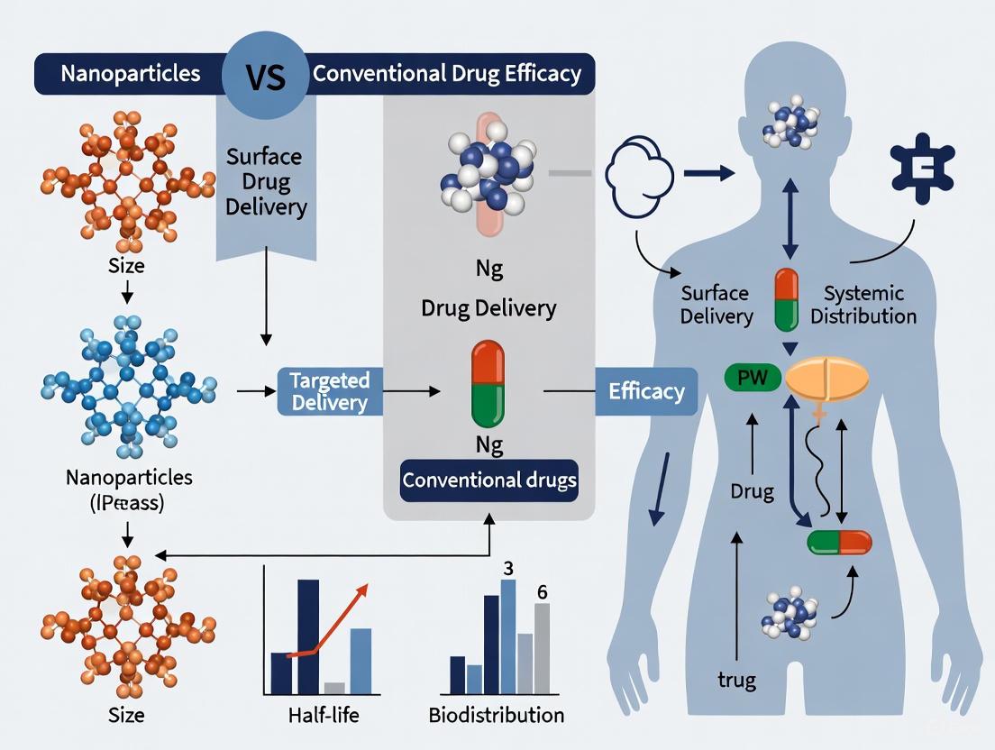
Nanoparticle Drug Delivery vs. Conventional Delivery: Comparative Efficacy
The fundamental properties of nanoparticles translate directly to enhanced therapeutic performance when compared to conventional drug delivery approaches. Nanoparticle-based systems address multiple limitations of traditional formulations through several key mechanisms [31] [32]:
Table 3: Comparative Analysis: Nanoparticle vs Conventional Drug Delivery
| Parameter | Conventional Drug Delivery | Nanoparticle-Based Delivery | Experimental Evidence |
|---|---|---|---|
| Targeting Efficiency | Limited biodistribution, poor specificity | Enhanced permeability and retention (EPR) effect + active targeting | 4-fold increase in tumor-to-normal tissue ratio [32] |
| Cellular Uptake | Variable depending on drug properties | Enhanced cellular internalization | 3-fold increase in cellular uptake with antibody-conjugated NPs [32] |
| Therapeutic Efficacy | Limited by off-target distribution | Improved tumor growth inhibition | 75% reduction in tumor volume vs conventional forms [32] |
| Toxicity Profile | Significant off-target effects | Reduced systemic toxicity | Improved survival rates (45-day median increase) [32] |
| Drug Encapsulation | N/A | High loading capacity | 60-85% encapsulation efficiency across drug types [32] |
Mechanisms Underlying Enhanced Efficacy
The superior performance of nanoparticle-based drug delivery systems stems from several interconnected mechanisms [31] [32]:
Passive Targeting via EPR Effect: The leaky vasculature and impaired lymphatic drainage characteristic of tumor tissues enable selective accumulation of nanocarriers (typically 10-200 nm), while minimizing distribution to healthy tissues [31] [32]. This phenomenon results in drug concentrations at the target site that are significantly higher than achievable with conventional formulations.
Active Targeting Capabilities: Surface functionalization with targeting ligands (antibodies, peptides, aptamers) enables specific recognition of and binding to receptors overexpressed on target cells [31] [32]. This active targeting mechanism enhances cellular internalization and further improves therapeutic specificity.
Controlled Release Kinetics: Nanoparticles can be engineered to provide sustained, localized drug release through various mechanisms, including polymer degradation, diffusion, and stimuli-responsive triggering [31] [32]. pH-sensitive systems demonstrate rapid drug release in the acidic tumor microenvironment (pH 5.5) while maintaining stability at physiological pH (7.4) [32].
The Scientist's Toolkit: Essential Research Reagents and Materials
Successful nanoparticle research and development requires specialized materials and reagents tailored to specific applications:
Table 4: Essential Research Reagents for Nanoparticle Development
| Reagent Category | Specific Examples | Function and Application | Key Considerations |
|---|---|---|---|
| Ionizable Lipids | AMG1541, DLin-MC3-DMA | Form core structure of LNPs, enable endosomal escape | Degradability enhances clearance; tail length affects mRNA delivery [29] [33] |
| Structural Lipids | Cholesterol, Phospholipids | Stabilize LNP structure, modulate fluidity and integrity | Impact transfection efficiency and in vivo stability [29] |
| PEGylated Lipids | DMG-PEG, DSG-PEG | Reduce protein adsorption, extend circulation half-life | Concentration affects size and polydispersity during formulation [29] |
| Biodegradable Polymers | PLGA, PCL, Chitosan | Form polymeric nanoparticle matrix, control drug release | Molecular weight and copolymer ratio determine degradation rate [31] [32] |
| Targeting Ligands | Antibodies, Peptides, Hyaluronic Acid | Enable active targeting to specific tissues or cells | Conjugation chemistry must preserve ligand activity [31] [32] |
| Characterization Standards | Latex beads, Molecular weight markers | Instrument calibration, method validation | Essential for quantitative comparison across studies [29] |
Experimental Protocol: Formulation of Targeted Chitosan Nanoparticles
Purpose: To synthesize chitosan-based nanoparticles for targeted drug delivery to colon cancer using hyaluronic acid conjugation [31].
Materials:
- Chitosan (medium molecular weight)
- Sodium tripolyphosphate (TPP)
- Hyaluronic acid (HA)
- 5-Fluorouracil (5-FU) as model drug
- Acetic acid solution (1% v/v)
- EDC/NHS coupling reagents
Synthesis Method (Ionotropic Gelation):
- Polymer Solution Preparation: Dissolve chitosan in 1% acetic acid solution to obtain 0.1-0.5 wt% concentration [31].
- Cross-linker Solution: Prepare TPP solution in purified water at equivalent concentrations [31].
- Drug Loading: Add 5-FU to chitosan solution under constant stirring [31].
- Nanoparticle Formation: Add TPP solution dropwise to chitosan-drug mixture under magnetic stirring (ratio 5:1 chitosan:TPP) [31].
- Surface Functionalization: Conjugate HA to pre-formed nanoparticles using carbodiimide chemistry (EDC/NHS) [31].
- Purification: Centrifuge at 12,000 rpm for 30 minutes and resuspend in phosphate buffer [31].
Characterization Results:
- Particle size: 135 nm (increased to 150 nm after HA conjugation) [31]
- Drug encapsulation efficiency: 60-85% depending on drug properties [32]
- Enhanced cellular uptake: 3-fold increase with HA-conjugated nanoparticles [31]
This formulation demonstrates the advantage of nanoparticle systems in achieving targeted delivery while providing controlled release kinetics [31].
Future Perspectives and Challenges
While nanoparticle-based drug delivery systems show tremendous promise, several challenges must be addressed to advance clinical translation. These include scaling up production while maintaining batch-to-batch consistency, ensuring long-term stability, and conducting comprehensive toxicological assessments [32]. Emerging research focuses on developing increasingly sophisticated nanoparticles with enhanced targeting capabilities and stimuli-responsive behavior [33].
The continued evolution of characterization techniques, particularly solution-based biophysical methods with higher resolution, will be essential for establishing robust structure-function relationships [29]. These advances will facilitate the creation of design rules for next-generation nanotherapeutics with precisely controlled interactions at the bio-nano interface [29] [30].
As nanotechnology continues to mature, interdisciplinary collaboration between materials science, biology, and medicine will be crucial for fully realizing the potential of nanomedicine in addressing unmet clinical needs and improving patient outcomes across diverse therapeutic areas [14] [32].
A fundamental paradox exists in modern pharmacology: many therapeutic compounds demonstrate potent efficacy in vitro but fail to achieve clinical success due to inadequate pharmacokinetic profiles. Conventional drug formulations often face significant challenges, including rapid clearance by the renal system or mononuclear phagocyte system, poor aqueous solubility, and non-specific distribution leading to systemic toxicity [13] [6]. These limitations severely restrict the amount of active pharmaceutical ingredient (API) that reaches the target site, diminishing therapeutic potential while increasing adverse effects.
Nanoparticle-based drug delivery systems represent a paradigm shift in addressing these pharmacokinetic barriers. By engineering carriers at the nanometer scale (typically 1-1000 nm), researchers can fundamentally alter the liberation, absorption, distribution, metabolism, and excretion (LADME) profiles of therapeutic compounds [34] [35]. The unique physicochemical properties of nanoparticles—including their high surface area-to-volume ratio, tunable surface chemistry, and engineered size and shape—enable precise control over drug release kinetics, biodistribution patterns, and cellular uptake mechanisms [13] [36]. This review systematically compares the pharmacokinetic performance of nanoparticle-based drug delivery systems against conventional formulations, providing experimental data and methodologies relevant to researchers and drug development professionals.
Comparative Analysis: Nanoparticles vs. Conventional Formulations
Key Pharmacokinetic Parameters
Table 1: Comparative Pharmacokinetic Parameters of Conventional Formulations vs. Nanoparticle-Based Systems
| Pharmacokinetic Parameter | Conventional Formulations | Nanoparticle Systems | Experimental Evidence |
|---|---|---|---|
| Oral Bioavailability | Low for BCS Class II/IV drugs due to poor solubility and permeability [13] | Enhanced solubility and mucosal adhesion improve absorption [37] [35] | CLA-BSA NPs showed controlled release in reductive media; Chitosan NPs enhance mucoadhesion [4] |
| Circulation Half-life | Short (minutes to hours) due to rapid renal clearance and metabolism [6] | Prolonged (hours to days) through evasion of RES and reduced renal filtration [37] [36] | PEGylated liposomal doxorubicin (Doxil) exhibits significantly prolonged circulation vs. free drug [37] |
| Volume of Distribution | Often widespread, leading to systemic toxicity [6] | Selective accumulation in target tissues via EPR effect and active targeting [36] | PLD demonstrates reduced cardiotoxicity vs. free doxorubicin in clinical use [37] |
| Clearance Rate | High renal and hepatic clearance [34] | Reduced clearance through RES evasion and protection from metabolic enzymes [34] [36] | Methotrexate-loaded nanoformulations showed lower clearance values vs. free solution [34] |
| Tumor Accumulation | Limited by physiological barriers and non-specific distribution [6] | Enhanced via EPR effect (passive) and ligand-receptor interactions (active) [36] | Magnetic SFPs showed enhanced tumor-specific accumulation with magnetic guidance in breast cancer models [4] |
Impact on Therapeutic Outcomes
The improved pharmacokinetic profile of nanoparticle-based systems translates directly to enhanced therapeutic outcomes. In oncology, nanoformulations of chemotherapeutic agents have demonstrated reduced systemic toxicity while maintaining or improving anti-tumor efficacy [37] [36]. For instance, the landmark comparison between free doxorubicin and its liposomal encapsulated form (Doxil) revealed dramatically different clinical profiles: while both forms effectively kill cancer cells, the nanoparticle formulation significantly reduces cardiotoxicity—a dose-limiting side effect of the conventional formulation—through altered tissue distribution patterns [37].
Similarly, nanoparticle systems have revolutionized the delivery of nucleic acid therapeutics, which face immense pharmacokinetic challenges including nuclease degradation, rapid renal clearance, and inefficient cellular uptake. Lipid nanoparticles (LNPs) successfully addressed these limitations for COVID-19 mRNA vaccines and are now being adapted for cancer therapy, with clinical trials demonstrating successful mRNA delivery to hepatic cells and robust protein expression [37] [4]. The versatility of nanocarriers enables encapsulation of diverse therapeutic payloads—from small molecules to proteins and nucleic acids—each benefiting from improved pharmacokinetic profiles [13] [36].
Mechanisms Underlying Improved Pharmacokinetics
Enhanced Bioavailability Strategies
The diagram below illustrates the primary mechanisms through which nanoparticles enhance drug bioavailability.
Nanoparticle Mechanisms for Enhanced Bioavailability
Nanoparticles overcome bioavailability challenges through multiple complementary mechanisms. For poorly soluble drugs (BCS Class II and IV), the hydrophobic cores of polymeric nanoparticles and lipid-based systems create protective microenvironments that enhance apparent solubility and dissolution rates [13]. This was demonstrated in clarithromycin-loaded bovine serum albumin nanoparticles (CLA-BSA NPs), where the nanoformulation significantly improved delivery of the poorly soluble macrolide antibiotic [4]. Similarly, cannabidiol (CBD) composites with tailored carbon supports demonstrated superior release profiles under simulated digestive conditions, addressing the compound's notorious bioavailability challenges [4].
Beyond solubility enhancement, nanoparticles protect therapeutic payloads from presystemic metabolism. The encapsulation of drugs within nanocarriers creates a physical barrier against digestive enzymes and harsh pH conditions, particularly crucial for biological therapeutics like peptides, proteins, and nucleic acids [13] [36]. Additionally, surface engineering with mucoadhesive polymers (e.g., chitosan) prolongs gastrointestinal residence time, further enhancing absorption opportunities through sustained release at mucosal surfaces [13].
Prolonged Circulation and Targeted Distribution
The following diagram outlines the key strategies nanoparticles employ to achieve prolonged circulation and targeted distribution.
Nanoparticle Circulation and Targeting Mechanisms
Extended circulation time represents a cornerstone of nanoparticle pharmacokinetic advantages. The reticuloendothelial system (RES), primarily comprising liver and spleen macrophages, rapidly clears conventional drugs and foreign particles from circulation [36]. Nanoparticles evade this clearance through surface engineering strategies, most notably PEGylation—the covalent attachment of polyethylene glycol chains that create a hydrophilic protective layer around the nanoparticle [37] [36]. This steric hindrance reduces opsonization (the adsorption of plasma proteins that mark particles for phagocytosis), significantly extending circulatory half-life [36].
Beyond circulation extension, nanoparticles achieve superior tissue distribution through passive and active targeting mechanisms. The Enhanced Permeability and Retention (EPR) effect leverages the pathological anatomy of diseased tissues—particularly tumors—which feature leaky vasculature with endothelial gaps (100 nm to 2 μm) and impaired lymphatic drainage [37] [36]. This combination allows nanoparticles to extravasate and accumulate preferentially in target tissues, while their optimized size (typically 50-200 nm) prevents rapid renal clearance (which filters particles <5-10 nm) [36].
Active targeting strategies further enhance specificity through surface functionalization with targeting ligands including antibodies, peptides, aptamers, and small molecules that recognize disease-specific biomarkers [13] [36]. These ligands facilitate receptor-mediated endocytosis, increasing cellular internalization at the target site while minimizing non-specific distribution. The evolution of targeting sophistication has progressed to stimuli-responsive systems that release their payload in response to pathological cues such as pH shifts, enzyme activity, or redox gradients [36].
Experimental Models and Methodologies
Standardized Protocols for Pharmacokinetic Assessment
Robust pharmacokinetic evaluation of nanoparticle systems requires specialized methodologies that account for their unique biological behavior. The following experimental approaches represent current best practices in the field:
In Vitro Release Kinetics Protocol: Dissolution testing under biomimetic conditions provides initial screening data. For example, researchers developing silk fibroin particles (SFPs) for breast cancer therapy conducted release studies over 72 hours in physiological buffers, demonstrating sustained release profiles for both curcumin (37% encapsulation efficiency) and 5-FU (82% encapsulation efficiency) [4]. This methodology typically involves: (1) Incubation of nanoparticle formulation in release media at physiological temperature (37°C) with constant agitation; (2) Time-point sampling with replacement of release media to maintain sink conditions; (3) Quantification of released drug via HPLC or UV-Vis spectroscopy; (4) Mathematical modeling of release kinetics (zero-order, first-order, Higuchi, Korsmeyer-Peppas) [4] [34].
Cellular Uptake and Transport Studies: Assessment of nanoparticle internalization and transcellular transport utilizes cell culture models representing biological barriers. The methodology for evaluating chlorambucil-functionalized mesoporous silica nanoparticles (MSNs) against lung adenocarcinoma (A549) and colon carcinoma (CT26WT) cells exemplifies this approach [4]. Key steps include: (1) Culture of relevant cell lines on permeable supports for transport studies; (2) Fluorescent labeling of nanoparticles or therapeutic payload; (3) Incubation of nanoparticles with cells for predetermined timepoints; (4) Quantification of uptake via flow cytometry, confocal microscopy, or LC-MS/MS; (5) Measurement of transepithelial electrical resistance (TEER) to monitor barrier integrity [4] [38].
In Vivo Biodistribution and Pharmacokinetics: Animal studies remain essential for comprehensive pharmacokinetic profiling. The protocol typically involves: (1) Administration of nanoparticle formulation via relevant route (IV, oral, etc.) with free drug as control; (2) Serial blood collection at predetermined timepoints; (3) Tissue harvesting at endpoint (typically including target organs plus liver, spleen, kidney); (4) Drug quantification in biological matrices via validated bioanalytical methods; (5) Compartmental or non-compartmental pharmacokinetic analysis [34]. For instance, in vivo studies with magnetic SFPs demonstrated enhanced tumor-specific accumulation and increased tumor necrosis when magnetic guidance was applied [4].
Advanced Model Systems
Conventional two-dimensional cell cultures often fail to accurately predict nanoparticle behavior in humans. To address this limitation, researchers are developing more physiologically relevant models [38]:
Three-Dimensional (3D) Culture Systems: Spheroids, organoids, and tissue-engineered models better recapitulate the diffusion barriers, cell-cell interactions, and heterogeneous microenvironments encountered in vivo. These models are particularly valuable for studying nanoparticle penetration in solid tumors [38].
Dynamic Flow Systems: Microfluidic devices that simulate blood flow and vascular dynamics provide more realistic conditions for assessing nanoparticle extravasation and targeting under physiological shear stresses [38].
Co-culture Models: Systems incorporating multiple cell types (e.g., epithelial cells with immune cells or fibroblasts) better mimic the complex biological interactions that influence nanoparticle biodistribution and clearance [38].
The Scientist's Toolkit: Essential Research Reagents
Table 2: Key Research Reagents and Materials for Nanoparticle Pharmacokinetic Studies
| Reagent/Material | Function | Application Examples |
|---|---|---|
| Poly(lactic-co-glycolic acid) (PLGA) | Biodegradable polymer for controlled drug release | Nanoparticle core matrix for sustained release formulations [37] [36] |
| Polyethylene glycol (PEG) | Stealth coating to reduce protein adsorption and RES clearance | Surface functionalization to extend circulation half-life (PEGylation) [37] [36] |
| Chitosan | Mucoadhesive polymer for enhanced mucosal retention | Oral and nasal drug delivery systems [13] |
| Lipids (Ionizable, Phospholipids) | Core components of liposomes and lipid nanoparticles | mRNA delivery systems (COVID-19 vaccines), liposomal chemotherapeutics [37] [4] |
| Targeting Ligands (Antibodies, Peptides, Aptamers) | Active targeting to specific cells or tissues | Surface conjugation for receptor-mediated drug delivery [13] [36] |
| Fluorescent Probes (DiD, FITC, Quantum Dots) | Tracking and visualization of nanoparticles | In vitro and in vivo biodistribution studies [4] [34] |
| Mesoporous Silica | High surface area scaffold for drug loading | Mesoporous silica nanoparticles (MSNs) for enhanced drug loading capacity [4] |
Clinical Translation and Future Perspectives
Despite promising preclinical results, the translation of nanoparticle-based therapies faces significant challenges. The translational gap in nanomedicine is striking: while over 100,000 scientific articles on nanomedicines were published in the past decade, only approximately 90 nanomedicine products have obtained global marketing approval by 2023 [37]. This discrepancy highlights the complex biological, manufacturing, and regulatory hurdles facing nanoparticle therapeutics.
Key challenges include batch-to-batch variability in GMP-scale production, potential immune activation (particularly anti-PEG antibodies that can accelerate clearance upon repeated administration), and limitations of the EPR effect which demonstrates significant heterogeneity in human tumors compared to animal models [37]. The failure of BIND-014 (targeted docetaxel nanoparticles) in Phase II trials despite promising early activity signals exemplifies the difficulties in translating sophisticated targeting strategies to clinical benefit [37].
Future directions focus on multifunctional systems that combine targeting, diagnostic, and therapeutic capabilities, and personalized approaches based on patient-specific transport biomarkers [37] [36]. Artificial intelligence is emerging as a powerful tool for nanoparticle design, with recent demonstrations of AI-powered platforms successfully creating improved nanoparticle formulations for venetoclax and trametinib [39]. Additionally, advanced formulation strategies—including integration of nanoparticles into secondary delivery systems like hydrogels, microspheres, and implants—are gaining attention for bridging the formulation gap between nanoparticle design and clinically viable drug products [37].
As nanotechnology continues to evolve, the focus must shift from merely demonstrating efficacy in model systems to addressing the complex practical requirements of clinical translation, including scalable manufacturing, regulatory compliance, and demonstrated therapeutic superiority over conventional approaches.
Nanoparticle Platforms in Action: From Design to Targeted Therapeutic Applications
The evolution of modern medicine is increasingly dependent on the precision and efficacy of drug delivery systems. Traditional drug formulations, characterized by their simple chemical compositions and direct administration, often face significant challenges including poor bioavailability, rapid degradation, and non-specific distribution leading to systemic toxicity [40]. These limitations are particularly problematic in treating complex diseases such as cancer and chronic inflammatory conditions, where the therapeutic window is narrow and the biological barriers are formidable [13] [16]. The advent of nanocarriers represents a paradigm shift in pharmaceutical sciences, offering innovative solutions to these longstanding problems. Nanoparticle-based drug delivery systems utilize materials at the nanoscale (typically 1-1000 nm) to encapsulate, protect, and transport therapeutic agents to specific target sites within the body [13] [40]. This approach fundamentally enhances the therapeutic index of drugs—increasing their efficacy while minimizing adverse effects—by leveraging unique nanoscale properties such as high surface area-to-volume ratio and the ability to navigate biological barriers more effectively than conventional formulations [13] [40] [16]. The following analysis provides a comprehensive comparison of five major nanocarrier classes—liposomes, polymeric nanoparticles, solid lipid nanoparticles, dendrimers, and metallic nanoparticles—within the broader thesis that nano-engineered delivery systems substantially outperform conventional drug delivery in key metrics of therapeutic efficacy, safety, and targeting capability.
Comparative Analysis of Nanocarrier Systems
Table 1: Comprehensive Comparison of Major Nanocarrier Types
| Nanocarrier Type | Core Composition | Size Range | Key Advantages | Primary Limitations | Therapeutic Applications |
|---|---|---|---|---|---|
| Liposomes | Phospholipid bilayer surrounding aqueous core [41] | ~50-200 nm [41] | High biocompatibility; ability to deliver both hydrophilic & hydrophobic drugs; FDA-approved formulations [40] [41] | Potential stability issues in bloodstream; rapid clearance by immune system without PEGylation [13] | Cancer therapy (e.g., doxorubicin delivery); targeted brain delivery [16] [41] |
| Polymeric NPs | Natural (chitosan, alginate) or synthetic (PLA, PLGA) polymers [13] [40] | 10-1000 nm [13] [40] | Tunable degradation rates; sustained/controlled drug release; high functionalization capacity [13] | Batch-to-batch variability; potential residual solvent toxicity from synthesis [13] | Controlled release formulations; cancer therapy; transdermal delivery [13] [16] |
| Solid Lipid NPs (SLNs) | Solid lipid matrix stabilized by surfactants [40] | ~30-1000 nm (typically ~30 nm) [16] | Improved stability over liposomes; high biocompatibility; good scalability and industrial production [40] | Limited drug loading capacity; potential drug expulsion during storage [40] | Oral and transdermal drug delivery; natural product encapsulation [40] |
| Dendrimers | Highly branched, tree-like synthetic polymers with defined architecture [16] | 1-10 nm | Monodisperse size distribution; multifunctional surface for high drug loading; precise control over structure [16] | Potential cytotoxicity at higher generations; complex and costly synthesis [16] | Targeted drug delivery; combination therapy; diagnostic imaging [16] |
| Metallic NPs | Gold, silver, iron oxide, or other metal cores [13] [16] | Varies by type and application | Unique optical/magnetic properties for imaging & therapy; efficient surface plasmon resonance; external stimulus responsiveness [13] | Long-term toxicity concerns; potential for bioaccumulation [13] | Hyperthermia cancer treatment; diagnostic imaging; antimicrobial applications [13] [16] |
Table 2: Performance Comparison of Nanocarriers vs. Conventional Drug Delivery
| Performance Metric | Conventional Drug Delivery | Nanocarrier Systems | Experimental Evidence |
|---|---|---|---|
| Bioavailability | Low, especially for poorly soluble drugs [40] | Significantly enhanced via nano-encapsulation [13] [40] | Thymoquinone in lipid nanocarriers showed 6x higher bioavailability vs. free drug [40] |
| Targeting Efficiency | Primarily passive distribution | Active targeting (ligand-receptor) and passive (EPR effect) [13] [16] | TfR-targeted liposomes achieved specific brain delivery in murine models [41] |
| Controlled Release | Limited, often burst release | Tunable sustained release over days to weeks [13] [16] | Polymeric nanoparticles demonstrated prolonged release >72 hours [16] |
| Solubility Enhancement | Challenge for hydrophobic drugs | Significant improvement via nanoformulation [40] | Nanostructures aid delivery of sparingly water-soluble drugs [40] |
| Toxicity Profile | Systemic exposure and side effects | Reduced side effects via targeted delivery [13] [16] | Nanoparticle-encapsulated indomethacin showed safer gastrointestinal profile [16] |
Experimental Protocols in Nanocarrier Research
Protocol 1: Evaluating Targeted Brain Delivery of Liposomes
Objective: To characterize the receptor-mediated transcytosis of transferrin receptor (TfR)-targeted liposome nanoparticles across the blood-brain barrier (BBB) in vivo [41].
Methodology Details:
- Nanoparticle Formulation: Liposomes composed of distearoylphosphatidylcholine (DSPC) and cholesterol with polyethylene glycol (PEG) coating were prepared. Targeting was enabled by conjugating high-affinity anti-TfR antibodies (clone RI7217) to the surface. The lipid bilayer was tagged with Atto 550 or Atto 488 fluorophores [41].
- Animal Model: Mice with cranial windows implanted over the somatosensory cortex were utilized for real-time imaging [41].
- Administration: A single bolus injection of nanoparticles (70 nmol lipid/gram animal) was administered intravenously [41].
- Imaging & Analysis: Two-photon fluorescence microscopy was performed in anesthetized and awake mice. Vessels were classified by type (arterioles, capillaries, post-capillary venules) based on anatomical features and blood flow direction. Nanoparticle association and transcytosis were quantified across different vascular segments [41].
Key Findings: TfR-targeted nanoparticles associated with capillary and venule endothelium but not arterioles. Despite association at multiple sites, transcytosis-mediated brain entry occurred predominantly at post-capillary venules, challenging the conventional view of capillaries as the primary transport site [41].
Protocol 2: Assessing Anti-inflammatory Efficacy of Polymeric Nanoparticles
Objective: To evaluate the enhanced therapeutic efficacy of anti-inflammatory drugs encapsulated in polymeric nanocarriers [16].
Methodology Details:
- Nanoparticle Formulation: Amphiphilic poly-N-vinylpyrrolidone nanoparticles were synthesized and loaded with indomethacin. Alternatively, chitosan-coated bilosomes were prepared for berberine loading (BER-CTS-BLS) for transdermal delivery [16].
- In Vitro Release: Nanoparticles were incubated in physiological buffers, and drug release profiles were measured over time using appropriate analytical methods [16].
- In Vivo Models: Acute, subchronic, and chronic inflammation models (e.g., carrageenan-induced inflammation in rodents) were employed. For transdermal systems, skin permeability and irritation tests were conducted ex vivo [16].
- Pharmacokinetic & Efficacy Measurements: Drug concentrations in plasma and tissues were quantified via HPLC or MS. Anti-inflammatory activity was assessed by measuring reduction in edema and inflammatory markers compared to free drug controls [16].
Key Findings: Nanoparticle-encapsulated indomethacin exhibited controlled prolonged release, improved pharmacokinetics, and enhanced anti-inflammatory activity with reduced accumulation in liver and kidneys. BER-CTS-BLS gel demonstrated increased skin permeability without irritation and significant in vivo anti-inflammatory effects [16].
Visualization of Nanocarrier Mechanisms
Workflow for Evaluating Nanocarrier Brain Delivery
Mechanism of Targeted Nanocarrier Drug Delivery
The Scientist's Toolkit: Essential Research Reagents
Table 3: Key Reagents for Nanocarrier Development and Evaluation
| Reagent/Material | Function in Research | Specific Examples from Literature |
|---|---|---|
| DSPC/Cholesterol | Forms stable lipid bilayer in liposome formulations [41] | Used in TfR-targeted liposomes for brain delivery studies [41] |
| Polyethylene Glycol (PEG) | Provides "stealth" properties to reduce immune clearance and enhance circulation time [13] [41] | PEG coating on liposomes ensured stability in bloodstream [41] |
| Targeting Ligands (Antibodies, Peptides) | Enables active targeting to specific cells or receptors [13] [16] | Anti-TfR antibodies (RI7217) for brain targeting; Tat-Beclin peptide for autophagy induction [16] [41] |
| Biodegradable Polymers | Forms matrix for controlled drug release in polymeric NPs [13] [40] | PLGA, PLA, chitosan, poly-N-vinylpyrrolidone [13] [40] [16] |
| Fluorescent Tags (Atto Dyes) | Allows tracking and visualization of nanoparticles in vitro and in vivo [41] | Atto 550 and Atto 488 for two-photon microscopy of liposomes [41] |
| Metallic Nanoprecursors | Forms core of metallic nanoparticles for therapeutic and diagnostic applications [13] [16] | Silver nitrate for AgNPs; iron salts for magnetic nanoparticles [13] [16] |
The comprehensive comparison of nanocarrier systems presented herein substantiates the superior performance of nano-engineered drug delivery approaches over conventional methods. Each class of nanocarrier—from the clinically established liposomes to the emerging metallic nanoparticles—offers distinct advantages in addressing the fundamental limitations of traditional drug formulations. The enhanced bioavailability, precise targeting capabilities, and controlled release profiles demonstrated across multiple experimental models highlight the transformative potential of nanotechnology in medicine [13] [40] [16]. Particularly noteworthy is the growing understanding of nanocarrier behavior at biological barriers, such as the recent revelation that post-capillary venules rather than capillaries serve as the primary gateway for nanoparticle transport across the blood-brain barrier [41]. While challenges remain in standardization, scalability, long-term toxicity assessment, the strategic development of nanocarrier systems continues to push the boundaries of therapeutic possibility. As research advances, the rational design of next-generation nanocarriers will undoubtedly play a pivotal role in realizing personalized medicine and effective treatments for currently intractable diseases.
Passive Targeting Exploiting the Enhanced Permeability and Retention (EPR) Effect
The Enhanced Permeability and Retention (EPR) effect represents a foundational principle in cancer nanomedicine, describing a pathophysiological phenomenon whereby macromolecules and nanoparticles preferentially accumulate in solid tumor tissue compared to conventional small-molecule drugs [42] [43]. This passive targeting mechanism forms a critical thesis point in the comparison between nanoparticle-based drug delivery systems and conventional chemotherapy, offering the potential to enhance therapeutic efficacy while reducing systemic toxicity [13].
The EPR effect arises from key abnormalities in the tumor microenvironment: hyperpermeability of tumor blood vessels due to structural defects in endothelial cells and poor lymphatic drainage that impairs clearance of accumulated particles [42] [44]. Tumor vasculature exhibits significant abnormalities, with endothelial gaps ranging from 100-780 nm in size, allowing extravasation of nanomedicines that would be excluded from normal tissues [44]. Meanwhile, the lack of functional lymphatic drainage in tumors leads to prolonged retention of these accumulated particles [42]. This combination creates the EPR effect, first observed by Maeda and colleagues in 1986, which has since become a guiding principle for designing tumor-targeted nanomedicines in the 40-200 kDa molecular weight range [42] [43].
Comparative Analysis: EPR-Based Nanomedicines vs. Conventional Chemotherapy
Pharmacokinetic and Efficacy Comparison
Table 1: Comparative performance of EPR-based nanomedicines versus conventional chemotherapy
| Parameter | Conventional Chemotherapy | EPR-Based Nanomedicines | Experimental Evidence |
|---|---|---|---|
| Tumor Accumulation | Limited by rapid diffusion and clearance | 10-15-fold higher concentration in tumors [42] | Pegylated liposomal doxorubicin shows 10-15× higher tumor concentration vs. free doxorubicin [42] |
| Circulation Half-Life | Short (minutes to hours) | Significantly prolonged | Doxil exhibits substantially longer blood circulation half-life [42] [45] |
| Therapeutic Efficacy | Moderate, dose-limited by toxicity | Improved tumor response in preclinical models | Phase III trial: 45.9% overall response rate for Doxil vs. conventional doxorubicin [42] |
| Side Effect Profile | Significant systemic toxicity | Reduced cardiotoxicity, though hand-foot syndrome reported | Doxil shows less cardiotoxicity than free doxorubicin despite similar tumor response [42] [45] |
| Penetration Depth | Good initial penetration | Heterogeneous, primarily peripheral tumor regions | Limited penetration to well-vascularized peritumoral areas due to interstitial fluid pressure [42] |
Clinical Translation Challenges
Despite promising preclinical results, the clinical translation of EPR-based nanomedicines has demonstrated limitations. While nanomedicines like Doxil and Abraxane show improved pharmacokinetics and reduced toxicity, their impact on overall survival remains modest [45]. For example, Abraxane required a 50% higher dose than free paclitaxel and provided only a 0.9-month survival benefit in metastatic non-small cell lung cancer [45]. This performance gap between preclinical expectations and clinical outcomes highlights significant challenges in EPR-based targeting, primarily due to tumor heterogeneity and variable EPR effectiveness across patient populations [42] [46].
Methodologies for Evaluating EPR Effect
Standard Experimental Protocol for EPR Quantification
Table 2: Key methodological approaches for EPR effect evaluation
| Method | Protocol Details | Applications | Limitations |
|---|---|---|---|
| Evans Blue Dye Assay | Evans blue binds plasma albumin; intravenous administration with measurement of tumor accumulation over 145 hours [43] | Demonstration of selective macromolecule concentration in tumors (~10-fold higher than blood) [43] | Does not directly measure therapeutic agents |
| Radiolabeled Protein Tracking | Use of radio-labeled plasma proteins (transferrin - 90 kDa, IgG - 160 kDa); measurement of tumor accumulation [43] | Establishing molecular weight threshold for EPR effect (≥40 kDa) [43] | Requires specialized handling and equipment |
| Fluorescent Nanoparticle Tracking | Fluorescently labeled nanoparticles administered intravenously; ex vivo tissue analysis or in vivo imaging [47] | Quantification of tumor accumulation, extravasation, and penetration depth [47] | Potential for dye leakage and photobleaching |
| ICP-OES Analysis of Inorganic Nanoparticles | Metal-based nanoparticles (e.g., PSiNPs) measured in tissues via Inductively Coupled Plasma-Optical Emission Spectroscopy [47] | Precise quantification of nanoparticle biodistribution and tumor accumulation | Limited to nanoparticles with detectable elements |
| Multi-stage Factorial Design | Two-factor factorial experiments comparing delivery route (IV vs. IP) and formulation (nanomedicine vs. free drug) [45] | Evaluation of EPR bypassing strategies and dual pharmacokinetic advantages | Complex experimental design requiring larger animal cohorts |
EPR Enhancement Methodologies
Several approaches have been developed to enhance the EPR effect:
Pharmacological Modulation: Angiotensin II-induced hypertension increases systolic blood pressure, resulting in 2-6-fold selective increase in tumor blood flow and enhanced nanoparticle accumulation [43]. Bradykinin potentiation through ACE inhibitors (e.g., enalapril) enhances vascular permeability even under normotensive conditions [43].
Physical Priming: Application of heat, radiation, or ultrasound (particularly with microbubbles) mechanically untightens vessel walls and adjacent ECM, improving nanoparticle extravasation [48]. This approach is being clinically evaluated for brain tumors [48].
Nanoparticle Optimization: Machine learning approaches using deep neural networks can predict tissue distribution and tumor delivery efficiency based on nanoparticle physicochemical properties, with determination coefficients (R²) of 0.41-0.87 for major tissues [49].
Visualization of EPR Mechanisms and Experimental Evaluation
Diagram 1: Mechanism of EPR-mediated passive targeting. Nanoparticles preferentially extravasate through leaky tumor vasculature and are retained due to impaired lymphatic drainage.
Diagram 2: Comprehensive workflow for evaluating EPR effect and therapeutic efficacy in preclinical models.
The Scientist's Toolkit: Essential Research Reagents and Materials
Table 3: Key research reagents and materials for EPR effect studies
| Reagent/Material | Function | Application Examples |
|---|---|---|
| PEGylated Liposomal Doxorubicin (Doxil) | Gold standard EPR-based nanomedicine comparator | Positive control for EPR efficacy; benchmark for new formulations [42] [45] |
| Evans Blue Dye | Plasma protein binding tracer for vascular permeability | Visual and quantitative assessment of macromolecular leakage into tumors [43] |
| Angiotensin II | Vasoconstrictor for hypertension-induced EPR enhancement | Augments tumor blood flow and nanoparticle accumulation (2-6-fold increase) [43] |
| ACE Inhibitors (Enalapril) | Bradykinin potentiation through degradation inhibition | Enhances vascular permeability under normotensive conditions [43] |
| Ultrasound Microbubbles | Physical priming of tumor vasculature | Mechanical disruption of vessel walls and ECM to improve nanoparticle extravasation [48] |
| Porous Silicon Nanoparticles (PSiNPs) | Biocompatible, biodegradable drug carrier with high payload capacity | Model nanoparticle for studying autophagy-mediated exocytosis and tumor penetration [47] |
| Radiolabeled Proteins (¹²⁵I-albumin) | Quantitative tracking of macromolecular distribution | Establishing molecular weight thresholds for EPR effect (≥40 kDa) [43] |
| CD63 Antibody | Exosome surface marker identification | Verification of exosome-coated nanoparticles in biomimetic delivery systems [47] |
| 3-Methyladenine (3-MA) | Autophagy inhibitor | Investigation of autophagy role in nanoparticle exocytosis and trafficking [47] |
The EPR effect remains a significant yet imperfect mechanism for tumor-targeted drug delivery. While it provides a rational foundation for nanomedicine design, clinical outcomes have demonstrated that passive targeting alone is insufficient for transformative therapeutic advances. The future of EPR-based strategies lies in combination approaches that address tumor heterogeneity through patient stratification using imaging and histological biomarkers [48], and integration with active targeting modalities [46].
The emerging concept of Active Transport and Retention (ATR) suggests complementary mechanisms beyond passive extravasation, involving active endothelial transport processes that may be engineered for improved delivery [46]. Furthermore, biomimetic approaches utilizing tumor-derived exosomes show promise for enhancing tumor accumulation and penetration while maintaining biocompatibility [47].
For researchers developing nanoparticle drug delivery systems, success will likely require a multifaceted strategy that incorporates EPR optimization through pharmacological and physical priming, nanoparticle design informed by machine learning predictions [49], and careful patient selection based on EPR-relevant biomarkers. This integrated approach will maximize the potential of passive targeting while addressing its limitations, advancing the central thesis that nanoparticle delivery can significantly improve upon conventional drug efficacy.
Glioblastoma (GBM) is the most prevalent and aggressive primary malignant brain tumor in adults, accounting for approximately 50% of all malignant brain tumors and presenting a dire prognosis with a median survival of only 12 to 15 months following standard treatment [50]. The standard of care involves surgical resection followed by radiotherapy and chemotherapy, typically with the alkylating agent temozolomide (TMZ) [51]. However, therapeutic efficacy remains severely limited by two fundamental challenges: the presence of the blood-brain barrier (BBB), which restricts most chemotherapeutic drugs from reaching the tumor site in effective concentrations, and the development of tumor resistance mechanisms [50].
The BBB is a highly selective interface composed of endothelial cells tightly sealed by continuous tight junctions, along with pericytes, astrocytes, and extracellular matrix components that collectively maintain brain homeostasis [52]. This protective barrier restricts nearly all large molecules and over 98% of small-molecule drugs, presenting a formidable obstacle for brain tumor therapy [53]. Conventional chemotherapeutic administration often results in subtherapeutic drug concentrations at the tumor site alongside significant systemic toxicity, highlighting the urgent need for more advanced drug delivery platforms [50].
Nanoparticle (NP)-based delivery systems have emerged as a transformative strategy to overcome these challenges. These systems leverage nanoscale carriers (typically 1-1000 nm) engineered to traverse the BBB, target tumor cells specifically, and release therapeutic payloads in a controlled manner, thereby enhancing drug efficacy while minimizing off-target effects [52] [54]. This case study provides a comprehensive comparison of nanoparticle-mediated drug delivery against conventional chemotherapy in glioblastoma treatment, with a specific focus on mechanisms for BBB penetration, therapeutic efficacy, and experimental validation.
Comparative Analysis: Nanoparticle vs. Conventional Drug Delivery
Table 1: Key Characteristics of Conventional Drug Delivery versus Nanoparticle-Based Systems for Glioblastoma Therapy
| Characteristic | Conventional Drug Delivery | Nanoparticle-Mediated Delivery |
|---|---|---|
| BBB Penetration | Limited; <5% of drugs cross effectively [55] | Enhanced via receptor-mediated transcytosis and other active mechanisms [56] [53] |
| Targeting Specificity | Low; relies on passive diffusion | High; active targeting via surface ligands (e.g., transferrin, folate) [56] [53] |
| Systemic Toxicity | High due to non-specific distribution | Reduced through targeted delivery [50] |
| Drug Stability | Often poor; rapid degradation/clearance | Improved protection of encapsulated payloads [50] |
| Therapeutic Payload | Limited to single agents typically | Capacity for combination therapy (drugs, genes, sensitizers) [57] [51] |
| Circulation Time | Short half-life for many compounds | Prolonged through surface modification (e.g., PEGylation) [51] |
| Overcoming Resistance | Limited ability | Can co-deliver chemosensitizers (e.g., MGMT inhibitors) [57] |
Table 2: Quantitative Comparison of Therapeutic Efficacy in Preclinical GBM Models
| Delivery System | Drug Payload | Targeting Ligand | Median Survival (Days) | Tumor Reduction | Reference Model |
|---|---|---|---|---|---|
| Conventional TMZ | TMZ | None | 22-31 | Baseline | [56] |
| Polymeric NPs | TMZ | Transferrin | 38-57 | 2-5x higher tumor drug concentration | [56] [53] |
| Gold NPs | TMZ | Anti-EphA3 | 46 (1.64x increase) | Significant reduction with photothermal enhancement | [55] |
| Liposomal NPs | Doxorubicin | Angiopep-2 | ~28 | ~10x reduction in tumor volume | [53] |
| Solid Lipid NPs | Curcumin | None | Extended | Significant tumor size reduction | [50] |
Nanoparticle Platforms for Glioblastoma Therapy
Various nanoparticle architectures have been investigated for GBM therapy, each offering distinct advantages and capabilities for drug delivery across the BBB.
Table 3: Comparison of Major Nanoparticle Platforms for Glioblastoma Therapy
| NP Platform | Composition Examples | Key Advantages | BBB Crossing Mechanism | Therapeutic Cargo |
|---|---|---|---|---|
| Lipid-Based | Liposomes, SLNs | High biocompatibility, hydrophobic drug encapsulation | Receptor-mediated transcytosis, passive diffusion | TMZ, doxorubicin, paclitaxel [50] |
| Polymeric | PLGA, Chitosan | Controlled release, biodegradability, surface functionalization | Ligand-mediated active transport | Chemotherapeutics, nucleic acids [50] [52] |
| Metallic | Gold NPs, Iron oxides | Multimodal capabilities (therapy & imaging), photothermal properties | Enhanced permeability, receptor-mediated transport | TMZ, radiosensitizers, photothermal agents [55] |
| Dendrimers | PAMAM | Monodisperse structure, multifunctional surface | Receptor-mediated transcytosis | Drugs, genes, contrast agents [52] |
Evolution of Nanoparticle Generations
Nanoparticle technology for GBM has evolved through distinct generations with increasing sophistication:
First-Generation NPs: Rely on passive targeting through the Enhanced Permeability and Retention (EPR) effect due to leaky tumor vasculature. These systems improve drug solubility and stability but exhibit limited BBB penetration and non-specific targeting [51].
Second-Generation NPs: Incorporate active targeting ligands (e.g., transferrin, folate) for receptor-mediated transport across the BBB and specific binding to GBM cells. Surface modifications like PEGylation extend circulation time and reduce immune recognition [56] [51].
Third-Generation NPs: Feature stimuli-responsive designs that release payloads upon internal (pH, enzymes) or external (light, magnetic fields) triggers. These theranostic platforms combine therapeutic and diagnostic capabilities for real-time monitoring and treatment [51].
Blood-Brain Barrier Crossing Mechanisms
Structural and Functional Composition of the BBB
The BBB is a dynamic and highly selective interface composed primarily of a monolayer of non-fenestrated endothelial cells tightly sealed by continuous tight junctions [52]. These endothelial cells are surrounded by pericytes embedded within the basement membrane, astrocyte end-foot processes that ensheath over 90% of the vascular surface, and extracellular matrix components [52]. The BBB endothelial cells exhibit exceptionally high transendothelial electrical resistance (exceeding 1,000-2,000 Ω·cm² in vivo), which effectively limits paracellular diffusion of water-soluble and charged molecules [52]. The restrictive nature is governed by tight junction complexes composed of transmembrane proteins (claudins, occludin, junctional adhesion molecules) anchored intracellularly by cytoplasmic scaffold proteins (zonula occludens-1, ZO-2, cingulin) [52].
Diagram 1: BBB Structural Composition
Nanoparticle Transport Mechanisms Across the BBB
Despite its restrictive nature, nanoparticles exploit several physiological pathways to cross the BBB, with receptor-mediated transcytosis being the most promising for targeted delivery [52].
Diagram 2: NP Transport Mechanisms
Receptor-Mediated Transcytosis (RMT)
RMT has emerged as the most promising and widely utilized mechanism for NP transport across the BBB. This approach exploits the natural nutrient transport systems of the brain endothelium by functionalizing NPs with specific ligands that bind to receptors expressed on BBB endothelial cells [53]. The process involves receptor-ligand binding, clathrin-mediated endocytosis, vesicular trafficking across the endothelial cell, and exocytosis on the brain side [52].
Table 4: Major Receptor Systems Exploited for Nanoparticle Transport Across the BBB
| Receptor System | Targeting Ligand | Expression Profile | NP Platform Examples | Experimental Outcomes |
|---|---|---|---|---|
| Transferrin Receptor (TfR) | Transferrin, anti-TfR antibodies | Highly expressed on BBB endothelial and GBM cells | Tf-functionalized polymeric NPs [53] | 2-5x enhanced brain accumulation; significant tumor growth inhibition [56] [53] |
| Lactoferrin Receptor | Lactoferrin | Overexpressed on BBB and glioma cells | Lf-modified mesoporous organosilica NPs [53] | Superior BBB permeation compared to Tf; enhanced cytotoxicity in GBM models [53] |
| LDL Receptor Family | Angiopep-2, ApoE | BBB endothelial cells | Angiopep-2-modified Au NPs [53] | 10x reduction in tumor volume; significantly extended survival [53] |
| Folate Receptor | Folic acid | BBB and glioma cells | Folate/iRGD-modified NPs [53] | Enhanced targeting and uptake; inhibition of tumor proliferation [53] |
| EGFR/EGFRvIII | Cetuximab, panitumumab | 40-70% of GBM patients (EGFR); 25-50% (EGFRvIII) | Panitumumab-conjugated PLGA NPs [53] | Enhanced antitumor efficacy; promotion of apoptotic cell death [53] |
Experimental Models and Methodologies
Standard Protocols for Evaluating NP Efficacy in GBM
In Vitro BBB Models
Transwell Assay for BBB Permeability Assessment
- Objective: Quantify NP transport across a simulated BBB.
- Methodology:
- Culture brain endothelial cells (e.g., hCMEC/D3, bEnd.3) on porous Transwell filters until tight junction formation (confirmed by transendothelial electrical resistance >150-200 Ω·cm²).
- Apply fluorescently labeled NPs to the apical compartment (blood side).
- Collect samples from the basolateral compartment (brain side) at predetermined time points.
- Quantify NP concentration using fluorescence measurements, HPLC, or other analytical techniques.
- Calculate apparent permeability coefficients (Papp) and transport percentage [56] [52].
Cellular Uptake and Targeting Studies
- Objective: Evaluate NP internalization and specificity in GBM cells.
- Methodology:
- Culture GBM cell lines (e.g., U87, U251) and relevant control cells.
- Incubate cells with targeted and non-targeted NPs at various concentrations.
- Analyze internalization using flow cytometry or confocal microscopy.
- Assess targeting specificity by comparing uptake in receptor-positive vs. receptor-negative cells.
- Perform competitive inhibition assays with free ligands to confirm receptor-mediated uptake [56] [53].
In Vivo Efficacy Studies
Orthotopic GBM Mouse Models
- Objective: Evaluate NP therapeutic efficacy in physiologically relevant settings.
- Methodology:
- Establish orthotopic tumors by stereotactic injection of human GBM cells (e.g., U87-MG, patient-derived xenografts) into nude or immunodeficient mice.
- Randomize animals into treatment groups upon tumor confirmation (typically by MRI):
- Conventional drug therapy (e.g., oral TMZ)
- NP-formulated drug
- Saline control
- Administer treatments via intravenous injection at predetermined schedules.
- Monitor tumor growth by longitudinal MRI or bioluminescence imaging.
- Record survival times and assess toxicity through body weight monitoring, blood chemistry, and histopathology [56] [55].
Biodistribution and Pharmacokinetic Studies
- Objective: Quantify NP accumulation in tumors and major organs.
- Methodology:
- Administer radiolabeled or fluorescently labeled NPs via tail vein injection.
- Euthanize animals at predetermined time points post-injection.
- Collect organs (brain, heart, liver, spleen, kidneys) and blood samples.
- Quantify NP concentrations using gamma counting, fluorescence imaging, or mass spectrometry.
- Calculate pharmacokinetic parameters: elimination half-life, area under the curve, maximum concentration.
- Determine tumor-to-normal brain ratios and tumor-to-blood ratios [56] [50].
The Scientist's Toolkit: Essential Research Reagents
Table 5: Key Research Reagents for Nanoparticle GBM Research
| Reagent Category | Specific Examples | Research Function | Application Notes |
|---|---|---|---|
| NP Core Materials | PLGA, PEG, gold, iron oxide, lipids | Form NP structural foundation | Determines physicochemical properties, drug release kinetics, and biocompatibility [50] [54] |
| Targeting Ligands | Transferrin, lactoferrin, angiopep-2, folate, EGFR antibodies | Enable active targeting to BBB and GBM cells | Critical for receptor-mediated transcytosis; conjugation efficiency must be optimized [56] [53] |
| GBM Cell Lines | U87-MG, U251, T98G, patient-derived GSCs | In vitro screening and mechanism studies | Patient-derived cells offer more clinically relevant models [56] [51] |
| BBB Model Systems | hCMEC/D3, bEnd.3 cells, in vitro BBB models | Assess BBB penetration capability | TEER measurement essential for barrier integrity validation [52] |
| Imaging Agents | DiR, Cy5.5, quantum dots, radiolabels (⁹⁹mTc, ⁶⁴Cu) | Track NP biodistribution and tumor accumulation | Enable quantitative pharmacokinetic and bioistribution studies [55] [58] |
| Animal Models | Immunodeficient mice (nu/nu, NSG), orthotopic xenografts | Preclinical efficacy and safety evaluation | Orthotopic models essential for assessing BBB crossing capability [56] [55] |
Nanoparticle-mediated drug delivery represents a paradigm shift in glioblastoma therapy, directly addressing the fundamental limitation of conventional chemotherapy: ineffective drug delivery across the blood-brain barrier. The comparative data presented in this case study demonstrate that surface-engineered nanoparticles consistently outperform conventional drug administration across critical parameters, including BBB penetration efficiency, tumor-specific targeting, therapeutic payload capacity, and preclinical efficacy outcomes.
The translational potential of nanoparticle-based systems is particularly evident in their ability to employ multiple targeting strategies simultaneously, overcome drug resistance mechanisms through combination therapy, and respond to specific tumor microenvironment stimuli. However, despite remarkable preclinical success, challenges remain in scaling manufacturing, ensuring long-term safety profiles, and validating efficacy in clinically relevant models.
As nanoparticle engineering continues to evolve through increasingly sophisticated generations—progressing from simple passive carriers to smart, multifunctional theranostic platforms—their potential to fundamentally improve glioblastoma management appears increasingly promising. The integration of nanoparticle technology with emerging therapeutic modalities and precision medicine approaches will likely shape the next frontier in neuro-oncology, potentially transforming the prognosis for this devastating disease.
The multifaceted nature of cancer and its resistance to conventional monotherapies necessitates innovative treatment strategies that can simultaneously address multiple disease pathways. Multifunctional nanoparticle systems have emerged as a transformative solution, enabling the coordinated delivery of chemotherapeutic agents, genetic materials, and diagnostic tools within a single platform. This approach represents a significant advancement over conventional drug delivery, which often suffers from uncoordinated pharmacokinetics, insufficient tumor targeting, and dose-limiting toxicities [59]. By encapsulating diverse therapeutic modalities, nanocarriers ensure simultaneous delivery to the same cellular targets, overcoming critical limitations of traditional combination therapy where differences in physicochemical and pharmacokinetic properties between different drugs lead to uncoordinated targeting, efficacy, and toxicity [59]. The integration of theranostic capabilities further enhances treatment precision by allowing real-time monitoring of drug distribution and therapeutic response [60].
The superiority of this coordinated approach is substantiated by comprehensive meta-analyses of preclinical studies. A systematic analysis of 273 tumor growth inhibition studies demonstrated that multi-drug nanotherapy outperforms single-drug therapy, multi-drug combination therapy, and single-drug nanotherapy by 43%, 29%, and 30%, respectively [59]. Furthermore, combination nanotherapy results in the best overall survival rates, with 56% of studies demonstrating complete or partial survival, versus 20-37% for control regimens [59]. This review provides a comprehensive comparison of multifunctional nanoparticle platforms against conventional alternatives, with detailed experimental data and methodologies to guide researchers in optimizing these advanced therapeutic systems.
Comparative Efficacy: Multifunctional Nanotherapy Versus Conventional Approaches
Quantitative Analysis of Therapeutic Outcomes
Table 1: Comparative Efficacy of Treatment Modalities in Preclinical Tumor Growth Inhibition Studies [59]
| Treatment Modality | Tumor Growth (% of Control) | Superiority Over Single Free Drug | Studies Showing Complete/Partial Survival |
|---|---|---|---|
| Single Free Drug | 66.9% | - | 20% |
| Free Drug Combination | 53.4% | 13.5% | 26% |
| Single Drug Nanotherapy | 54.3% | 12.6% | 37% |
| Multi-Drug Nanotherapy | 24.3% | 42.6% | 56% |
The meta-analysis further revealed that co-encapsulating two different drugs in the same nanoformulation reduces tumor growth by a further 19% compared with the combination of two individually encapsulated nanomedicines [59]. This benefit was consistently observed across different cancer types, in both sensitive and resistant tumors, and in xenograft and allograft models, substantiating the robust value of multi-drug nanomedicine as a potent strategy to improve cancer therapy [59].
Analysis of Nanocarrier Materials and Administration Strategies
Table 2: Impact of Nanocarrier Design on Multi-Drug Delivery Efficacy [59]
| Design Parameter | Prevalence in Studies | Key Findings |
|---|---|---|
| Carrier Material | ||
| Lipids | High | Excellent biocompatibility; numerous clinical products (e.g., Doxil, Vyxeos) |
| Polymers | Highest | Versatile drug release kinetics; high drug loading capacity |
| Inorganic NPs | Moderate | Unique capabilities for hyperthermia, imaging, and triggered release |
| Administration Strategy | ||
| Co-encapsulation in single NP | More frequent | 19% superior tumor growth inhibition vs. mixed single-drug NPs |
| Mixed single-drug NPs | Less frequent | Requires careful ratio matching; potential for disparate biodistribution |
| Targeting Approach | ||
| Passive (EPR) | Predominant | Foundation for tumor accumulation |
| Active targeting | Emerging | Enhanced cellular internalization and specificity |
Beyond carrier materials, the integration of stimulus-responsive elements has further advanced the specificity of multifunctional systems. For instance, lactate-gated nanoparticles exploit the "Warburg effect" in cancer cells, releasing drugs specifically in lactate-rich tumor environments [61]. This approach achieved a 10-fold higher drug concentration in tumors compared to direct drug injection and significantly enhanced survival in mouse models [61].
Integrated Nanoplatforms for Chemotherapy and Gene Therapy
Synergistic Loading and Delivery Mechanisms
The combination of chemotherapeutic agents with gene therapies in single nanocarriers represents a sophisticated approach to overcome cancer resistance mechanisms. Lipid and polymer-based nanoparticles have demonstrated exceptional utility in this domain, protecting nucleic acids from degradation while enabling coordinated delivery with chemotherapeutic drugs [60]. These platforms address the fundamental challenge of delivering materials with vastly different physicochemical properties—hydrophobic small molecule drugs and hydrophilic nucleic acids—to the same cellular targets [59].
The strategic advantage of co-encapsulation was elegantly demonstrated in mRNA delivery studies, where two mRNAs encoding for different fluorescent proteins were encapsulated in lipid nanoparticles either together or separately. Co-encapsulation enabled the delivery of both mRNAs into the same cells at the desired ratio, whereas separate encapsulation led to dissimilar cellular uptake and variable protein expression [59]. Similarly, a bottlebrush polymer prodrug co-formulating three multiple myeloma drugs (bortezomib, pomalidomide, and dexamethasone) potently outperformed co-administration of three single-drug polymer prodrugs, even when lower total doses of the co-formulation were applied [59].
Experimental Models and Methodologies
Experimental Protocol for Evaluating Multi-Drug Nanotherapy Efficacy [59]
Nanoparticle Formulation: Prepare lipid or polymer-based nanoparticles using emulsion or self-assembly techniques. For co-encapsulation studies, dissolve chemotherapeutic drug(s) and nucleic acids (siRNA, miRNA, or plasmid DNA) in the aqueous phase prior to nanoparticle formation.
Characterization: Determine particle size (target 20-200 nm for optimal EPR effect), zeta potential, drug loading efficiency, and nucleic acid integrity using dynamic light scattering, HPLC, and gel electrophoresis.
In Vitro Validation:
- Treat cancer cell lines (e.g., 4T1 triple-negative breast cancer, HCT116 colorectal cancer) with multi-drug formulations.
- Assess cellular uptake via flow cytometry and confocal microscopy.
- Evaluate cytotoxicity using MTT or ATP-based viability assays.
- Measure gene expression changes via qRT-PCR and Western blot.
In Vivo Efficacy:
- Utilize xenograft (immunodeficient mice with human cancer cells) or allograft (immunocompetent mice with murine cancer cells) models.
- Administer nanoparticles intravenously at predetermined doses (e.g., 5-10 mg/kg chemotherapeutic agent).
- Monitor tumor volume regularly using caliper measurements.
- Assess survival outcomes over 60-90 days.
Biodistribution Analysis:
- Use in vivo imaging systems (IVIS) to track fluorescently labeled nanoparticles.
- Quantify drug concentrations in tumors and major organs via HPLC-MS/MS at designated endpoints.
Diagram: Integrated platform for combination therapy and theranostics. Multifunctional nanoparticles enable co-delivery of diverse payloads to tumor cells through sequential biological processes.
Advanced Theranostic Integration for Real-Time Monitoring
Multimodal Imaging and Therapeutic Monitoring
Nanotheranostics—where nanoscale materials serve both diagnostic and therapeutic functions—are transforming cancer treatment by tackling critical delivery challenges and enabling real-time monitoring of therapeutic efficacy [60]. By combining imaging agents (e.g., fluorescent dyes, magnetic nanoparticles, or PET tracers) with gene-delivery vehicles and chemotherapeutic drugs, clinicians can track biodistribution, endosomal escape, and gene expression within targeted tissues [60]. This synergy is especially important for genetic interventions that require localized, controllable release over a defined period.
Quantum dots and metallic nanoparticles provide unique optical, electronic, and physical properties due to their tunable plasmonic resonance, enabling them to serve as both contrast agents and therapeutic components [19]. For example, gold nanoparticles can be engineered to target tumor cells through surface functionalization and, once localized at the tumor site, can be irradiated with near-infrared light, which they absorb and convert into localized heat for photothermal therapy while simultaneously releasing chemotherapeutic drugs [19]. This combination of targeted drug delivery, photothermal therapy, and imaging capability maximizes tumor destruction while enabling real-time treatment monitoring.
Representative Experimental Workflow for Theranostic Applications
Protocol for Theranostic Nanoparticle Development and Evaluation [60] [19]
Multifunctional Formulation:
- Synthesize hybrid nanoparticles containing chemotherapeutic drugs, nucleic acids, and imaging agents (e.g., encapsulate iron oxide nanoparticles for MRI contrast with chemotherapeutic drugs in a lipid-polymer hybrid structure).
- Functionalize surface with targeting ligands (e.g., folate, RGD peptides, or antibodies) for active targeting.
In Vitro Characterization:
- Confirm imaging capability using appropriate modalities (fluorescence microscopy, MRI, etc.).
- Validate co-localization of therapeutic and diagnostic components in cancer cells.
- Assess photothermal conversion efficiency for light-activated systems.
In Vivo Tracking:
- Administer theranostic nanoparticles to tumor-bearing models via intravenous injection.
- Monitor real-time distribution using non-invasive imaging (fluorescence imaging, MRI, or PET).
- Correlate nanoparticle accumulation with subsequent therapeutic response.
Therapeutic Assessment:
- Evaluate combination therapy efficacy against tumor growth compared to single-modality treatments.
- Conduct histological analysis to confirm targeted delivery and minimal off-target effects.
The Scientist's Toolkit: Essential Research Reagents and Materials
Table 3: Key Research Reagent Solutions for Multifunctional Nanoparticle Development
| Reagent/Material | Function | Application Examples |
|---|---|---|
| Lipid Formulations | ||
| DSPC, Cholesterol, PEG-lipids | Liposome formation; prolonged circulation | Doxil-like formulations; mRNA encapsulation [62] [63] |
| Cationic lipids (DLin-MC3-DMA) | Nucleic acid complexation; enhanced cellular uptake | siRNA/mRNA delivery in lipid nanoparticles [60] |
| Polymeric Materials | ||
| PLGA [poly(lactic-co-glycolic acid)] | Biodegradable polymer for sustained drug release | Controlled release nanoparticles; combination therapy systems [64] [19] |
| Chitosan | Mucoadhesive polymer; nucleic acid complexation | Gene delivery; mucosal tissue targeting [19] |
| PEI (polyethylenimine) | High cationic charge density for nucleic acid condensation | Gene transfection; "proton sponge" endosomal escape [60] |
| Inorganic Nanoparticles | ||
| Gold nanoparticles (AuNPs) | Photothermal therapy; contrast agent; drug carrier | Hyperthermia-based combination therapy; theranostic applications [65] [19] |
| Iron oxide nanoparticles (SPIONs) | Magnetic targeting; MRI contrast agent | Magnet-guided drug delivery; theranostic platforms [65] |
| Quantum dots (QDs) | Fluorescent imaging; photosensitizer | Cellular tracking; photodynamic therapy [19] |
| Targeting Ligands | ||
| Folate | Targeting folate receptor-overexpressing cancers | Active targeting in various nanocarriers [64] |
| RGD peptides | Targeting αvβ3 integrin in tumor vasculature | Enhanced tumor accumulation [64] |
| Hyaluronic acid | Targeting CD44 receptor | Tumor-specific delivery [19] |
| Characterization Tools | ||
| Dynamic Light Scattering (DLS) | Particle size and zeta potential analysis | Standard nanoparticle characterization [63] |
| HPLC-MS/MS | Drug quantification in biological samples | Pharmacokinetic and biodistribution studies [59] |
| In Vivo Imaging Systems (IVIS) | Real-time nanoparticle tracking | Theranostic applications; biodistribution analysis [60] |
Diagram: Development workflow for multifunctional nanotherapeutics. The process integrates material science, pharmaceutical formulation, and biological validation through iterative optimization.
The integration of chemotherapy, gene therapy, and theranostics within unified nanoparticle platforms represents a paradigm shift in cancer treatment, addressing fundamental limitations of conventional drug delivery. The quantitative evidence from extensive preclinical studies consistently demonstrates the superior efficacy of multi-drug nanotherapy, particularly when therapeutic agents are co-encapsulated within the same carrier system [59]. As the field advances, key areas of focus include the development of stimuli-responsive systems that activate specifically in the tumor microenvironment [61], the refinement of biomimetic coatings to evade immune clearance [63], and the integration of artificial intelligence to optimize nanocarrier design [64]. The ongoing challenge of scaling manufacturing processes and ensuring regulatory compliance must be addressed through interdisciplinary collaboration to fully realize the potential of these sophisticated therapeutic platforms in clinical oncology [64] [66].
Stimuli-Responsive Systems for Controlled Drug Release in the Tumor Microenvironment
The pursuit of effective cancer therapy is increasingly focused on precision—delivering therapeutics specifically to tumor sites while sparing healthy tissues. Conventional chemotherapeutic agents are limited by nonspecific biodistribution, leading to systemic toxicity and suboptimal efficacy due to inadequate drug accumulation within tumors. [67] This fundamental challenge has propelled the development of nanoparticle-based drug delivery systems, particularly those engineered to respond to the unique pathophysiological conditions of the tumor microenvironment (TME).
The TME exhibits distinctive biochemical characteristics, including weak acidity (pH 6.5-6.9), hypoxia, elevated reactive oxygen species (ROS) concentrations, and overexpressed enzymes such as matrix metalloproteinases (MMPs). [68] [69] These inherent features provide ideal triggers for designing stimulus-responsive nanocarriers that remain stable during circulation but undergo structural changes or disassembly upon reaching the tumor site, enabling controlled drug release precisely where needed. [70] This targeted approach represents a paradigm shift from conventional chemotherapy, offering the potential to significantly enhance therapeutic efficacy while minimizing dose-limiting side effects.
This guide systematically compares the performance of various stimulus-responsive drug delivery systems against conventional formulations and details the experimental methodologies underpinning their evaluation, providing researchers with a practical framework for advancing this transformative technology.
Comparative Performance of Stimuli-Responsive Nanosystems
Stimuli-responsive drug delivery systems (DDSs) are broadly categorized as endogenous (responding to internal TME cues) or exogenous (responding to externally applied energy). The tables below summarize their designs, release mechanisms, and therapeutic performance against melanoma and other solid tumors.
Table 1: Endogenous Stimuli-Responsive Drug Delivery Systems
| Stimulus Type | Sensitive Component/ Bond | Released Therapeutic Agent(s) | Key Experimental Findings | Reference |
|---|---|---|---|---|
| pH | Hydrazone bond | All-trans retinal, Ovalbumin (OVA) | Significantly enhanced antigen presentation and cytotoxic T-cell responses in melanoma models. | [68] |
| pH | Schiff base | 1-Methyltryptophan (1-MT), OVA | Effectively reversed IDO-mediated immunosuppression and induced potent anti-tumor immunity. | [68] |
| pH | Protonation (tertiary amines) | HPPH (Photosensitizer), Indoximod (IND) | >60% drug release at pH 5.0 vs. minimal release at pH 7.4; synergized photodynamic therapy with IDO inhibition. | [68] |
| ROS | Phenylboronic acid groups | SN38 (chemotherapeutic), aPD-L1 (antibody) | Achieved synergistic chemo-immunotherapy, overcoming immune checkpoint resistance. | [68] |
| Redox (GSH) | Disulfide bonds | PD-1 inhibitory polypeptide AUNP12, Indocyanine Green (ICG) | Demonstrated enhanced tumor penetration and potent checkpoint blockade in vivo. | [68] |
| Enzymes (MMP-2) | MMP-2 responsive peptide | Chlorin e6 (Ce6), SB-3CT (MMP-2 inhibitor) | Dual action: enabled site-specific drug release and simultaneous inhibition of tumor metastasis. | [68] |
| Dual (pH+ROS) | CaCO3 matrix | Zebularine (Zeb), aPD-1 antibody | Successfully remodeled the immunosuppressive TME and enhanced checkpoint inhibitor efficacy. | [68] |
Table 2: Exogenous and Multi-Stimuli Responsive Drug Delivery Systems
| Stimulus Type | Sensitive Component/ Material | Released Therapeutic Agent(s) | Key Experimental Findings | Reference |
|---|---|---|---|---|
| Light | PEGylated bilirubin | Chlorin e6 (Ce6), Doxorubicin (DOX) | Spatiotemporally controlled release via near-infrared light irradiation enabled synergistic photodynamic and chemotherapy. | [68] |
| Ultrasound | Ultrasmall barium titanate (BTO) nanoparticles | aPD-L1 antibody | Ultrasound-triggered disruption enhanced tumor penetration and T-cell infiltration in immune-excluded tumors. | [68] |
| Magnetic | Iron oxide nanoparticles | Immunostimulatory adjuvant CpG-1826 | Served as both a contrast agent for MRI and a controlled-release platform for immune activation. | [68] |
| Dual (pH+Light) | Acid-labile bonds, Iron oxide nanoparticles | R848 (Toll-like receptor agonist) | Combined magnetic targeting and pH-/photo-triggered release for potent innate immune activation. | [68] |
| Dual (pH+Enzyme) | Tertiary amines, GFLG peptide | Imidazoquinoline (IMDQ) | Achieved multi-stage targeting: initial TME accumulation via EPR, followed by pH- and enzyme-specific cellular internalization and drug release. | [68] |
Experimental Protocols for Evaluating Stimuli-Responsive Systems
In Vitro Drug Release Kinetics
Objective: To quantify the controlled release profile of a therapeutic agent from a nanocarrier in response to a specific stimulus, mimicking physiological versus TME conditions. [68]
Protocol:
- Nanoparticle Dialysis: Place a precise volume of the drug-loaded nanoparticle suspension (e.g., 1 mL) into a dialysis membrane tube (e.g., MWCO 12-14 kDa).
- Release Media Immersion: Immerse the sealed dialysis bag in a large volume (e.g., 200 mL) of release buffer under sink conditions. Use different buffers to simulate various environments:
- Physiological pH: Phosphate-buffered saline (PBS), pH 7.4.
- Acidic TME: Acetate buffer, pH 5.0, to simulate endosomal/lysosomal conditions or the extracellular TME.
- Redox Conditions: PBS, pH 7.4, with the addition of 10 mM glutathione (GSH) to mimic the intracellular reducing environment.
- Enzymatic Conditions: PBS, pH 7.4, containing a specific enzyme (e.g., 100 µg/mL Hyaluronidase or MMP-2).
- Controlled Incubation: Maintain the entire system at 37°C under constant agitation.
- Sampling and Analysis: At predetermined time intervals, withdraw aliquots (e.g., 1 mL) from the external release medium and replace with an equal volume of fresh buffer to maintain sink conditions. Quantify the drug concentration in the aliquots using a validated method, such as High-Performance Liquid Chromatography (HPLC) with UV or fluorescence detection.
- Data Processing: Calculate the cumulative drug release percentage over time and plot the release profile. A well-designed stimuli-responsive system will show minimal release at pH 7.4 but a significant and rapid increase in release upon exposure to the target stimulus. [68]
In Vivo Antitumor Efficacy
Objective: To evaluate the therapeutic efficacy and targeted capability of the stimuli-responsive DDS in a live animal model, typically mice bearing subcutaneous tumor xenografts or syngeneic grafts. [68]
Protocol:
- Tumor Inoculation: Implant relevant cancer cells (e.g., B16-F10 melanoma cells for a syngeneic C57BL/6 mouse model) subcutaneously into the flank of mice.
- Group Randomization: Once tumors reach a palpable volume (e.g., ~50-100 mm³), randomize mice into experimental groups (n=5-8):
- Group 1: Saline control.
- Group 2: Free drug (conventional formulation).
- Group 3: Non-responsive nanoparticles.
- Group 4: Stimuli-responsive nanoparticle formulation.
- (Optional) Group 5: Stimuli-responsive nanoparticles + external trigger (e.g., light irradiation).
- Dosing and Administration: Administer treatments via intravenous injection (e.g., tail vein) at predetermined doses and schedules (e.g., every 3 days for 4 cycles).
- Tumor Monitoring and Trigger Application: Measure tumor dimensions with digital calipers every 2-3 days. Calculate tumor volume using the formula: V = (Length × Width²)/2. For externally activated systems (e.g., light), expose the tumor region to the specific stimulus (e.g., NIR laser at a defined power density and time) at a set interval post-injection.
- Endpoint Analysis: At the end of the study, euthanize the animals and collect tumors and major organs for further analysis. Key metrics include:
- Tumor growth curves and calculation of tumor growth inhibition (%TGI).
- Immunohistochemical (IHC) staining of tumor sections for markers of cell proliferation (Ki-67), apoptosis (TUNEL), and immune cell infiltration (CD4+, CD8+ T cells).
- Biodistribution Studies: In a parallel study, inject mice with dye-labeled (e.g., DiR) or drug-loaded nanoparticles. At various time points, image animals using an in vivo imaging system (IVIS) and subsequently quantify fluorescence or drug concentration in excised tissues to confirm tumor-specific accumulation. [68]
Signaling Pathways and Workflows in Stimuli-Responsive Therapy
pH-Responsive Drug Release Mechanism
The diagram below illustrates the dominant mechanism for achieving tumor-specific drug release using acid-labile chemical bonds.
This mechanism leverages acid-labile bonds—such as hydrazone, imine, Schiff base, or acetal groups—which are stable at neutral blood pH but undergo hydrolysis in the acidic TME. [68] [70] This cleavage disrupts the nanoparticle's integrity, leading to the controlled release of the encapsulated drug payload directly within the tumor.
Experimental Workflow for System Development
The diagram below outlines a standard R&D pipeline for creating and validating a novel stimuli-responsive drug delivery system.
This workflow begins with the design and synthesis of smart materials, progressing through rigorous in vitro and in vivo testing to establish efficacy and safety before potential clinical translation. [68]
The Scientist's Toolkit: Essential Research Reagents
Table 3: Key Reagents for Developing Stimuli-Responsive Drug Delivery Systems
| Reagent / Material | Function / Role | Specific Example(s) |
|---|---|---|
| pH-Sensitive Polymers | Protonate or change conformation in acidic TME, triggering drug release. | Poly(acrylic acid) (PAA), Chitosan, Polyethylene glycol-b-cationic polypeptide (PEG-b-cPPT). [68] [70] |
| Acid-Labile Linkers | Chemical bonds that hydrolyze in acidic conditions, serving as a trigger for release. | Hydrazone, Imine, Schiff base, Acetal/Ketal bonds. [68] [70] |
| Redox-Sensitive Linkers | Cleave in the high-glutathione (GSH) environment of tumor cells. | Disulfide bonds, Diselenide bonds. [68] [70] |
| ROS-Responsive Moieties | Oxidized and degraded by high levels of reactive oxygen species in the TME. | Phenylboronic acid / esters, Sulfide/sulfoxide-containing groups. [68] |
| Enzyme-Substrate Peptides | Peptide sequences cleaved by TME-overexpressed enzymes (e.g., MMPs). | MMP-2/9 responsive peptide (e.g., GPLGIAGQ). [68] [69] |
| Inorganic Nanocarriers | Provide multifunctional platforms for drug delivery, imaging, and stimulus-response. | Mesoporous Silica Nanoparticles (MSNs), Iron Oxide Nanoparticles (IONPs), Gold Nanocages (AuNCs). [68] [67] |
| Amphiphilic Copolymers | Self-assemble into core-shell nanostructures (e.g., micelles) for drug encapsulation. | PLGA-PEG, PEG-b-Poly(amino acids). [67] [69] |
Stimuli-responsive drug delivery systems represent a cornerstone of modern precision nanomedicine, effectively addressing critical limitations of conventional chemotherapy. By exploiting the unique biochemical signature of the tumor microenvironment—through pH, redox, enzymatic, or other triggers—these advanced platforms ensure highly specific drug release at the disease site. The experimental data and methodologies outlined in this guide demonstrate their superior performance in enhancing therapeutic efficacy while reducing systemic toxicity. As research progresses, the integration of multi-stimuli responsiveness, biomimetic designs, and personalized therapeutic approaches will further solidify the role of these intelligent systems in shaping the future of oncology therapeutics.
Navigating the Hurdles: Biocompatibility, Scalability, and Optimization of Nanomedicines
The integration of nanotechnology into drug delivery represents a paradigm shift in therapeutic intervention, offering innovative solutions to overcome the limitations of conventional drugs. Nanoparticle-based drug delivery systems have demonstrated remarkable capabilities in enhancing therapeutic efficacy by improving drug solubility, enabling targeted tissue delivery, facilitating controlled release, and overcoming biological barriers that often impede conventional drugs [14]. These advanced systems range from lipid-based and polymeric nanoparticles to inorganic structures, each engineered to navigate the complex biological landscape with precision. However, the very properties that make nanoparticles so therapeutically compelling—their small size, large surface area-to-volume ratio, and high surface reactivity—also raise important toxicological considerations that must be thoroughly characterized [71].
The field of nanotoxicology has emerged to address these safety concerns, systematically evaluating the interactions between nanoparticles and biological systems. Unlike conventional drugs, whose safety profiles are primarily governed by molecular structure and dosage, nanoparticle toxicity is influenced by a complex array of physicochemical parameters including size, shape, surface charge, chemical composition, and stability [72]. This comparison guide objectively examines the current state of nanotoxicological research, providing researchers and drug development professionals with experimental data and methodologies to assess the safety of nanoparticle-based delivery systems relative to conventional therapeutic approaches. By framing this discussion within the broader context of nanoparticle versus conventional drug efficacy research, we aim to facilitate the rational design of safer nanomedicines without compromising their therapeutic potential.
Cytotoxicity Profiles of Engineered Nanomaterials
Mechanistic Insights into Nanoparticle-Induced Cellular Toxicity
Nanoparticles can induce cytotoxicity through multiple interconnected pathways that differ significantly from the mechanisms typically associated with conventional small-molecule drugs. The primary mechanisms include oxidative stress, genomic instability, organellar dysfunction, and ultimately, programmed or necrotic cell death. Reactive oxygen species (ROS) generation represents a central mechanism in nanoparticle-induced cytotoxicity, with studies demonstrating that multi-walled carbon nanotubes (MWCNTs) trigger ROS production in 3T3 fibroblasts, bronchial epithelial cells, and RAW macrophages as early as six hours post-exposure [73]. This oxidative stress precedes other cellular responses, including lysosomal membrane destabilization, mitochondrial permeability transition, and eventual activation of apoptotic pathways.
The physical characteristics of nanoparticles, including their structural morphology, significantly influence their toxicological profiles. Comparative studies have revealed that nanographite (NG) induces stronger cellular toxicity than carbon nanotubes in RAW264.7 cells, as evidenced by remarkable lactate dehydrogenase (LDH) release at high concentrations [73]. Similarly, single-walled carbon nanohorns (SWCNHs) cause cell death through both apoptotic and necrotic mechanisms when internalized at high concentrations (0.3 mg/mL) by macrophage cells [73]. The chemical composition further modulates these effects, with acid-functionalized single-walled carbon nanotubes (af-SWCNTs) demonstrating concentration-dependent cytotoxicity, while fullerene (C60) exhibits notably low cytotoxicity in human macrophages and does not significantly provoke inflammatory reactions [73].
Table 1: Comparative Cytotoxicity Profiles of Carbon-Based Nanomaterials
| Nanomaterial Type | Cell Models Tested | Key Cytotoxicity Findings | Primary Mechanisms | Comparative Severity |
|---|---|---|---|---|
| Single-walled Carbon Nanotubes (SWCNTs) | Human epidermal keratinocytes, RAW macrophages | Concentration-dependent cytotoxicity; reduced phagocytic activity | ROS generation, lysosomal damage, mitochondrial dysfunction | Moderate to High |
| Multi-walled Carbon Nanotubes (MWCNTs) | 3T3 fibroblasts, bronchial epithelial cells, RAW264.7 cells | Variable effects based on functionalization; cell-type specific responses | ROS generation, apoptosis via MAPK/TGF-β pathways | Variable (Low to High) |
| Fullerene (C60) | Alveolar macrophages, human macrophages | Very low cytotoxicity; no significant inflammation | Minimal oxidative stress | Low |
| Carbon Black Nanoparticles | Human alveolar macrophages, RAW264.7 cells | Cell enlargement, membrane rupture, LDH leakage, IL-1β release | Caspase-1 activation, pyroptosis | Moderate to High |
| Nanographite (NG) | RAW264.7 cells | Strong LDH release at high doses | Apoptosis and necrosis | High |
| Single-walled Carbon Nanohorns (SWCNHs) | RAW264.7 cells | Cell death at high uptake (0.3 mg/mL) | Apoptosis and necrosis | High at high concentrations |
Experimental Protocols for Cytotoxicity Assessment
Standardized methodologies are essential for generating reproducible and comparable toxicity data across different nanoparticle systems. The following experimental protocols represent current best practices in nanotoxicology assessment:
ROS Detection Assay:
- Cell Preparation: Seed appropriate cell lines (e.g., RAW264.7 macrophages, A549 epithelial cells) in 96-well plates at optimal density and culture for 24 hours to achieve 70-80% confluence.
- Nanoparticle Treatment: Prepare serial dilutions of nanoparticle suspensions in culture medium. Include controls (vehicle alone) and antioxidant controls (e.g., N-acetylcysteine, NAC).
- DCFDA Staining: Replace medium with DCFDA solution (20 μM in PBS) and incubate for 45 minutes at 37°C.
- Fluorescence Measurement: Measure fluorescence intensity (excitation 485 nm, emission 535 nm) at regular intervals (0, 1, 3, 6, 12, 24 hours) using a microplate reader.
- Data Analysis: Express results as fold-increase in fluorescence relative to untreated controls [73].
LDH Release Assay:
- Cell Culture: Seed cells in 96-well plates and allow to adhere overnight.
- Nanoparticle Exposure: Treat cells with varying nanoparticle concentrations for 24 hours. Include lysis buffer control for maximum LDH release.
- Sample Collection: Centrifuge plates at 250 × g for 10 minutes and transfer 50 μL of supernatant to a fresh 96-well plate.
- Reaction Mixture: Add 50 μL of reconstituted LDH substrate mix to each well and incubate for 30 minutes protected from light.
- Termination and Measurement: Add 50 μL of stop solution and measure absorbance at 490 nm and 680 nm (reference wavelength) [73].
Mitochondrial Membrane Potential (ΔΨm) Assessment:
- Cell Staining: Incubate nanoparticle-treated cells with JC-1 dye (5 μg/mL) for 20 minutes at 37°C.
- Washing: Rinse cells twice with PBS to remove excess dye.
- Fluorescence Measurement: Analyze using flow cytometry or fluorescence microscopy (JC-1 aggregates: excitation 560 nm, emission 595 nm; JC-1 monomers: excitation 514 nm, emission 529 nm).
- Data Interpretation: Calculate red/green fluorescence ratio, with decreased ratio indicating mitochondrial depolarization [73].
Immunological Effects of Nanoparticles
Innate and Adaptive Immune Responses
The interaction between nanoparticles and the immune system represents a critical consideration in nanotoxicology, with implications for both safety and therapeutic efficacy. Unlike conventional drugs, nanoparticles are frequently recognized as foreign bodies by the immune system, triggering complex response pathways. The innate immune system serves as the first line of defense, with pulmonary macrophage activation representing a well-documented response to carbon-based nanomaterials [73]. These interactions can lead to inflammasome activation, pro-inflammatory cytokine production (IL-1β, IL-6, TNF-α), and chemokine release that collectively establish an inflammatory microenvironment.
The adaptive immune system also engages with nanoparticle exposure, though these responses are less characterized. T and B lymphocytes can undergo activation or functional modulation upon nanoparticle exposure, potentially influencing immunological memory and long-term responses to both the nanoparticles and co-administered therapeutic agents [73]. Importantly, the immunological effects of nanoparticles are not universally detrimental; certain formulations can be engineered to specifically modulate immune responses for therapeutic benefit, such as in vaccine development or cancer immunotherapy.
Table 2: Immunological Effects of Nanoparticles Versus Conventional Drugs
| Parameter | Nanoparticle-Based Therapeutics | Conventional Drugs |
|---|---|---|
| Immune Recognition | Often recognized by pattern recognition receptors; opsonization by plasma proteins | Typically unrecognized unless conjugated to carriers |
| Macrophage Uptake | High phagocytic uptake; tissue-dependent accumulation (liver, spleen) | Variable uptake based on chemical properties |
| Inflammatory Potential | Can trigger inflammasome activation and cytokine release | Specific to drug mechanism; often off-target effect |
| Immunogenicity | Can be engineered for low immunogenicity; potential adjuvant properties | Generally low unless protein/peptide-based |
| Complement Activation | Surface-dependent complement activation possible | Rare except for specific biologic agents |
| Long-Term Immunological Effects | Potential for granuloma formation with persistent particles | Typically transient effects resolving after discontinuation |
Experimental Framework for Immunotoxicity Assessment
Comprehensive evaluation of nanoparticle immunotoxicity requires integrated methodological approaches:
Cytokine Profiling Assay:
- Cell Culture: Primary macrophages or whole blood cultures exposed to nanoparticles at relevant concentrations.
- Sample Collection: Collect supernatants at multiple time points (6, 24, 48 hours).
- Multiplex Analysis: Use Luminex-based multiplex assays or ELISA to quantify pro-inflammatory cytokines (IL-1β, IL-6, TNF-α, IL-8).
- Data Interpretation: Compare cytokine levels to positive controls (LPS) and evaluate dose-response relationships [73].
Macrophage Phagocytosis Assay:
- Cell Preparation: Differentiate THP-1 cells into macrophages or use primary macrophages.
- Nanoparticle Exposure: Treat cells with sub-cytotoxic nanoparticle concentrations for 24 hours.
- Phagocytosis Measurement: Incubate with fluorescently-labeled E. coli particles or zymosan for 2 hours.
- Flow Cytometry: Analyze phagocytic activity by measuring internalized fluorescence following trypan blue quenching [73].
Complement Activation Assay:
- Serum Incubation: Incubate nanoparticles with human serum at 37°C for 1 hour.
- Detection: Use ELISA kits to quantify complement activation products (C3a, C5a, SC5b-9).
- Controls: Include zymosan (positive control) and PBS (negative control) [71].
Methodologies in Nanotoxicology Research
Advanced Characterization Techniques
Accurate nanoparticle characterization represents a fundamental prerequisite for meaningful toxicological assessment, as size, surface properties, and composition profoundly influence biological interactions. A comparative analysis of characterization methods reveals distinctive advantages and limitations for each approach:
Table 3: Nanoparticle Characterization Methods in Toxicological Studies
| Method | Measured Parameters | Sample Preparation | Key Advantages | Toxicology Relevance |
|---|---|---|---|---|
| Transmission Electron Microscopy (TEM) | Core size, morphology, dispersion state | Drying on grids, staining possible | Direct visualization; high resolution | Correlates structure with toxicity |
| Scanning Electron Microscopy (SEM) | Surface morphology, size distribution | Sputter coating for conductivity | 3D surface information | Reveals aggregation state |
| Dynamic Light Scattering (DLS) | Hydrodynamic size, size distribution | Dispersion in relevant medium | Measures behavior in solution | Predicts biological behavior |
| Zeta Potential Measurement | Surface charge, stability | Dilution in appropriate buffer | Predicts colloidal stability | Indicates protein adsorption potential |
| Inductively Coupled Plasma Mass Spectrometry (ICP-MS) | Elemental composition, quantification | Acid digestion of samples | Extreme sensitivity; quantification | Biodistribution and clearance studies |
| X-ray Diffraction (XRD) | Crystallite size, crystal structure | Powder or thin film preparation | Crystal structure information | Relates to dissolution and persistence |
The Scientist's Toolkit: Essential Research Reagents
The following reagents and materials represent essential components for nanotoxicology research:
- *Dynamic Light Scattering (DLS) Instrumentation:* Zetasizer systems (Malvern Panalytical) or equivalent for determining hydrodynamic diameter and size distribution of nanoparticles in physiological solutions [74].
- *JC-1 Dye (Mitochondrial Membrane Potential Assay):* Fluorescent probe for assessing mitochondrial health through potential-dependent accumulation in mitochondria [73].
- *DCFDA Cellular ROS Detection Assay Kit:* Contains 2',7'-dichlorofluorescin diacetate for detecting intracellular reactive oxygen species formation [73].
- *LDH Cytotoxicity Detection Kit:* Colorimetric assay for quantifying lactate dehydrogenase release from damaged cells [73].
- *Luminex Multiplex Cytokine Panels:* Bead-based immunoassays for simultaneous quantification of multiple cytokines in cell culture supernatants [73].
- *Enhanced Dark-Field Hyperspectral Microscope:* Advanced optical system for identifying and mapping nanoparticles within biological samples without labeling [75].
- *ICP-MS Standard Reference Materials:* Certified nanoparticle standards for calibration and validation of elemental analysis procedures [74].
Long-Term Safety and Regulatory Considerations
Biodistribution, Persistence, and Chronic Toxicity
The long-term safety profiles of nanoparticle-based drug delivery systems differ fundamentally from conventional drugs in terms of biodistribution patterns, persistence in biological compartments, and potential for chronic toxicities. Nanoparticles exhibit tissue-specific accumulation profiles, with preferential uptake in mononuclear phagocyte system (MPS) organs such as the liver, spleen, and bone marrow [71]. This distribution pattern contrasts with conventional small molecules, which typically display more homogeneous tissue distribution or specific target organ accumulation based on their physicochemical properties.
The persistence of nanoparticles in biological systems represents another distinguishing feature. Certain inorganic nanoparticles can resist biodegradation and remain in tissues for extended periods, potentially leading to chronic inflammatory responses, granuloma formation, or progressive tissue fibrosis [72]. Single-walled carbon nanotubes (SWCNTs) have been shown to induce acute and chronic pulmonary pathologies following respiratory exposure, with systematic damage detected in the liver and cardiovascular system [73]. These findings highlight the importance of designing nanoparticles with appropriate biodegradation profiles or clearance pathways to minimize long-term accumulation.
Assessment Methodologies for Long-Term Safety
Evaluating the long-term safety of nanoparticles requires specialized methodological approaches:
Histopathological Analysis:
- Tissue Collection: Harvest organs (liver, spleen, kidneys, lungs) at multiple time points following repeated nanoparticle administration.
- Tissue Processing: Fix in neutral buffered formalin, embed in paraffin, section at 4-5 μm thickness.
- Staining: Hematoxylin and eosin staining for general morphology; special stains (Masson's trichrome, Sirius red) for fibrosis.
- Scoring System: Use semi-quantitative scoring (0-4) for inflammatory infiltration, tissue degeneration, and architectural changes [71] [73].
Biodistribution Studies:
- Nanoparticle Labeling: Incorporate radiolabels (111In, 99mTc, 64Cu) or fluorescent tags (Cy dyes, quantum dots).
- Whole-Body Imaging: Utilize SPECT/CT or PET/CT imaging at multiple time points.
- Tissue Quantification: Perform gamma counting or LC-MS/MS analysis of digested tissues for quantitative biodistribution data [76].
Hyperspectral Imaging of Tissue Sections:
- Sample Preparation: Prepare fresh-frozen or formalin-fixed tissue sections.
- Image Acquisition: Use enhanced dark-field microscopy with hyperspectral imaging capability.
- Spectral Analysis: Compare nanoparticle spectra to reference libraries for identification and mapping within tissue architecture [75].
Figure 1: Nanotoxicity Pathways: This diagram illustrates the sequential molecular and cellular events in nanoparticle-induced toxicity, from initial cellular uptake through to chronic pathological outcomes.
Integrated Safety Assessment Framework
Correlation of In Vitro and In Vivo Findings
Establishing robust correlations between in vitro screening data and in vivo outcomes remains a significant challenge in nanotoxicology. Current evidence suggests that in vitro systems can effectively identify hazard potential, particularly for mechanisms such as ROS generation, mitochondrial dysfunction, and inflammasome activation [71]. However, quantitative extrapolation to in vivo responses requires careful consideration of dosimetry, exposure kinetics, and immune system complexity. The development of advanced in vitro models—including 3D organoids, microfluidic devices, and co-culture systems—shows promise for improving predictive capability while addressing ethical concerns associated with animal testing.
The regulatory landscape for nanoparticle-based therapeutics continues to evolve, with current frameworks adapting conventional drug approval processes to address nanomaterial-specific considerations. Key regulatory priorities include standardization of characterization methods, validation of toxicity testing protocols, and development of specific guidance for complex nano-formulations [72]. Unlike conventional drugs, which primarily require characterization of identity, strength, quality, and purity, nanoparticle therapeutics demand additional assessment of physicochemical properties such as size distribution, surface characteristics, encapsulation efficiency, and drug release kinetics.
Risk-Benefit Analysis in Context of Therapeutic Application
The safety assessment of nanoparticle-based drug delivery systems must ultimately be contextualized within their therapeutic benefit. While certain nanoparticle formulations may demonstrate higher toxicity profiles than conventional drugs in preclinical models, their enhanced efficacy, targeted delivery, and ability to overcome biological barriers may justify these risks in specific clinical contexts [77]. For example, lipid nanoparticles used for mRNA delivery, while potentially triggering inflammatory responses, provide unparalleled protection and intracellular delivery of nucleic acid therapeutics, enabling previously impossible treatment modalities [71].
The risk-benefit calculus differs significantly between disease contexts—the safety profile acceptable for an oncology application would be inappropriate for a chronic condition requiring repeated administration. This nuanced assessment framework emphasizes the importance of application-specific safety evaluation rather than universal toxicity classification. As the field progresses toward precision nanomedicine, with nanoparticles engineered for specific patient populations or even individualized therapies, safety assessment paradigms must similarly evolve to accommodate this personalized approach [77].
Figure 2: Nanoparticle Safety Assessment Workflow: This diagram outlines the comprehensive testing framework for evaluating nanoparticle safety, progressing from physicochemical characterization through clinical monitoring.
The toxicological profile of nanoparticle-based drug delivery systems presents both challenges and opportunities when compared to conventional therapeutic approaches. While nanoparticles introduce additional complexity in safety assessment due to their unique physicochemical properties and biological interactions, they also offer mechanisms to enhance therapeutic efficacy and reduce off-target effects that frequently limit conventional drugs. The continued advancement of nanotoxicology as a discipline requires standardized methodologies, robust characterization techniques, and application-specific risk assessment frameworks that balance potential risks against therapeutic benefits.
As the field progresses toward increasingly sophisticated nanoparticle designs—including targeted delivery systems, stimulus-responsive materials, and combination therapies—safety evaluation must similarly evolve to address these complexities. The integration of advanced in vitro models, computational approaches, and high-throughput screening methodologies will enhance our ability to predict and mitigate potential adverse effects. Ultimately, the thoughtful integration of safety considerations throughout the nanoparticle design process will enable the full realization of nanomedicine's potential to address unmet clinical needs while ensuring patient safety.
Scalability and Manufacturing Challenges in Nanoparticle Production and Quality Control
The transition of nanoparticle-based drug delivery systems from promising laboratory results to commercially viable therapeutics represents one of the most significant challenges in modern pharmaceutical science. While nanoparticles (NPs) demonstrate remarkable advantages over conventional drug formulations—including enhanced solubility, improved bioavailability, targeted delivery, and reduced side effects—their manufacturing at commercial scale introduces complex engineering and quality control obstacles [13]. The inherent conflict between the sophisticated nanostructures required for therapeutic efficacy and the practical demands of industrial production creates a critical bottleneck in the translation of nanomedicine from research to clinical application [78]. This guide systematically examines the scalability challenges of prominent nanoparticle production technologies, provides comparative performance data, and details advanced characterization methods essential for rigorous quality control, all within the broader context of demonstrating therapeutic advantages over conventional drug delivery systems.
For researchers and drug development professionals, understanding these challenges is paramount. The batch-to-batch consistency of nanocarriers directly impacts their therapeutic performance, safety profile, and regulatory approval pathway [79]. As the field progresses toward more complex active targeting nanoparticles and nucleic acid delivery systems, the limitations of traditional manufacturing and characterization methods become increasingly apparent, necessitating innovative approaches to both production and analysis [80] [79].
Nanoparticle Production Methods: Scalability Analysis
Multiple nanoparticle production technologies have been developed, each presenting distinct advantages and limitations for industrial-scale manufacturing. The selection of an appropriate method requires careful consideration of throughput, particle quality, cost, and compatibility with both the carrier material and therapeutic payload.
Table 1: Comparison of Nanoparticle Production Methods and Scalability
| Production Method | Scalability Potential | Particle Size Range | Key Advantages | Major Scaling Challenges |
|---|---|---|---|---|
| Microfluidizer Technology [78] | High | Varies by configuration | Precise size control, continuous process | High energy consumption, numerous cycles required (50-100) |
| Supercritical Fluid (SCF) Technology [78] | Medium-High | Narrow distribution | Minimal solvent residue, mild temperatures | Poor solvent power of CO₂, high infrastructure cost |
| Pulsed Laser Ablation in Liquid (PLAL) [81] | Medium | 1-100 nm | Sterile, ligand-free particles, simple instrumentation | Limited throughput, relatively new method |
| Nanoprecipitation [78] | Medium | Colloidal range | Simple, cost-effective, fast | Limited to lipophilic drugs, difficult particle growth control |
| Solvent Emulsification-Evaporation [78] | Medium | Low polydispersity | Avoids thermal stress, continuous process | Organic solvent removal, drug loss during processing |
| High-Pressure Homogenization [78] | High | Larger particles with broader distribution | No organic solvents, scale-up feasibility | Not ideal for thermolabile drugs (hot process) |
| Milling Method [78] | Medium | Nanocrystals | Established technology for nanocrystals | Potential product contamination, long processing times |
Critical Analysis of Methodologies
Microfluidizer Technology: This method utilizes high-pressure (up to 1,700 bar) frontal collision of fluid streams to generate nanoparticles through shear and cavitation forces [78]. While it offers excellent control over particle size and is suitable for both polymeric and lipid nanoparticles, its scalability is hampered by the requirement for numerous processing cycles (potentially 50-100 passes) to achieve target particle sizes, resulting in significant energy consumption [78].
Supercritical Fluid Technology: SCF methods, particularly those using carbon dioxide as an anti-solvent, produce nanoparticles with narrow size distributions and minimal organic solvent residues [78]. This technology operates at mild temperatures, making it suitable for thermolabile compounds. However, scaling is constrained by the poor solvent power of CO₂ for many pharmaceutical polymers and the substantial capital investment required for high-pressure equipment [78].
Pulsed Laser Ablation in Liquid (PLAL): An emerging green synthesis approach, PLAL generates sterile, ligand-free colloidal nanoparticles directly in solution, eliminating many post-synthesis purification steps [81]. Its compact instrumentation and clean production process make it promising for biomedical applications, though productivity rates currently limit industrial-scale implementation [81].
Quality Control Challenges in Nanoparticle Manufacturing
The complex architecture of nanoparticle drug delivery systems introduces multifaceted quality control challenges that extend far beyond conventional pharmaceutical analysis. Traditional characterization methods that rely on ensemble-average analysis often fail to detect critical heterogeneities in nanoparticle populations that significantly impact therapeutic performance [79].
The Characterization Gap
For active targeting nanoparticles—those decorated with specific ligands for precise cellular targeting—inconsistencies in surface coating efficiency and drug payload distribution represent the most significant quality control hurdles [79]. Current ensemble methods like dynamic light scattering (DLS) provide only population averages and can be biased toward larger particles in polydisperse samples, masking the true heterogeneity of the formulation [79]. This limitation is particularly problematic for complex RNA-lipid nanoparticle (LNP) therapeutics, where comprehensive characterization requires multiple analytical methodologies to assess critical quality attributes including encapsulation efficiency, particle size distribution, and RNA integrity [80].
Table 2: Quality Control Challenges in Nanoparticle Manufacturing
| Quality Attribute | Traditional Analysis Methods | Limitations | Impact on Therapeutic Performance |
|---|---|---|---|
| Drug/Gene Loading Efficiency [79] | HPLC, UV-Vis spectroscopy | Ensemble averages, indirect measurement | Inconsistent dosing, variable efficacy |
| Surface Ligand Coating [79] | Various ensemble techniques | Cannot quantify uncoated particles or ligand density | Reduced targeting specificity, altered biodistribution |
| Particle Size Distribution [80] [79] | Dynamic Light Scattering (DLS) | Biased toward larger particles, poor resolution in polydisperse systems | Variable cellular uptake, altered clearance kinetics |
| Encapsulation Efficiency [80] | Modified RiboGreen assay | Hindered by sample heterogeneity | Premature drug release, reduced efficacy, increased toxicity |
Advanced Characterization Solutions
Emerging single-molecule analysis technologies are revolutionizing nanoparticle quality control by providing detailed information on population heterogeneity that ensemble methods cannot detect [79]. Techniques such as mass photometry (e.g., Refeyn's instruments) enable direct characterization of individual nanoparticles, revealing variations in molecular mass, drug content, and surface modifications that significantly impact therapeutic performance [79]. These advanced methods facilitate the detection of rare subpopulations—such as oversized particles or completely uncoated nanoparticles—that may pose safety risks or reduce treatment efficacy but remain undetectable by conventional ensemble approaches [79].
For RNA-LNP therapeutics, analytical separation techniques including chromatographic, electrophoretic, and field-based separation methods are increasingly employed to overcome the limitations of batch-based assays [80]. These approaches provide superior resolution of complex nanoparticle formulations, enabling more reliable characterization and quality control throughout the manufacturing process [80].
Nanoparticle Drug Delivery vs. Conventional Drug Efficacy: Comparative Analysis
The manufacturing challenges of nanoparticle-based drug delivery systems must be evaluated against their demonstrated therapeutic advantages over conventional formulations. When successfully produced with appropriate quality control, nanoparticles provide significant pharmacological benefits that justify their development complexity.
Table 3: Therapeutic Performance: Nanoparticles vs. Conventional Formulations
| Therapeutic Attribute | Conventional Drug Formulations | Nanoparticle Drug Delivery Systems | Experimental Evidence |
|---|---|---|---|
| Bioavailability [13] | Often poor for insoluble drugs | Enhanced solubility and bioavailability | CLA-BSA NPs showed improved delivery of clarithromycin [4] |
| Targeting Capability [13] [79] | Limited, relies on drug properties | Active targeting possible with surface ligands | Hyaluronic acid-coated NPs target CD44 in breast cancer [79] |
| Therapeutic Index [13] | Often narrow due to off-target effects | Improved efficacy with reduced side effects | Diclofenac in chitosan-coated vesicles showed superior anti-inflammatory effects with enhanced antioxidant activity [4] |
| Drug Stability [82] | Subject to degradation and clearance | Protection from enzymatic degradation | Silk fibroin particles provided sustained release over 72 hours [4] |
| Cellular Uptake [13] | Passive diffusion | Enhanced permeability and retention (EPR) effect | Magnetic SFPs showed enhanced tumor accumulation with magnetic guidance [4] |
The fundamental therapeutic advantage of nanoparticle systems lies in their ability to overcome biological barriers that limit conventional drug efficacy. Nanoparticles improve drug stability against enzymatic degradation, enhance permeability across cellular membranes, and increase retention time at target sites [13]. Furthermore, their surface functionality enables active targeting strategies that significantly improve precision medicine approaches for conditions like cancer, cardiovascular diseases, and osteoporosis [79].
Experimental Protocols for Critical Quality Attributes
Protocol: Determining Nanoparticle Encapsulation Efficiency
Principle: This protocol describes a method for quantifying the efficiency of drug encapsulation within nanoparticles, a critical quality attribute directly impacting therapeutic efficacy and safety [80].
Materials:
- Ultrafiltration devices (e.g., Amicon Ultra centrifugal filters with appropriate molecular weight cutoff)
- Analytical instrument (HPLC system with UV detection or fluorescence spectrophotometer)
- Release medium (appropriate buffer, typically phosphate-buffered saline at physiological pH)
- Lysis buffer (for lipid nanoparticles: 1% Triton X-100 in PBS; for polymeric NPs: appropriate organic solvent)
Procedure:
- Dilute the nanoparticle suspension to an appropriate concentration in release medium.
- Separate unencapsulated drug using ultrafiltration centrifugation at 3,000 × g for 15 minutes.
- Collect the filtrate containing free drug and analyze using validated analytical methods.
- Lyse a separate aliquot of the nanoparticle suspension using lysis buffer to release all encapsulated drug.
- Analyze the lysed sample to determine total drug content.
- Calculate encapsulation efficiency using the formula: EE% = (Total drug - Free drug) / Total drug × 100 [80]
Quality Control Consideration: For RNA-loaded nanoparticles, the modified RiboGreen assay is commonly employed, though it can be hindered by sample heterogeneity. Separation-based methods are increasingly used to improve accuracy [80].
Protocol: Surface Coating Efficiency Analysis via Single-Particle Methods
Principle: This protocol utilizes emerging single-particle characterization technologies to quantify targeting ligand density and distribution on active targeting nanoparticles, addressing a critical limitation of ensemble methods [79].
Materials:
- Mass photometry instrument (e.g., Refeyn TwoMP)
- Purified nanoparticle sample (requires minimal sample preparation)
- Reference standards (uncoated nanoparticles for baseline measurement)
- Imaging buffer (compatible with the nanoparticle formulation)
Procedure:
- Calibrate the mass photometer using protein standards of known molecular weight.
- Deposit uncoated nanoparticles onto the measurement surface and record molecular mass distribution to establish baseline.
- Deposit ligand-coated nanoparticles using the same preparation method.
- Analyze the mass distribution shifts to determine the average number of ligands per nanoparticle.
- Calculate coating efficiency by comparing the measured ligand density to theoretical maximum.
- Assess population heterogeneity by analyzing the distribution width of mass measurements [79].
Quality Control Consideration: This method directly quantifies the stochastic variations in ligand decoration that significantly impact targeting efficacy but are undetectable by ensemble techniques [79].
Visualization of Nanoparticle Characterization Workflows
Figure 1: Comparative workflows for nanoparticle quality control showing the informational advantage of single-particle analysis methods over traditional ensemble approaches.
The Scientist's Toolkit: Essential Research Reagents and Technologies
Table 4: Essential Research Reagents and Technologies for Nanoparticle Development
| Reagent/Technology | Function | Application Examples |
|---|---|---|
| Mass Photometry [79] | Single-particle molecular mass determination | Quantifying ligand density, detecting population heterogeneity |
| Microfluidizer System [78] | High-pressure nanoparticle formation | Producing uniform liposomes and polymeric nanoparticles |
| Dynamic Light Scattering [80] [79] | Ensemble hydrodynamic size measurement | Initial size characterization, stability assessment |
| HPLC with UV/FLD Detection [79] | Drug quantification and stability testing | Encapsulation efficiency analysis, drug release profiling |
| Supercritical Fluid Technology [78] | Solvent-free nanoparticle production | Generating particles with narrow size distributions |
| Analytical Ultracentrifugation [80] | Separation and analysis of complex nanoparticles | Resolving subpopulations in RNA-LNP formulations |
| Capillary Electrophoresis [80] | High-resolution separation of nanoparticles | Assessing purity and surface charge heterogeneity |
The scalability and manufacturing challenges in nanoparticle production represent significant but surmountable obstacles in the advancement of nanomedicine. The comparative analysis presented in this guide demonstrates that while nanoparticle production methods each present distinct scalability limitations, emerging technologies in both manufacturing and characterization are progressively addressing these constraints. The therapeutic advantages of nanoparticle-based drug delivery systems—including enhanced targeting, improved bioavailability, and reduced side effects—provide compelling justification for overcoming these manufacturing complexities.
For researchers and drug development professionals, the integration of advanced single-particle characterization methods with traditional quality control approaches represents the most promising path forward. This multifaceted analytical strategy enables comprehensive understanding of critical quality attributes that directly impact therapeutic efficacy and safety. As the field continues to evolve, the convergence of innovative production technologies, sophisticated characterization platforms, and deeper mechanistic understanding of nanoparticle-biological interactions will undoubtedly accelerate the translation of nanomedicine from promising research to clinical reality.
Process Analytical Technologies (PAT) and Quality-by-Design (QbD) for Reproducible Manufacturing
The manufacturing of pharmaceuticals, particularly for complex formulations like nanoparticle-based drug delivery systems, is undergoing a fundamental transformation. The traditional approach, Quality by Testing (QbT), relies on rigorous end-product testing to ensure quality. However, this method is reactive, often leading to costly batch failures and providing limited insight into the manufacturing process itself [83]. In response, a proactive framework integrating Quality by Design (QbD) and Process Analytical Technology (PAT) has been championed by regulatory agencies worldwide to build quality directly into the product through meticulous design and control of the manufacturing process [84] [85]. For nanoparticle drug delivery systems, which are highly complex and susceptible to variability, this paradigm is not just an improvement but a necessity for achieving the reproducibility required for clinical and commercial success [13] [86]. This guide objectively compares the performance of this modern QbD/PAT framework against conventional approaches, providing experimental data and methodologies relevant to researchers and drug development professionals.
Core Concepts: QbD and PAT Explained
The Quality-by-Design (QbD) Framework
QbD is a systematic, scientific, and risk-based approach to pharmaceutical development that begins with predefined objectives. It emphasizes profound product and process understanding and rigorous process control [87] [84]. The implementation of QbD follows a structured sequence:
- Define the Quality Target Product Profile (QTPP): The QTPP is a prospective summary of the quality characteristics of the drug product that must be achieved to ensure the desired safety and efficacy. For a nanoparticle product, this typically includes attributes like particle size, polydispersity index (PDI), zeta potential, drug loading efficiency, and in vitro release profile [87].
- Identify Critical Quality Attributes (CQAs): CQAs are physical, chemical, biological, or microbiological properties that must be within an appropriate limit, range, or distribution to ensure the QTPP. These are derived from the QTPP [83] [87].
- Link Material Attributes and Process Parameters to CQAs: Using risk assessment tools, Critical Material Attributes (CMAs) and Critical Process Parameters (CPPs) that significantly impact CQAs are identified [87].
- Establish a Design Space: The design space is the multidimensional combination and interaction of input variables (e.g., CMAs and CPPs) that have been demonstrated to provide assurance of quality. Operating within the design space is not considered a change, providing operational flexibility [87].
- Implement a Control Strategy: This is a planned set of controls, derived from current product and process understanding, that ensures process performance and product quality. The strategy often includes controls for input materials, monitoring of CPPs, and verification of CQAs [87] [84].
The following diagram illustrates the logical workflow and relationships in a QbD framework.
The Role of Process Analytical Technology (PAT)
PAT is defined as "a system for designing, analyzing, and controlling manufacturing through timely measurements of critical quality and performance attributes of raw and in-process materials and processes" [83] [84]. It is the technological enabler of QbD. PAT tools provide the real-time data necessary to understand and control the process, moving away from offline testing. These tools can be deployed in several ways [83]:
- In-line: The analyzer is directly in the process stream.
- On-line: The analyzer measures a sidestream diverted from the main process flow.
- At-line: Measurement is performed near the process line, requiring a short sample removal.
The core objective of PAT is to facilitate Real-Time Release (RTR), where the final product can be approved based on process data meeting predefined criteria, potentially eliminating the need for end-product testing [83] [84].
Comparative Analysis: QbD/PAT vs. Conventional Manufacturing
The table below provides a direct, data-driven comparison of the two paradigms, highlighting key performance differences.
Table 1: Objective Comparison of Conventional (QbT) and QbD/PAT Manufacturing Paradigms
| Aspect | Conventional (Quality by Testing) | QbD/PAT Framework | Supporting Data & Implications |
|---|---|---|---|
| Quality Assurance | Reactive; relies on end-product testing. | Proactive; quality built into the process design and controls. | QbD minimizes defective products, which account for up to 20% of annual pharmaceutical sales in the conventional model [83]. |
| Process Understanding | Limited, based on one-factor-at-a-time (OFAT) studies. | Deep, multivariate understanding via Design of Experiments (DoE). | DoE reveals interaction effects between parameters, leading to a robust design space. Enables "right-first-time" manufacturing [87] [84]. |
| Process Monitoring | Off-line lab analysis; slow, delayed data. | Real-time, in-line/on-line monitoring with PAT tools. | PAT can monitor CQAs like concentration with an error margin <5% in real-time vs. offline methods [88]. |
| Batch Release | Based on final product testing (Quality by Testing). | Real-Time Release (RTR) possible using PAT data as a surrogate for quality. | RTR shortens production cycles from days/weeks to hours, accelerating time-to-market [83] [84]. |
| Regulatory Flexibility | Fixed process; changes require regulatory submission. | Flexible operation within the approved design space. | Operating within the pre-approved design space is not considered a change, allowing for continuous improvement without regulatory filings [85]. |
| Cost & Efficiency | High cost of batch failures, slower development. | Reduced waste, shorter development cycles, lower cost of goods. | PAT implementation in UF/DF steps provides deeper process understanding, reducing development time and cost [88]. |
PAT Tools and Applications in Nanoparticle Manufacturing
A variety of PAT tools are employed to monitor different Critical Quality Attributes during nanomedicine production. The selection below represents key technologies with proven applications.
Table 2: Key PAT Tools for Monitoring Nanoparticle CQAs
| PAT Tool | Working Principle | Measurable CQAs (Nanoparticles) | Deployment Mode |
|---|---|---|---|
| NIR Spectroscopy [89] | Absorption of NIR light (780-2500 nm) by molecular overtones and combinations (C-H, O-H, N-H bonds). | Chemical composition, moisture content, API concentration, excipient concentration. | In-line, On-line |
| Raman Spectroscopy [89] | Inelastic scattering of monochromatic light, providing a molecular "fingerprint". | Polymorphic form, chemical identity, drug loading, protein structure (amide I band). | In-line, On-line |
| Mid-Infrared (MIR) Spectroscopy [88] | Absorption of MIR light (400-4000 cm⁻¹) due to fundamental molecular vibrations. | Protein concentration (Amide I/II bands: 1600-1700 cm⁻¹, 1450-1580 cm⁻¹), excipient concentration (e.g., trehalose: 950-1100 cm⁻¹). | In-line |
| Ultrasonic Spectroscopy [89] | Analysis of high-frequency sound wave attenuation and velocity through a medium. | Particle size, particle density, solid content, microstructural changes. | In-line |
| UV-Vis Spectroscopy [83] | Absorption of ultraviolet or visible light by molecules causing electronic transitions. | Protein concentration, drug concentration, aggregation (turbidity). | At-line, On-line |
Case Study: Real-Time Monitoring of UF/DF for Monoclonal Antibodies
Experimental Protocol:
- Objective: To monitor in real-time the concentration of a therapeutic protein (IgG4 mAb) and excipients (trehalose) during an Ultrafiltration/Diafiltration (UF/DF) step [88].
- PAT Tool: In-line Mid-Infrared (MIR) spectrometer (Monipa, Irubis GmbH).
- Process Phases:
- Ultrafiltration 1 (UF1): Concentration of the mAb from a low concentration to ~25 g/L.
- Diafiltration (DF): Buffer exchange into a formulation buffer (20 mM histidine, 8% trehalose, pH 6.0).
- Ultrafiltration 2 (UF2): Final concentration to a target of 90 g/L.
- Methodology: The MIR probe was inserted directly into the process flow. The specific absorption bands for the protein (Amide I and II) and trehalose were monitored continuously. The spectral data was converted to concentration values using pre-developed chemometric models [88].
- Results and Comparison:
- The MIR-PAT system tracked the protein up-concentration with an accuracy of <5% compared to the reference method (SoloVPE).
- It accurately monitored the trehalose concentration during DF with an error margin of ±1%, providing a direct, real-time indicator of buffer exchange completion.
- Advantage: This PAT approach replaces multiple offline HPLC or SoloVPE measurements, which are time-consuming and provide only discrete data points, with a continuous, accurate, and non-destructive monitoring solution [88].
The workflow for this specific PAT application is detailed below.
The Scientist's Toolkit: Essential Research Reagents and Materials
For researchers developing and implementing PAT methods for nanoparticle systems, a specific toolkit is required. The following table lists key solutions and their functions.
Table 3: Research Reagent Solutions for PAT and QbD in Nanoparticle Development
| Research Reagent / Material | Function in PAT/QbD Context |
|---|---|
| Chemometric Software | Used to build multivariate calibration models (e.g., PLS Regression) that convert complex spectral data (NIR, Raman) into quantitative values for CQAs like concentration and particle size [89] [83]. |
| Standardized Buffer & Excipient Solutions | Critical for developing accurate PAT calibration models. Solutions with known, precise concentrations of histidine, trehalose, etc., are used to train the system to recognize specific spectral fingerprints [88]. |
| Stable Reference Materials | Well-characterized nanoparticle samples with known CQAs (size, PDI, zeta potential) are essential for validating the accuracy and precision of PAT methods against traditional techniques like Dynamic Light Scattering (DLS) [13]. |
| Design of Experiments (DoE) Software | Facilitates the systematic study of process parameters and their interactions to define the design space, a core QbD element. Modern software often integrates AI/ML for predictive modeling [87] [86]. |
| PAT Probe Calibration Kits | Specific kits provided by instrument manufacturers to ensure the analytical probe (MIR, NIR) is functioning correctly and providing metrologically traceable data, which is crucial for regulatory compliance [89] [84]. |
The integration of QbD and PAT represents a foundational shift from a reactive, quality-checking paradigm to a proactive, quality-building one. For nanoparticle drug delivery systems, this is particularly critical. The inherent complexity and sensitivity of nanosystems to process variations make traditional QbT approaches inadequate for ensuring batch-to-batch reproducibility [13]. The experimental data and case studies presented demonstrate that the QbD/PAT framework offers superior process understanding, control, and efficiency.
The future lies in further digitalization. Concepts like Quality by Digital Design (QbDD) are emerging, which leverage artificial intelligence, machine learning, and digital twins to create in-silico models of the nanoparticle manufacturing process [86]. These models can predict optimal formulations and process parameters, drastically reducing experimental trial-and-error. As these digital tools converge with established QbD principles and advanced PAT, they pave the way for fully automated, continuously verified, and highly reproducible manufacturing of next-generation nanomedicines, ultimately ensuring their consistent therapeutic efficacy and safety for patients.
The advent of nanomedicine has fundamentally shifted the paradigm of therapeutic design from conventional drug formulations to sophisticated nanoparticle-based delivery systems. While conventional drugs, administered in forms such as tablets, capsules, or simple injections, often suffer from non-specific distribution, poor bioavailability, and rapid clearance, nanotechnology offers a powerful means to overcome these limitations [90]. The efficacy of a drug is not merely a function of its inherent pharmacological activity but is profoundly influenced by its ability to reach the target site at a therapeutic concentration and remain there for a sufficient duration. Nanoparticles (NPs) address this by enhancing drug stability, enabling targeted delivery, and providing controlled release profiles, thereby improving the therapeutic index and reducing off-target effects [91] [16].
The core of optimizing these advanced delivery systems lies in the precise engineering of their fundamental physicochemical properties: particle size, surface charge, and surface functionalization. These parameters are not independent but are deeply interconnected, collectively governing the nanoparticle's behavior in the complex biological environment. This guide provides a comparative analysis of how these properties impact nanoparticle performance, drawing on experimental data to offer a objective resource for researchers and drug development professionals.
Comparative Impact of Physicochemical Properties on Nanoparticle Performance
Particle Size: Influence on Diffusion and Biodistribution
Particle size is a primary determinant of a nanoparticle's journey from administration to its cellular target. It critically influences circulation time, biodistribution, and the ability to navigate biological barriers.
Table 1: Impact of Nanoparticle Size on Key Biological Processes
| Biological Process/Barrier | Optimal Size Range | Effect of Size on Performance |
|---|---|---|
| Blood-Brain Barrier (BBB) Penetration | A few nanometres [92] | Smaller particles have a higher potential to cross the BBB. |
| Diffusion in Brain White Matter | Up to a size threshold [92] | Effective diffusion coefficient is positively correlated with size for negatively charged NPs until a threshold is reached. |
| Diffusion in Extracellular Space (ECS) | < 38-64 nm [92] | The width of the brain tissue ECS suggests this is the upper limit for uncharged NPs. |
| Enhanced Permeability and Retention (EPR) Effect | Up to 150-200 nm [16] | Enables nanoparticles to accumulate in tissues with leaky vasculature (e.g., tumors). |
| General Systemic Delivery | Up to 150-200 nm [16] | Protects drugs from degradation and rapid renal clearance. |
Experimental data from a modeling study of brain white matter showed that for negatively charged nanoparticles, the effective diffusion coefficient (D) initially increases with particle size before reaching a threshold, challenging the simplistic view that smaller always equals better diffusion [92]. Furthermore, Nance et al. demonstrated that through strategic surface coating, nanoparticles as large as 114 nm in diameter could successfully transport within rat and human brain tissue, a finding that would be unexpected based on ECS width data alone [92]. This highlights that size thresholds are not absolute but are modulated by other surface properties.
Surface Charge: The Role of Zeta Potential
Surface charge, typically quantified as zeta potential (Zp), dictates nanoparticle interactions with biological components, affecting stability, cellular uptake, and toxicity.
Table 2: Impact of Surface Charge on Nanoparticle Behavior
| Parameter | Positive Surface Charge | Negative Surface Charge | Near-Neutral Surface Charge |
|---|---|---|---|
| Interaction with Cell Membranes | Strong electrostatic attraction (membranes are generally negatively charged) [90] | Electrostatic repulsion [90] | Limited electrostatic interaction |
| Cellular Uptake | Typically enhanced [90] | Can be reduced without targeting ligands [90] | Variable |
| Circulation Time | Can be shorter due to non-specific binding | Can be longer with moderate charge | Often prolonged (e.g., PEGylation) |
| Aggregation Tendency | High without stabilization | Lower due to electrostatic repulsion [93] | Low with steric stabilization |
| Diffusion in Brain White Matter | Not explicitly studied in source | Positively correlated with Zp (more negative charge) until a threshold [92] | Limited difference from free diffusion when Zp > -10 mV [92] |
| Protein Corona Formation | Composition differs significantly from negative NPs [93] | Composition differs significantly from positive NPs [93] | Can minimize opsonization |
A key finding from computational modeling is that when the zeta potential is less negative than -10 mV, the difference between the diffusion coefficient of nanoparticles in brain white matter and in pure fluid is limited, suggesting a reduced hindrance from the tissue microstructure [92]. This indicates that a nearly neutral surface might be optimal for deep tissue penetration in some contexts. Furthermore, surface charge is a primary factor controlling the formation of the protein corona, which in turn defines the nanoparticle's biological identity and fate in vivo [93].
Surface Functionalization: Active Targeting and Controlled Release
Surface functionalization involves modifying the nanoparticle surface with various agents to impart specific functionalities, such as active targeting, enhanced stability, and controlled release.
Table 3: Common Surface Functionalization Strategies and Their Applications
| Functionalization Strategy | Key Function | Experimental Findings |
|---|---|---|
| PEG (Poly(ethylene glycol)) | "Stealth" coating; reduces immune recognition, prolongs circulation [92] [77] | Enabled 114 nm NPs to diffuse in brain; charged NPs ~ -5 mV [92]. |
| COOH (Carboxyl groups) | Confers negative surface charge; allows for further bioconjugation [93] | Creates a negatively charged surface for electrostatic adsorption of biomolecules [93]. |
| Chitosan (Polymer) | Biocompatible, mucoadhesive; confers positive charge [91] [93] | Used in cosmetics to enhance transdermal delivery; as a coating to create positively charged surfaces [91] [93]. |
| Targeting Ligands (e.g., Antibodies, Folates) | Active targeting to specific cell receptors [16] [90] | Enhances specific cellular uptake and accumulation at the disease site. |
| Polyethyleneimine (PEI) | Confers strong positive charge; enhances adsorption of nucleic acids [93] | Renders NP surface positively charged for adsorption of DNA, RNA, and acidic proteins [93]. |
| 11-mercaptoundecanoic acid | Confers negative surface charge via thiol bonding (gold NPs) [94] | Leads to a higher negative surface charge, influencing aggregation kinetics [94]. |
Functionalization can decouple the effects of size and charge. For instance, while PEG coating minimally affects charge, it significantly increases hydrodynamic size and provides steric stabilization [92]. Conversely, a COOH group strongly increases negative charge but may only slightly increase size [92]. The choice of capping agent was shown to be critical for aggregation kinetics, with long-chain agents providing steric repulsion and higher critical coagulation concentration (CCC) [94].
Experimental Protocols for Key Studies
Protocol 1: Investigating Nanoparticle Diffusion in Brain White Matter
This protocol is derived from a computational study that developed a framework to investigate NP transport in the brain [92].
1. Stochastic Geometric Model Construction:
- Objective: Recreate a realistic 3D microstructure of brain white matter.
- Method: Use programming and image analysis methodologies to reconstruct the ordered distribution of axons, simulating the confined extracellular space (ECS) where diffusion occurs.
2. Mathematical Particle Tracing Model:
- Objective: Simulate the trajectory of individual nanoparticles within the geometric model.
- Forces Modeled:
- Brownian Motion: Modeled as a Gaussian white noise process using the formula: ( FB = \Phi \sqrt{\frac{12 \pi kB \mu T rp}{\Delta t}} ), where ( \Phi ) is a Gaussian random number, ( kB ) is Boltzmann's constant, ( \mu ) is viscosity, ( T ) is temperature, ( r_p ) is particle radius, and ( \Delta t ) is the time step [92].
- Viscous Drag Force: Calculated using a modified Stokes' law: ( FD = 6 \pi \mu rp (v{flow} - v{particle}) / C{slip} ), where ( C{slip} ) is a coefficient accounting for slip boundary effects at the nanoscale [92].
- Electrostatic (Coulomb) Forces: Accounted for repulsion between charged nanoparticles to prevent aggregation: ( FC = \frac{q qi}{4 \pi \epsilon0} \sum{i=1}^n \frac{r - ri}{|r - ri|^3} ) [92].
- 3. Data Analysis and Diffusion Coefficient Calculation:
- Output: The MSD is used to calculate the effective diffusion coefficient (D) using the equation: ( D = \frac{}{6t} ), where ( = \sum{i=1}^n (dxi^2 + dyi^2 + dzi^2) ) [92].
Protocol 2: Studying Aggregation Kinetics of Functionalized Nanoparticles
This protocol is based on an experimental study investigating the aggregation kinetics of functionalized gold nanoparticles (FAuNPs) [94].
1. Nanoparticle Preparation and Characterization:
- Samples: Use FAuNPs of different sizes (e.g., 30 nm and 100 nm) and with different capping agents (e.g., citrate vs. 11-mercaptoundecanoic acid).
- Characterization: Measure the initial hydrodynamic size and zeta potential of the NPs in suspension.
2. Environmental Parameter Variation:
- Systematically vary key environmental parameters to mimic different conditions:
- pH: Adjust across a physiologically relevant range.
- Ionic Strength: Use different concentrations of NaCl or divalent electrolytes like CaCl₂.
- Natural Organic Matter (NOM): Add NOM at various concentrations to study its stabilizing or destabilizing effect.
3. Time-Resolved Dynamic Light Scattering (DLS):
- Method: Expose the FAuNP suspensions to the varied conditions and use DLS to monitor the change in hydrodynamic size over time.
- Measurement: The initial rate of change in the average hydrodynamic diameter is used to determine the aggregation rate constant (( k_{11} )).
4. Data Analysis:
- Objective: Establish the initial aggregation rate constants as a function of the environmental parameters.
- Output: Determine the critical coagulation concentration (CCC) for each type of FAuNP, which is the salt concentration at which rapid aggregation begins.
The Scientist's Toolkit: Essential Research Reagents and Materials
Table 4: Key Reagents for Nanoparticle Synthesis and Functionalization
| Reagent/Material | Function in Research | Application Context |
|---|---|---|
| Poly(ethylene glycol) (PEG) | "Stealth" coating; reduces opsonization, increases circulation half-life [92] [77]. | Commonly used in NP formulations for systemic drug delivery. |
| Chitosan | Natural biopolymer; confers positive charge, mucoadhesive properties [91] [93]. | Used in oral, nasal, and transdermal drug delivery studies. |
| Polyethyleneimine (PEI) | Cationic polymer; enhances condensation and delivery of nucleic acids (DNA, siRNA) [93]. | A standard transfecting agent in gene delivery research. |
| Citrate | Anionic capping agent; provides colloidal stability via electrostatic repulsion [94]. | Commonly used in the synthesis and stabilization of gold nanoparticles. |
| 11-mercaptoundecanoic acid | Thiol-based capping agent; confers a strong negative charge and allows for further conjugation [94]. | Used for functionalizing gold surfaces and creating stable, charged NPs. |
| Poly(lactic-co-glycolic acid) (PLGA) | Biodegradable polymer; forms the core matrix of NPs for controlled drug release [92] [91]. | Widely used for encapsulating small molecule drugs. |
| Silane Coupling Agents (e.g., APTES) | Covalent surface modification; introduces amine (-NH₂) groups for positive charge or further conjugation [93]. | Used to functionalize silica and metal oxide nanoparticles. |
| Natural Organic Matter (NOM) | Representative of environmental humic/fulvic acids; studies NP behavior in environmental or biological fluids [94]. | Used in aggregation and stability experiments. |
Integrated Optimization Guidelines
The experimental data reveals that successful nanoparticle design requires a holistic, integrated approach rather than optimizing each parameter in isolation. The following guidelines synthesize the key findings:
- Size and Charge Coupling for Brain Delivery: For central nervous system targets, smaller nanoparticles (a few nanometers) are generally required for initial BBB penetration [92]. Once in the parenchyma, a moderately negative surface charge can enhance diffusion through the extracellular space of white matter, but a nearly neutral surface (Zp > -10 mV) may minimize hindrance and facilitate deeper penetration [92].
- Functionalization to Overcome Size Limits: Size thresholds can be overcome with strategic functionalization. Dense PEG coating allowed for the diffusion of 114 nm nanoparticles in brain tissue, which is larger than the estimated width of the extracellular space, likely due to a combination of steric repulsion and a specific surface charge [92].
- Charge and Functionalization for Stability: To prevent aggregation in biological fluids, a combination of electrostatic and steric stabilization is most effective. Using a long-chain capping agent (e.g., 11-mercaptoundecanoic acid vs. citrate) provides steric repulsion and results in a higher critical coagulation concentration, making the nanoparticles more resistant to salt-induced aggregation [94].
- Targeting Requires a Neutral "Stealth" Background: For active targeting strategies using ligands (e.g., antibodies, folates) to be effective, the nanoparticle must first have an extended circulation time to reach its target. This is best achieved by starting with a "stealth" foundation, typically through PEGylation or using a near-neutral surface charge, to minimize non-specific interactions and rapid clearance, before adding the targeting moiety [16] [90].
In conclusion, the move from conventional drugs to nanoparticle-based systems offers unprecedented control over drug fate in vivo. This comparison guide underscores that the optimization of size, surface charge, and functionalization is a delicate, interlinked process. The provided experimental data and protocols offer a foundation for making rational choices in the design of next-generation nanomedicines, with the ultimate goal of achieving higher efficacy and safety in therapeutic applications.
The efficacy of nanoparticle (NP)-based drug delivery systems is fundamentally governed by their ability to overcome a series of biological barriers that separate the point of administration from the intracellular target site. While conventional drug formulations often fail due to poor solubility, rapid clearance, or non-specific distribution, nanocarriers offer the potential to enhance therapeutic efficacy. However, their performance is critically limited by three interconnected challenges: the formation of a protein corona (PC) that alters their biological identity, rapid clearance by the mononuclear phagocyte system (MPS), and entrapment within the endo-lysosomal pathway preventing cytosolic delivery. This guide systematically compares the performance of different nanocarrier designs in overcoming these barriers, providing experimental data and methodologies essential for rational nanocarrier optimization in pharmaceutical development.
Protein Adsorption: The Protein Corona Challenge
Upon introduction into biological fluids, nanoparticles rapidly adsorb a layer of proteins, the "protein corona," which overwrites their synthetic surface and dictates subsequent biological interactions [95] [96]. This corona alters NP physicochemical properties, influencing targeting capability, cellular uptake, biodistribution, and immune recognition [97] [96].
Quantitative Analysis of Corona Composition
Table 1: Protein Corona Composition by Nanoparticle Material
| NP Material | Preferentially Adsorbed Proteins | Biological Consequence | Key Experimental Findings |
|---|---|---|---|
| Silica, Polystyrene, Lipid-based NPs (<100 nm, moderately negative ζ-potential) | APOE, APOB-100 [95] | Enhanced receptor-mediated uptake; Improved brain/liver delivery [95] | Meta-analysis of 817 NP formulations showed preferential binding linked to LDL receptor pathways [95] |
| Metal & Metal Oxide NPs (Highly negative surface charge) | Complement C3 (C3) [95] | Immune recognition; Complement activation; Enhanced clearance [95] | Enrichment indicates greater likelihood of immune recognition and clearance [95] |
| Lipid Nanoparticles (LNPs) | Varies by surface chemistry | Impacts cellular uptake and endosomal escape efficiency [98] | Segregation of ionizable lipid and RNA payload during endosomal sorting observed [98] |
Table 2: Impact of Nanoparticle Physicochemical Properties on Protein Adsorption
| Physicochemical Property | Impact on Protein Adsorption | Experimental Evidence |
|---|---|---|
| Size | Normalized protein adsorption per surface area increases with particle size [97] | Demonstrated for gold, PS, silica, and solid lipid NPs; Alters corona composition [97] |
| Surface Charge (ζ-potential) | Extreme charges (+/-) increase non-specific adsorption; Neutral surfaces resist fouling [95] | ML models identify ζ-potential as a top predictor of protein adsorption (ROC-AUC >0.85) [95] |
| Surface Modification | PEGylation and zwitterionic coatings reduce protein adsorption [97] | Pre-adsorption of specific "dysopsonins" (e.g., Clusterin) can create stealth properties [95] |
| Hydrophobicity | Hydrophobic surfaces typically adsorb more proteins than hydrophilic ones [96] | Increased serum protein adsorption on hydrophobic surfaces; Impacts opsonization [96] |
Experimental Protocols for Protein Corona Analysis
Methodology 1: Isolating and Characterizing the Hard Protein Corona
- Isolation: Incubate NPs with relevant biological fluid (e.g., human plasma, 50-100% concentration) for desired time (e.g., 1 hour, 37°C). Separate NP-PC complexes via centrifugation (e.g., 21,000 × g, 30 minutes) or magnetic separation [95] [97].
- Washing: Gently wash pellet 2-3 times with buffer (e.g., PBS, pH 7.4) to remove loosely bound proteins and isolate the "hard" corona [97].
- Characterization:
- SDS-PAGE: For initial protein separation and semi-quantitative analysis based on molecular weight [97].
- Liquid Chromatography-Tandem Mass Spectrometry (LC-MS/MS): For in-depth identification and quantification of corona proteins. This is the gold standard [95] [97].
- Dynamic Light Scattering (DLS) & ζ-potential: Measure the hydrodynamic diameter and surface charge change of NPs pre- and post-corona formation to evaluate its physicochemical impact [95].
Methodology 2: In Situ Corona Analysis via Field-Flow Fractionation (FFF)
- Principle: FFF separates NPs from unbound proteins in complex media without prior centrifugation, minimizing artifacts [97].
- Procedure: Inject the NP-plasma mixture into the FFF channel. The separation is based on the differential diffusion coefficients of NPs and proteins. Couple the FFF system online with multi-angle light scattering (MALS) and MS for real-time, in-situ characterization of the PC [97].
Rapid Clearance: The Battle against Systemic Elimination
Rapid clearance of nanoparticles from the bloodstream by the MPS limits their time to reach the target tissue, reducing therapeutic efficacy and potentially causing off-target toxicity in clearance organs.
Size-Dependent Clearance Mechanisms
Table 3: Nanoparticle Clearance Mechanisms by Size
| NP Size Range | Primary Clearance Mechanism | Impact on Pharmacokinetics | Design Strategies |
|---|---|---|---|
| >15 nm | Reticuloendothelial System (RES) in liver/spleen [99] | Short circulation half-life; High accumulation in liver/spleen [99] | Surface PEGylation; Biomimetic coatings (e.g., CD47) [99] |
| ~5.5 - 15 nm | Renal Clearance [99] | Fast elimination; Reduced long-term accumulation [99] | Tunable for diagnostics/therapeutics requiring rapid clearance [99] |
| <5.5 nm | Rapid Renal Clearance [99] | Very short circulation time; Often therapeutically irrelevant [99] | Increase size slightly or alter shape to hinder glomerular filtration [99] |
Experimental Models for Studying Clearance
- In Vivo Biodistribution Studies: Utilize small animal models (e.g., mice, rats). Inject NPs labeled with a fluorophore (e.g., Cy5, DiR) or radioisotope (e.g., ⁹⁹mTc). At predetermined time points, sacrifice animals, collect blood and major organs (liver, spleen, kidney, heart, lung, tumor). Quantify signal intensity in each organ using imaging systems (e.g., IVIS) or gamma counters to create pharmacokinetic (PK) and biodistribution profiles [99].
- In Vitro MPS Uptake Assays: Differentiate macrophage cell lines (e.g., THP-1, RAW 264.7) and co-incubate with fluorescently labeled NPs. Use flow cytometry to quantify cellular association and confocal microscopy to confirm internalization. This provides a high-throughput model for predicting in vivo clearance by MPS [97].
Endo-Lysosomal Trapping: The Final Intracellular Hurdle
Most nanocarriers are internalized by cells via endocytosis, leading to trafficking through the endo-lysosomal pathway. Failure to escape this compartment results in cargo degradation and therapeutic failure [98] [100].
Mechanisms and Efficiency of Endosomal Escape
Table 4: Strategies and Efficiencies for Endosomal Escape
| Escape Mechanism | Nanocarrier Type | Experimental Escape Efficiency | Key Evidence |
|---|---|---|---|
| Proton Sponge Effect | Cationic polymers (e.g., PEI), LNPs with ionizable lipids [98] [100] | Variable; Often <5% cargo release [98] | Buffering capacity leads to osmotic swelling and vesicle rupture [100] |
| Membrane Destabilization / Pore Formation | pH-responsive polymers, fusogenic lipids/peptides [100] | Correlated with galectin-3/9 recruitment [98] | Live-cell imaging shows galectin-9 recruitment precedes siRNA/mRNA release [98] |
| Photochemical Disruption | Photosensitizers (e.g., porphyrins) [100] | Spatially and temporally controllable | Light-induced reactive oxygen species cause endosomal membrane rupture [100] |
A recent pivotal study using live-cell microscopy of MC3-based LNPs revealed major inefficiencies: only a fraction of internalized LNPs trigger galectin-9-positive endosomal damage, and of those damaged endosomes, only a small fraction of the nucleic acid cargo is released. Furthermore, segregation of the ionizable lipid and RNA payload during endosomal sorting was observed, creating a significant barrier to delivery [98].
Experimental Protocols for Quantifying Endosomal Escape
Methodology 1: Galectin Recruitment Assay (Membrane Damage Sensor)
- Procedure: Transfect cells with a fluorescently tagged galectin protein (e.g., Galectin-9-mGreenLantern). Treat cells with NPs for various times. Use live-cell confocal microscopy to image and quantify the co-localization of galectin foci with fluorescently labeled NPs [98].
- Data Analysis: The number of galectin-positive endosomes per cell and the fraction of galectin-positive endosomes that contain NP cargo ("hit rate") are key quantitative metrics of membrane damage and its correlation with cargo presence [98].
Methodology 2: Cytosolic Access Assay Using Split Luciferase/GFP
- Principle: A small peptide tag (e.g., HiBIT) is fused to the cargo (e.g., mRNA). The complementary larger fragment (e.g., LgBIT) is expressed in the cytosol. Upon endosomal escape and cargo delivery to the cytosol, the fragments complement to form active luciferase, producing a quantifiable signal [100].
- Procedure: Use engineered cells stably expressing the cytosolic fragment. Transfert with NP-loaded cargo fused to the small tag. Measure luminescence over time as a direct indicator of functional cytosolic delivery, normalized to total cellular uptake [100].
Diagram 1: Intracellular trafficking and major barriers to endosomal escape. The pathway highlights key inefficiencies, including lipid/RNA segregation and low cargo release rates from damaged endosomes [98].
The Scientist's Toolkit: Essential Reagents and Materials
Table 5: Key Research Reagents for Barrier Investigation
| Reagent / Material | Function / Application | Example Use Case |
|---|---|---|
| Ionizable Lipids (e.g., MC3) | Core component of LNPs; promotes endosomal escape via membrane disruption [98] | Formulating siRNA/mRNA delivery systems [98] |
| Polyethylene Glycol (PEG)-lipids | Surface shield to reduce protein adsorption and extend circulation time [97] [99] | Creating "stealth" nanoparticles; modulating PK properties [97] |
| Fluorescently Labeled Payloads (e.g., Cy5-siRNA, AF647-mRNA) | Tracking NP uptake, intracellular trafficking, and cargo release [98] | Live-cell imaging to quantify endosomal escape efficiency [98] |
| Galectin-9 Fluorescent Protein Constructs | Biomarker for detecting endosomal membrane damage [98] | Microscopy assays to identify NP-induced endosomal rupture [98] |
| pH-Sensitive Dyes (e.g., LysoTracker) | Staining acidic organelles (late endosomes/lysosomes) [100] | Co-localization studies to track NP position in endocytic pathway [100] |
| Size-Exclusion Chromatography / FFF | Isolating NP-PC complexes from unbound proteins [97] | Preparing samples for downstream corona analysis by MS [97] |
Integrated Analysis: Nanoparticle Performance vs. Conventional Drugs
The defining advantage of nanoparticles over conventional drugs lies in their multifunctionality and tunability. While a small molecule drug is a single entity with fixed properties, a nanoparticle is a modular platform where size, charge, material, surface chemistry, and cargo can be independently optimized to navigate biological barriers.
- Targeting Specificity vs. Passive Distribution: Conventional drugs rely on passive diffusion and often require high doses to achieve therapeutic concentrations at the target site, leading to systemic toxicity. Nanoparticles can leverage both passive targeting (EPR effect in tumors) and active targeting (surface ligands) to enhance specificity [13] [64]. However, the protein corona can mask these targeting ligands, a challenge not faced by conventional drugs [95] [96].
- Controlled Release vs. Instant Exposure: Conventional drugs typically release their payload immediately upon administration. Nanoparticles can be engineered for sustained, stimuli-responsive release (e.g., pH, enzymes in the tumor microenvironment), protecting the cargo until the target is reached and improving therapeutic index [64] [100].
- Barrier Navigation vs. Simple Diffusion: The greatest distinction is in intracellular delivery. Small molecules can often diffuse across membranes, while biologics (mRNA, siRNA, proteins) encapsulated in NPs are susceptible to endo-lysosomal trapping. Advanced NP designs are essential to overcome this final hurdle, which is irrelevant for most conventional small-molecule drugs [98] [100].
In conclusion, the transition from conventional drugs to nanoparticle-based delivery systems represents a paradigm shift from simple pharmacology to complex bio-interface engineering. The experimental data and methodologies presented here provide a framework for directly comparing nanocarrier performance and guiding the rational design of next-generation delivery systems capable of predictively overcoming biological barriers. The future of this field hinges on the adoption of robust, standardized assays—particularly for evaluating endosomal escape and protein corona in physiologically relevant conditions—to bridge the gap between promising in vitro data and successful clinical translation.
Head-to-Head: Comparative Efficacy, Toxicity, and Clinical Outcomes Analysis
The efficacy and safety of any therapeutic agent are fundamentally governed by its behavior within the biological system. Key among the parameters defining this behavior are bioavailability, or the proportion of a drug that reaches systemic circulation to exert its effect; the therapeutic index (TI), which defines the window between a drug's effective dose and its toxic dose; and off-target accumulation, which dictates unintended, potentially adverse, effects in non-target tissues. Conventional drug formulations, particularly for challenging diseases like cancer and neurological disorders, often perform poorly on these metrics due to issues such as poor solubility, rapid clearance, and non-specific distribution [101] [102] [17].
Nanoparticle-based drug delivery systems (NDDS) have emerged as a transformative strategy to overcome these limitations. By encapsulating drugs within nanoscale carriers, NDDS can alter the pharmacokinetics and biodistribution of their payload [36] [13]. This guide provides a objective, data-driven comparison of key performance metrics between nanoparticle-delivered drugs and their conventional counterparts, framing the analysis within the broader thesis that nanotechnology offers a powerful means to enhance drug efficacy and safety.
Comparative Performance Metrics: NDDS vs. Conventional Drugs
The following tables summarize quantitative data comparing the performance of nanoparticle-based drug delivery systems against conventional drug formulations across the critical metrics of bioavailability, therapeutic index, and off-target accumulation.
Table 1: Comparative Bioavailability and Therapeutic Index
| Metric | Conventional Drug Formulations | Nanoparticle-Based Formulations | Key Supporting Evidence |
|---|---|---|---|
| Oral Bioavailability | Low, especially for BCS Class IV drugs (low solubility, low permeability) [102]. Significantly limited by gastrointestinal barriers [102]. | Enhanced via increased drug stability, mucoadhesion, and permeation enhancement [102]. Nanoparticles improve solubility via size reduction, complexation, and encapsulation [102]. | Nano-encapsulation (e.g., phosphatidylcholine liposomes for Vitamin C) significantly improves bioavailability compared to free supplements [7]. |
| Brain Bioavailability | Extremely low for most therapeutics due to the blood-brain barrier (BBB) [25]. | Significantly improved via receptor-mediated (e.g., transferrin, insulin receptors) and adsorptive-mediated transcytosis [25]. | Targeted nanoparticles (NPs) significantly improve brain drug bioavailability for Alzheimer's disease treatment [25]. |
| Therapeutic Index (TI) | Narrow for many chemotherapeutics (e.g., doxorubicin, paclitaxel) due to systemic toxicity [101] [36]. | Substantially widened by reducing off-target toxicity and enhancing tumor-specific drug delivery [36] [13]. | Liposomal doxorubicin (Doxil) maintains antitumor efficacy while drastically reducing cardiotoxicity compared to free doxorubicin [36]. CPX-351 (Vyxeos) improves TI in acute myeloid leukemia by co-encapsulating cytarabine and daunorubicin [101] [36]. |
Table 2: Comparative Off-Target Effects and Tumor Accumulation
| Metric | Conventional Drug Formulations | Nanoparticle-Based Formulations | Key Supporting Evidence |
|---|---|---|---|
| Off-Target Accumulation | High, leading to dose-limiting side effects (e.g., myelosuppression, nephrotoxicity) [101] [17]. | Reduced through passive (EPR effect) and active targeting strategies [19] [36]. | PEGylation and ligand conjugation (e.g., with antibodies, peptides) enhance circulation time and targeting specificity, sparing healthy tissues [25] [36]. |
| Tumor Drug Accumulation | Low (typically <10% of injected dose) due to non-specific distribution and efflux pumps [17]. | Enhanced intracellular accumulation by bypassing efflux transporters and leveraging the EPR effect [17] [36]. | NP systems can co-deliver chemotherapeutic agents with efflux pump inhibitors (e.g., P-gp inhibitors) to increase intracellular drug concentration in resistant cancers [17]. |
Experimental Protocols for Key Metrics
Protocol: Evaluating Bioavailability and Pharmacokinetics
Objective: To quantitatively compare the bioavailability and circulation half-life of a drug delivered via nanoparticles versus a conventional solution formulation.
Methodology:
- Animal Model: Use a relevant animal model (e.g., Sprague-Dawley rats or nude mice).
- Dosing: Administer a single dose of the drug in both conventional and nano-formulations via the desired route (e.g., intravenous or oral). Maintain equivalent drug dosages across groups.
- Sample Collection: Collect blood plasma samples at predetermined time intervals (e.g., 5 min, 30 min, 1, 2, 4, 8, 12, 24 hours post-administration).
- Drug Quantification: Analyze plasma samples using techniques like High-Performance Liquid Chromatography (HPLC) or Mass Spectrometry (LC-MS) to determine drug concentration over time.
- Data Analysis: Calculate key pharmacokinetic parameters:
Protocol: Assessing Therapeutic Index and Efficacy
Objective: To determine the therapeutic index and antitumor efficacy in a xenograft model of cancer.
Methodology:
- Model Establishment: Implant human cancer cells (e.g., MCF-7 for breast cancer) in immunodeficient mice to form solid tumors.
- Treatment Groups: Randomize mice into groups receiving: (a) Vehicle control, (b) Conventional drug, (c) Nano-formulated drug. Multiple dose levels are required for TI calculation.
- Dosing Regimen: Administer treatments via intravenous injection when tumors reach a specific volume (e.g., 100 mm³).
- Endpoint Measurements:
- Efficacy: Monitor tumor volume twice weekly using calipers. Calculate the dose required to achieve 50% tumor growth inhibition (ED₅₀).
- Toxicity: Monitor body weight loss, organ weight, and serum biochemical markers (e.g., liver enzymes, creatinine). Perform histopathological analysis of key organs (liver, kidney, heart) post-study. Determine the dose lethal to 50% of animals (LD₅₀).
- Therapeutic Index Calculation: TI = LD₅₀ / ED₅₀. A higher TI indicates a safer drug [17] [36].
Protocol: Quantifying Off-Target vs. Target Tissue Accumulation
Objective: To visualize and quantify the biodistribution and specific accumulation of a drug in tumors versus off-target organs like the liver and spleen.
Methodology:
- Fluorescent or Radioactive Labeling: Label the drug or the nanoparticle carrier with a near-infrared dye (e.g., Cy5.5 or DIR) or a radioactive isotope (e.g., ¹¹In or ⁹⁹mTc).
- Administration and Imaging: Inject the labeled formulations into tumor-bearing mice.
- In Vivo Imaging: Use Non-Invasive Imaging systems at various time points:
- Fluorescence Molecular Tomography (FMT) or IVIS Imaging for fluorescent probes.
- Single-Photon Emission Computed Tomography (SPECT) for radioactive probes.
- Ex Vivo Analysis: At the endpoint (e.g., 24 or 48 hours), euthanize the animals, collect tumors and major organs (liver, spleen, kidney, heart, lung), and image them ex vivo to quantify signal intensity. This provides a direct measure of drug accumulation in each tissue [25] [36].
- Data Expression: Results are typically presented as percentage of injected dose per gram of tissue (%ID/g) or as tumor-to-background ratios.
Visualizing Key Mechanisms and Workflows
Nanoparticle Delivery and EPR Effect Mechanism
Mechanism of Targeted Nanoparticle Delivery. This diagram illustrates how nanoparticles leverage the Enhanced Permeability and Retention (EPR) effect to accumulate in tumor tissue. Due to the disorganized, leaky vasculature and poor lymphatic drainage in tumors, nanoparticles can extravasate and are retained, leading to higher drug concentrations at the target site compared to healthy tissue with intact tight junctions [36].
Experimental Workflow for Biodistribution
Biodistribution Study Workflow. The standard experimental protocol for assessing drug delivery involves preparing and labeling the formulation, administering it to an animal model, conducting non-invasive imaging at multiple time points to track distribution, and finally, analyzing excised organs for precise quantification of accumulation [25] [36].
The Scientist's Toolkit: Research Reagent Solutions
Table 3: Essential Reagents for Nanoparticle Drug Delivery Research
| Reagent / Material | Function in Research | Example Application |
|---|---|---|
| Poly(Lactic-co-Glycolic Acid) (PLGA) | A biocompatible and biodegradable polymer forming the core of nanoparticles for controlled drug release [19] [103]. | Sustained release of chemotherapeutics; forming pH-sensitive NPs for tumor-targeted release [19]. |
| DSPC / Cholesterol Liposomes | Phospholipids forming lipid bilayer vesicles to encapsulate hydrophilic (in core) and hydrophobic (in bilayer) drugs [19] [36]. | Model nanocarrier for studying EPR effect; core component of FDA-approved Doxil [36]. |
| Polyethylene Glycol (PEG) | A polymer used for "PEGylation" of NP surfaces to impart stealth properties, reduce opsonization, and extend circulation half-life [25] [36]. | Coating on liposomal and polymeric NPs to evade the immune system and enhance tumor accumulation [25] [36]. |
| Targeting Ligands (e.g., Antibodies, Peptides) | Molecules conjugated to NP surface for active targeting of overexpressed receptors on specific cells (e.g., cancer cells) [25] [36]. | Anti-EGFR antibodies for targeting solid tumors; Transferrin receptor ligands for crossing the blood-brain barrier [25] [36]. |
| Near-Infrared (NIR) Dyes (e.g., DiR, Cy5.5) | Fluorescent probes for non-invasive, real-time tracking of NP biodistribution and tumor accumulation using optical imaging [36]. | Labeling NPs for in vivo and ex vivo imaging in biodistribution studies [36]. |
The transition from conventional chemotherapeutic agents to nanoparticle-based drug delivery systems represents a paradigm shift in oncology research. Conventional chemotherapy is often limited by non-specific biodistribution, inadequate intratumoral accumulation, and severe off-target toxicity, ultimately restricting therapeutic efficacy and compromising patient quality of life [104]. Nanoparticle (NP) platforms have emerged as powerful tools to overcome these limitations by enhancing drug solubility, prolonging circulation time, and facilitating targeted delivery to tumor tissue through both passive and active mechanisms [104] [36]. This guide objectively compares the preclinical performance of nanoparticle-based drug delivery systems against conventional drug formulations, presenting key experimental data and methodologies that demonstrate the superior anti-tumor efficacy of nanomedicines in animal models.
Quantitative Comparison of Therapeutic Efficacy
A comprehensive meta-analysis of 273 preclinical tumor growth inhibition studies provides robust, quantitative evidence supporting the superior efficacy of multi-drug cancer nanotherapy compared to various control regimens [59]. The analysis systematically compared four treatment groups against PBS/vehicle controls, with results summarized in the table below.
Table 1: Meta-Analysis of Preclinical Tumor Growth Inhibition Efficacy Across Treatment Modalities
| Treatment Modality | Average Tumor Growth (% of Control) | Efficacy Enhancement vs. Single Free Drugs | Efficacy Enhancement vs. Free Drug Combinations | Efficacy Enhancement vs. Single-Drug Nanotherapy |
|---|---|---|---|---|
| Single Free Drugs | 66.9% | - | - | - |
| Free Drug Combinations | 53.4% | 20.2% improvement | - | - |
| Single-Drug Nanotherapy | 54.3% | 18.8% improvement | Comparable efficacy | - |
| Multi-Drug Nanotherapy | 24.3% | 42.6% improvement | 29.1% improvement | 30.0% improvement |
This data demonstrates that multi-drug nanotherapy robustly outperforms all other treatment regimens, reducing tumor growth to just 24.3% of control levels [59]. Importantly, the analysis revealed that co-encapsulating two different drugs within the same nanoformulation reduced tumor growth by an additional 19% compared to administering a combination of two individually encapsulated nanomedicines, highlighting the critical importance of coordinated delivery [59].
The therapeutic superiority of nanotherapy extends beyond tumor growth inhibition to overall animal survival. Combination nanotherapy resulted in the best survival outcomes, with 56% of studies demonstrating complete or partial survival, compared to 20-37% for control regimens [59]. This survival benefit was consistently observed across different cancer types, in both chemotherapy-sensitive and resistant tumors, and in various animal models including xenografts and allografts [59].
Experimental Models and Methodologies
Animal Models and Tumor Induction
Preclinical evaluation of nanotherapy efficacy employs standardized animal models, primarily immunodeficient mice bearing human cancer xenografts or immunocompetent mice with syngeneic allografts [59] [36]. The 4T1 triple-negative breast cancer model is the most frequently utilized system due to its robust, rapidly growing nature, spontaneous metastatic potential, and close resemblance to human disease [59]. Tumor growth is typically monitored through caliper measurements, with tumor volume calculated using the formula: V = (length × width²)/2. Survival is tracked as the primary endpoint, with studies often reporting complete or partial survival rates [59].
Dosing Regimens and Administration
Nanoparticle formulations are predominantly administered via intravenous injection, mirroring the intended clinical route [59]. The optimal size range for nanoparticles is 10-100 nm, as particles smaller than 10 nm are rapidly cleared by renal filtration, while those larger than 100 nm are prone to clearance by phagocytes [104]. Surface modification with hydrophilic polymers like polyethylene glycol (PEG) extends circulation half-life by reducing opsonization and immune clearance [104] [36].
Gold Nanoparticle Therapeutic Protocol
Beyond drug delivery vehicles, some nanoparticles exhibit intrinsic therapeutic properties. A specific experimental protocol utilizing unmodified gold nanoparticles (AuNPs) demonstrates their self-therapeutic capability against ovarian cancer [105]:
- Nanoparticle Synthesis and Characterization: AuNPs of four different sizes (5, 20, 50, and 100 nm) are synthesized via the citrate reduction method and characterized using transmission electron microscopy (TEM), dynamic light scattering (DLS), and zeta potential measurements [105].
- Cell Culture and Treatment: Ovarian cancer cell lines (A2780, OVCAR5, SKOV3-ip) and normal ovarian surface epithelial (OSE) cells are cultured under standard conditions. For proliferation assays, cells are treated with various concentrations of differently sized AuNPs (0-20 µg/mL) for 72 hours under serum-starved conditions [105].
- Proliferation Assessment: Cell proliferation is quantified using ³[H]-Thymidine incorporation assays, with measurements taken at 24, 48, and 72-hour timepoints [105].
- MAPK Signaling Analysis: Following AuNP treatment (20 µg/mL of 20 nm particles for 48 hours), cells are lysed and subjected to Western blot analysis using antibodies against phosphorylated p42/44 MAPK and total MAPK to assess pathway inhibition [105].
- In Vivo Therapeutic Evaluation: Orthotopic ovarian cancer models are established in mice. AuNPs (20 nm, 20 µg/mL equivalent) are administered via intravenous injection once weekly for four weeks. Tumor growth and metastasis are monitored, with subsequent Western blot analysis of tumor tissues for E-Cadherin, Snail, and phospho-MAPK expression [105].
This protocol revealed that 20 nm AuNPs exhibited the highest efficacy in inhibiting cancer cell proliferation and abrogating MAPK signaling, with effects correlating with intracellular uptake levels [105].
Mechanisms of Action and Signaling Pathways
The enhanced efficacy of nanoparticle-based therapies stems from their ability to leverage specific biological mechanisms and signaling pathways. The following diagrams illustrate key pathways and experimental workflows that underpin nanotherapy performance.
Enhanced Permeability and Retention (EPR) Effect
Diagram 1: EPR Effect Mechanism. The EPR effect enables passive tumor targeting. Nanoparticles enter circulation after administration and extravasate through leaky tumor vasculature. Defective lymphatic drainage in tumors promotes nanoparticle accumulation [104] [36].
Gold Nanoparticle Mechanism in Ovarian Cancer
Diagram 2: AuNP Anti-Tumor Mechanism. Unmodified 20 nm gold nanoparticles (AuNPs) inhibit tumor growth and metastasis by binding heparin-binding growth factors (HB-GFs), abrogating MAPK signaling, and reversing epithelial-mesenchymal transition (EMT) in ovarian cancer models [105].
Research Reagent Solutions Toolkit
Table 2: Essential Research Reagents for Nanoparticle Cancer Therapy Studies
| Reagent/Category | Specific Examples | Research Application and Function |
|---|---|---|
| Nanoparticle Materials | PLGA, PEG, Lipids, Albumin, Gold | Form nanoparticle core structure; determine biodegradability, drug release kinetics, and biocompatibility [104] [23] [106]. |
| Targeting Ligands | Transferrin, Antibodies, Peptides, Aptamers | Functionalize nanoparticle surface for active targeting; enable specific binding to tumor cell receptors [23] [106] [36]. |
| Therapeutic Payloads | Doxorubicin, Paclitaxel, siRNA, mRNA | Primary anti-tumor agents; encapsulated within nanoparticles for targeted delivery [104] [59] [36]. |
| Characterization Instruments | DLS, TEM, Zetasizer | Measure nanoparticle size, distribution, surface charge, and morphology; ensure quality and reproducibility [105] [23]. |
| Cell Culture Models | A2780, OVCAR5, SKOV3-ip, 4T1 | Provide in vitro screening platforms for efficacy and mechanism studies; represent different cancer types [105] [59]. |
| Animal Models | Mouse Xenografts, Syngeneic Allografts | Evaluate in vivo efficacy, biodistribution, and toxicity; bridge between cell culture and clinical applications [59] [36]. |
| Analytical Assays | Western Blot, ELISA, INAA, Histology | Analyze molecular mechanisms, drug distribution, and treatment effects at cellular and tissue levels [105] [23]. |
The collective preclinical evidence robustly demonstrates that nanoparticle-based drug delivery systems significantly enhance tumor growth inhibition and improve survival outcomes compared to conventional chemotherapeutic approaches. The superior performance of nanotherapeutics stems from their ability to leverage the EPR effect for tumor-selective accumulation, co-deliver multiple therapeutic agents in a coordinated manner, and in some cases, exert intrinsic therapeutic effects by modulating key cancer signaling pathways. These findings provide a compelling rationale for the continued development and clinical translation of nanoparticle-based cancer therapeutics, with the ultimate goal of improving outcomes for cancer patients.
The translation of nanomedicine from theoretical promise to clinical reality represents a paradigm shift in therapeutic intervention, particularly in oncology. Nanomedicines are defined as products at the nanoscale range (approximately 1–100 nm) or products outside this range that exhibit properties or phenomena attributable to their dimensions [107]. By altering the pharmacokinetics and biodistribution profiles of drugs, nanomedicines are engineered to reduce toxicity and enhance overall therapeutic indices, creating a distinct profile compared to conventional drugs [108]. The clinical adoption of nanomedicine is evidenced by the approval of over 100 nanomedicines globally, with an additional 563 in various stages of clinical development as of 2021 [107] [109]. This growth is propelled by significant market potential, with the global nanomedicine market projected to grow from US$139 billion in 2022 to US$358 billion in 2032, reflecting a compound annual growth rate of 10.2% [109]. This review systematically compares the clinical performance of nanomedicines against conventional alternatives, analyzes the current clinical trial landscape, and details experimental methodologies driving this innovative field forward.
Approved Nanomedicines: A Comparative Analysis with Conventional Therapeutics
Clinical Advantages and Performance Data
Nanomedicines demonstrate distinct clinical advantages over conventional formulations, primarily through enhanced drug targeting and reduced systemic toxicity. The foundational principle involves exploiting the Enhanced Permeability and Retention (EPR) effect, where the leaky vasculature and impaired lymphatic drainage in tumors allow selective accumulation of nano-sized particles [107] [6]. This targeted approach translates to quantifiable clinical benefits, as evidenced by several approved agents.
Liposomal Formulations: Doxil (liposomal doxorubicin), the first FDA-approved nanomedicine in 1995, exemplifies these advantages. Compared to conventional doxorubicin, Doxil demonstrates significantly reduced cardiotoxicity while maintaining comparable efficacy in AIDS-related Kaposi's sarcoma, ovarian cancer, and multiple myeloma [107] [110]. The liposomal encapsulation with surface-bound polyethylene glycol (PEG) protects the drug from clearance by the mononuclear phagocyte system, prolonging circulation time and facilitating tumor accumulation [107]. Similarly, Onivyde (liposomal irinotecan), approved for pancreatic cancer, shows improved therapeutic outcomes by increasing drug delivery to tumor sites while minimizing systemic exposure [110].
Protein-Based and Polymeric Nanoparticles: Abraxane (albumin-bound paclitaxel nanoparticles) addresses the solubility limitations of conventional paclitaxel, which requires Cremophor EL as a vehicle associated with severe hypersensitivity reactions. Abraxane eliminates the need for premedication and demonstrates superior efficacy in metastatic breast cancer, non-small cell lung cancer, and pancreatic cancer [110]. Polymer-drug conjugates, such as Calaspargase pegol (PEGylated L-asparaginase), reduce immunogenicity while extending half-life, providing a more favorable pharmacokinetic profile for acute lymphoblastic leukemia treatment [107].
Table 1: Select Approved Nanomedicines and Their Clinical Impact
| Product Name | Nanoparticle Type | Indication(s) | Year Approved | Key Clinical Advantage Over Conventional Therapy |
|---|---|---|---|---|
| Doxil/Caelyx | Liposome | Kaposi's sarcoma, Ovarian cancer, Multiple myeloma | 1995 | Reduced cardiotoxicity; prolonged circulation [107] [110] |
| Abraxane | Protein nanoparticle (Albumin-bound) | Breast cancer, NSCLC, Pancreatic cancer | 2005 | Improved solubility; no premedication required; enhanced efficacy [110] |
| Onivyde | Liposome | Pancreatic cancer | 2015 | Increased tumor delivery; reduced systemic toxicity [110] |
| Vyxeos | Liposome | Acute myeloid leukemia | 2017 | Co-encapsulation of daunorubicin/cytarabine in synergistic ratio; improved efficacy [107] [111] |
| Calaspargase pegol | Polymer-protein conjugate | Acute lymphoblastic leukemia | 2019 | Reduced immunogenicity; longer half-life [107] |
| Feraheme | Inorganic (Iron oxide) | Iron deficiency anemia in chronic kidney disease | 2009 | Prolonged release; reduced dosing frequency [110] |
Quantitative Efficacy and Toxicity Comparisons
Direct head-to-head comparisons in clinical trials provide the most compelling evidence for the superior profile of nanomedicines. For instance, nanoparticle-based chemotherapy has demonstrated a 50% higher response rate in cancer patients compared to traditional chemotherapy [109]. Furthermore, patients treated with nanoparticle-based therapies have shown a 25% increase in 5-year survival rates for certain cancer types [109].
The ability of nanomedicines to enhance drug delivery efficiency is staggering, with some systems achieving up to 1000-fold improvement compared to conventional drug delivery methods [109]. This enhanced efficiency directly contributes to reduced off-target effects, as approximately 85% of nanoparticle-based cancer therapies are specifically designed for targeted delivery to minimize damage to healthy tissues [109].
Table 2: Therapeutic Efficacy: Nanomedicines vs. Conventional Chemotherapy
| Performance Parameter | Nanomedicine | Conventional Chemotherapy | Source |
|---|---|---|---|
| Drug Delivery Efficiency | Up to 1000x higher | Baseline | [109] |
| Therapeutic Response Rate | 50% higher | Baseline | [109] |
| 5-Year Survival Rate (select cancers) | 25% increase | Baseline | [109] |
| Targeted Delivery Design | ~85% of therapies | Minimal | [109] |
| Tumor Accumulation | Enhanced via EPR effect and/or active targeting | Low (typically <0.7% of injected dose) | [12] |
The Clinical Trial Landscape: Current Trends and Formulations
Phase Distribution and Therapeutic Focus
The clinical pipeline for nanomedicines is robust and expanding. Analysis of the 563 nanomedicines in clinical development reveals that the majority are in early-stage trials, with 33% in Phase I and 21% in Phase II [107]. This distribution indicates a continuous influx of innovative nanomedicine candidates entering the clinical evaluation pathway.
The therapeutic focus of nanomedicine clinical trials is predominantly oriented toward oncology, which constitutes 53% of all investigations [107] [109]. Infectious diseases represent the second largest category at 14%, fueled in part by the successful application of lipid nanoparticles in COVID-19 mRNA vaccines [107] [112]. The remaining trials are distributed across diverse therapeutic areas, including blood disorders, nervous system diseases, immunological diseases, and cardiovascular conditions, demonstrating the broadening application of nanotechnology in medicine [107].
Figure 1: Clinical Trial Landscape for Nanomedicines. Data sourced from Shan et al. (2022) [107].
Dominant Nanocarrier Platforms in Clinical Development
Among the various nanocarrier platforms in clinical development, lipid-based systems maintain dominance. Liposomes and lipid-based nanoparticles constitute 33% of nanomedicines in the market or clinical trials, making them the most prevalent category [107]. Their success stems from biodegradability, biocompatibility, and versatility in delivering both hydrophilic and hydrophobic drugs [107].
Other significant categories include antibody-drug conjugates (15%), polymer-drug/protein conjugates (10%), and polymeric nanoparticles (10%) [107]. Contemporary clinical trials encompass a wide spectrum of nanomedicine types, including virus-like particles, micelles, inorganic nanoparticles, and dendrimers, reflecting continuous innovation in nanocarrier design [109].
Experimental Protocols for Preclinical Evaluation
Standardized Benchmarking for Translational Research
The translation of nanomedicines from preclinical research to clinical application requires standardized benchmarking to enable meaningful comparisons across different platforms. A proposed protocol for in vivo preclinical studies of drug delivery platforms for cancer therapy includes critical parameters summarized in Table 3 [113].
Table 3: Recommended Benchmarking Protocol for Preclinical Evaluation of Cancer Nanomedicines
| Guideline | Recommended Standard | Reporting Requirements |
|---|---|---|
| Animal Model | LS174T subcutaneous xenografts in athymic nu/nu mice | Mouse weight (g), tumor diameter (mm) [113] |
| Tumor Size | 8–10 mm in diameter | Tumor mass (g), smallest and largest dimensions [113] |
| Physicochemical Properties | Size, shape, composition, surface chemistry, zeta potential | Diameter (nm), shape, composition, surface chemistry, zeta potential (mV) [113] |
| Dose | 10¹³ nanoparticles per mouse | Number of nanoparticles, mg of drug per kg body weight [113] |
| Pharmacokinetics & Tumor Accumulation | Time points at 6, 24, and 48 hours post-injection | % Injected Dose (%ID), %ID per gram of tissue (%ID/g) [113] |
This standardized approach addresses the critical problem of variability in experimental design that has hampered the development of definitive design rules for cancer nanomedicines. By implementing these benchmarking protocols, researchers can generate comparable data that elucidates the relationship between the physico-chemical properties of nanocarriers and their biological performance [113].
Advanced Methodologies: Machine Learning in Nanomedicine Development
Cutting-edge methodologies are emerging to optimize nanomedicine design. Machine learning (ML) models are being deployed to predict the biodistribution of nanoparticles, addressing the challenge of low delivery efficiency to tumor sites, which typically is less than 0.7% of the loaded drug dose [12]. Studies have utilized ML models such as Bayesian Ridge Regression (BRR), Kernel Ridge Regression (KRR), and K-Nearest Neighbors (KNN) to analyze complex datasets incorporating nanoparticle properties (size, zeta potential, composition), administration parameters, and resulting distribution across organs [12].
These models, enhanced with feature selection techniques like Recursive Feature Elimination (RFE) and hyperparameter tuning via the Firefly Algorithm, have demonstrated high efficiency in representing the non-linear characteristics of nanoparticle biodistribution. The Kernel Ridge Regression (KRR) model, in particular, has shown superior performance in predicting biodistribution outcomes based on higher R² and lower RMSE values [12]. This data-driven approach enables more rational design of nanoparticles with improved delivery efficiency.
The Scientist's Toolkit: Essential Research Reagents and Materials
Successful nanomedicine development relies on specialized reagents and materials that enable the fabrication, functionalization, and characterization of nanocarriers. The following table details essential components for nanomedicine research.
Table 4: Essential Research Reagent Solutions for Nanomedicine Development
| Reagent/Material | Function/Application | Examples/Notes |
|---|---|---|
| Phospholipids | Form lipid bilayers for liposomes; biodegradable and biocompatible | Various synthetic and natural phospholipids for vesicle formation [107] |
| Biocompatible Polymers (PLGA, PEG) | Form polymeric nanoparticles; PEGylation improves circulation half-life | PLGA for controlled release; PEG for stealth properties [107] [112] |
| Targeting Ligands | Enable active targeting to specific cell types or receptors | Antibodies, peptides, aptamers, small molecules [107] [6] |
| Characterization Standards | Assess physicochemical properties (size, charge, composition) | Standards for DLS, NTA, zeta potential, electron microscopy [113] |
| Process Analytical Technologies (PAT) | Real-time monitoring and control of manufacturing processes | Critical for ensuring consistent quality and performance [112] |
Figure 2: Workflow for Nanomedicine Development from Design to Characterization. PAT: Process Analytical Technologies [112] [113].
The clinical translation of nanomedicines has established a new therapeutic paradigm with demonstrated advantages over conventional drugs, particularly in oncology. The continued expansion of the clinical pipeline, with over 500 candidates in various development phases, signals sustained innovation in this field. The ongoing transition from passive targeting systems to actively targeted, multifunctional, and responsive nanomedicines promises to further enhance therapeutic precision. However, successful translation requires adherence to standardized preclinical benchmarking, embracement of advanced computational approaches like machine learning, and rigorous attention to manufacturing and regulatory considerations. As the field matures, the integration of patient stratification biomarkers and personalized nanomedicine approaches will be crucial for maximizing clinical impact and fulfilling the potential of nanotechnology in medicine.
The pursuit of more effective and safer cancer therapies has catalyzed a significant shift from conventional drug formulations toward advanced nanoparticle-based delivery systems. Conventional chemotherapy, while often effective, is fundamentally limited by its lack of specificity, leading to widespread systemic toxicity and suboptimal drug concentrations at the tumor site [62]. These limitations arise from the rapid clearance, inadequate solubility, and non-specific biodistribution of free drugs, which severely compromise their therapeutic window and frequently result in dose-limiting side effects that hinder treatment continuity and diminish patient quality of life [6] [1].
Nanoparticle-based drug delivery systems represent a paradigm shift in oncology, designed to overcome these hurdles through sophisticated engineering. These systems encompass a diverse array of structures—including liposomes, polymeric nanoparticles, solid lipid nanoparticles (SLNs), and dendrimers—that enhance therapeutic efficacy by improving drug solubility, modulating release kinetics, and leveraging both passive and active targeting mechanisms to augment drug accumulation within tumors [6] [4] [114]. The core premise of this comparison is to objectively evaluate the experimental evidence demonstrating how nanotechnology mitigates the drawbacks of conventional formulations and expands the potential of cancer therapeutics.
Comparative Mechanisms of Action
The fundamental differences in efficacy between conventional and nano-formulations are rooted in their distinct pharmacokinetic behaviors and interactions with the tumor microenvironment.
Conventional Formulations: Passive Diffusion and Systemic Limitations
Conventional chemotherapeutic agents are typically administered as free drugs in solution or simple salt forms. Their distribution throughout the body is largely unguided, relying on passive diffusion from the circulation into tissues. This process is inefficient and non-selective; the drugs penetrate both healthy and cancerous tissues, causing collateral damage to rapidly dividing normal cells and leading to common side effects such as myelotoxicity, cardiotoxicity, and gastrointestinal damage [62]. Furthermore, many potent anticancer agents exhibit poor water solubility, creating significant formulation challenges and reducing their bioavailability [1]. The body's rapid clearance of these small molecules further shortens their circulation time, limiting their window of opportunity to extravasate into tumors.
Nanoparticle Formulations: Engineered Targeting and Retention
Nanoparticles employ a multi-faceted strategy to enhance drug delivery, primarily utilizing the Enhanced Permeability and Retention (EPR) effect for passive targeting. The disorganized, leaky vasculature and impaired lymphatic drainage characteristic of solid tumors allow nanoparticles ranging from 50-200 nm to extravasate and accumulate selectively within the tumor interstitium [114] [36]. This provides the foundational advantage.
Beyond passive targeting, surface-functionalized nanoparticles enable active targeting. By conjugating ligands such as antibodies, peptides, or aptamers to their surface, nanoparticles can specifically bind to receptors overexpressed on cancer cells, promoting receptor-mediated endocytosis and enhancing cellular uptake [114] [106]. Additionally, surface modifications like PEGylation (coating with polyethylene glycol) create a "stealth" effect, reducing opsonization and recognition by the mononuclear phagocyte system. This significantly prolongs systemic circulation time, thereby increasing the probability of tumor accumulation [36].
Finally, nanoparticles can be engineered as "smart" responsive systems that release their payload upon encountering specific internal tumor microenvironment (TME) stimuli (e.g., low pH, elevated enzymes) or external triggers (e.g., heat, light) [106]. This controlled release mechanism ensures that the drug is predominantly unloaded at the desired site, further minimizing off-target effects.
The following diagram illustrates the primary mechanisms of tumor accumulation for nanoparticle-based drug delivery systems.
Quantitative Efficacy and Toxicity Comparison
Robust experimental data from both preclinical and clinical studies consistently demonstrate the superior profile of nano-formulations over their conventional counterparts. The table below summarizes key comparative metrics.
Table 1: Direct Comparison of Conventional vs. Nanoparticle Formulations
| Comparative Metric | Conventional Formulation | Nanoparticle Formulation | Experimental Support & Key Examples |
|---|---|---|---|
| Tumor Drug Accumulation | Low, non-specific | Significantly enhanced (2-5 fold increase in some models) via EPR effect [62] [114] | Doxil (liposomal doxorubicin) shows higher tumor drug concentration vs. free doxorubicin [62] |
| Therapeutic Index | Narrow | Broadened, allowing for higher effective dosing [62] [36] | Abraxane (albumin-bound paclitaxel) permits higher paclitaxel doses without solvent-related toxicity [36] |
| Systemic Toxicity Profile | High (e.g., cardiotoxicity, neurotoxicity, myelosuppression) | Substantially reduced | Doxil significantly reduces cardiotoxicity and myelotoxicity compared to free doxorubicin [62] |
| Circulation Half-life | Short (minutes to hours) | Prolonged (hours to days) due to evasion of RES clearance [114] [36] | PEGylated liposomes exhibit circulation half-lives >10x longer than free drug [36] |
| Solubility & Bioavailability | Poor for many chemotherapeutics (e.g., paclitaxel) | Greatly enhanced for hydrophobic drugs | Abraxane improves paclitaxel solubility without toxic Cremophor EL solvent [36] |
The data in Table 1 is supported by specific experimental findings. For instance, in the case of Doxil, the liposomal encapsulation fundamentally alters the drug's pharmacokinetics and biodistribution. A pivotal study demonstrated that compared to free doxorubicin, Doxil achieved higher drug concentrations in tumors while simultaneously reducing cardiotoxicity and myelotoxicity [62]. This directly translates to a wider therapeutic index, meaning a more effective and safer drug.
Similarly, the development of Abraxane addressed the critical limitation of conventional paclitaxel, which required the toxic solvent Cremophor EL to solubilize it, causing severe hypersensitivity reactions and neutropenia. The albumin-based nanoparticle platform of Abraxane eliminates the need for this solvent, allowing for administration of 50% higher paclitaxel doses and significantly improving safety and patient tolerance [36].
Detailed Experimental Protocols for Efficacy Evaluation
To generate the comparative data cited above, researchers employ standardized, rigorous experimental methodologies. The following workflow and protocol details are central to direct efficacy comparisons.
Standardized In Vivo Tumor Model Workflow
The gold standard for evaluating anti-cancer drug efficacy involves well-established in vivo models. The following diagram outlines a typical study workflow.
Key Methodological Components
Formulation Preparation & Characterization:
- Nanoparticle Synthesis: For polymeric NPs like those made from PLGA, the double emulsion solvent evaporation method is commonly used. This involves dissolving the drug and polymer in an organic solvent, emulsifying in an aqueous phase, and then evaporating the solvent to form solid nanoparticles [106]. Liposomes are typically prepared by lipid film hydration and extrusion, where lipids are dissolved in chloroform, a thin film is formed, and then hydrated with an aqueous buffer containing the drug, followed by extrusion through polycarbonate membranes to achieve a uniform size [4].
- Critical Characterization Parameters: Post-synthesis, nanoparticles are characterized for size (dynamic light scattering), surface charge (zeta potential), drug loading capacity, and encapsulation efficiency [112]. These properties are critical as they directly influence circulation time, biodistribution, and EPR-based targeting.
In Vivo Dosing and Pharmacokinetics (PK)/Biodistribution Studies:
- Animals are treated with equimolar doses of the nano-formulation and its conventional counterpart via a relevant route (typically intravenous).
- For PK analysis, blood samples are collected at serial time points. The plasma concentration of the drug is measured using techniques like HPLC-MS/MS to calculate key parameters: maximum concentration (Cmax), area under the curve (AUC), half-life (t1/2), and clearance (CL). Nanoparticles consistently demonstrate a higher AUC and longer t1/2 [62] [36].
- For biodistribution, animals are euthanized at predetermined times. Tissues (tumor, heart, liver, spleen, kidneys) are harvested, homogenized, and analyzed for drug content. Results are often expressed as % injected dose per gram of tissue (%ID/g). A significantly higher %ID/g in tumors and often a lower %ID/g in sensitive organs like the heart confirm targeted delivery and reduced potential for off-target toxicity [62].
Efficacy and Toxicity Endpoints:
- Primary Efficacy: Tumor volume is measured 2-3 times weekly using digital calipers. Volume is calculated as (length × width²)/2. Tumor Growth Inhibition (TGI) is calculated versus the control group. In some studies, the Final Tumor Weight is measured at endpoint.
- Overall Survival: The study tracks the survival of animals from treatment initiation to a predefined humane endpoint.
- Toxicity Assessment: This includes monitoring body weight loss (a surrogate for systemic toxicity), hematological analysis (e.g., white blood cell counts for myelosuppression), and histological examination of key organs (e.g., heart for vacuolization indicating cardiotoxicity from anthracyclines).
The Scientist's Toolkit: Essential Research Reagents and Materials
The development and evaluation of nano-formulations rely on a specific set of reagents and analytical tools. The following table details key items central to this field.
Table 2: Essential Research Reagents and Materials for Nanoparticle Cancer Therapy Research
| Reagent / Material | Function and Role in Research | Specific Examples and Applications |
|---|---|---|
| Biodegradable Polymers | Form the matrix of polymeric nanoparticles for drug encapsulation and controlled release. | PLGA: Widely used FDA-approved polymer for sustained release [106]. Chitosan: Natural polymer used for mucoadhesive and permeation-enhancing properties [4]. |
| Phospholipids & Cholesterol | Fundamental building blocks for constructing lipid-based nanocarriers like liposomes. | DSPC, DPPC: Provide structural integrity to the lipid bilayer. Cholesterol modulates membrane fluidity and stability [4] [62]. |
| PEGylated Lipids | Impart "stealth" properties to nanoparticles by reducing protein adsorption and RES clearance. | DSPE-PEG: A commonly used PEG-lipid conjugate to prolong systemic circulation half-life [62] [36]. |
| Targeting Ligands | Enable active targeting by binding to specific receptors on cancer cell surfaces. | Antibodies/Fragments (e.g., anti-HER2), Peptides (e.g., RGD), Folic Acid: Conjugated to nanoparticle surface for specific cell recognition and uptake [114] [106]. |
| Stimuli-Responsive Materials | Confer "smart" release capabilities in response to specific triggers in the tumor microenvironment. | pH-sensitive linkers (e.g., hydrazone): Degrade in acidic TME. Thermo-sensitive polymers (e.g., pNIPAM): Release drug upon hyperthermia [106]. |
Challenges and Future Directions in Clinical Translation
Despite the clear preclinical advantages, the translation of cancer nanomedicines from the laboratory to the clinic faces several significant hurdles. A primary challenge is the heterogeneity of the EPR effect across different cancer types and individual patients, which can lead to inconsistent therapeutic outcomes [114] [36]. The complexity and high cost of large-scale, reproducible manufacturing of nanoparticles under Good Manufacturing Practice (GMP) standards also present a substantial barrier [6] [112].
Furthermore, a comprehensive understanding of the long-term biocompatibility and potential nanotoxicity of these materials is still evolving. Immune responses, such as the accelerated blood clearance (ABC) phenomenon against PEGylated nanoparticles, and the formation of a "protein corona" that can alter the nanoparticle's biological identity, are active areas of investigation [4] [112].
Future research is focused on next-generation "smart" nanoparticles that respond to multiple stimuli and incorporate advanced targeting modalities [106]. The integration of artificial intelligence (AI) in nanoparticle design and the application of real-time imaging to guide therapy are emerging frontiers [6] [112]. There is also a strong push towards developing personalized nanomedicine approaches, where nanoparticle properties are tailored based on a patient's specific tumor biology to maximize efficacy [36]. Finally, the success of lipid nanoparticles (LNPs) in mRNA vaccine delivery has opened new avenues for their use in cancer therapy, including mRNA-based cancer vaccines and gene therapies [4] [115].
Analysis of Reduced Systemic Toxicity and Improved Patient Outcomes with Nano-DDS
Conventional drug delivery systems often pose significant clinical challenges, including nonspecific biodistribution, uncontrolled release, and substantial off-target toxicity, which collectively compromise therapeutic efficacy and patient quality of life [82] [116]. These limitations are particularly problematic in oncology, where chemotherapeutics administered via conventional formulations exhibit poor specificity, high toxicity, and frequently induce drug resistance [116]. Nanocarrier-based drug delivery systems (nano-DDS) represent a paradigm shift in therapeutic administration, engineered to overcome these barriers through enhanced targeting, improved pharmacokinetic profiles, and reduced systemic exposure [117] [118]. By exploiting pathophysiological features of diseased tissues and enabling precise delivery of therapeutic agents, nano-DDS significantly improve the therapeutic index of drugs—enhancing efficacy while minimizing adverse effects [116]. This analysis comprehensively evaluates the demonstrated advantages of nano-DDS over conventional formulations through structured comparison of experimental data, mechanistic insights, and clinical outcomes.
Comparative Analysis: Nano-DDS Versus Conventional Formulations
Quantitative Advantages of Nano-DDS
Table 1: Comparative Performance of Conventional versus Nano-Based Drug Delivery Systems
| Performance Parameter | Conventional Delivery Systems | Nano-Drug Delivery Systems | Experimental Evidence |
|---|---|---|---|
| Bioavailability & Solubility | Low solubility leading to suboptimal absorption [118] | Increased dissolution rates and enhanced solubility via high surface-to-volume ratio [118] | Nanocarriers improve bioavailability of poorly water-soluble drugs per BCS classification [118] |
| Targeting Specificity | Non-specific distribution throughout body [116] | Passive targeting via EPR effect; active targeting via surface ligands [118] [116] | Liposomal doxorubicin: reduced volume of distribution from 1,000 to 2.8 L/m² [116] |
| Systemic Toxicity | Significant off-target side effects [116] | Preferential accumulation at disease sites reduces systemic exposure [116] [119] | Doxorubicin-loaded PEGylated liposomes show reduced cardiac toxicity compared to conventional formulation [116] |
| Drug Release Profile | Immediate burst release; inconsistent dosing [118] | Controlled, sustained release maintaining therapeutic concentrations [118] [116] | Polymer-based nanoparticles enable tunable release kinetics from days to weeks [116] |
| Therapeutic Efficacy | Limited by poor target engagement and toxicity-driven dose limitations | Enhanced efficacy through improved target site accumulation | NP-based therapies demonstrate increased survival in metastatic cancer models [119] |
Table 2: Clinical Translation of Selected Nano-Drug Delivery Systems
| Nanocarrier Platform | Therapeutic Agent | Clinical Advantages | Key Experimental Findings |
|---|---|---|---|
| PEGylated Liposomes | Doxorubicin | Reduced cardiotoxicity; prolonged circulation half-life [116] | Significantly lower volume of distribution (2.8 L/m² vs. 1,000 L/m² for free drug) [116] |
| Solid Lipid Nanoparticles (SLNs) | Docetaxel, Paclitaxel | Enhanced drug payload for lipophilic drugs; improved stability [116] | Controlled drug delivery with lack of biotoxicity; economical large-scale production [116] |
| Polymeric Nanoparticles | Various chemotherapeutics | Tunable release kinetics; surface functionalization capability [117] [116] | Ability to incorporate both hydrophilic and ionic anticancer drugs [116] |
| Dendrimers | Multiple drug conjugates | Monodispersed macromolecules; distinct molecular weight [116] | Covalent drug attachment enables prodrug strategies; multivalency enhances target site concentration [116] |
Mechanisms Underlying Reduced Systemic Toxicity
The fundamental advantage of nano-DDS lies in their ability to alter drug pharmacokinetics and biodistribution through several interconnected mechanisms. First, passive targeting leverages the Enhanced Permeability and Retention (EPR) effect, a phenomenon particularly prominent in malignant tumors characterized by leaky vasculature and impaired lymphatic drainage [118] [119]. This enables nanocarriers (typically 1-1000 nm in size) to extravasate and accumulate preferentially in tumor tissue while minimizing distribution to healthy organs [120] [116]. Second, active targeting incorporates surface ligands (antibodies, peptides, aptamers) that recognize specific receptors overexpressed on target cells, further enhancing site-specific delivery and cellular uptake [117] [25] [116]. Third, controlled release mechanisms enable maintenance of therapeutic drug levels at target sites while preventing peak-and-trough kinetics associated with conventional administration, thereby reducing toxic exposures to non-target tissues [116].
The blood-brain barrier (BBB) represents a particular challenge for neuropsychiatric and neurological therapeutics. Nanoparticles functionalized with targeting ligands exploit endogenous transport mechanisms, including receptor-mediated transcytosis (e.g., via transferrin or insulin receptors) and adsorptive-mediated transcytosis, to achieve enhanced CNS delivery while limiting systemic exposure [117] [25]. In Alzheimer's disease, targeted nanoparticles have demonstrated significantly improved brain drug bioavailability, enabling stage-specific therapeutic interventions while minimizing peripheral adverse effects [25].
Experimental Protocols and Methodologies
Standardized Characterization of Nano-DDS
Rigorous characterization of nanocarrier properties is essential for predicting in vivo behavior and therapeutic performance. The following methodologies represent standard approaches in the field:
Physicochemical Characterization: Dynamic Light Scattering (DLS) determines particle diameter based on Brownian motion and light scattering properties, typically measuring particles between 1 nm and 10 μm [121]. Samples must be in liquid state with known viscosity. Atomic Force Microscopy (AFM) provides ultra-high resolution topographic mapping of samples based on force between a sharp probe and sample surface, enabling imaging of delicate biological and polymeric nanocarriers without special treatment [121]. Zeta potential measurement applies electrical current through samples while recording nanocarrier movement via laser Doppler velocimetry to determine surface charge, which significantly influences bioavailability, stability, cellular uptake, and biodistribution [121].
In Vitro Release Kinetics: Studies employ dialysis membrane methods in physiologically relevant media (pH 7.4 PBS for systemic circulation, pH 6.5 for tumor microenvironment) with continuous stirring at 37°C. Samples are collected at predetermined intervals and analyzed via HPLC or UV-Vis spectroscopy to quantify drug release profiles [116].
Cellular Uptake and Cytotoxicity: Flow cytometry and confocal microscopy with fluorescently labeled nanocarriers quantify cellular internalization kinetics. Standard MTT or Alamar Blue assays measure cell viability after exposure to nanoformulations versus free drugs, generating dose-response curves and IC50 values [116].
In Vivo Efficacy and Toxicity Assessment
Pharmacokinetic and Biodistribution Studies: Animal models (typically rodents) receive nanoformulations or conventional drugs via relevant administration routes. Plasma concentration-time profiles are generated through serial blood collection, with tissues harvested at endpoint for drug quantification. Targeting efficiency is calculated as (AUCtarget/AUCnon-target)nano-DDS ÷ (AUCtarget/AUCnon-target)conventional [116] [119].
Toxicity Evaluation: Comprehensive assessment includes hematological parameters, serum biochemistry (liver enzymes, renal function markers), and histopathological examination of major organs (heart, liver, spleen, lungs, kidneys). Specific toxicity endpoints depend on the conventional drug's known adverse effects (e.g., cardiac troponin levels for doxorubicin, neurological scoring for neurotoxic agents) [116].
Therapeutic Efficacy: Tumor-bearing models measure tumor volume regression over time, progression-free survival, and overall survival compared to conventional formulations. In bone metastasis models, micro-CT imaging quantifies osteolytic lesion area and bone volume fraction [119].
Figure 1: Mechanism of Action for Nano-DDS Mediated Therapeutic Improvement
The Scientist's Toolkit: Essential Research Reagents and Materials
Table 3: Key Research Reagents for Nano-DDS Development and Evaluation
| Reagent/Material | Function/Purpose | Examples & Applications |
|---|---|---|
| Polymeric Materials | Form biodegradable nanoparticle matrix; control drug release kinetics | PLGA (poly lactic-co-glycolic acid) - FDA-approved for controlled release; PEG for stealth properties [116] |
| Lipids | Create lipid bilayers for vesicular systems; improve biocompatibility | Phospholipids, cholesterol for liposomes; solid lipids for SLNs [116] |
| Targeting Ligands | Enable active targeting to specific cells/tissues | Antibodies, peptides, aptamers, transferrin for BBB penetration [117] [25] |
| Characterization Tools | Quantify size, charge, stability of nanocarriers | DLS for hydrodynamic diameter; AFM for topography; HPLC for drug release [121] |
| Cell Culture Models | Assess cytotoxicity, cellular uptake, mechanism of action | Caco-2 for intestinal absorption; hCMEC/D3 for BBB studies; cancer cell lines for efficacy [25] |
| Animal Models | Evaluate in vivo efficacy, biodistribution, and toxicity | Tumor xenograft models; transgenic models; bone metastasis models [119] |
The comprehensive analysis of nano-drug delivery systems reveals their substantial advantages over conventional formulations in mitigating systemic toxicity while enhancing therapeutic outcomes. Through precise engineering of size, surface properties, and targeting functionalities, nano-DDS achieve superior biodistribution profiles, reduced off-target effects, and improved patient quality of life metrics across multiple therapeutic areas [117] [118] [116]. The integration of artificial intelligence in nanocarrier design, coupled with advanced targeting strategies responsive to pathological stimuli, promises to further enhance the precision and efficacy of next-generation nano-DDS [117] [118] [121]. As regulatory frameworks evolve to accommodate the unique properties of nanopharmaceuticals, and manufacturing capabilities advance to ensure reproducible large-scale production, nano-DDS are poised to increasingly transform therapeutic paradigms across oncology, neurology, and infectious diseases [118] [3]. The continued translation of these technologies from preclinical validation to clinical implementation holds significant promise for achieving personalized medicine with optimized efficacy-toxicity profiles.
Conclusion
The comparative analysis unequivocally demonstrates that nanoparticle drug delivery systems represent a paradigm shift over conventional methods, offering superior efficacy through enhanced targeting, reduced systemic toxicity, and the ability to overcome formidable biological barriers like the blood-brain barrier. The successful clinical application of various nano-platforms validates their potential to improve therapeutic outcomes in complex diseases, particularly oncology. However, the full translational potential hinges on resolving key challenges in scalable manufacturing, long-term toxicological safety, and regulatory standardization. The future of nanomedicine is poised for transformation through the integration of artificial intelligence for rational nanoparticle design, the development of personalized nanotherapies, and advanced multifunctional theranostic platforms. These innovations promise to further bridge the gap between preclinical promise and clinical reality, ultimately revolutionizing treatment paradigms for researchers and drug development professionals worldwide.
References
- 1. Advances in drug delivery systems, challenges and future ... [https://pmc.ncbi.nlm.nih.gov/articles/PMC10320272/]
- 2. Advances and Emerging Trends in Drug Delivery Systems [https://juniperpublishers.com/jpcr/JPCR.MS.ID.555803.php]
- 3. Nanotechnology Drug Delivery Market: A Deep-Dive ... [https://www.pharmiweb.com/press-release/2025-07-17/nanotechnology-drug-delivery-market-a-deep-dive-analysis-2025-2035]
- 4. Advances in Nanotechnology-Based Drug Delivery Systems [https://pmc.ncbi.nlm.nih.gov/articles/PMC12389492/]
- 5. Nano-formulations in disease therapy: designs, advances ... [https://jnanobiotechnology.biomedcentral.com/articles/10.1186/s12951-025-03442-7]
- 6. Advances in nanoparticles in targeted drug delivery–A review [https://www.sciencedirect.com/science/article/pii/S2666845925001163]
- 7. Nanoparticles as Drug Delivery Systems: A Review of the ... [https://www.mdpi.com/2073-4360/15/7/1596]
- 8. Data-driven optimization of nanoparticle size using the ... [https://pubs.rsc.org/en/content/articlehtml/2025/nr/d5nr01664a]
- 9. Data-Driven Prediction of Nanoparticle Biodistribution from ... [https://pmc.ncbi.nlm.nih.gov/articles/PMC12312149/]
- 10. Nanotechnology Drug Delivery Market Size to Reach USD ... [https://www.biospace.com/press-releases/nanotechnology-drug-delivery-market-size-to-reach-usd-228-82-billion-by-2032-on-back-of-personalized-healthcare-advancements]
- 11. Challenges in drug delivery to the tumors—nanoparticles ... [https://www.explorationpub.com/Journals/eds/Article/1008126]
- 12. Intelligence analysis of drug nanoparticles delivery ... [https://www.nature.com/articles/s41598-024-84450-9]
- 13. Review Nanoparticle-enhanced drug delivery systems [https://www.sciencedirect.com/science/article/abs/pii/S0167732225001576]
- 14. Nanoparticle-Based Drug Delivery Systems [https://pubmed.ncbi.nlm.nih.gov/40947723/]
- 15. Advances in nanomaterials for precision drug delivery [https://pmc.ncbi.nlm.nih.gov/articles/PMC12008503/]
- 16. Nanotechnology-Based Drug Delivery Systems - PMC [https://pmc.ncbi.nlm.nih.gov/articles/PMC12299597/]
- 17. Nanoparticle-Based Delivery Strategies for Combating ... [https://www.mdpi.com/2072-6694/17/16/2628]
- 18. Toxicology of Nanoparticles in Drug Delivery - PMC [https://pmc.ncbi.nlm.nih.gov/articles/PMC8611175/]
- 19. Nanoparticle-based drug delivery systems - RSC Publishing [https://pubs.rsc.org/en/content/articlehtml/2025/nr/d4nr05114a]
- 20. The blood–brain barrier: Structure, regulation and drug ... [https://www.nature.com/articles/s41392-023-01481-w]
- 21. Precision targeting of the CNS: recent progress in brain ... [https://pubs.rsc.org/en/content/articlehtml/2025/ra/d5ra03578c]
- 22. Nanoparticle transport across the blood brain barrier - PMC [https://pmc.ncbi.nlm.nih.gov/articles/PMC4836460/]
- 23. Comparison of Drug Delivery Systems with Different Types of ... [https://pmc.ncbi.nlm.nih.gov/articles/PMC11679882/]
- 24. Blood–Brain Barrier-Targeting Nanoparticles - PubMed Central [https://pmc.ncbi.nlm.nih.gov/articles/PMC11123901/]
- 25. Targeted Nanoparticles for Drug Delivery Across the Blood ... [https://pmc.ncbi.nlm.nih.gov/articles/PMC12627166/]
- 26. Nanoparticle classification, physicochemical properties, characterization, and applications: a comprehensive review for biologists | Journal of Nanobiotechnology | Full Text [https://jnanobiotechnology.biomedcentral.com/articles/10.1186/s12951-022-01477-8]
- 27. A review on nanoparticles: characteristics, synthesis, applications, and challenges - PMC [https://pmc.ncbi.nlm.nih.gov/articles/PMC10168541/]
- 28. Physicochemical Properties of Nanomaterials: Implication in Associated Toxic Manifestations - PMC [https://pmc.ncbi.nlm.nih.gov/articles/PMC4140132/]
- 29. Elucidating lipid nanoparticle properties and structure through biophysical analyses | Nature Biotechnology [https://www.nature.com/articles/s41587-025-02855-x]
- 30. Understanding the synergistic effect of physicochemical properties of nanoparticles and their cellular entry pathways | Communications Biology [https://www.nature.com/articles/s42003-020-0917-1]
- 31. Nanoparticle-based drug delivery systems for cancer therapy [https://pmc.ncbi.nlm.nih.gov/articles/PMC8455119/]
- 32. Enhancing Therapeutic Efficacy and Minimizing Toxicity [https://www.mdpi.com/2673-4591/109/1/19]
- 33. Articles in 2025 | Nature Nanotechnology [https://www.nature.com/nnano/articles?year=2025]
- 34. Pharmacokinetics of nanoparticles: current knowledge, future ... [https://fjps.springeropen.com/articles/10.1186/s43094-023-00569-y]
- 35. Nanoparticles (NPs) Based Drug Delivery System [https://jddtonline.info/index.php/jddt/article/view/7040]
- 36. Developments in nanotechnology approaches for the ... [https://ehoonline.biomedcentral.com/articles/10.1186/s40164-025-00656-1]
- 37. Bridging the Gap: The Role of Advanced Formulation ... [https://pmc.ncbi.nlm.nih.gov/articles/PMC12579833/]
- 38. Comparison of conventional and advanced in vitro models ... [https://pmc.ncbi.nlm.nih.gov/articles/PMC6214528/]
- 39. Creating New Drug Delivery Techniques With AI | Duke Today [https://today.duke.edu/2025/10/creating-new-drug-delivery-techniques-ai]
- 40. Nano based drug delivery systems: recent developments and ... [https://pmc.ncbi.nlm.nih.gov/articles/PMC6145203/]
- 41. Post-capillary venules are the key locus for transcytosis- ... [https://www.nature.com/articles/s41467-021-24323-1]
- 42. The Enhanced Permeability and Retention (EPR) Effect [https://pmc.ncbi.nlm.nih.gov/articles/PMC8402171/]
- 43. Passive targeting of nanoparticles to cancer [https://pmc.ncbi.nlm.nih.gov/articles/PMC4179822/]
- 44. Enhancement of EPR Effect for Passive Tumor Targeting [https://www.mdpi.com/2076-3417/15/6/3189]
- 45. Bypassing the EPR effect with a nanomedicine harboring a ... [https://pmc.ncbi.nlm.nih.gov/articles/PMC4386769/]
- 46. It may all come down to the mechanisms of nanoparticle ... [https://www.nature.com/articles/s44222-024-00165-6]
- 47. Tumor exosome-based nanoparticles are efficient drug ... [https://www.nature.com/articles/s41467-019-11718-4]
- 48. Image-guided strategies to improve drug delivery to tumors ... [https://www.sciencedirect.com/science/article/pii/S0006291X25010617]
- 49. Predicting tissue distribution and tumor delivery of ... [https://www.sciencedirect.com/science/article/pii/S0168365924005546]
- 50. Progress in Drug Delivery Systems Based on Nanoparticles ... [https://pmc.ncbi.nlm.nih.gov/articles/PMC11852615/]
- 51. Chemotherapeutic nanoparticles for glioblastoma - PMC [https://pmc.ncbi.nlm.nih.gov/articles/PMC12375642/]
- 52. Nanoparticle‑based delivery systems for targeted therapy ... [https://www.spandidos-publications.com/10.3892/ijo.2025.5789]
- 53. Blood–Brain Barrier Conquest in Glioblastoma Nanomedicine [https://pmc.ncbi.nlm.nih.gov/articles/PMC11475686/]
- 54. Innovative nanocarriers: Synthetic and biomimetic strategies ... [https://pmc.ncbi.nlm.nih.gov/articles/PMC12362117/]
- 55. Nanotechnology in brain cancer treatment: The role of gold ... [https://pmc.ncbi.nlm.nih.gov/articles/PMC12177680/]
- 56. Advancing glioblastoma therapy with surface-modified ... [https://pmc.ncbi.nlm.nih.gov/articles/PMC12537612/]
- 57. Blood-brain barrier penetrating nanosystems enable ... [https://www.sciencedirect.com/science/article/pii/S1748013224001658]
- 58. Nanoplatform-Based Theranostics in Glioblastoma [https://www.sciencedirect.com/science/article/pii/S266697812500087X]
- 59. Analysis of multi-drug cancer nanomedicine [https://www.nature.com/articles/s41565-025-01932-1]
- 60. Nanotheranostics Revolutionizing Gene Therapy [https://www.mdpi.com/2624-845X/6/2/10]
- 61. Nanoparticle-based targeted delivery unleashes the full ... [https://www.uchicagomedicine.org/forefront/cancer-articles/2025/may/nanoparticle-based-targeted-delivery-unleashes-the-full-power-of-anti-cancer-drugs]
- 62. Biological Nanocarriers in Cancer Therapy [https://pmc.ncbi.nlm.nih.gov/articles/PMC12191216/]
- 63. Nano-formulations in disease therapy: designs, advances ... [https://pmc.ncbi.nlm.nih.gov/articles/PMC12123787/]
- 64. Nanotechnology‐Driven Drug Delivery Systems for Lung Cancer [https://pmc.ncbi.nlm.nih.gov/articles/PMC12274165/]
- 65. Nanotechnology in oncology: advances in biosynthesis, ... [https://link.springer.com/article/10.1007/s12672-025-02664-3]
- 66. 2025 Top Technology Trends: Nanotherapeutics [https://parolaanalytics.com/blog/2025-top-technology-trend-nanotherapeutics/]
- 67. Advances in Nano-Drug Delivery for Tumor ... [https://pmc.ncbi.nlm.nih.gov/articles/PMC12298278/]
- 68. Stimuli-Responsive Drug Delivery Systems for Enhanced ... [https://pmc.ncbi.nlm.nih.gov/articles/PMC12338099/]
- 69. Tumor Microenvironment-Responsive Drug Delivery Based ... [https://www.mdpi.com/2227-9059/12/2/417]
- 70. Tumor Microenvironment‐Responsive Nanomedicines for ... [https://pmc.ncbi.nlm.nih.gov/articles/PMC12561478/]
- 71. Experimental Methods for the Biological Evaluation of ... [https://pmc.ncbi.nlm.nih.gov/articles/PMC9959606/]
- 72. of Nanomaterials: Using Cytotoxicity to Address the... Nanotoxicology [https://pubmed.ncbi.nlm.nih.gov/29065848/]
- 73. Cellular Toxicity and Immunological Effects of Carbon-based... [https://particleandfibretoxicology.biomedcentral.com/articles/10.1186/s12989-019-0299-z]
- 74. How to determine the correct size of nanoparticles?- A review [https://www.sciencedirect.com/science/article/pii/S0925838825058645?dgcid=rss_sd_all]
- 75. Hyperspectral imaging of nanoparticles in biological samples [https://stacks.cdc.gov/view/cdc/202863]
- 76. Nanomaterial-based contrast agents - PMC [https://pmc.ncbi.nlm.nih.gov/articles/PMC10732545/]
- 77. Engineering precision nanoparticles for drug delivery [https://www.nature.com/articles/s41573-020-0090-8]
- 78. Nanomedicine Scale-up Technologies: Feasibilities and ... [https://pmc.ncbi.nlm.nih.gov/articles/PMC4245446/]
- 79. Current and Near-Future Technologies to Quantify ... [https://pmc.ncbi.nlm.nih.gov/articles/PMC12025210/]
- 80. Challenges and advances in analytical separation ... [https://pubmed.ncbi.nlm.nih.gov/40940519/]
- 81. Overview of modern methods for nanoparticle production [https://chemrxiv.org/engage/chemrxiv/article-details/68e30770bc2ac3a0e08aa56e]
- 82. Nano-based drug delivery systems: Conventional ... [https://www.sciencedirect.com/science/article/pii/S259009862200015X]
- 83. Current PAT Landscape in the Downstream Processing of ... [https://pmc.ncbi.nlm.nih.gov/articles/PMC12120361/]
- 84. Process Analytical Technology Tools for Monitoring ... [https://pmc.ncbi.nlm.nih.gov/articles/PMC8234957/]
- 85. Quality by design | European Medicines Agency (EMA) [https://www.ema.europa.eu/en/human-regulatory-overview/research-development/quality-design]
- 86. Quality by digital design for accelerated sustainable ... [https://www.sciencedirect.com/science/article/pii/S0928098725002386]
- 87. Quality by Design (QbD) and Design of Experiments (DOE) as ... [https://pmc.ncbi.nlm.nih.gov/articles/PMC10604315/]
- 88. Drug Development Study | Real-Time Monitoring with PAT [https://www.agcbio.com/case-studies/pat-technology-real-time-process-monitoring]
- 89. Trends and new process analytical technologies in ... [https://www.sciencedirect.com/science/article/abs/pii/S037851732500794X]
- 90. Nanomaterials in Drug Delivery: Strengths and Opportunities ... [https://pmc.ncbi.nlm.nih.gov/articles/PMC11173789/]
- 91. Nanoparticles as Drug Delivery Systems: A Review of the ... [https://pmc.ncbi.nlm.nih.gov/articles/PMC10096782/]
- 92. Effect of Particle Size and Surface Charge on Nanoparticles ... [https://pmc.ncbi.nlm.nih.gov/articles/PMC9090877/]
- 93. Surface Functionalization of Nanoparticles for Enhanced ... [https://www.mdpi.com/1420-3049/30/15/3206]
- 94. Influence of surface functionalization and particle size on ... [https://www.sciencedirect.com/science/article/abs/pii/S0045653512001245]
- 95. Meta-Analysis and Machine Learning Prediction of Protein ... [https://pmc.ncbi.nlm.nih.gov/articles/PMC12593346/]
- 96. Nano–bio interactions and drug delivery using soft nanoparticles [https://pubs.rsc.org/en/content/articlehtml/2025/pm/d4pm00170b]
- 97. Protein Adsorption on Nano- and Microparticles [https://www.mdpi.com/2079-4991/15/13/1013]
- 98. Cellular and biophysical barriers to lipid nanoparticle ... [https://www.nature.com/articles/s41467-025-60959-z]
- 99. Sub-15 nm Nanoparticles for Drug Delivery [https://www.mdpi.com/1422-0067/26/22/10842]
- 100. Achieving Endo/Lysosomal Escape Using Smart ... [https://www.mdpi.com/1420-3049/29/13/3131]
- 101. Nanotechnology in Hematology: Enhancing Therapeutic ... [https://pmc.ncbi.nlm.nih.gov/articles/PMC12086657/]
- 102. Nanoparticle tools for maximizing oral drug delivery - PMC [https://pmc.ncbi.nlm.nih.gov/articles/PMC12172155/]
- 103. Characterisation of polymeric nanoparticles for drug delivery [https://pubs.rsc.org/en/content/articlehtml/2025/nr/d5nr00071h]
- 104. Nanoparticle-Based Drug Delivery in Cancer Therapy and ... [https://pmc.ncbi.nlm.nih.gov/articles/PMC7468194/]
- 105. Inhibition of tumor growth and metastasis by a self-therapeutic ... [https://pmc.ncbi.nlm.nih.gov/articles/PMC3637766/]
- 106. Smart nanoparticles for cancer therapy [https://www.nature.com/articles/s41392-023-01642-x]
- 107. Current approaches of nanomedicines in the market and ... [https://pmc.ncbi.nlm.nih.gov/articles/PMC9293719/]
- 108. Clinical cancer nanomedicines [https://pubmed.ncbi.nlm.nih.gov/40582644/]
- 109. Nanomedicine Statistics and Facts (2025) [https://media.market.us/nanomedicine-statistics/]
- 110. Nanomedicine and Clinical Research List - CD Bioparticles [https://www.cd-bioparticles.net/supports/nanomedicine-and-clinical-research-list?srsltid=AfmBOorNx8a5n6IHOf2cED_PLmnvOMIx82M7ulrYf9xwsVxbF_HWePaV]
- 111. Q&A Translational Cancer Nanomedicine [https://www.nature.com/articles/s41467-025-63488-x]
- 112. the transformative potential of nanotechnology in medicine [https://pmc.ncbi.nlm.nih.gov/articles/PMC12360437/]
- 113. Recommendations for benchmarking pre-clinical studies of ... [https://pmc.ncbi.nlm.nih.gov/articles/PMC4592474/]
- 114. Optimizing Cancer Treatment: A Comprehensive Review of ... [https://pmc.ncbi.nlm.nih.gov/articles/PMC12584355/]
- 115. Key oncology trends to watch in 2025 [https://www.labiotech.eu/in-depth/oncology-trends-2025/]
- 116. Effective use of nanocarriers as drug delivery systems for the ... [https://pmc.ncbi.nlm.nih.gov/articles/PMC5634382/]
- 117. The Impact of Drug Delivery Systems on Pharmacokinetics ... [https://pmc.ncbi.nlm.nih.gov/articles/PMC12237080/]
- 118. Therapeutic potential and clinical translation of nanodrugs [https://www.sciencedirect.com/science/article/abs/pii/S1773224725006550]
- 119. Nano-Drug Delivery Systems for Bone Metastases [https://www.mdpi.com/1999-4923/17/5/603]
- 120. A systematic review of nanocarriers used in medicine ... - PMC [https://pmc.ncbi.nlm.nih.gov/articles/PMC11804063/]
- 121. Nanocarrier Drug Delivery Systems: Characterization ... [https://www.mdpi.com/1999-4923/14/4/883]