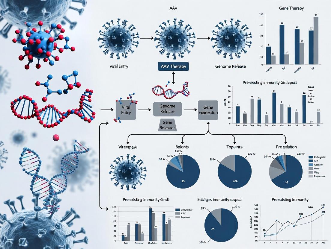
Overcoming Pre-existing Immunity to AAV: Strategies for Next-Generation Gene Therapies
This article provides a comprehensive analysis of the challenge posed by pre-existing immunity to Adeno-Associated Virus (AAV) vectors in gene therapy.
Overcoming Pre-existing Immunity to AAV: Strategies for Next-Generation Gene Therapies
Abstract
This article provides a comprehensive analysis of the challenge posed by pre-existing immunity to Adeno-Associated Virus (AAV) vectors in gene therapy. Targeting researchers, scientists, and drug development professionals, we explore the origins and impact of neutralizing antibodies (NAbs), detail current and emerging methodologies to circumvent immune responses, evaluate optimization and troubleshooting techniques for clinical translation, and compare the validation of novel capsids and adjunctive strategies. The synthesis offers a roadmap for developing safer, more effective, and broadly applicable AAV-based therapeutics.
Understanding the AAV Immunity Barrier: Prevalence, Origins, and Clinical Impact
The Prevalence of Pre-existing Neutralizing Antibodies (NABs) in Global Populations.
Technical Support Center
Welcome to the Technical Support Center for AAV Pre-existing Immunity Research. This resource provides troubleshooting guides and FAQs for key experimental challenges in characterizing pre-existing neutralizing antibodies (NAbs) against adeno-associated virus (AAV) vectors.
FAQs & Troubleshooting
Q1: Our in vitro NAb assay shows high variability between replicates. What could be the cause and how can we improve consistency? A: High variability often stems from cell passage number, serum sample handling, or AAV vector titer inconsistency.
- Troubleshooting Steps:
- Standardize Cells: Use low-passage-number cells (e.g., HEK293T, HepG2) and ensure consistent seeding density and viability.
- Handle Serum/Plasma Properly: Avoid repeated freeze-thaw cycles of samples. Heat-inactivation (56°C, 30 min) can reduce complement interference but may also affect some antibodies; include a consistent protocol.
- Titer Vector Accurately: Use digital droplet PCR (ddPCR) for genomic titer determination instead of less precise methods like ELISA or spectrophotometry. Aliquot working stocks to avoid freeze-thaw.
- Recommended Protocol (Standard In Vitro Luciferase-based NAb Assay):
- Day 1: Seed cells in a 96-well plate at a density optimized for 90-95% confluence at transduction (e.g., 1.5x10^4 HEK293T cells/well).
- Day 2: Prepare serum/plasma dilutions (e.g., 1:2 to 1:50 in culture medium) in a separate plate. Mix equal volumes of each serum dilution with a fixed dose of AAV-luciferase vector (e.g., 1e8 vg/well final MOI). Incubate at 37°C for 1 hour.
- Apply the serum-vector mixture to cells after removing old medium. Include controls: cells only, vector only, and a known positive control serum.
- Day 3/4: Lyse cells and measure luciferase activity. Normalize values to the "vector only" control (100% transduction). The NAb titer is often reported as the dilution that inhibits transduction by 50% (IC50 or ND50).
Q2: How do we interpret discordant results between different NAb assay formats (e.g., in vitro transduction inhibition vs. total AAV-binding ELISA)? A: Discordance is common and informative. ELISA detects total binding antibodies (IgG, IgM, non-neutralizing), while cell-based assays specifically measure functional neutralization.
- Action Plan:
- Run Parallel Assays: Perform both a cell-based neutralization assay and a total IgG AAV-capsid ELISA on the same sample set.
- Analyze Correlation: Use the data to determine the correlation in your cohort. A weak correlation suggests a significant proportion of binding antibodies are non-neutralizing.
- Key Insight: High ELISA signal with low neutralization may not preclude gene therapy. The cell-based assay is clinically more relevant for predicting vector inactivation.
Q3: What are the key considerations when establishing a new animal model to study pre-existing AAV immunity? A: The choice depends on the research question (natural vs. induced immunity).
- Key Considerations:
- Natural Prevalence: Screen the animal colony for pre-existing AAV NAbs. Specific Pathogen Free (SPF) facilities may have lower rates.
- Immunization Model: To induce consistent NAb titers, immunize with wild-type AAV capsid (not recombinant vector) plus adjuvant (e.g., Freund's adjuvant, Alum). Characterize the kinetics and durability of the response.
- Cross-Reactivity: Test for cross-neutralization across relevant AAV serotypes (e.g., AAV2, AAV5, AAV8, AAV9).
Data Presentation: Global Prevalence of Pre-existing AAV NAbs
The prevalence of pre-existing NAbs varies significantly by serotype and geography. The table below summarizes recent meta-analyses and regional studies.
Table 1: Global Prevalence of Pre-existing Neutralizing Antibodies (NAb Titers ≥1:50)
| AAV Serotype | North America | Europe | Asia | Global Average (Estimated) | Key Notes |
|---|---|---|---|---|---|
| AAV2 | 30-50% | 30-40% | 50-70% | 40-55% | Most studied; highest prevalence globally. |
| AAV5 | ~10-20% | ~15-25% | 15-30% | 15-25% | Generally lower seroprevalence. |
| AAV8 | 25-40% | 20-35% | 30-50% | 30-40% | High cross-reactivity with AAV2 reported. |
| AAV9 | 20-35% | 15-30% | 40-60% | 25-45% | Geographic variability is significant. |
Note: Prevalence rates are highly dependent on the assay cut-off titer. The ≥1:50 threshold is commonly used in clinical screening. Data compiled from recent cohort studies (2020-2023).
Experimental Protocols
Protocol: Digital Droplet PCR (ddPCR) for AAV Genome Titering Objective: To accurately quantify the genomic titer (vg/mL) of an AAV vector stock.
- Sample Prep: Treat AAV vector with DNase I to remove unpackaged DNA. Then inactivate DNase and degrade the capsid with Proteinase K.
- Digestion: Heat-inactivate, and dilute sample appropriately (typically 1e4-1e5 fold).
- Droplet Generation: Mix diluted DNA with ddPCR supermix and primers/probe targeting a conserved region (e.g., polyA signal, promoter). Generate droplets using a droplet generator.
- PCR Amplification: Transfer droplets to a PCR plate and run amplification: 95°C (10 min), then 40 cycles of 94°C (30 sec) and 60°C (1 min), with a final 98°C (10 min) enzyme deactivation.
- Reading & Analysis: Read plate in a droplet reader. Use Poisson statistics to calculate the concentration of target DNA molecules in the original sample (copies/µL). Convert to vg/mL.
Protocol: AAV-Capsid Specific Total IgG ELISA Objective: To quantify total anti-AAV capsid IgG in human serum/plasma.
- Coating: Coat a 96-well ELISA plate with purified AAV capsids (e.g., 1e9 vg/well) in carbonate buffer overnight at 4°C.
- Blocking: Block with 5% non-fat milk or BSA in PBS-T for 2 hours at room temperature (RT).
- Sample Incubation: Add serial dilutions of test serum and a standard curve of a known positive control (e.g., human anti-AAV IgG) to the plate. Incubate 2 hours at RT.
- Detection: Add horseradish peroxidase (HRP)-conjugated anti-human IgG antibody. Incubate 1 hour at RT.
- Development: Add TMB substrate, incubate in the dark, then stop the reaction with H2SO4.
- Analysis: Read absorbance at 450 nm. Plot the standard curve and interpolate sample concentrations (in arbitrary units/mL relative to the standard).
Mandatory Visualizations
Diagram Title: Assay Selection Workflow for AAV NAb Detection
Diagram Title: Antibody-Mediated Neutralization of AAV Pathways
The Scientist's Toolkit: Research Reagent Solutions
Table 2: Essential Reagents for AAV NAb Research
| Reagent/Material | Function & Rationale | Example/Notes |
|---|---|---|
| Purified AAV Capsids (Multiple Serotypes) | Coating antigen for ELISA; immunogen for animal models. Essential for specificity. | Wild-type (empty) capsids preferred for ELISA to avoid transgene interference. |
| Reference Standard Serum | Positive control for assay normalization and inter-lab comparison. | Pooled human serum with characterized high-titer NAbs. Commercial or in-house qualified. |
| Reporter AAV Vectors | Engineered AAV expressing a reporter gene for functional cell-based neutralization assays. | AAV2-Luciferase, AAV8-GFP, etc. Use the serotype matching your therapy vector. |
| Susceptible Cell Line | Permissive cells for AAV transduction in neutralization assays. | HEK293T (high permissiveness), HepG2 (liver-relevant), primary hepatocytes (gold standard). |
| ddPCR Mastermix & Assays | For absolute quantification of AAV vector genome titer, critical for assay consistency. | Bio-Rad QX200 or equivalent. Target sequence should be within the ITR or a conserved vector backbone region. |
| HRP-conjugated Anti-Human IgG | Detection antibody for ELISA to quantify total anti-AAV antibodies. | Must have specificity for all human IgG subclasses. Pre-adsorbed if using animal sera. |
| Adjuvants (for Animal Models) | To boost immune response when generating NAbs in preclinical models. | Complete/Incomplete Freund's Adjuvant, Alum, or modern alternatives like AddaVax. |
Technical Support Center: Troubleshooting Pre-Existing Immunity in AAV Gene Therapy
Troubleshooting Guides & FAQs
FAQ 1: How do I determine if my animal model or patient cohort has pre-existing neutralizing antibodies (NAbs) against my AAV serotype of interest?
Answer: Pre-existing NAbs are typically measured using an in vitro cell-based transduction inhibition assay.
- Sample Collection: Collect serum or plasma.
- Assay Setup: Serially dilute the sample and incubate it with a known titer of your AAV vector (e.g., 1e9 vg/well) encoding a reporter gene (e.g., GFP, Luciferase) for 1 hour at 37°C.
- Transduction: Add the mixture to permissive cells (e.g., HEK293, HepG2).
- Quantification: After 48-72 hours, quantify reporter expression. The NAb titer is reported as the highest dilution that inhibits transduction by 50% (IC50 or ID50) compared to a no-serum control.
Troubleshooting: High background or inconsistent results can be caused by:
- Issue: Serum cytotoxicity.
- Fix: Heat-inactivate serum at 56°C for 30 minutes prior to assay. Include a cell viability control.
- Issue: Low signal-to-noise from reporter.
- Fix: Titrate your AAV control virus to achieve a robust, linear signal in the absence of serum. Use a highly sensitive detection method.
FAQ 2: My in vivo gene transfer efficiency is low despite low in vitro NAb titers. What could be the cause?
- Answer: This discrepancy often points to the involvement of cellular immunity (AAV-specific T-cells) or non-neutralizing antibodies that mediate clearance via Fc-receptor or complement-dependent mechanisms, which are not captured in standard NAb assays.
- Investigation Path:
- Analyze Cellular Immunity: Perform IFN-γ ELISpot or intracellular cytokine staining on peripheral blood mononuclear cells (PBMCs) stimulated with AAV capsid peptides.
- Consider Total Binding Antibodies: Use an ELISA to measure total anti-AAV IgG/IgM, which may correlate with clearance mechanisms independent of neutralization.
- Investigation Path:
FAQ 3: How can I address serotype cross-reactivity in my study design?
- Answer: Cross-reactivity occurs because antibodies against one AAV serotype (e.g., from natural infection with AAV2) can neutralize other serotypes (e.g., AAV3, AAV6).
- Actionable Steps:
- Screen Broadly: Test subject sera against a panel of potential therapeutic serotypes, not just your primary candidate.
- Consider Engineered Capsids: Explore data on synthetic or engineered capsids designed to evade common cross-reactive antibodies.
- Empirical Testing: The gold standard is to test the actual subject serum against your final clinical vector lot, as manufacturing nuances can affect antigenicity.
- Actionable Steps:
Key Quantitative Data on AAV Seroprevalence and Cross-Reactivity
Table 1: Global Seroprevalence of Common AAV Serotypes
| Serotype | Regional Prevalence (Approx. % NAb Positive, Titer ≥1:5) | Key Notes & Cross-Reactivity |
|---|---|---|
| AAV1 | 30-40% (Global) | Significant cross-reactivity with AAV6. |
| AAV2 | 30-70% (Varies widely) | Highest natural seroprevalence. Antibodies often cross-react with AAV3, AAV5*, AAV6, AAV13. |
| AAV5 | 30-50% (Global) | More distinct; lower cross-reactivity with AAV2/8/9, but not absent. |
| AAV6 | 30-45% (Global) | High homology with AAV1; strong cross-reactivity. |
| AAV8 | 30-55% (Global) | Significant cross-reactivity with AAV2 in some populations. |
| AAV9 | 30-60% (Global) | Cross-reactivity observed with AAV2 and AAV8. |
| Note: Prevalence is age and geography-dependent. Cross-reactivity can be asymmetric and titer-dependent. |
Table 2: Common Experimental Assays for Characterizing Pre-Existing Immunity
| Assay Type | Measures | Output | Time | Key Limitation |
|---|---|---|---|---|
| Cell-Based Neutralization | Functional NAbs | IC50/ID50 Titer | 3-4 days | Misses non-neutralizing mechanisms. |
| Total IgG/IgM ELISA | Binding Antibodies | ELISA Titer / OD | 1 day | Does not indicate function. |
| PBMC-based ELISpot | Capsid-specific T-cells | Spot-Forming Units (SFU) | 2 days | Requires fresh cells; complex assay. |
| ADCVI / ADCD Assays | Antibody-dependent cellular/ complement inhibition | % Inhibition of Transduction | 4-5 days | Complex; not yet standardized. |
Experimental Protocols
Protocol: Standard In Vitro Neutralizing Antibody Assay
Objective: Determine the 50% inhibitory dilution (ID50) of serum against an AAV vector.
Reagents: HEK293 cells, DMEM+10% FBS, AAV-GFP vector (1e12 vg/mL stock), test serum, poly-L-lysine coated 96-well plates.
Methodology:
- Day 0: Plate HEK293 cells at 1.5e4 cells/well in 100 µL complete media.
- Day 1: Prepare serum dilutions (e.g., 1:2 to 1:1024) in serum-free media in a separate plate. Mix 50 µL of each dilution with 50 µL of AAV-GFP vector (diluted to 2e9 vg/well in final assay). Incubate 1 hr at 37°C.
- Transduction: Remove media from cell plate. Add 100 µL of the serum-vector mixture to corresponding wells. Include AAV-only (no serum) and cell-only controls.
- Day 3: Analyze GFP expression via flow cytometry or fluorescence microscopy.
- Analysis: Calculate % transduction inhibition relative to AAV-only control. Fit data with a 4-parameter logistic curve to calculate the ID50 value.
Diagrams
Diagram Title: AAV Pre-Existing Immunity Troubleshooting Workflow
Diagram Title: Humoral Immune Mechanisms Against AAV Vectors
The Scientist's Toolkit: Research Reagent Solutions
Table 3: Essential Reagents for Studying AAV Pre-Existing Immunity
| Reagent / Material | Function in Experiments | Example / Note |
|---|---|---|
| AAV Reference Standards (e.g., AAV2, AAV8, AAV9) | Positive controls for serology assays; ensure assay consistency. | Available from ATCC, Vigene, or internal production. |
| Reporter AAV Vectors (GFP, Luciferase) | Enable quantification of transduction inhibition in NAb assays. | Use a ubiquitous promoter (CAG, CBh). |
| Validated Positive Control Sera (Animal or Human) | Essential for normalizing NAb assays between runs. | Sera from AAV-immunized animals; commercial human Ig preparations. |
| AAV Capsid Peptide Pools (15mer overlapping) | Stimulate AAV-specific T-cells in ELISpot/ICS assays to probe cellular immunity. | Custom synthesis covering entire VP1/2/3 region. |
| Fc Receptor Blocking Reagent | Used in assays to distinguish neutralization from Fc-mediated effects. | e.g., Human Fc Block (BD Biosciences). |
| Standardized Cell Line (HEK293, HepG2) | Critical for reproducible, sensitive NAb assays. | Use a low-passage, mycoplasma-free master bank. |
| Anti-AAV Capsid Monoclonal Antibodies | Tools for developing quantitative immunoassays (ELISA, MSD). | e.g., ADK8, ADK1b (Progen). |
Troubleshooting & FAQs: A Technical Support Center for NAb-Related AAV Research
FAQ 1: Pre-Assay & Experimental Design
Q: How do I determine the critical threshold for NAb titer that will block my specific AAV serotype? A: The inhibitory threshold (IT) is serotype, target cell, and transgene-dependent. The general rule is that an ID50 (50% inhibitory dose) titer ≥ 1:5 can significantly reduce transduction in vivo, but you must establish this empirically for your system.
- Table 1: Reported Neutralizing Antibody (NAb) Thresholds for Common AAV Serotypes
AAV Serotype Typical Reported Critical Titer (ID50) Key Determinants Primary References AAV2 1:2 - 1:20 Highly prevalent; sensitive to low NAb levels. Depends on route of administration (intravenous vs. local). Boutin et al., 2010; Calcedo et al., 2009 AAV5 1:5 - 1:50 Lower seroprevalence; generally more resistant to NAbs. Boutin et al., 2010 AAV8 1:10 - 1:100 Moderate seroprevalence; relatively resistant but high titers block. Wang et al., 2011; Calcedo et al., 2011 AAV9 1:20 - 1:200 Similar to AAV8; shows some resistance but is not fully evasive. Calcedo et al., 2011
Troubleshooting Guide: If your in vivo transduction is unexpectedly low despite low pre-screened NAb titers:
- Verify Assay: Ensure your NAb detection assay (e.g., GFP-reduction, Luciferase-based) uses the same AAV capsid and promoter as your therapeutic vector.
- Check Cell Type: The permissiveness of your target cell type affects the threshold. Primary cells may be more susceptible to blockade.
- Consider Total IgG: High total AAV-specific IgG, even without strong neutralizing capacity, can enhance clearance via Fc receptor-mediated phagocytosis, confounding results.
FAQ 2: Mechanisms & Molecular Interactions
Q: What are the precise molecular steps by which a NAb blocks AAV cellular transduction? A: NAbs interfere at multiple steps, primarily before internalization. The core mechanism is steric hindrance.
Detailed Mechanism:
- Binding: NAbs, typically IgG, bind to epitopes on the AAV capsid's VP proteins.
- Steric Hindrance: The large antibody molecule physically obstructs the essential interaction between the AAV capsid and its primary cellular receptor (e.g., AAV2 with HSPG).
- Aggregation: NAbs can cross-link multiple AAV particles, forming large aggregates that are inefficient for cell binding.
- Post-Binding Block: Even if receptor binding occurs, NAbs can block subsequent steps like co-receptor interaction (e.g., AAV2 with αvβ5 integrin) or conformational changes needed for endocytosis.
- Clearance: Opsonized vectors (coated with antibodies) are marked for rapid clearance by phagocytic cells (e.g., Kupffer cells in the liver) before reaching target tissue.
Diagram Title: NAb Blockade vs. Normal AAV Transduction Pathway
FAQ 3: Protocols & Validation
Q: What is a robust protocol for determining NAb titer in mouse or NHP serum? A: Here is a standardized in vitro GFP-reduction neutralization assay protocol.
Protocol: In Vitro Neutralization Assay (96-well format) Objective: To quantify the serum dilution that inhibits AAV transduction by 50% (ID50).
Materials & Reagent Solutions: Table 2: Key Research Reagent Solutions for NAb Assays
| Reagent/Material | Function/Role | Example Product/Catalog |
|---|---|---|
| Target Cells | Permissive cells for AAV transduction. | HEK293 cells (ATCC CRL-1573), HepG2 cells. |
| AAV Reporter Vector | Expresses quantifiable protein (GFP, Luc). Must match capsid of study. | AAV2-CB-GFP (Vector Biolabs, #7002). |
| Control Serotype | AAV with different capsid to test specificity. | AAV5-CB-GFP. |
| Negative Control Serum | Pre-immune or confirmed NAb-negative serum. | Commercial fetal bovine serum (FBS). |
| Positive Control Serum | High-titer anti-AAV serum (polyclonal). | AAV2 neutralizing antibody (PROGEN, #6513). |
| Dilution Buffer | Maintains AAV & antibody stability. | DMEM + 2% FBS (no supplements). |
| Detection Reagent | Quantifies transgene expression. | Flow cytometry buffer (PBS + 1% BSA). |
| Data Analysis Software | Calculates ID50 from dose-response. | GraphPad Prism (4-parameter logistic fit). |
Step-by-Step Method:
- Serum Heat-Inactivation: Incubate serum samples at 56°C for 30 minutes to inactivate complement.
- Serum Dilution: Prepare 2-fold serial dilutions of serum in dilution buffer (e.g., 1:2 to 1:512) in a separate dilution plate.
- Virus-Antibody Incubation: Mix a fixed dose of AAV reporter vector (e.g., 1e4 vg/cell, MOI ~10,000) with an equal volume of each serum dilution. Include virus-only (no serum) and cell-only controls.
- Incubation: Incubate mixtures at 37°C for 1 hour.
- Cell Infection: Plate target cells at 70-80% confluence 24 hours prior. Aspirate medium and add 100µL of each virus-serum mixture to cells (in triplicate).
- Transduction: Incubate cells at 37°C for 48-72 hours.
- Analysis (Flow Cytometry):
- Harvest cells (trypsinization).
- Resuspend in cold detection buffer.
- Analyze percentage of GFP-positive cells using a flow cytometer.
- ID50 Calculation:
- Normalize data: (GFP+ % with serum / GFP+ % virus-only control) * 100 = % Transduction.
- Plot % Transduction (Y) vs. Serum Dilution (log10, X).
- Fit a 4-parameter logistic curve (in GraphPad Prism).
- The ID50 titer is the serum dilution at which transduction is reduced to 50%.
Troubleshooting: If the curve fit is poor:
- Flag: Insufficient virus dose. Repeat with higher MOI.
- Flag: Serum toxicity at low dilutions. Include a serum-only control on cells (no virus) to check for cytotoxicity.
FAQ 4: Addressing Pre-Existing Immunity in Research
Q: What experimental strategies can I use to overcome pre-existing NAbs in my gene therapy model? A: Several direct and indirect strategies are under active investigation.
- Table 3: Experimental Strategies to Circumvent Pre-Existing NAbs
Strategy Mechanism Key Experimental Considerations Current Efficacy (Animal Models) Capsid Switching Use a serotype with low seroprevalence or different antigenicity. Screen human/NHP serum panels in vitro. AAV5, AAV8, AAVrh.10 show partial success. Empty Capsid Decoy Co-administer excess empty capsids to adsorb NAbs. Requires precise, high dose of decoys; may affect total vector yield. Can increase transduction 2-10 fold in mice with low-medium NAb titers. Plasmapheresis / Immunoadsorption Physically remove immunoglobulins prior to vector administration. Transient effect; requires specialized equipment and clinical setting. Rapid NAb reduction, but rebound possible. Efficacy proven in some clinical cases. Capsid Engineering Develop novel, engineered capsids resistant to NAb binding. Use directed evolution or structure-guided design in presence of pooled human IgG. Shown to evade high-titer human NAbs in murine & NHP models. Leading approach. Immunosuppression Use transient B-cell depletion (e.g., anti-CD20) or drugs. Targets the source, not immediate NAb pool. Risk of general immunosuppression. Effective in preventing de novo NAb formation; less effective for pre-existing.
Diagram Title: Decision Workflow for Overcoming Pre-existing NAbs
Technical Support Center: FAQs & Troubleshooting for AAV Pre-existing Immunity Research
Q1: How do I accurately measure pre-existing neutralizing antibody (NAb) titers against AAV serotypes? A: Inconsistent NAb titer measurements are often due to variability in the reporter gene assay. Ensure you use a standardized reference standard (e.g., WHO International Standard for AAV2 NAbs, if available for your serotype) to calibrate your assay. Use a minimum of eight serial dilutions of serum in duplicate. The titer is typically reported as the reciprocal of the highest serum dilution that inhibits transduction by 50% (IC50 or ID50). Validate your cell line (e.g., HEK293) for consistent permissiveness and transducibility.
Q2: What is the critical NAb titer threshold for patient exclusion in clinical trials? A: Published thresholds vary by serotype and route of administration. The table below summarizes current consensus from recent literature and trial protocols:
Table 1: Representative AAV NAb Titer Exclusion Thresholds in Clinical Development
| AAV Serotype | Route of Administration | Reported Critical Titer (Reciprocal) | Clinical Phase | Associated Risk |
|---|---|---|---|---|
| AAV2 | Intravenous (IV) | ≥ 1:5 | I/II | Reduced transduction efficacy |
| AAV8 | IV (Liver-directed) | ≥ 1:5 to ≥ 1:10 | III | Complete ablation of gene transfer |
| AAV9 | Intravenous (CNS-directed) | ≥ 1:10 | I/II | Potential for reduced biodistribution |
| AAV5 | Intraocular | ≥ 1:2 | II/III | Local immune response risk |
| AAVrh74 | Intramuscular | ≥ 1:40 (some studies) | I/II | Impact on muscle transduction |
Note: Thresholds are protocol-specific and evolving. Always refer to the latest regulatory guidance.
Q3: Our in-vivo model shows gene transfer despite high pre-existing NAbs. How can this be reconciled with clinical trial failures? A: Animal models may not fully recapitulate human humoral immunity. Key troubleshooting steps:
- Verify Serotype Specificity: Confirm your assay measures NAbs against the exact capsid variant used in your trial.
- Assay Sensitivity: Compare your assay's limit of detection to the clinical assay used. Consider implementing a total antibody (binding) assay as a complementary screen, as non-neutralizing antibodies can also impact pharmacokinetics.
- Cohort Analysis: Re-analyze clinical failure data stratifying patients by both NAb titer and pre-existing AAV-specific T-cell responses. The interaction is critical.
Q4: What are the best practices for developing a robust NAb assay to support regulatory submissions? A: Follow a fit-for-purpose validation strategy:
- Precision: Intra- and inter-assay CV should be <20% (ideally <15%) around the cut point.
- Selectivity/Specificity: Test interference from common serum factors (lipids, hemoglobin, rheumatoid factor).
- Cut-Point Determination: Use at least 50 individual sera from relevant disease population (minus AAV treatment) to establish a statistically derived (e.g., 95% percentile) screening cut point.
- Sample Stability: Establish stability under conditions mimicking sample collection, shipment, and storage.
Experimental Protocol: Standardized Luciferase-Based Neutralizing Antibody Assay
Purpose: To quantify serum neutralizing activity against recombinant AAV vectors.
- Day 1 - Plate Cells: Seed HEK293 cells in 96-well plates at a density of 1.5 x 10^4 cells/well in complete growth medium. Incubate at 37°C, 5% CO2 for 20-24 hours.
- Day 2 - Prepare Serum-Vector Mix:
- Heat-inactivate test serum samples at 56°C for 30 minutes.
- Perform 2-fold serial dilutions of serum in assay medium (e.g., DMEM + 2% FBS) in a separate dilution plate.
- Dilute the AAV-luciferase vector (of target serotype) to a pre-titered working concentration (e.g., 1x10^8 vg/mL) in assay medium.
- Mix equal volumes of diluted serum and diluted vector. Include controls: No-serum control (vector only), no-vector control, and a positive control (serum with known high NAb titer).
- Incubate at 37°C for 1 hour.
- Day 2 - Transduction: Remove growth medium from cell plate. Add 100µL of serum-vector mixture to respective wells. Incubate at 37°C, 5% CO2 for 24 hours.
- Day 3 - Readout: Aspirate medium, lyse cells with 50µL passive lysis buffer (per manufacturer's instructions). Transfer lysate to a white plate, inject luciferase substrate, and measure luminescence.
- Analysis: Normalize luminescence to the no-serum control (100% transduction). Fit dose-response curve (4-parameter logistic) to calculate the IC50/ID50 titer.
The Scientist's Toolkit: Key Research Reagent Solutions
Table 2: Essential Materials for AAV NAb & Immunity Studies
| Reagent / Material | Function & Importance |
|---|---|
| WHO International Standard (IS) for AAV2 Neutralizing Antibodies | Critical for assay standardization and cross-trial data comparability. Provides a universal unit (IU/mL). |
| Certified Reference Sera (Positive/Negative Controls) | Ensures inter-assay precision and validates assay performance over time. |
| Reporter AAV Vectors (Luciferase, GFP, SEAP) | Probes for functional neutralization. Must be produced to high purity (empty capsid <10%) and accurately titered (vg/mL). |
| Relevant Permissive Cell Line (e.g., HEK293T) | Consistent host for transduction. Must be routinely tested for mycoplasma and maintained at low passage. |
| Validated AAV-Specific ELISA or MSD Assay Kits | Quantifies total anti-capsid binding antibodies (IgG, IgM, IgA), complementing NAb data. |
| Multiplexed IFN-γ ELISpot Kits | Detects pre-existing AAV-specific T-cell responses, a key co-variable with NAbs for risk assessment. |
| High-Sensitivity Luminescence/Fluorescence Plate Reader | Essential for accurate, sensitive readout of reporter gene assays. |
Visualizations
Diagram 1: AAV NAb Impact on Clinical Trial Efficacy Pathway
Diagram 2: NAb Titer Assay & Analysis Workflow
Technical Support Center: Troubleshooting AAV-Specific T-Cell Assays
FAQs & Troubleshooting Guides
Q1: In our IFN-γ ELISpot assay for AAV-capsid specific T-cells, we consistently get high background noise in wells containing peptides from healthy donor PBMCs with no known AAV exposure. What could be the cause and how can we resolve it? A: High background in ELISpot is often due to non-specific activation or contaminants.
- Primary Cause: Peptide impurities or DMSO carryover from peptide stock solutions can be mitogenic.
- Troubleshooting Steps:
- Peptide Preparation: Ensure peptides are dissolved in sterile, endotoxin-free PBS or culture-grade DMSO at a stock concentration ≤10 mg/mL, followed by dilution in assay medium to a final DMSO concentration <0.1%.
- Peptide Titration: Perform a dose-response curve (e.g., 1-10 µg/mL) to identify the optimal, non-toxic concentration.
- Control Wells: Include critical controls: (i) Cells + DMSO vehicle at the same final concentration as test wells, (ii) Cells + a known immunogenic positive control peptide (e.g., CEF pool), (iii) Cells + medium only.
- Cell Quality: Isolate PBMCs using a density gradient centrifugation method with minimal platelet contamination, as platelets can secrete cytokines.
- Protocol: IFN-γ ELISpot for AAV Capsid Peptide Libraries.
- Coat Plate: Coat PVDF-backed 96-well plate with anti-human IFN-γ capture antibody (15µg/mL in sterile PBS) overnight at 4°C.
- Block: Block plate with assay medium (RPMI-1640, 10% human AB serum, 1% L-Glut) for 2 hours at 37°C.
- Plate Cells & Peptides: Add 2.5x10^5 PBMCs per well in 100µL assay medium. Add pre-diluted AAV peptide pools or single peptides in 100µL medium. Final peptide concentration: 2µg/mL per peptide. Run in triplicate.
- Incubate: Incubate plate for 40-48 hours at 37°C, 5% CO2 in a humidified incubator.
- Develop: Follow manufacturer's protocol for biotinylated detection antibody, streptavidin-ALP, and BCIP/NBT substrate.
- Analyze: Enumerate spots using an automated ELISpot reader.
Q2: When using a flow cytometry-based intracellular cytokine staining (ICS) assay to phenotype AAV-specific T-cells, our antigen-stimulated cells show poor viability and low cytokine signals compared to the positive control. How can we improve the response? A: This indicates suboptimal T-cell stimulation or excessive cell death during the prolonged assay.
- Primary Cause: Inadequate co-stimulatory signals during the peptide presentation phase.
- Troubleshooting Steps:
- Enhance Co-stimulation: Add soluble anti-CD28 and anti-CD49d antibodies (1µg/mL each) at the start of the stimulation. This is crucial when using exogenous peptides that bypass professional antigen-presenting cells.
- Optimize Duration: Reduce the stimulation period. For ICS, a 6-hour stimulation (with protein transport inhibitor added for the final 4-5 hours) often yields better viability than 12-16 hours for strong AAV responses.
- Use of CD137 (4-1BB) Assay: As an alternative, stain for CD137 surface expression after 24-hour stimulation. This identifies recently activated T-cells without requiring cytokine blockade and prolonged culture, improving viability assessment.
- Protocol: ICS for Phenotyping AAV-specific CD4+/CD8+ T-cells.
- Stimulate: Seed 1-2x10^6 PBMCs/mL in assay medium. Add AAV peptide pools (2µg/mL/peptide), anti-CD28/anti-CD49d (1µg/mL), and Brefeldin A/Monensin. Include PMA/Ionomycin positive control and SEB super-antigen control.
- Incubate: Stimulate for 6 hours at 37°C, 5% CO2.
- Surface Stain: Wash cells, stain with viability dye and surface antibodies (e.g., CD3, CD4, CD8, CD45RA, CCR7) for 30 min at 4°C.
- Fix/Permeabilize: Use a commercial cytofix/cytoperm kit.
- Intracellular Stain: Stain for cytokines (e.g., IFN-γ, TNF-α, IL-2) for 30 min at 4°C.
- Acquire & Analyze: Acquire on a flow cytometer. Gate on live, single CD3+ lymphocytes, then CD4+ or CD8+, and analyze cytokine co-expression.
Q3: Our data on AAV-neutralizing antibody (NAb) titers and T-cell responses seem discordant. Some subjects with high NAbs show no T-cell response, and vice versa. Is this expected? A: Yes, this is a key observation underscoring the independence of humoral and cellular arms of pre-existing immunity.
- Explanation: NAbs are directed against conformational capsid epitopes and block cellular entry. T-cell responses (especially CD8+) are typically directed against linear peptide sequences presented by MHC-I after intracellular processing of the capsid. Their immunodominance and memory persistence are governed by different factors.
- Data Correlation Table:
| Subject Profile | Typical NAb Titer (IU/mL)* | Likely T-cell Response (IFN-γ SFU/10^6 PBMCs)* | Immunological Interpretation |
|---|---|---|---|
| Natural AAV Exposure | Variable (Low to High: <5 to >100) | Low to Moderate (10-100) | Humoral and cellular memory established, but often non-overlapping epitopes. |
| AAV Gene Therapy Recipient | Very High (>500) | High (>100) | High-level exposure to full capsid antigen primes both arms strongly. |
| Seropositive, NAb Low | Low (<2) | Detectable (20-50) | Exposure primed T-cells but not high-affinity B-cell/NAb response. |
| Seronegative | Negative (<1) | Undetectable (<10) | No prior adaptive immunity. |
*Representative quantitative ranges. Actual values vary by assay sensitivity and patient history.
The Scientist's Toolkit: Key Reagent Solutions
| Reagent/Factor | Primary Function in AAV T-Cell Assays |
|---|---|
| Overlapping Peptide Libraries (15mer, 11aa overlap) | Covers the entire AAV capsid protein (VP1/2/3) sequence to detect CD4+ and CD8+ T-cell responses without MHC restriction. |
| Human AB Serum | Used in assay medium to provide essential nutrients and reduce non-specific background activation compared to FBS. |
| Protein Transport Inhibitors (Brefeldin A, Monensin) | Block cytokine secretion, allowing intracellular accumulation for detection by flow cytometry (ICS). |
| Co-stimulatory Antibodies (anti-CD28, anti-CD49d) | Provide critical Signal 2 to T-cells during peptide stimulation, enhancing sensitivity for weak memory responses. |
| MHC Multimers (Tetramers, Dextramers) | Directly stain T-cells with specific T-cell receptors for defined AAV capsid epitopes, enabling high-resolution phenotyping. |
| Cytokine Capture Assays (e.g., Miltenyi Cytokine Secretion Assay) | Allows for live sorting of antigen-specific T-cells based on secreted cytokine for downstream functional analysis. |
Experimental Workflow & Pathway Diagrams
Title: Workflow for Detecting AAV-Specific T-Cells
Title: AAV Capsid-Specific CD8+ T-Cell Activation Pathway
Bypassing the Immune System: Technical Strategies for AAV Gene Delivery
Capsid Engineering and Directed Evolution for Stealth Vectors
Technical Support Center
Troubleshooting Guides & FAQs
Q1: Our AAV library diversity post-selection is critically low. What are the primary causes and solutions? A: Low diversity is often due to a selection bottleneck or inadequate library size.
- Cause 1: Excessive selection pressure (e.g., high antibody/heparin concentration).
- Solution: Titrate the negative selective agent (e.g., neutralizing antibodies) during the panning round. Use concentrations that select for binders but do not eliminate >95% of the library. A pilot neutralization assay with pooled IVIG against your parental capsid can establish a baseline.
- Cause 2: Insufficient library transformation efficiency leading to low pre-selection diversity.
- Solution: Ensure library complexity is at least 10⁹-10¹¹ unique variants. Use high-efficiency electrocompetent cells (e.g., NEB 10-beta Electrocompetent E. coli), multiple electroporations, and extensive amplification. Quantify diversity by NGS of the input library.
- Protocol: Titering IVIG for In Vitro Selection
- Coat a 96-well plate with 5e10 vg/well of your parental AAV library in PBS overnight at 4°C.
- Block with 2% BSA in PBS for 1 hour.
- Incubate with serial dilutions of IVIG (e.g., 1:10 to 1:1000 in PBS) for 2 hours at 37°C.
- Wash and detect bound IgG with HRP-conjugated anti-human IgG. Plot OD vs. concentration.
- Use the IVIG concentration that yields 50-70% signal reduction for the first selection round.
Q2: We observe poor transduction efficiency with our newly evolved capsid in vivo, despite successful escape from NAbs in vitro. A: This disconnect often stems from neglecting other serum or tissue factors.
- Cause 1: Complement-mediated inactivation.
- Solution: Perform selection under active human complement (e.g., 10% normal human serum, verified for complement activity). Include a heat-inactivated serum control. Assay final candidates in a complement activation assay (C3a detection ELISA).
- Cause 2: Off-target tropism or reduced affinity for the target receptor.
- Solution: Incorporate a positive selection step post-negative selection. For liver tropism, incubate the post-antibody-selected pool with HepG2 cells. Isolate bound/transduced variants. Always include a parallel in vivo selection round in animal models with pre-existing immunity if possible.
- Protocol: Combined Negative/Positive In Vitro Selection
- Incubate your AAV peptide display library (1e11 vg) with a neutralizing IVIG pool (titered to bind ~80% of input) for 1h at 37°C in DMEM.
- Add the mixture to a confluent monolayer of your target cells (e.g., HepG2) in a 10cm dish. Incubate for 2h at 37°C.
- Wash rigorously 5x with PBS to remove unbound/antibody-bound virions.
- Harvest cells, extract genomic DNA, and recover the AAV cap gene via PCR for the next round or analysis.
Q3: Our NGS data from directed evolution is overwhelming. What are the key bioinformatic filters to identify true stealth candidates? A: Focus on enrichment and convergence.
- Step 1: Align sequences to parental capsid (e.g., AAV9) and call mutations. Calculate frequency per variant.
- Step 2: Calculate fold-enrichment (Round n frequency / Round 0 frequency) for each variant. Filter for variants with >10-fold enrichment.
- Step 3: Look for convergent mutations—amino acid changes appearing in multiple high-enrichment variants, especially in known antigenic regions (VRs) or heparin-binding sites.
- Step 4: Cross-reference with structural data (PDB: 3UX1, 6X1M) to map mutations onto the capsid surface.
Table 1: Key Quantitative Metrics for AAV Library Selection
| Metric | Target Value | Measurement Method |
|---|---|---|
| Pre-selection Library Diversity | >1 x 10¹¹ unique variants | NGS of plasmid library pre-packaging |
| Viral Particle (VP) to Genome-containing Particle (VG) Ratio | <100:1 | Digital PCR (genome titer) vs. ELISA (VP titer) |
| Neutralization Threshold in Selection | 70-90% neutralization of parent | In vitro transduction inhibition assay |
| Enrichment Fold (Post-Selection) | >10-fold for top variants | NGS count comparison (Round n / Round 0) |
| In Vivo Transduction Efficiency (vs. Parent) | >50% of parent in naïve mice; >10x in immunized mice | Bioluminescence or qPCR on target tissue |
Table 2: Common Pitfalls in Capsid Evolution for Stealth
| Pitfall | Consequence | Corrective Action |
|---|---|---|
| Over-selection with high IVIG | Loss of all infectivity; no variants | Titrate IVIG to partial neutralization. |
| No positive selection step | Capsids lose tropism | Alternate negative selection with cell binding. |
| Ignoring complement | In vitro escape but in vivo failure | Add active complement serum in selection. |
| Small library size | Limited solution space | Maximize transformation efficiency; use multiple shuffling techniques. |
The Scientist's Toolkit: Research Reagent Solutions
| Item | Function & Application |
|---|---|
| Pooled Human Intravenous Immunoglobulin (IVIG) | Source of diverse, pre-existing neutralizing antibodies for in vitro selection pressure. |
| Normal Human Serum (Complement-Active) | Provides complement factors for selection against the complete innate immune response. |
| HEK293T/AAV Producer Cell Line | Standard cell line for high-titer production of AAV variant libraries. |
| Heparin Sulfate Affinity Column | Purifies functional capsids based on heparan sulfate proteoglycan binding, a key initial attachment factor. |
| High-Efficiency Electrocompetent E. coli (e.g., NEB 10-beta) | Essential for achieving the high transformation efficiency required to maintain library diversity. |
| Next-Generation Sequencing (NGS) Service/Primers | For deep sequencing of the cap gene to track variant enrichment across selection rounds. |
| Anti-AAV Capsid ELISA Kit (e.g., Progen) | Quantifies total viral particles (VP/mL) irrespective of genome content. |
| Digital PCR (ddPCR) Kit for ITR Sequence | Accurately quantifies genome-containing vector genomes (vg/mL). |
Experimental Protocols
Protocol: Creation of a Shuffled AAV Capsid Library via DNA Family Shuffling Objective: Generate a diverse library of chimeric AAV capsid genes from multiple serotype parents. Materials: Purified cap genes from AAV1, 2, 6, 8, 9; DNase I (RNase-free); S1 Nuclease; Taq DNA Polymerase; DpnI; Cloning vector with AAV2 inverted terminal repeat (ITR) flanking sequence.
- Fragment Generation: Combine ~100 ng each of purified cap plasmids. Add 0.15 U of DNase I in 10 µL reaction with Mn²⁺ buffer. Incubate at 15°C for 10-15 min to generate random 50-200 bp fragments. Heat-inactivate.
- Reassembly PCR: Purify fragments. Perform PCR without primers: 0.2 mM dNTPs, 2.5 U Taq polymerase, in 50 µL. Cycle: 94°C 2 min; then 40 cycles of [94°C 30s, 55°C 30s, 72°C 30s]; final 72°C 5 min. Fragments prime each other based on homology, reassembling full-length chimeric genes.
- Amplification: Add outer primers (homologous to ITR regions) to 1 µL of reassembly product. Run standard PCR to amplify full-length, shuffled cap genes.
- Cloning: Digest PCR product and vector with appropriate enzymes. Ligate and transform into high-efficiency electrocompetent cells. Aim for >10⁹ colonies to ensure diversity.
Protocol: In Vivo Selection in Murine Model of Pre-existing Immunity Objective: Directly evolve stealth capsids in an immune-competent mouse. Materials: C57BL/6 mice, AAV library (shuffled or peptide-insert), AAV9 empty capsid for immunization, Adjuvant (e.g., Freund's incomplete), qPCR tissue DNA extraction kit.
- Immunization: Inject 6-8 week-old mice intraperitoneally with 1e11 vg of empty AAV9 capsids in adjuvant. Boost at day 14.
- Serum Verification: At day 28, collect tail bleed. Verify high-titer neutralizing antibodies against AAV9 in vitro.
- Library Challenge: Inject 1e11 vg of your AAV capsid library (carrying a reporter genome like GFP) intravenously into immunized mice.
- Harvest and Recovery: After 7 days, harvest target organ (e.g., liver). Extract genomic DNA. Use primers outside the ITRs to rescue and amplify the AAV genomes via PCR.
- Iteration: Clone rescued cap genes into packaging plasmid to produce the next round's viral pool. Repeat steps 3-4 for 2-3 rounds.
Visualizations
Diagram 1: Workflow for Iterative Stealth Capsid Evolution
Diagram 2: Immune Barriers to AAV Transduction
Diagram 3: Directed Evolution Cycle for AAV
Serotype Switching and Screening of Rare Human/Non-Human AAV Isolates
Technical Support Center: Troubleshooting Guides & FAQs
Q1: During serotype switching via capsid DNA shuffling, my library diversity is consistently low. What are the primary causes? A: Low diversity often results from insufficient template fragmentation or suboptimal reassembly PCR conditions.
- Troubleshooting Steps:
- Verify Fragmentation: Analyze fragmented parental AAV capsid DNA on a high-sensitivity gel (e.g., Agilent Bioanalyzer). Ideal fragment size range is 50-300 bp.
- Optimize Reassembly PCR: Use a proofreading polymerase without 3’->5’ exonuclease activity for the primary reassembly. Perform the reaction with limited cycles (15-20) and no primers, then add outer primers for 5-10 cycles of amplification.
- Quantify Input: Ensure equimolar amounts of all parental serotype DNA templates.
Q2: My cell-based screening of a rare AAV isolate library shows unexpectedly high background (false-positive) signal. How can I reduce this? A: High background is frequently due to non-specific transduction or insufficient washing.
- Troubleshooting Steps:
- Increase Wash Stringency: Post-transduction, include washes with PBS containing 0.5% sodium deoxycholate or heparin (5-10 IU/mL) to remove non-internalized virions.
- Use Control Cells: Always include cells treated with a known non-infectious vector or transduction inhibitor (e.g., heparin) as a background control.
- Optimize Detection Window: For fluorescent reporter screens, ensure you are not measuring signal past the linear phase of reporter expression.
Q3: Neutralizing antibody (NAb) assays using rare isolates show high variability between replicates. What protocols improve reproducibility? A: Reproducibility hinges on consistent serum/virus pre-incubation and standardized cell infectivity readouts.
- Detailed Protocol:
- Serum-Virus Incubation: Dilute heat-inactivated test serum in DMEM. Mix a fixed dose of AAV (e.g., 1e9 vg) with an equal volume of serum dilution. Incubate at 37°C for 1 hour in a thermal cycler (not a water bath) for consistent temperature.
- Cell Seeding: Seed HEK293 or HeLa cells in a 96-well plate at a precise density (e.g., 2e4 cells/well) 24 hours prior, using a multichannel pipette.
- Internal Control: Include a wells with virus + no serum (100% transduction control) and cells only (background control).
- Quantitative Readout: Use a luciferase reporter system instead of GFP for a wider dynamic range and higher sensitivity. Measure luminescence 48 hours post-transduction.
Q4: How do I efficiently quantify cross-reactivity of NAbs against a panel of rare AAV isolates? A: Perform a high-throughput neutralization assay and analyze the half-maximal inhibitory dilution (ID₅₀).
Table 1: Example Neutralization Assay Data for Serum Sample #A101
| AAV Isolate | ID₅₀ Titer | Fold-Change vs. AAV2 | Interpretation |
|---|---|---|---|
| AAV2 (Reference) | 1:850 | 1.0 | High pre-existing immunity |
| AAV5 | 1:95 | 0.11 | Low cross-reactivity |
| Rh.10 (Non-Human) | 1:12 | 0.01 | Very low cross-reactivity |
| AAV-DJ (Shuffled) | 1:210 | 0.25 | Moderate cross-reactivity |
Experimental Protocol for NAb Cross-Reactivity:
- Prepare serial dilutions of test serum (1:2 to 1:2048) in duplicate.
- Incubate each dilution with a standardized MOI of each AAV isolate (expressing the same reporter, e.g., luciferase) for 1 hr at 37°C.
- Apply mixtures to susceptible cells in a 96-well format.
- At 48 hours, lyse cells and measure reporter activity.
- Normalize data to the no-serum control (100% transduction). Calculate ID₅₀ using a 4-parameter logistic curve fit in software like GraphPad Prism.
Q5: What is the recommended workflow for in vivo pre-screening of lead rare AAV isolates for tissue tropism? A: A dual-fluorescent reporter system in a mouse model allows simultaneous tracking of biodistribution and transduction efficiency.
Diagram Title: In Vivo Screening Workflow for AAV Tissue Tropism
The Scientist's Toolkit: Research Reagent Solutions
Table 2: Essential Reagents for Serotype Switching & Screening
| Reagent / Material | Function & Rationale |
|---|---|
| DNase I (Fragmentase) | Creates random fragments of parental capsid genes for DNA shuffling. Critical for generating diversity. |
| Proofreading Polymerase (e.g., Q5) | Used for the reassembly PCR step to minimize mutations while joining fragments. |
| HEK293T/AAV2 Rep-Cap Stable Cell Line | Allows packaging of shuffled AAV libraries; provides Rep/Cap in trans for production. |
| Heparin Sepharose Column | Standardized purification method for many AAV serotypes; useful for initial library recovery. |
| Porcine Heparin | Used in wash buffers to elute non-specifically bound virions during cell-based screening, reducing background. |
| Firefly Luciferase Reporter Plasmid | Quantifiable, sensitive readout for in vitro and ex vivo transduction efficiency and NAb assays. |
| Anti-AAV Capsid Monoclonal Antibody (ADK8) | Standardized ELISA quantification of viral titers across different serotypes. |
| Mouse Anti-AAV NAbs Positive Control Serum | Critical positive control for validating neutralization assay performance. |
| Next-Generation Sequencing (NGS) Services | For deep sequencing of input vs. output libraries to identify enriched capsid variants post-screening. |
Technical Support Center
Troubleshooting Guides & FAQs
Q1: During in vivo AAV gene therapy studies in pre-immune models, we observe poor transgene expression despite high vector genome copy numbers. Could pre-existing neutralizing antibodies (NAbs) be the cause, and which immunosuppressive regimen is most appropriate to test?
A: Yes, this is a classic sign of pre-existing humoral immunity. Corticosteroids (e.g., Prednisone) are a first-line intervention due to their broad anti-inflammatory and B-cell suppressive effects. However, for a more targeted approach, consider monoclonal antibodies like Rituximab (anti-CD20) to deplete B-cell precursors. An mTOR inhibitor (e.g., Sirolimus) can be added to modulate T-follicular helper cells and memory B-cell responses. A recommended experimental protocol is below.
Protocol 1: Evaluating Rituximab + Sirolimus on Pre-existing AAV NAb Titers.
- Pre-Immunization: Mice (n=10/group) are administered AAV8 (empty capsid, 1e11 vg/mouse, IV) to induce NAbs. Wait 4 weeks.
- Baseline Titer: Collect serum. Determine AAV NAb titer via an in vitro GFP neutralization assay (see Protocol 3).
- Treatment Groups:
- Group 1: Isotype control IgG (10 mg/kg, IP, weekly).
- Group 2: Rituximab (10 mg/kg, IP, weekly).
- Group 3: Sirolimus (1.5 mg/kg, oral gavage, daily).
- Group 4: Rituximab + Sirolimus (doses as above).
- Duration: Treat for 3 weeks.
- Challenge & Readout: Administer AAV8 expressing luciferase (5e10 vg/mouse, IV). Perform in vivo imaging at day 7 and 14 post-injection. Re-measure serum NAb titers at endpoint (day 28).
Q2: Our lab is investigating cytokine release syndrome (CRS) risk in subjects with pre-existing AAV immunity receiving gene therapy. Which regimen best mitigates pro-inflammatory cytokines like IL-6 and IFN-γ?
A: Monoclonal antibodies are most direct. Tocilizumab (anti-IL-6R) is FDA-approved for CRS and can be proactively incorporated into protocols. Corticosteroids (e.g., Methylprednisolone) provide rapid, broad cytokine suppression. mTOR inhibitors (Sirolimus) indirectly reduce IFN-γ production by inhibiting T-cell activation. A combination of a short steroid taper with Tocilizumab is a common clinical strategy for high-risk profiles.
Protocol 2: Monitoring Cytokine Profiles Under Immunosuppression.
- Model Setup: Use a humanized mouse model or murine model with adoptively transferred AAV-reactive T-cells.
- AAV Administration: Deliver AAV therapeutic vector at research dose.
- Immunosuppression Administration (start 1 day pre-vector):
- Group A: Vehicle control.
- Group B: Methylprednisolone (20 mg/kg, IP, daily for 5 days).
- Group C: Tocilizumab (10 mg/kg, IP, single dose).
- Group D: Sirolimus (1 mg/kg, oral, daily).
- Sample Collection: Collect plasma via submandibular bleed at 6, 24, 48, and 72 hours post-vector.
- Analysis: Use a multiplex Luminex assay (e.g., Mouse Cytokine 32-Plex Panel) to quantify IL-6, IFN-γ, TNF-α, IL-2, etc.
Q3: We see variability in AAV transduction efficiency when using mTOR inhibitors. What are the key pharmacokinetic/pharmacodynamic (PK/PD) parameters we must monitor to ensure proper dosing?
A: mTOR inhibitors have a narrow therapeutic index. Critical PK/PD parameters to track are summarized in the table below. Trough concentration (Cmin) is the most common clinical metric for dose adjustment.
Table 1: Key PK/PD Monitoring Parameters for Common Immunosuppressants
| Drug Class | Example Agent | Key PK Parameter (Blood) | Target Therapeutic Range (Human) | Critical PD Assay | Common Side Effect to Monitor |
|---|---|---|---|---|---|
| mTOR Inhibitor | Sirolimus | Trough Concentration (Cmin) | 4-12 ng/mL (transplant) | pS6RP phosphorylation (WB/IHC) | Hyperlipidemia, Thrombocytopenia |
| Corticosteroid | Prednisone | Area Under Curve (AUC) | N/A (dose/weight based) | NF-κB activity assay | Hyperglycemia, Leukocytosis |
| Monoclonal Antibody | Rituximab | Peripheral B-cell Count | >95% CD19+ depletion | Flow cytometry (CD19/CD20) | Infusion reactions, Hypogammaglobulinemia |
Protocol 3: In Vitro AAV Neutralizing Antibody (NAb) Assay.
- Cell Seeding: Seed HEK293 cells in 96-well plates at 2e4 cells/well in growth media. Incubate 24h.
- Serum/Inhibitor Prep: Heat-inactivate test serum (56°C, 30 min). Prepare 2-fold serial dilutions (1:2 to 1:256) in infection media.
- Virus Neutralization: Mix a constant dose of AAV-GFP (MOI ~10^4 vg/cell) with each serum dilution. Incubate at 37°C for 1h.
- Infection: Aspirate media from cells. Add 100µL of serum/virus mixture to wells. Include virus-only (no serum) and cell-only controls.
- Incubation: Infect for 48-72h.
- Analysis: Quantify GFP+ cells via flow cytometry. The NAb titer is reported as the highest serum dilution that reduces transduction by ≥50% (IC50) compared to virus-only control.
The Scientist's Toolkit: Research Reagent Solutions
Table 2: Essential Reagents for AAV Pre-Immunity & Immunosuppression Studies
| Reagent / Material | Function & Application | Example Vendor/Cat. # (for reference) |
|---|---|---|
| Anti-CD20 mAb (Mouse specific) | Depletes B-cells in vivo to model Rituximab effect. | Bio X Cell, BE0101 (clone 5D2) |
| Sirolimus (Rapamycin) | mTOR inhibitor for in vitro T-cell modulation or in vivo dosing. | LC Laboratories, S-8500 |
| Recombinant IL-6 Protein | Positive control for cytokine assays and CRS model development. | PeproTech, 200-06 |
| AAV Empty Capsids (Multiple Serotypes) | For pre-immunization without transgene effects. | Vigene Biosciences, custom order |
| Luminex Cytokine Multiplex Assay | Quantifies multiple cytokine/chemokine profiles from small sample volumes. | Thermo Fisher, EPX280-26097-901 (Mouse) |
| Phospho-S6 Ribosomal Protein (Ser235/236) Antibody | Key readout for mTOR pathway inhibition via WB/IHC. | Cell Signaling Tech, #4858 |
| Prednisolone Acetate | Water-soluble corticosteroid for in vivo administration. | Sigma-Aldrich, P6004 |
| Anti-IL-6R mAb (Tocilizumab analog) | For in vivo blockade of IL-6 signaling in murine models. | Bio X Cell, BE0047 (clone 15A7) |
Experimental Pathway & Workflow Diagrams
Diagram 1: Immunosuppression Strategy for AAV Pre-Immunity
Diagram 2: In Vivo Efficacy Testing Workflow
Diagram 3: mTOR Inhibitor (Sirolimus) Mechanism in T-Cells
Plasmapheresis and Immunoadsorption for NAb Reduction Pre-Dosing
Technical Support & Troubleshooting Center
Troubleshooting Guide: Common Experimental Issues
Q1: During plasmapheresis column preparation, I observe poor antibody binding. What could be the cause? A: This is often due to improper column conditioning or flow rate issues. Ensure the immunoadsorption column (e.g., with immobilized protein A, anti-IgG, or synthetic peptide ligands) is equilibrated with at least 10 column volumes of binding buffer (e.g., PBS, pH 7.4). Verify the sample's pH and ionic strength match the binding conditions. Excessive flow rates (>5 mL/min for a 10 mL column) can reduce contact time and binding efficiency. Pre-filter the plasma through a 0.22 µm filter to prevent clogging.
Q2: My patient samples show a significant drop in total protein and coagulation factors post-procedure. Is this expected, and how can it be managed? A: Yes, non-selective plasmapheresis removes all plasma components. To mitigate this, use selective immunoadsorption columns where available. Monitor patient albumin levels and have a protocol for albumin replacement if levels fall below 3.0 g/dL. For coagulation factors, measure PT/INR and PTT pre- and post-procedure; fresh frozen plasma (FFP) may be administered if coagulation is significantly impaired. The goal is to balance NAb reduction with the maintenance of essential plasma proteins.
Q3: After immunoadsorption, I detect a rapid rebound of neutralizing antibody (NAb) titers. What strategies can prevent this? A: Rebound is common due to antibody redistribution and ongoing B-cell activity. Implement a tightly scheduled gene therapy dosing protocol, ideally within 24-48 hours post-procedure. Consider concurrent, transient B-cell suppression (e.g., with a single dose of rituximab or corticosteroids) as per approved clinical protocols. Sequential treatment (e.g., two sessions 48 hours apart) pre-dosing may also dampen rebound.
Q4: How do I validate the success of NAb reduction before administering AAV vector? A: Employ a validated, cell-based neutralization assay. Immediately pre- and post-procedure, titrate patient serum against the specific AAV serotype. Run the assay in triplicate. A successful reduction is typically defined as a drop in neutralizing titer to below a pre-defined threshold (e.g., <1:1 for sensitive muscle-directed therapies, or <1:16 for liver-directed therapies). Include a known positive control and an internal standard serum in each assay batch.
Q5: The immunoadsorption system is generating high back-pressure. How should I respond? A: Immediately stop the pump. High pressure indicates a clog, often from aggregated proteins or lipids. Carefully back-flush the column according to the manufacturer's instructions if the resin allows it. If back-flushing is not possible or ineffective, replace the column. To prevent recurrence, ensure thorough plasma separation via centrifugation or filtration prior to loading, and avoid introducing air bubbles into the system.
Frequently Asked Questions (FAQs)
Q: What is the key mechanistic difference between plasmapheresis and immunoadsorption in this context? A: Plasmapheresis is a non-selective bulk removal of plasma (and all its components, including antibodies, albumin, clotting factors). Immunoadsorption is a selective process where plasma is passed over a column with ligands (e.g., protein A, specific antigens) that bind and remove primarily immunoglobulins, offering greater specificity for antibody depletion and better preservation of other plasma proteins.
Q: For which AAV serotypes and therapy areas is this pre-dosing strategy most critical? A: It is most critical for systemic administration of AAV vectors where high pre-existing seroprevalence exists in the target population, notably AAV2, AAV5, AAV8, and AAV9. This is especially pertinent in adult populations for liver-directed (e.g., for hemophilia), muscle-directed, or CNS-directed therapies. Localized administration (e.g., intraretinal) may be less affected by systemic NAbs.
Q: What are the standard efficacy benchmarks for NAb reduction pre-dosing? A: Benchmarks are protocol-dependent but aim for a minimum 16-fold reduction in neutralizing titer. The ultimate goal is to reduce the titer below the clinically relevant threshold for the specific therapy, often to ≤1:1 (undetectable) or <1:10.
Q: Can these techniques be used to enable re-dosing of AAV gene therapy? A: Currently, this is a major area of research but is not standard clinical practice. The primary barrier to re-dosing is not humoral immunity (NAbs) but the expansion of capsid-specific T-cells upon first exposure. While plasmapheresis/immunoadsorption can lower circulating NAbs, it does not eliminate memory B-cells, and the anamnestic response upon re-exposure to the vector remains a significant challenge.
Q: How do I choose between a protein A/G column and a specific antigen column? A: Use Protein A/G columns for broad IgG depletion (most NAbs are IgG). They are robust, well-characterized, and effective for general NAb reduction. Use Specific Antigen Columns (e.g., with immobilized AAV capsids or specific peptides) if you need to deplete only a subset of antibodies (e.g., anti-AAV antibodies while preserving other protective antibodies), or if the target antibody is of a class that binds poorly to protein A (e.g., some IgG3, IgA, IgM).
Table 1: Comparative Performance of NAb Reduction Techniques
| Technique | Selectivity | Avg. Reduction in Anti-AAV NAb Titer (Fold) | Key Advantage | Key Limitation | Approx. Cost per Session |
|---|---|---|---|---|---|
| Plasmapheresis | Non-selective | 8-32 | Rapid, widely available | Removes all plasma proteins; high rebound | High |
| Immunoadsorption (Protein A) | Semi-selective (IgG) | 32-128 | High efficiency for IgG; preserves some factors | Can deplete beneficial IgG; expensive equipment | Very High |
| Immunoadsorption (Antigen-Specific) | Highly Selective | 16-64 (for target) | Preserves non-target antibodies; minimal protein loss | Requires known antigen; complex column production | Highest |
Table 2: Clinical Thresholds for AAV Dosing Post-NAb Reduction
| Target Tissue | Common AAV Serotypes | Typical "Permissive" NAb Titer Threshold (Post-Reduction) | Clinical Goal of Procedure |
|---|---|---|---|
| Liver | AAV5, AAV8 | ≤1:1 to <1:10 | Enable transduction in >90% of treated patients |
| Skeletal Muscle | AAV1, AAV6, AAV9 | ≤1:1 | Critical for efficacy in systemic muscular disorders |
| Central Nervous System | AAV9, AAV-PHP.eB | <1:10 to <1:50 | Allow sufficient vector to cross BBB |
| Retina (Intravitreal) | AAV2 | <1:100 | Overcome high baseline seroprevalence |
Experimental Protocols
Protocol 1: In Vitro Validation of Immunoadsorption Column Efficiency
Objective: To quantify the depletion efficiency of anti-AAV antibodies from human serum using a specific immunoadsorption column.
Materials: Human serum with known high anti-AAV NAb titer; Immunoadsorption column (e.g., Protein A Sepharose); Peristaltic pump; Binding Buffer (PBS, pH 7.4); Elution Buffer (0.1 M Glycine-HCl, pH 2.7); Neutralization Buffer (1 M Tris-HCl, pH 9.0); Collection tubes.
Methodology:
- Column Preparation: Pack the column with 5 mL of resin. Equilibrate with 50 mL of Binding Buffer at a flow rate of 2 mL/min.
- Sample Application: Load 10 mL of pre-filtered (0.45 µm) human serum onto the column at 1 mL/min. Collect the flow-through (FT).
- Wash: Wash the column with 30 mL of Binding Buffer at 2 mL/min to remove unbound proteins. Collect wash fractions.
- Elution: Apply 25 mL of Elution Buffer at 1 mL/min, collecting 2 mL fractions directly into tubes containing 0.2 mL Neutralization Buffer.
- Re-equilibration: Re-equilibrate the column with 50 mL of Binding Buffer.
- Analysis: Measure IgG concentration in the starting serum, FT, and elution fractions by spectrophotometry (A280) or ELISA. Measure anti-AAV NAb titer in the starting serum and FT using a cell-based neutralization assay (see Protocol 2).
- Calculation: Depletion Efficiency (%) = [1 - (NAb titer in FT / NAb titer in starting serum)] x 100.
Protocol 2: Cell-Based AAV Neutralization Assay
Objective: To determine the neutralizing antibody titer in serum samples pre- and post-immunoadsorption/plasmapheresis.
Materials: HEK293 or HeLa cells; 96-well tissue culture plates; Relevant AAV vector (e.g., AAV2-CMV-GFP); Test serum samples (heat-inactivated at 56°C for 30 min); Growth medium (DMEM + 10% FBS); Assay medium (DMEM + 2% FBS); Fluorescence microscope or flow cytometer.
Methodology:
- Cell Seeding: Seed cells at 20,000 cells/well in 100 µL growth medium. Incubate for 24 hrs at 37°C, 5% CO2.
- Serum-Vector Mix Preparation: Perform 2-fold serial dilutions of each test serum in assay medium in a separate plate. Mix each serum dilution with an equal volume of AAV vector containing a known, pre-titered MOI (e.g., 10,000 vg/cell for GFP expression). Incubate at 37°C for 1 hour.
- Infection: Remove growth medium from cell plate. Add 100 µL of the serum-vector mixture to the cells (in triplicate). Include controls: cells only (negative), vector only (positive transduction), and a reference serum with known high NAb titer.
- Incubation: Incubate for 48-72 hrs.
- Analysis: For GFP vectors, quantify transduction by counting fluorescent cells via microscopy or measuring fluorescence intensity via flow cytometry.
- Titer Determination: The NAb titer is defined as the highest serum dilution that inhibits transduction (e.g., GFP+ cells) by ≥50% compared to the vector-only control.
Diagrams
Diagram 1: NAb Reduction and Gene Therapy Workflow
Title: Workflow for Pre-Dosing NAb Reduction in AAV Gene Therapy
Diagram 2: Mechanism of Selective vs. Non-Selective Antibody Removal
Title: Mechanism of Antibody Removal: Plasmapheresis vs. Immunoadsorption
The Scientist's Toolkit: Research Reagent Solutions
Table 3: Essential Materials for NAb Reduction and Analysis Experiments
| Item / Reagent | Function / Application | Key Considerations |
|---|---|---|
| Protein A, G, or A/G Sepharose | Ligand for immunoadsorption columns to bind and remove IgG antibodies. | Choose based on species and IgG subclass binding profile. Protein A/G offers broadest capture. |
| Synthetic AAV Capsid Peptides | Ligand for antigen-specific immunoadsorption columns to selectively remove anti-AAV antibodies. | Must match immunodominant epitopes of the target AAV serotype. High coupling efficiency required. |
| Apheresis/Plasmapheresis Kit | For non-selective plasma exchange in clinical or large-animal models. | Ensure compatibility with the centrifuge or filtration system being used. Sterility is critical. |
| Anti-Human IgG (H+L) ELISA Kit | To quantify total IgG depletion efficiency in column flow-through. | A rapid, quantitative alternative to A280 measurement. |
| Validated AAV Neutralization Assay Kit | Gold-standard for measuring functional, serotype-specific NAb titers. | Use a kit with a reporter (e.g., luciferase, GFP) and a standardized, permissive cell line. |
| AAV Reference Standard Serum | Positive control for neutralization assays with a known, standardized NAb titer. | Essential for inter-experiment and inter-lab comparison of results. |
| Glycine-HCl Buffer (pH 2.5-3.0) | Low-pH elution buffer for stripping antibodies from protein A/G columns. | Must be neutralized immediately after elution to preserve antibody integrity for analysis. |
| Size-Exclusion Chromatography (SEC) Columns | For analyzing the aggregate status of AAV vectors pre- and post-incubation with serum. | Ensures NAb effects are not confounded by vector aggregation. |
Empty Capsid Decoy Strategies and Dose Escalation Protocols
Troubleshooting Guides and FAQs
Q1: What are common signs of ineffective decoy neutralization in a preclinical model, and how can I troubleshoot this?
A: Signs include reduced transgene expression in target tissues despite high vector genome doses, and persistent anti-AAV neutralizing antibodies (NAbs) in serum. Troubleshooting steps:
- Verify Decoy Dose: Ensure the empty capsid to full capsid ratio is sufficient (e.g., 10:1 to 100:1). Re-titer both components.
- Characterize NAb Profile: Use an in vitro neutralization assay against the specific AAV serotype to confirm the NAbs are directed against the capsid, not other components. High NAb titers (>1:100) may overwhelm the decoy.
- Check Timing: Administer the decoy dose immediately (within 1-5 minutes) before the full vector. Delayed administration reduces efficacy.
- Assess Decoy Quality: Analyze empty capsid prep via analytical ultracentrifugation (AUC) or electron microscopy to confirm purity and absence of partial genomes.
Q2: During a dose escalation study, we observe a loss of transgene expression at higher doses. What could be the cause?
A: This is often indicative of a cytotoxic T lymphocyte (CTL) response against transduced cells, triggered by high vector doses.
- Troubleshoot: Monitor peripheral blood mononuclear cells (PBMCs) for AAV capsid-specific T-cells using ELISpot. Implement immunosuppression protocols (e.g., corticosteroids) in parallel cohorts.
- Protocol - Mouse PBMC ELISpot for IFN-γ: Isolate mouse splenocytes. Plate 2x10^5 cells/well in an IFN-γ pre-coated ELISpot plate. Stimulate with AAV capsid peptide pools (e.g., 1 µg/mL). Incubate 24-48h at 37°C. Develop spots per manufacturer's instructions. Count spots to quantify capsid-specific T-cell frequency.
Q3: How do I determine the optimal empty:full capsid ratio for a new AAV serotype?
A: This requires an in vivo titration study in a pre-immunized animal model.
- Protocol - Ratio Optimization in Mice:
- Immunize mice with wild-type AAV (e.g., 1e10 vg/mouse, IM) to induce NAbs.
- At Day 28, group mice (n=5-8) and pre-administer empty capsids at varying ratios (e.g., 0:1, 1:1, 10:1, 100:1) relative to a fixed dose of DNA-containing vector (e.g., 1e11 vg/mouse).
- At Day 35, sacrifice and quantify transgene expression (e.g., luciferase bioluminescence, ELISA for hFIX) in target tissue.
- Measure serum NAb titers at Days 0, 28, and 35 to correlate neutralization.
Q4: Our empty capsid preparations are contaminated with partial genomes. How does this impact decoy function and how can we improve purification?
A: Contamination can potentially prime anti-transgene immune responses, undermining the decoy's safety profile.
- Solution: Implement a dual-purification strategy.
- Iodixanol Gradient Ultracentrifugation: Initial separation based on buoyant density.
- Anion-Exchange (AEX) or Affinity Chromatography: Follow with AEX HPLC (e.g., using a POROS HQ column) to separate empty (less negative charge) from full/partial (more negative charge due to DNA) capsids. Analyze fractions by AUC.
Table 1: Efficacy of Empty Capsid Decoys in Pre-Immunized NHP Models
| Study Reference | AAV Serotype | Pre-existing NAb Titer (IC50) | Empty:Full Ratio | Transgene Expression Outcome (vs No Decoy) | Key Finding |
|---|---|---|---|---|---|
| Meliani et al., 2018 | AAV8 | ~1:100 | 100:1 | Restored to 85% of naive levels | Decoys effective at moderate NAb titers. |
| Börner et al., 2020 | AAV2 | >1:1000 | 100:1 | Minimal restoration (<10%) | High NAb titers can saturate decoy approach. |
| Current Trials | AAV5 | <1:10 | 50:1 | Ongoing | Evaluating lower ratios in seroprevalent populations. |
Table 2: Summary of Clinical Dose Escalation Protocols in Cardiac Gene Therapy
| Trial Identifier | Transgene (Target) | Starting Dose (vg/kg) | Escalation Steps (Multiples) | Empty Capsid Co-administration | Immunosuppression Regimen |
|---|---|---|---|---|---|
| NCT05995933 | GALGT2 (DMD) | 5e12 | 4 steps (2.5x) | Yes (1:1 ratio) | Prednisone taper (4 weeks) |
| NCT05638659 | SERCA2a (HF) | 6e12 | 3 steps (5x) | No | Methylprednisolone (peri-infusion) |
Experimental Protocols
Protocol 1: In Vitro Neutralization Assay for AAV NAbs Purpose: Quantify serum neutralizing antibody (NAb) titers. Method:
- Heat-inactivate test serum (56°C, 30 min).
- Perform 2-fold serial dilutions of serum in culture medium.
- Incubate a fixed amount of AAV-luciferase vector (e.g., MOI 10^4) with each serum dilution for 1hr at 37°C.
- Add mixture to HEK293 cells in a 96-well plate. Include no-serum and no-vector controls.
- After 48-72h, lyse cells and measure luciferase activity.
- The NAb titer (IC50 or IC90) is the serum dilution that reduces luciferase activity by 50% (or 90%) compared to the no-serum control.
Protocol 2: Biodistribution Analysis of Empty vs. Full Capsids Purpose: Compare tissue tropism and clearance kinetics. Method:
- Labeling: Label empty and full capsids separately with fluorescent dyes (e.g., Cy5, Cy7) using a commercial protein labeling kit.
- Administration: Co-inject labeled empty and full capsids via the intended route (e.g., IV) in mice. Include a group for full capsid alone.
- Imaging: Perform longitudinal in vivo optical imaging at 1, 6, 24, 48, and 72 hours post-injection.
- Ex Vivo Analysis: At terminal timepoints, harvest tissues (liver, spleen, heart, muscle, etc.). Quantify fluorescence intensity and extract DNA for qPCR analysis of vector genomes to correlate signal with physical presence.
Visualizations
Diagram 1: Decoy Mechanism & Immune Interaction
Diagram 2: Dose Escalation Clinical Trial Workflow
The Scientist's Toolkit: Research Reagent Solutions
| Item | Function in Decoy/Dose Studies |
|---|---|
| AAV Empty Capsid Reference Standard | Quantified, high-purity standard for calibrating decoy doses and analytical methods. |
| AAVx Titration ELISA Kit | Serotype-specific kit for rapid, reproducible quantification of total viral particles (VP/mL). |
| Recombinant AAV Receptor (AAVR) | Soluble AAVR protein for in vitro binding/neutralization studies to dissect entry mechanisms. |
| Capsid Peptide Megapools | Overlapping peptides spanning the VP1/2/3 capsid proteins for T-cell ELISpot assays. |
| Anti-AAV Neutralizing Antibody (Positive Control Serum) | High-titer standardized serum for validating in vitro NAb assay performance. |
| Iodixanol (OptiPrep Density Gradient Medium) | Medium for ultracentrifugation-based separation of empty, full, and partial AAV capsids. |
| DNase I, RNase A | Enzymes for pre-treatment of samples to degrade unencapsidated nucleic acids prior to genome titering. |
| qPCR Kit for ITR/Transgene Sequence | For quantifying vector genomes (vg/mL) in purified vector or tissue DNA extracts. |
Technical Support Center: Troubleshooting AAV Delivery in Pre-Existing Immunity Contexts
FAQs & Troubleshooting Guides
Q1: During a study comparing intramuscular (IM) vs. intravenous (IV) AAV delivery in pre-immunized mice, we observe no transgene expression with IV but minimal expression with IM. What is the likely cause and how can we troubleshoot? A: This strongly suggests neutralization by pre-existing anti-AAV antibodies. Upon IV administration, the entire viral dose is exposed to circulating neutralizing antibodies (NAbs). Local IM injection may partially evade this due to slower drainage and lower local antibody concentration.
- Troubleshooting Steps:
- Quantify NAb Titers: Use an in vitro transduction inhibition assay on serum collected pre-injection. Correlate titer with in vivo results.
- Modify Delivery Protocol: For IV, consider plasmapheresis analogs (e.g., saline flush pre-injection) or use empty capsid decoys. For IM, confirm injection technique to avoid rapid leakage into circulation.
- Alternative Serotypes: Screen a panel of AAV serotypes (AAV8, AAV9, Rh74, etc.) in vitro against the serum to identify one with lower cross-reactivity.
Q2: We are planning a biodistribution study for local vs. systemic AAV9 delivery. What are the key tissues to analyze, and how do we interpret off-target data in the context of pre-existing immunity? A: Pre-existing immunity can drastically alter biodistribution by enhancing clearance in the liver and spleen.
- Key Tissues for Analysis:
- Local Delivery (e.g., Intramuscular, Intracerebral): Target tissue, draining lymph node, liver, spleen, serum.
- Systemic Delivery (IV): Liver, heart, skeletal muscle, central nervous system (if relevant), spleen, serum.
- Interpretation Guide: High vector genome copies in the spleen and low copies in the target organ post-IV suggest immune complex formation and Fc receptor-mediated clearance. Compare ratios (target organ/liver) between naive and pre-immunized animals.
Q3: For local administration to the eye (intravitreal) or brain (intraparenchymal), we see inflammatory responses. How do we determine if this is capsid-specific immunity or a response to the transgene? A: This requires controlled experiments to isolate the variables.
- Experimental Protocol:
- Control Group 1: Inject PBS or formulation buffer.
- Control Group 2: Inject AAV containing a null or GFP expression cassette.
- Test Group: Inject AAV containing your therapeutic transgene cassette.
- Assay Timeline: Monitor (e.g., weekly) for 4-8 weeks using:
- In vivo imaging (if reporter present).
- Histology (H&E, IHC for CD4+/CD8+ T-cells, macrophages) at endpoint.
- ELISpot for IFN-γ on splenocytes re-stimulated with capsid peptides vs. transgene protein.
- Conclusion: Inflammation in Groups 2 & 3 implicates the capsid. Inflammation only in Group 3 suggests a transgene-specific response.
Q4: Our data shows that empty capsids co-administered with IV AAV can enhance transduction in pre-immunized models. What is the optimal ratio and administration protocol? A: Recent studies indicate optimal ratios are serotype and titer-dependent.
- Summary of Quantitative Data from Recent Studies:
| Serotype | Pre-Existing NAb Model | Optimal Full:Empty Capsid Ratio | Reported Transduction Enhancement (vs. Full Only) | Key Note |
|---|---|---|---|---|
| AAV8 | Mouse (Passive Immunization) | 1:10 to 1:100 | 3-5 fold (in liver) | Ratio critical; too high can sequester dose. |
| AAV9 | Mouse (Active Immunization) | 1:1 to 1:5 | ~2 fold (in heart) | Less effective at very high NAb titers. |
| AAVrh.74 | NHP (Natural) | 1:1 | Variable (muscle) | Requires precise mixing and co-formulation. |
- Detailed Protocol:
- Prepare Capsid Mixtures: Purify full and empty capsids via ultracentrifugation or chromatography. Quantify by genome titration (qPCR) for full and protein assays (ELISA, AUC) for total. Mix to desired ratio.
- Administration: Administer the mixed preparation as a single bolus via IV injection. Do not pre-inject empty capsids separately, as this may saturate antibodies unevenly.
- Control: Include a group receiving only full capsids at the same genomic dose.
Key Experimental Protocols
Protocol 1: In Vitro Serum Neutralization Assay for Route Selection Purpose: To predict in vivo efficacy of local vs. systemic routes by measuring neutralizing antibody (NAb) titers. Method:
- Serum Heat-Inactivation: Incubate serum samples at 56°C for 30 min.
- Serial Dilution: Prepare 2-fold serial dilutions of serum in culture medium (e.g., DMEM+2% FBS) in a 96-well plate.
- Virus Incubation: Add a fixed dose of AAV (e.g., 1e4 vg/cell) expressing a reporter (e.g., luciferase) to each well. Incubate at 37°C for 1 hour.
- Cell Infection: Add HeLa or HEK293 cells to each well.
- Quantification: After 48-72 hrs, measure reporter signal. The NAb titer (NT50) is the serum dilution that inhibits transduction by 50%.
Protocol 2: Evaluating Capsid Immunogenicity via ELISpot Purpose: To assess T-cell responses against capsid following different routes of administration. Method:
- Animal Immunization: Administer AAV via local (IM) or systemic (IV) route.
- Splenocyte Harvest: Isolate splenocytes 2-3 weeks post-injection.
- Peptide Stimulation: Plate cells on IFN-γ coated ELISpot plates. Stimulate with overlapping 15-mer peptides spanning the VP1-3 capsid proteins.
- Development & Analysis: Develop spots per manufacturer's protocol. Count spot-forming units (SFU). A significant increase over PBS control indicates a capsid-specific T-cell response.
The Scientist's Toolkit: Research Reagent Solutions
| Item | Function in AAV Route Optimization Studies |
|---|---|
| Anti-AAV Capsid Neutralizing Antibody Titer Kit | Standardized in vitro kit to quantify NAbs in serum/plasma, critical for screening subjects and animal models. |
| AAV Serotype Panel (e.g., 1, 2, 5, 8, 9, Rh74) | For screening the most evasive serotype against patient sera to inform route and vector choice. |
| Recombinant Empty AAV Capsids | Used as decoys to adsorb NAbs, enabling more effective systemic delivery in pre-immunized hosts. |
| In Vivo Imaging System (IVIS) | Enables longitudinal, non-invasive tracking of bioluminescent reporter gene expression to compare local vs. systemic kinetics. |
| Species-Specific IFN-γ ELISpot Kit | Measures capsid-specific cellular immune responses, a key safety readout for both delivery routes. |
| Digital Droplet PCR (ddPCR) Assay for AAV Genome Titering | Provides absolute quantification of vector biodistribution with high precision in complex tissues. |
| Next-Generation Sequencing (NGS) Capsid Library | For directed evolution of novel capsids with reduced seroprevalence and optimized tissue tropism. |
Pathway & Workflow Diagrams
Title: Decision Pathway: AAV Delivery Route Under Pre-Existing Immunity
Title: Workflow: Comparing AAV Delivery Routes Experimentally
Navigating Clinical Hurdles: Optimization, Assay Development, and Risk Mitigation
Troubleshooting Guides & FAQs
Q1: In our cell-based NAb assay using HEK293 cells, we are experiencing high variability in infection rates between replicates. What could be the cause and how can we mitigate this?
A: High variability often stems from inconsistent cell seeding density or passage number. Adhere to a strict protocol:
- Cell Preparation: Use cells between passages 15-30. Detach with accurate, gentle enzyme (e.g., Accutase), count with an automated cell counter, and seed at a precise density (e.g., 20,000 cells/well in a 96-well plate) in a minimal volume of pre-warmed medium. Allow cells to adhere for at least 4 hours before assay.
- AAV Serum Incubation: Dilute serum/plasma samples 1:50 in DMEM+2% FBS. Heat-inactivate at 56°C for 30 min to degrade complement. Mix a fixed AAV vector dose (e.g., 1e9 vg/well) with diluted serum in a separate plate. Incubate at 37°C for 1 hour.
- Infection: Remove growth medium from cells. Add the AAV-serum mixture directly to cells. Centrifuge the plate at 1000 x g for 1 hour at 37°C (spinoculation) to synchronize infection. Add complete medium after incubation.
- Quantification: Harvest cells 48-72 hours post-infection. For transduction-based assays, lyse cells and measure transgene (e.g., luciferase) activity. Normalize data to no-serum (positive) and no-AAV (negative) controls.
Q2: Our ELISA-based NAb detection shows consistently high background signal, obscuring low-titer positives. How can we improve specificity?
A: High background is typically due to non-specific binding or insufficient blocking.
- Plate Coating: Coat high-binding plates with intact AAV capsids (1e10 vg/well in PBS) overnight at 4°C. Do not use fragmented or over-purified capsids.
- Blocking: Block for 2 hours at RT with 5% non-fat dry milk + 0.05% Tween-20 in PBS (PBS-T). Do not use BSA if serum samples contain anti-BSA antibodies.
- Sample & Detection: Dilute test samples in blocking buffer. Incubate for 2 hours at RT. Wash 5x with PBS-T. Use a high-affinity, pre-adsorbed anti-human IgG (Fc-specific) secondary antibody conjugated to HRP. Develop with a sensitive, low-background substrate like TMB. Stop reaction with 1M H2SO4.
- Critical Step: Include a "capsid-only" control (no serum) and a "serum-only" control (no capsids) on every plate. The absorbance of these must be below 0.1 at 450nm.
Q3: When using a luciferase reporter system, our signal-to-noise ratio is poor. What optimization steps are crucial?
A: Poor S/N indicates suboptimal vector dose or lysis conditions.
- Vector Titration: Perform a dose-response curve using the AAV-luciferase vector on your cell line without serum. Identify the linear range of the curve (typically between 1e8 - 5e9 vg/well). Select a dose in the middle of this linear range for all NAb assays.
- Lysis & Detection: Use a 1X passive lysis buffer (Promega) and shake plates for 15 minutes at RT. Transfer lysate to a white, flat-bottom plate. Inject luciferase assay reagent containing D-luciferin. Read immediately on a luminometer. Ensure reagent is at room temperature.
- Data Analysis: Calculate % neutralization as:
[1 - (Sample RLU - Negative Control RLU) / (Positive Control RLU - Negative Control RLU)] * 100. The negative control is cells only; the positive control is AAV vector without serum.
Q4: How do we validate that our assay specifically measures neutralizing antibodies and not total binding antibodies?
A: This requires a parallel assessment using a functional vs. a binding assay.
- Protocol: Run the same set of patient samples (n≥20) in your cell-based neutralization assay and in a total IgG binding ELISA (as described in Q2).
- Validation Criterion: The correlation (Pearson's r) between the two assays should be less than 0.7. A high correlation (>0.9) suggests your cell-based assay is simply detecting binding antibodies, possibly due to insufficient vector dose or an insensitive readout. A low-to-moderate correlation confirms the cell-based assay is measuring a distinct, functional neutralization event.
Q5: What is the recommended cutoff for defining seropositivity in AAV NAb assays for gene therapy?
A: There is no universal standard, but consensus is emerging from clinical trial data. The cutoff must be determined empirically for each assay format.
| Assay Type | Common Cutoff (Titer) | Basis for Determination |
|---|---|---|
| In vitro Cell-Based | ≥ 1:5 to ≥ 1:50 | The lowest dilution that inhibits transduction by 50% (IC50 or ND50) compared to the positive control. Often set based on the limit of detection of the assay. |
| ELISA (Total IgG) | ≥ 1:50 to ≥ 1:500 | Statistical analysis (e.g., mean + 3 SD) of a confirmed negative population. Often much higher than cell-based cutoffs. |
- Protocol for Determining Cutoff: Test at least 30-50 samples from individuals with no known AAV exposure (e.g., newborns, specific geographic populations). For cell-based assays, calculate the ND50 for each. The 95th or 99th percentile of this negative population is often set as the positive cutoff. Re-test samples near the cutoff multiple times to establish a grey zone.
Research Reagent Solutions
| Item | Function in NAb Assays |
|---|---|
| AAV Reference Standard (e.g., serotype-specific) | Provides a consistent, titered viral stock for assay validation and inter-lab comparison. Critical for standardization. |
| Validated Positive Control Serum | Serum with a known, high NAb titer. Used as an assay control and for generating standard curves. |
| Validated Negative Control Serum | Serum confirmed to lack AAV NAbs (e.g., from AAV-naïve individuals). Serves as the baseline for infection/transduction. |
| Cell Line with High Transduction Efficiency | Engineered cell line (e.g., HEK293, HeLa) highly susceptible to AAV infection, ensuring a robust signal. |
| Stable Reporter Cell Line | Cell line with an integrated, AAV-inducible reporter (luciferase, GFP). Reduces variability from transient transfection. |
| Standardized Luciferase Assay Kit | Provides optimized buffers and substrate for consistent, sensitive luminescence quantification. |
| Anti-AAV Capsid Monoclonal Antibody | Used as a reference binding agent in ELISA development and for capsid detection in Western blots. |
| HRP-conjugated Anti-Human IgG (Fc-specific) | Secondary antibody for ELISA detection. Must be pre-adsorbed to minimize non-specific reactivity. |
Experimental Workflow & Pathways
Diagram 1: Cell-Based vs ELISA NAb Assay Workflow
Diagram 2: AAV Cellular Entry & Neutralization Pathways
Technical Support Center: AAV Neutralizing Antibody (NAb) Assay Troubleshooting
This support center addresses common experimental challenges in establishing and interpreting NAb titer cut-offs for AAV gene therapy, framed within the thesis of overcoming pre-existing immunity.
Frequently Asked Questions (FAQs)
Q1: Our cell-based NAb assay shows high inter-assay variability. What are the key factors to control? A1: High variability often stems from inconsistent cell state, virus batch, or reporter readout. Key controls:
- Cell Passage & Confluency: Use cells within a narrow passage range (e.g., 5-20) and seed at consistent density (e.g., 70-80% confluency).
- Virus Titer Stability: Aliquot and titer each new AAV-luciferase/GFP batch. Always include a virus-only control (no serum) to normalize infection efficiency across plates.
- Serum/Plasma Handling: Avoid repeated freeze-thaw cycles. Heat-inactivation (56°C, 30 min) must be consistent but can sometimes reduce NAb activity; validate its necessity for your sample type.
Q2: How do we determine if a statistically derived cut-off (e.g., mean+2SD of negative controls) is clinically relevant? A2: A statistical cut-off is a starting point. Clinical relevance must be established through in vivo correlation.
- Protocol: Perform the NAb assay on sera from your target species (e.g., NHP/mouse) prior to AAV administration. Then, administer the therapeutic AAV vector at the intended dose and measure transgene expression (e.g., by ELISA or imaging). Correlate pre-dose NAb titer with the reduction in transgene expression.
- Analysis: Establish the titer threshold where a significant (>80%) reduction in transduction is consistently observed. This biologically validated threshold is your clinically relevant cut-off.
Q3: We see discordance between ELISA-based total antibody titers and cell-based NAb titers. Which should we use? A3: The cell-based NAb assay is functionally relevant for predicting transduction inhibition. Discordance occurs because ELISA detects non-neutralizing binding antibodies. Rely on the cell-based NAb titer for clinical decision-making. Use ELISA data to understand total immunogenicity.
Q4: What is the recommended approach for validating a clinically relevant cut-off for patient screening? A4: Follow a multi-tier validation strategy:
- Assay Precision: Determine intra-/inter-assay CV% for low-positive samples near the proposed cut-off.
- Dilutional Linearity: Demonstrate that serially diluted high-positive samples yield expected titer reductions.
- Confirmatory Assay: Use an alternative method (e.g., a different reporter gene or cell line) to test samples around the cut-off. Agreement confirms robustness.
Troubleshooting Guide
| Symptom | Possible Cause | Solution |
|---|---|---|
| Poor Signal-to-Noise | Low MOI; Inactive Reporter; Weak Promoter | Re-titer virus. Include a positive control antibody. Test promoter strength in your cell line. |
| High Background in No-Virus Controls | Serum Cytotoxicity | Dilute serum further. Ensure FBS is in media to neutralize toxicity. Check cell health microscopically. |
| Non-linear Dose Response | Antibody/Serum Matrix Effects | Increase serum dilution range. Use a standardized buffer for all dilutions. |
| Plate-to-Plate Inconsistency | Variable Incubation Times/Temperatures | Use calibrated equipment. Standardize all incubation steps (±5 mins). |
Key Quantitative Data for Cut-off Establishment
Table 1: Common Statistical Methods for Defining Initial Assay Cut-off (Screen)
| Method | Formula/Description | Best Use Case | Limitation |
|---|---|---|---|
| Mean + 2 or 3 SD | Cut-off = Mean(neg) + (2 or 3)*SD(neg) | Large, normally distributed negative population. | Sensitive to outliers. Assumes normal distribution. |
| Percentile-based | 95th or 99th percentile of negative population. | Non-normal negative population. | Requires large negative sample set (>100). |
| Receiver Operating Characteristic (ROC) | Optimizes sensitivity & specificity vs. a known positive set. | When known positive/negative samples are available. | Requires validated true positives. |
Table 2: Example In Vivo Validation Data Linking NAb Titer to Transduction Inhibition
| Pre-dose NAb Titer (Sample ID) | In Vivo Transgene Expression (% of NAb-negative control) | Clinical Interpretation |
|---|---|---|
| < 1:5 (N=5) | 95% ± 10% | Eligible. Full transduction expected. |
| 1:10 (N=3) | 75% ± 15% | Gray Zone. May require dose adjustment. |
| 1:50 (N=4) | 20% ± 8% | Ineligible. Significant transduction blockade. |
| >1:200 (N=3) | <5% | Ineligible. Complete blockade. |
Experimental Protocol: Cell-Based Luciferase Reporter Neutralization Assay
1. Principle: Patient serum/plasma is incubated with AAV-luciferase vector. Residual infectivity is measured on permissive cells (e.g., HEK293) via luciferase activity. Reduction vs. controls indicates neutralizing activity.
2. Reagents & Materials:
- HEK293AAV cells
- AAV-luciferase vector (serotype-matched), aliquoted at known titer (vg/mL)
- Test sera/plasma (heat-inactivated)
- Negative control (pooled human serum from AAV-naive donors)
- Positive control (monoclonal AAV NAb or high-titer patient serum)
- Dulbecco’s Modified Eagle Medium (DMEM) + 10% FBS
- Bright-Glo Luciferase Assay System
- 96-well tissue culture plate, white-walled
- Plate reader (luminometer)
3. Procedure:
- Day 0: Seed HEK293 cells at 1x10^4 cells/well in 80µL complete medium. Incubate 24h (37°C, 5% CO2).
- Day 1: Serum-Virus Incubation: Prepare 2-fold serial dilutions of test serum in medium (e.g., 1:5 to 1:1280). Mix equal volumes (e.g., 25µL) of each serum dilution with AAV-luciferase (pre-diluted to 2x10^8 vg/mL in medium). Include virus-only (100% transduction) and cell-only (0% transduction) controls. Incubate 1h at 37°C.
- Infection: Remove 80µL medium from seeded plate. Add 80µL of serum-virus mixture to cells (in triplicate). Final MOI ~2x10^4 vg/cell.
- Day 3: Luciferase Readout: Equilibrate plate to room temp. Add 50µL Bright-Glo reagent per well. Shake 5 min, measure luminescence (RLU).
4. Analysis:
- Calculate % Neutralization for each well:
[1 - (RLUsample - RLUcell only) / (RLUvirus only - RLUcell only)] * 100. - Plot % Neutralization vs. serum dilution (log2). Fit a 4-parameter logistic curve.
- Reported Titer: The dilution at which 50% neutralization (IC50 or ND50) is achieved, interpolated from the curve.
The Scientist's Toolkit: Research Reagent Solutions
| Item | Function & Importance |
|---|---|
| AAV Reporter Vectors (Luc/GFP) | Standardized reagents for functional NAb detection. Must be matched to therapeutic capsid serotype. |
| Reference Standard NAb | Monoclonal or purified polyclonal antibody for inter-assay normalization and positive control. |
| AAV-Naive Serum Matrix | Provides the negative control background for determining baseline and cut-offs. |
| Validated Cell Line | Permissive for AAV infection (e.g., HEK293AAVR). Consistent passage history is critical for reproducibility. |
| Luciferase Assay Reagent | Highly sensitive, linear-range detection of residual transduction. Homogeneous format saves steps. |
Visualizations
Managing Immunogenicity in Repeat-Dosing Scenarios
Technical Support Center
Troubleshooting Guides & FAQs
Q1: After initial AAV dosing, our neutralizing antibody (NAb) titers are high. What strategies can we employ to enable a second dose? A: High pre-existing NAb titers (>1:5 to 1:100, depending on serotype) typically block re-administration. Current strategies include:
- Serotype Switching: Use a different AAV serotype for the second dose. Success depends on the cross-reactivity of NAbs.
- Immunosuppression: Transient regimens (e.g., mTOR inhibitors like Sirolimus, anti-CD20 like Rituximab) can lower NAb titers.
- Plasmapheresis/Immunoadsorption: Physically remove antibodies immediately before re-dosing.
- Empty Capsid Decoy: Co-administer empty capsids to adsorb NAbs.
Q2: Our T-cell ELISpot assays show capsid-specific responses after re-dosing. How do we mitigate this? A: Capsid-specific CD8+ T-cells can eliminate transduced cells. Mitigation involves:
- Prophylactic Corticosteroids: Standard care (e.g., prednisone) starting before vector infusion.
- Targeted Immunomodulation: Drugs blocking costimulation (e.g., Abatacept) or specific cytokines (e.g., anti-IL-6, Tocilizumab).
- Engineered Capsids: Use capsids with mutated immunodominant epitopes (e.g., specific mutations in AAV2) to evade T-cell recognition.
Q3: What is the impact of empty vs. full capsid ratio on immunogenicity in repeat dosing? A: High empty capsid content (>10-20%) is strongly immunogenic, promoting both humoral and cellular responses. For re-dosing, aim for a high full-to-empty ratio (>99% full capsids via improved purification like affinity chromatography + AUC/SEC analytics).
Q4: How do we monitor for total antibodies versus neutralizing antibodies? A: Use a two-tiered assay:
- Total Binding Antibody Assay (e.g., ELISA): Screens for all anti-AAV IgGs. A positive result necessitates a NAb assay.
- Neutralizing Antibody Assay (e.g., in vitro transduction inhibition): Quantifies functional blocking antibodies. Titers ≥1:5 are often considered inhibitory.
Q5: Are there reliable in vitro or in vivo models to predict repeat-dosing immunogenicity? A:
- In Vitro: Human PBMC-based assays can evaluate T-cell activation to capsid.
- In Vivo: Humanized mouse models (e.g., NOG-EXL, BLT) or non-human primate (NHP) studies are critical. NHPs best recapitulate human immune responses to AAV.
Summarized Quantitative Data
Table 1: Impact of Common Immunosuppressive Regimens on AAV Re-dosing Success in NHP Models
| Immunosuppressive Agent | Target | Dose Reduction in NAb Titer (Fold) | Successful Re-dose (Serotype Switch) | Key Study Duration |
|---|---|---|---|---|
| Rituximab + Sirolimus | CD20 B-cells + mTOR | 8-16 | Yes (AAV8→AAV9) | 12 weeks |
| Methylprednisolone | Broad anti-inflammatory | 2-4 | Partial (Low starting titer) | 4 weeks |
| Bortezomib | Plasma cells | 4-8 | Yes (Same serotype) | 8 weeks |
Table 2: Correlation Between Empty Capsid Ratio and Immunogenic Readouts
| Empty Capsid Content (%) | Anti-Capsid IgG Titer (ELISA) | Capsid-Specific IFN-γ+ T-cells (SFU/10^6 PBMCs) | Transduction Efficacy (Relative to 0% Empty) |
|---|---|---|---|
| <1% (High Purity) | Low (≤1:100) | 10-50 | 100% |
| 10% | Moderate (1:500) | 100-200 | 60-80% |
| 50% | High (≥1:5000) | 500-1000 | <30% |
Experimental Protocols
Protocol 1: In Vitro Neutralizing Antibody Assay Using a Reporter System
- Principle: Serum/plasma NAbs block AAV transduction of reporter genes in HEK293 cells.
- Steps:
- Heat-inactivate test serum at 56°C for 30 min.
- Serially dilute serum in culture medium (e.g., 1:5 to 1:320).
- Incubate a fixed titer of AAV (e.g., 1e4 vg/cell of AAV-GFP) with each serum dilution for 1hr at 37°C.
- Add complexes to HEK293 cells (seeded at 50k/well in a 96-well plate).
- After 48-72hrs, analyze GFP expression via flow cytometry.
- The NAb titer is the highest dilution causing ≥50% reduction in GFP+ cells vs. no-serum control.
Protocol 2: IFN-γ ELISpot for Capsid-Specific T-Cell Responses
- Principle: Detect T-cells secreting IFN-γ upon stimulation with AAV capsid peptides.
- Steps:
- Isolate PBMCs from blood via density gradient centrifugation.
- Plate PBMCs (2e5-4e5/well) in anti-IFN-γ antibody-coated ELISpot plates.
- Stimulate with overlapping 15-mer peptide pools spanning the VP1-3 capsid proteins (1-2 µg/mL/peptide). Include positive (PHA) and negative (media) controls.
- Incubate for 36-48hrs at 37°C, 5% CO2.
- Develop plate per manufacturer's protocol (biotinylated detection Ab, streptavidin-ALP, BCIP/NBT substrate).
- Count spots using an automated ELISpot reader. Results expressed as Spot-Forming Units (SFU) per million PBMCs.
Diagrams
Title: Immune Response Blocking Repeat AAV Dosing
Title: Clinical Decision Flow for Repeat AAV Dosing
The Scientist's Toolkit: Research Reagent Solutions
Table 3: Essential Reagents for Immunogenicity Assessment
| Item | Function & Application | Example Vendor/Cat. No (Representative) |
|---|---|---|
| AAV Serotype-Specific ELISA Kits | Quantify total anti-capsid binding antibodies in serum/plasma. | Progen, AAVance Bio |
| Pre-Packaged AAV NAb Assay Kits | Reporter-based (Luc/GFP) kits for standardized in vitro NAb titering. | Thermo Fisher Scientific, Vector Biolabs |
| AAV Capsid Peptide Pools | Overlapping peptides for T-cell ELISpot or Intracellular Cytokine Staining. | JPT Peptide Technologies |
| Human IFN-γ ELISpot Kit | Pre-coated plates & reagents for detecting capsid-specific T-cells. | Mabtech, Cellular Technology Limited |
| Immunomodulators (Small Molecules) | In vitro/vivo testing of regimens (Sirolimus, Mycophenolate). | Selleckchem, MedChemExpress |
| Recombinant AAV Reference Standards | Well-characterized full/empty capsids for assay calibration. | ATCC, NIST (RM 8666) |
| Humanized Mouse Models | In vivo study of human immune responses to AAV re-dosing. | The Jackson Laboratory (NSG, NOG-EXL) |
Troubleshooting Guide & FAQ for AAV Gene Therapy Research
Section 1: Hepatotoxicity in Systemic AAV Administration
Q1: During our NHP study, we observed elevated ALT/AST levels post-systemic AAV9 administration. What are the most likely causes and how can we troubleshoot this? A: Acute hepatocellular enzyme elevation is commonly linked to the innate immune response to the AAV capsid and/or transgene product toxicity. First, measure vector genome copy number in liver tissue via qPCR to confirm biodistribution. Concurrently, assay serum for anti-AAV capsid IgM/IgG and cytokines (e.g., IL-6, TNF-α). To mitigate, consider: 1) Pre-treatment with a short-course, tapering glucocorticoid protocol (e.g., Prednisolone at 1 mg/kg starting day -1), which has shown efficacy in reducing transaminitis in clinical trials. 2) Re-evaluate your vector dose in a dose-ranging study. 3) Explore engineered capsid variants with lower hepatotropism.
Q2: Our mouse model shows severe hepatotoxicity only with a specific transgene, but not with a null vector. How do we determine if it's an off-target effect or immune response to the protein? A: This strongly suggests transgene-specific toxicity. Implement the following protocol:
- RNA-Seq Analysis: Perform transcriptomic profiling of liver tissue from mice treated with null vector vs. transgene vector vs. PBS control. Look for dysregulated pathways indicative of ER stress, apoptosis, or unintended immune activation.
- ELISpot Assay: Isolate splenocytes and perform IFN-γ ELISpot using peptides spanning the transgene product to detect T-cell responses.
- Promoter/Construct Redesign: Test a liver-specific, lower-activity promoter (e.g., LP1 vs. strong ubiquitous CAG). Consider adding miRNA target sites (e.g., miR-122) to limit expression in hepatocytes.
Q3: What are the key biomarkers for monitoring hepatotoxicity in preclinical and clinical studies? A: Standard and emerging biomarkers are summarized below.
Table 1: Biomarkers for Hepatotoxicity Monitoring in AAV Gene Therapy
| Biomarker Category | Specific Marker | Sample Type | Indication & Notes |
|---|---|---|---|
| Standard Clinical Chemistry | ALT, AST | Serum | General hepatocellular injury. AAV trials often define stopping rules (e.g., ALT >5x ULN). |
| Total Bilirubin, ALP | Serum | Cholestatic or mixed injury. | |
| Coagulation | INR | Plasma | Synthetic liver function. |
| Emerging/Research | miR-122, Keratin-18 (K18) | Serum | More specific for hepatocyte injury. |
| GLDH (Glutamate Dehydrogenase) | Serum | Mitochondrial damage, specific to liver. | |
| Immune Monitoring | Anti-AAV Capsid IgG/IgM | Serum | Humoral immune activation. |
| Cytokines (IL-6, TNF-α, IP-10) | Serum/Plasma | Innate immune/inflammatory response. |
Section 2: Thrombotic Microangiopathy (TMA) Risk
Q4: Following high-dose systemic AAV delivery in a preclinical model, we observed signs of TMA (thrombocytopenia, schistocytes, elevated LDH). What is the hypothesized mechanism and immediate troubleshooting steps? A: The leading hypothesis is that supraphysiological doses of AAV vectors activate the classical complement pathway, leading to complement-mediated endothelial cell injury, platelet activation, and microvascular thrombosis. Immediate steps:
- Confirm Diagnosis: Run CBC (platelets), peripheral blood smear for schistocytes, serum LDH, and haptoglobin.
- Assess Complement Activation: Measure serum complement activity (CH50, AH50) and specific markers like soluble C5b-9 (terminal complement complex).
- Vector Characterization: Re-quantify your vector genome titer using ddPCR for accuracy, as overestimation by qPCR can lead to accidental overdose. Analyze vector prep for empty capsid content via AUC or SEC, as high empty capsid fraction may exacerbate this response.
Q5: How can we modify our vector design or administration protocol to potentially reduce TMA risk? A: Key strategies include:
- Dose Fractionation: Administer the total dose over several days (e.g., daily infusions over 3-5 days) rather than a single bolus.
- Empty Capsid Reduction: Purify the vector to maximize full capsid content (>90% target).
- Complement Inhibition: In a research setting, pre-treat with a complement inhibitor (e.g., anti-C5 antibody, recombinant Factor H) 1 hour before vector infusion. This is a proof-of-concept tool, not a routine solution.
- Enhanced Plasma Protein Binding Shield: Investigate the use of novel engineered capsids designed to evade natural pre-existing antibodies and potentially reduce complement activation.
Section 3: General Experimental Protocols
Protocol 1: Assessing Pre-existing Neutralizing Antibodies (NAbs) to AAV Title: In Vitro Neutralization Assay for Anti-AAV Antibodies. Method:
- Serum Heat-Inactivation: Heat patient/NHP/mouse serum at 56°C for 30 min.
- Serial Dilution: Perform 2-fold serial dilutions of serum in culture medium (e.g., DMEM+2% FBS) in a 96-well plate.
- Virus-Antibody Incubation: Mix a fixed titer of AAV vector encoding a reporter gene (e.g., GFP, Luciferase) with an equal volume of diluted serum. Incubate at 37°C for 1 hr.
- Cell Infection: Add the mixture to HEK293 or HeLa cells (80-90% confluent). Incubate for 48-72 hrs.
- Readout: Quantify reporter expression (flow cytometry for GFP, luminescence assay for Luciferase). The NAb titer is reported as the highest serum dilution that reduces transduction by ≥50% (IC50 or EC50) compared to no-serum control.
Protocol 2: Quantifying Vector Genome Copies in Tissue Title: ddPCR for Absolute Quantification of AAV Vector Genomes. Method:
- Tissue DNA Extraction: Homogenize ~20 mg tissue. Extract high-molecular-weight genomic DNA using a silica-column or magnetic bead-based kit. Include a DNase step during AAV purification to ensure only encapsidated genomes are measured.
- Probe Design: Design a TaqMan probe/primers targeting a specific sequence in the transgene (e.g., polyA signal) NOT found in the host genome.
- Droplet Digital PCR (ddPCR): Prepare reaction mix with DNA template, primers/probe, and ddPCR Supermix. Generate droplets using a droplet generator. Perform PCR: 95°C (10 min), then 40 cycles of 94°C (30 sec) and 60°C (1 min), followed by a 98°C (10 min) enzyme deactivation step.
- Analysis: Read plate on a droplet reader. Use Poisson statistics to calculate the absolute concentration of vector genomes per µg of total genomic DNA (vg/µg DNA).
Visualizations
Title: Immune & Toxicity Pathways in AAV Hepatotoxicity
Title: Proposed AAV-Induced Thrombotic Microangiopathy Pathway
Title: Neutralizing Antibody Assay Workflow
The Scientist's Toolkit: Research Reagent Solutions
Table 2: Essential Reagents for AAV Immunology & Toxicology Studies
| Reagent / Material | Function & Application | Example Vendor/Catalog |
|---|---|---|
| AAV Neutralization Assay Kit | Standardized system for detecting anti-AAV NAbs in serum/plasma. Includes control serum, reporter virus, and cells. | Promega (AAVanced), Vigene Biosciences |
| Mouse/Rat/NHP Cytokine Multiplex Panel | Simultaneously quantify multiple inflammatory cytokines (IL-6, TNF-α, IFN-γ, IP-10) from small sample volumes. | Meso Scale Discovery (MSD), Luminex |
| Digital Droplet PCR (ddPCR) Supermix | For absolute quantification of vector genome copies in tissue DNA without a standard curve. Critical for biodistribution. | Bio-Rad (ddPCR Supermix for Probes) |
| Complement Assay Kits (C5a, sC5b-9) | Measure key complement activation fragments in serum/plasma to investigate TMA-related mechanisms. | Quidel, Hycult Biotech |
| Recombinant Human/Mouse Factor H or Anti-C5 Antibody | Research tools for in vivo complement inhibition studies to probe mechanism and potential mitigation. | Complement Technology, Bio-Techne |
| Liver-Specific miRNA Assay (e.g., miR-122) | Sensitive biomarker for early hepatocyte injury in serum. More specific than ALT. | Thermo Fisher (TaqMan assays) |
| Anti-AAV Capsid Antibodies (for ELISA) | Capture/detection antibodies for quantifying total anti-AAV IgG/IgM titers by ELISA. | Progen, American Research Products |
Patient Stratification and Screening Protocols for Clinical Trials
Technical Support Center: Troubleshooting AAV Immunogenicity Assays
Frequently Asked Questions (FAQs)
Q1: Our neutralization assay shows high inter-assay variability in IC50 titers for the same serum samples. What are the primary sources of this variability and how can we mitigate them? A1: Primary sources include cell passage number variability, AAV vector batch inconsistency, and serum thawing cycles. Mitigation protocols: 1) Use cells between passages 5-20 only, 2) Aliquot AAV vectors into single-use vials from a master production batch, 3) Aliquot patient sera and avoid re-freeze/thaw cycles. Implement a standardized positive control serum on every plate.
Q2: When performing ELISpot for AAV-specific T-cells, we observe high background noise in negative controls. What steps should we take? A2: High background often stems from suboptimal cell density or non-specific activation. Follow this protocol: 1) Isolate PBMCs using Ficoll density gradient with centrifugation at 400 × g for 30 min with no brake, 2) Rest cells overnight in complete RPMI with 10% human AB serum, 3) Plate at 2×10^5 cells/well in triplicate, 4) Include positive control (PHA) and negative control (media only) wells. Use pre-coated PVDF plates and develop for exactly 7 minutes.
Q3: Our qPCR detection of anti-AAV antibodies consistently yields low signal in known positive samples. What could be wrong with our assay setup? A3: This typically indicates poor conjugate binding or suboptimal buffer conditions. Troubleshoot using this revised protocol: 1) Confirm AAV capsid coating concentration is 1×10^9 vg/well in carbonate buffer pH 9.6, 2) Use a secondary antibody conjugated to HRP at 1:5000 dilution in PBS with 0.05% Tween-20 and 1% BSA, 3) Ensure TMB substrate is fresh and room temperature before use, 4) Read absorbance at 450nm with 620nm reference within 5 minutes of stopping reaction.
Q4: How should we handle discordant results between neutralizing antibody assays and total antibody binding assays for patient stratification? A4: Discordance occurs in ~15-20% of samples. Implement this decision algorithm: 1) Re-test in both assays using fresh aliquots, 2) If discordance persists, prioritize neutralizing antibody result for enrollment exclusion, 3) For borderline cases (IC50 1:16 to 1:64), perform a confirmatory cell-based transduction inhibition assay using your therapeutic transgene. Document all discordant results for post-hoc analysis.
Key Assay Performance Metrics
Table 1: Standardized Assay Parameters for AAV Immunity Screening
| Assay Type | Target | Sample Type | Threshold for Positive | Coefficient of Variation | Turnaround Time |
|---|---|---|---|---|---|
| ELISA | Total anti-AAV IgG | Serum | OD > 0.2 above negative control | ≤15% intra-assay | 1 day |
| Neutralization (in vitro) | NAbs | Serum | IC50 ≥ 1:50 | ≤20% inter-assay | 3 days |
| ELISpot | IFN-γ T-cell response | PBMCs | ≥50 SFC/10^6 cells | ≤25% inter-assay | 2 days |
| Multiplex Bead Assay | Isotype profile (IgG1-4) | Serum | MFI > 500 above control | ≤18% intra-assay | 1 day |
Table 2: Clinical Stratification Based on Composite Immunity Profile
| Risk Category | NAb Titer | T-cell Response | Total IgG | Recommended Action | Expected Prevalence |
|---|---|---|---|---|---|
| High Risk | ≥1:100 | Positive (ELISpot) | High (>1:1000) | Exclude from trial | 30-40% population |
| Moderate Risk | 1:17-1:99 | Negative | Moderate | Consider with monitoring | 20-25% population |
| Low Risk | ≤1:16 | Negative | Low | Include without restriction | 35-45% population |
| Indeterminate | Borderline | Equivocal | Variable | Additional testing required | 5-10% population |
Detailed Experimental Protocols
Protocol 1: Cell-Based AAV Neutralization Assay
- Day 0: Plate HEK293 cells at 1×10^4 cells/well in 96-well plate in DMEM + 10% FBS.
- Day 1: Prepare serum dilutions (1:2 serial from 1:8 to 1:1024) in infection medium.
- Incubate AAV-CMV-GFP vector (MOI 10,000 vg/cell) with serum dilutions for 1 hour at 37°C.
- Remove cell media, add serum-vector complexes to cells, incubate 48 hours.
- Day 3: Analyze GFP expression by flow cytometry. Calculate IC50 as dilution inhibiting 50% transduction relative to no-serum control.
Protocol 2: AAV Capsid-Specific ELISpot
- Isolate PBMCs from 10mL whole blood using Ficoll-Paque PLUS density gradient.
- Coat ELISpot plate with anti-IFN-γ capture antibody (15μg/mL in PBS) overnight at 4°C.
- Block plate with RPMI + 10% human AB serum for 2 hours at 37°C.
- Add 2×10^5 PBMCs/well with AAV capsid peptides (2μg/mL) or controls.
- Incubate 40 hours at 37°C, 5% CO₂.
- Develop with biotinylated detection antibody, streptavidin-ALP, and BCIP/NBT substrate.
- Count spots using automated ELISpot reader.
Protocol 3: Multiplex Anti-AAV Isotype Profiling
- Couple AAV serotypes (1, 2, 5, 6, 8, 9) to distinct magnetic bead regions using carbodiimide chemistry.
- Incubate coupled beads with 1:100 serum dilution in assay buffer for 2 hours.
- Wash beads, incubate with PE-conjugated anti-human IgG1, IgG2, IgG3, IgG4 antibodies for 1 hour.
- Analyze on Luminex analyzer, report MFI for each serotype-isotype combination.
- Calculate seropositivity using 5-standard curve with blank + 6 points.
Research Reagent Solutions
Table 3: Essential Reagents for AAV Immunity Characterization
| Reagent | Vendor Examples | Function | Critical Quality Controls |
|---|---|---|---|
| AAV Reference Standards | ATCC, Vigene Biosciences | Positive controls for all assays | Verify titer by ddPCR, empty/full ratio by AUC |
| Anti-AAV Monoclonal Antibodies | PROGEN, American Research Products | ELISA/neutralization standards | Verify specificity by Western blot |
| Human AB Serum | Sigma-Aldrich, Gemini Bio | Cell culture supplement | Test for endotoxin (<0.5 EU/mL) |
| IFN-γ ELISpot Kit | Mabtech, R&D Systems | T-cell response detection | Validate with known PBMC donors |
| Neutralization Assay Cells | Thermo Fisher, Cell Biolabs | Consistent permissiveness | Test transduction efficiency monthly |
| Multiplex Bead Sets | Luminex, R&D Systems | Isotype profiling | Verify coupling efficiency weekly |
Visualizations
Patient Stratification Decision Pathway
AAV Pre-existing Immunity Impact Pathway
AAV Immunity Screening Workflow
Cost-Benefit Analysis of Complex Bypass Strategies
Technical Support Center
FAQs & Troubleshooting Guides for AAV Bypass Strategy Experiments
Q1: In our in vivo study using empty capsid pre-administration as a bypass strategy, we observe a significant reduction in primary AAV transduction in the target tissue, but therapeutic transgene expression remains lower than in naïve control animals. What could be the cause? A: This is a common issue. The reduction confirms successful capsid neutralization, but low expression suggests that the pre-administered empty capsids themselves may be triggering innate immune responses (e.g., TLR2/MyD88 pathway) or exhausting cellular uptake mechanisms, creating a non-permissive environment for the subsequent, gene-loaded vector. Troubleshooting Steps: 1) Quantify Innate Cytokines: Use a multiplex ELISA on serum collected 6-24 hours after empty capsid injection to measure IFN-γ, IL-6, TNF-α. 2) Modulate Timing: Increase the interval between empty capsid and full vector administration from 1-2 hours to 24-48 hours to allow immune activation to subside. 3) Use Immunomodulation: Co-administer a low-dose, non-steroidal anti-inflammatory drug (e.g., Ibuprofen) with the empty capsids.
Q2: When employing an organ-specific, drug-regulated "switch" system to bypass anti-capsid immunity, we see high background (leaky) transgene expression in the OFF state in immunized mouse models, but not in naïve mice. Why? A: This points to immune-cell-mediated inflammation as a potential confounder. In pre-immunized animals, capsid-specific T cell recognition of transduced cells can cause local inflammation and tissue damage. This inflammatory milieu can non-specifically activate certain promoter systems (e.g., synthetic promoters with NF-κB binding sites) that are intended to be drug-regulated. Troubleshooting Steps: 1) Analyze Infiltrate: Perform IHC on target tissue in the OFF state for CD4+ and CD8+ T cells. 2) Switch Promoters: Replace the promoter driving the transgene with one that has minimal residual activity and is insulated from inflammatory signals (e.g., use a core promoter with minimal upstream elements). 3) Employ a Different Bypass: Consider a parallel approach like species-switched or bioengineered capsids to reduce immune recognition.
Q3: Our data using alternative, non-AAV viral vectors (e.g., Lenti, HSV) as a bypass strategy shows high transduction efficiency in vitro, but poor in vivo delivery to the same target tissue (e.g., skeletal muscle) compared to AAV. How can we improve biodistribution? A: This highlights the intrinsic tropism differences between viral families. AAV naturally traffics to and persists in post-mitotic tissues like muscle, while Lentivirus integrates but has different entry receptors, and HSV has strong neural tropism. Troubleshooting Steps: 1) Pseudotype or Re-engineer: For Lentivirus, pseudotype the vector with alternate envelope proteins (e.g., VSV-G for broad tropism, or specific glycoproteins for muscle). 2) Optimize Delivery Route: For muscle, compare systemic intravenous delivery with isolated limb perfusion or direct intramuscular injection. 3) Use Biodistribution Tracers: Administer a radiolabeled or bioluminescent reporter vector to quantify and visualize organ targeting in real time.
Detailed Experimental Protocols
Protocol 1: Assessing Pre-existing Neutralizing Antibody (NAb) Titers for Bypass Strategy Selection Objective: To quantify serum NAb levels against standard and alternative AAV capsids to inform the choice of bypass strategy (e.g., empty capsid decoy vs. capsid switch). Methodology:
- Serum Heat-Inactivation: Incubate test serum at 56°C for 30 minutes to inactivate complement.
- Serial Dilution: Perform 2-fold serial dilutions of serum in DMEM (e.g., 1:2 to 1:512) in a 96-well plate.
- Virus-Serum Incubation: Mix each serum dilution with a fixed dose of AAV encoding a luciferase reporter (e.g., 2e9 vg/well of AAV9-Luc). Incubate at 37°C for 1 hour.
- Cell Infection: Add mixtures to HEK293T cells (70-80% confluent) in duplicate. Include virus-only and cell-only controls.
- Readout: After 48 hours, lyse cells and measure luciferase activity.
- Data Analysis: Calculate percentage transduction inhibition relative to virus-only control. The NAb titer is defined as the serum dilution that inhibits transduction by ≥50% (IC50).
Protocol 2: Evaluating Empty Capsid Decoy Efficacy In Vivo Objective: To determine the optimal ratio and timing of empty capsid pre-administration for restoring transgene expression in pre-immunized animals. Methodology:
- Mouse Immunization: Prime and boost C57BL/6 mice (n=5/group) with 1e11 vg of wild-type AAV8 capsids via IP injection, 14 days apart.
- Decoy Administration: 21 days post-boost, pre-administer empty AAV8 capsids at varying ratios (e.g., 10:1, 50:1, 100:1 empty:full vector) intravenously.
- Therapeutic Vector Delivery: At a defined timepoint post-decoy (e.g., 1 hour), administer the therapeutic AAV8 vector encoding a human FIX transgene (5e11 vg/kg).
- Analysis: At day 14 post-treatment, collect serum for 1) hFIX ELISA (therapeutic output) and 2) Anti-AAV8 total IgG ELISA (humoral immune status). Compare to immunized mice receiving no decoy and naïve mice.
Data Presentation
Table 1: Comparative Efficacy & Cost of Major Bypass Strategies
| Bypass Strategy | Avg. Transgene Expression Rescue (vs. Naïve) | Key Technical Hurdles | Estimated R&D Cost Increase | Time to Clinic Impact |
|---|---|---|---|---|
| Empty Capsid Decoy | 40-70% | Innate immune activation, dosing ratio optimization | Low (10-15%) | Low (Uses existing capsids) |
| Capsid Switching (Serotype) | 60-90% | Re-tropism & re-dosing studies, potential cross-reactivity | Medium (20-30%) | Medium (New biodistribution data) |
| Engineered Capsid (Mutant) | 70-95% | High-throughput screening, potential immunogenicity of new epitopes | High (40-60%) | High (Novel biologic) |
| Non-Viral Vector (e.g., LNP) | 30-80%* | Tissue targeting, persistence of expression, manufacturability | Very High (60-100%) | Very High (New CMC platform) |
| Highly variable by tissue and cargo. |
Table 2: Troubleshooting Summary: Observed Problem vs. Potential Root Cause
| Observed Problem | Most Likely Root Cause | Immediate Validation Experiment |
|---|---|---|
| Low expression post-decoy | Innate immune cytokine release | Serum cytokine array 6h post-decoy injection |
| High background in gene switches | Inflammatory promoter activation | IHC for T-cell markers in target tissue |
| Loss of tropism with new capsid | Altered receptor binding | In vitro transduction assay on primary target cells |
| Inconsistent NAb evasion | Cross-reactive anti-AAV memory B-cells | In vitro neutralization assay with new vs. old serum |
The Scientist's Toolkit: Research Reagent Solutions
| Reagent / Material | Function in Bypass Strategy Research |
|---|---|
| Recombinant Empty AAV Capsids (Multiple Serotypes) | Serve as decoys to neutralize pre-existing antibodies without delivering genetic cargo. |
| AAV Neutralizing Antibody Assay Kit (Luciferase-based) | Quantifies serum NAb titers against specific capsids to stratify subjects and measure strategy efficacy. |
| Multiplex Cytokine Profiling Assay (Mouse or Human) | Profiles innate (e.g., IL-6, IFN-α) and adaptive (IFN-γ, IL-2) immune responses to vectors and decoys. |
| Species-Switched or Synthetic AAV Capsid Libraries | Provides a pool of potential evade-and-replace vectors for in vitro and in vivo screening. |
| Drug-Inducible Gene Switch System (e.g., Doxycycline-inducible AAV) | Enables temporal control of transgene expression to separate dosing from therapy. |
| High-Capacity, Purified Adeno-Viral or Lentiviral Vector (Alternate Serotype) | Acts as a non-AAV viral bypass vehicle for cross-comparison studies. |
Mandatory Visualizations
Evaluating the Contenders: Comparative Analysis of AAV Immune Evasion Platforms
Technical Support Center: Addressing AAV Pre-existing Immunity in Gene Therapy Research
This support center provides troubleshooting guidance for researchers navigating the challenges of pre-existing immunity against Adeno-Associated Virus (AAV) vectors. The focus is on comparing and implementing two core strategic approaches: Engineered Capsids and Pharmacologic Interventions.
Frequently Asked Questions (FAQs) & Troubleshooting
Q1: Our pre-clinical model shows strong neutralization of our standard AAV9 vector. How do we decide between screening a new capsid library versus using a pharmacologic immunosuppressant like mTOR inhibitors? A: The decision matrix depends on your target indication and development timeline.
- Capsid Engineering (LIBRARY/Anc80/AARvh74) is preferred for long-term, repeatable therapies and when avoiding systemic immunosuppression is critical (e.g., in pediatric or chronic diseases). It offers a potentially permanent solution for the product.
- Pharmacologic Approaches (e.g., Rapamycin, Bortezomib) are suitable for acute, one-time administration support where transient immune modulation is acceptable. They can be rapidly deployed in the clinic with known drugs but carry ongoing side-effect profiles.
Q2: We are using an Anc80-like evolved capsid, but we still detect reduced transduction in non-human primates (NHPs) with high pre-existing neutralizing antibodies (NAbs). What are the likely failure points? A: Key troubleshooting steps:
- Verify Cross-Reactivity: Pre-existing NAb titers against the specific evolved capsid variant must be measured. Anc80-derived capsids may still share epitopes with wild-type AAVs. Use a cell-based neutralization assay with your specific capsid.
- Assay Sensitivity: Ensure your NAb assay threshold (typically IC50 or IC90) is clinically relevant. A low-titer, high-sensitivity read may overestimate neutralization.
- Cell-Type Specificity: The evolved capsid's tropism may differ. Confirm receptor expression in your target tissue. Reduced transduction may be a tropism issue, not solely an immunity issue.
- Complement Activation: Pre-existing immunity can also work through the complement system. Consider measuring C3a/C5a post-injection.
Q3: When implementing a prophylactic rapamycin (mTOR inhibitor) regimen to blunt capsid-specific T-cell responses, what are critical pharmacokinetic/pharmacodynamic (PK/PD) monitoring points? A:
- Timing: Initiate dosing 1-3 days before AAV administration. The goal is to have mTOR pathway inhibition active at the time of antigen presentation.
- Biomarkers: Monitor phospho-S6 ribosomal protein (pS6) in peripheral blood mononuclear cells (PBMCs) via flow cytometry to confirm pathway inhibition.
- Toxicity: Routinely check for metabolic disruptions (hyperglycemia, hyperlipidemia) and myelosuppression.
- Efficacy: Use IFN-γ ELISpot assays on PBMCs using AAV capsid peptides to quantify T-cell response suppression compared to non-treated controls.
Q4: For AAVrh74, which is considered a low-seroprevalence variant in humans, we are seeing unexpected neutralization in a global patient screening. How should we proceed? A: This indicates potential regional seroprevalence or cross-reactivity from other human primoviruses.
- Action 1: Segment your patient screening data by geographic region to identify hotspots.
- Action 2: Perform competition ELISA using sera pre-incubated with AAV8, AAV1, and AAVrh74 capsids. This can identify if neutralization is due to cross-reactive antibodies targeting shared epitopes.
- Solution: Consider creating a shuffled library using AAVrh74 as a parent template to further evolve variants that escape these regionally common antibodies.
Quantitative Data Comparison
Table 1: Prevalence of Neutralizing Antibodies (NAbs) Against Common Capsids in Human Populations
| Capsid | Approximate Global Seroprevalence (NAb Titers >1:50) | Key Geographic/Demographic Variances | Common Cross-Reactive With |
|---|---|---|---|
| AAV2 | 30-70% | Highly variable; often higher in older populations | AAV3, AAV13 |
| AAV8 | 30-55% | Lower in Europe vs. US/Asia | AAVrh10 |
| AAV9 | 40-60% | Relatively consistent across regions | AAVrh10 |
| AAVrh74 | ~15-25% | May be higher in specific regions (e.g., parts of Africa) | Weak with AAV8 |
| Anc80 | <5-10%* | Limited data; predicted low based on ancestral sequence | AAV1, AAV2, AAV8, AAV9 (minimal) |
| LIBRARY-derived | Variable (<1-20%)* | Dependent on screening and selection pressure | Designed to evade common serotypes |
*Estimated from preclinical and early-phase clinical studies. Actual human seroprevalence data for novel capsids is emerging.
Table 2: Pharmacologic Agents for Mitigating Pre-existing AAV Immunity
| Agent (Class) | Target/Mechanism | Primary Use Case | Key Efficacy Metric (Typical Reduction) | Major Risk/Consideration |
|---|---|---|---|---|
| Rapamycin/Sirolimus (mTORi) | mTOR pathway; inhibits T-cell proliferation & promotes Tregs | Prophylaxis of capsid CD8+ T-cell response | ~60-80% reduction in IFN-γ+ T-cells (ELISpot) | Metabolic toxicity, impaired wound healing |
| Bortezomib (Proteasome Inhibitor) | Plasma cells; depletes antibody-producing cells | Reduction of pre-existing NAbs | ~50% reduction in circulating NAbs (in vivo models) | Neuropathy, thrombocytopenia |
| Rituximab (anti-CD20 mAb) | CD20+ B cells; depletes B-cell lineage | Prevention of de novo humoral response | B-cell count depletion >95% | Infusion reactions, infection risk |
| IgG-degrading Enzymes (e.g., Imlifidase) | Cleaves IgG into Fab/c fragments | Rapid clearance of pre-existing NAbs | >90% reduction in IgG titer within hours | Immunogenicity of enzyme, rebound immunity |
Experimental Protocols
Protocol 1: In Vitro Neutralization Assay for Novel Capsid Variants Purpose: To determine the neutralization titer of human or NHP serum against a novel engineered capsid. Steps:
- Serum Heat-Inactivation: Incubate serum at 56°C for 30 minutes.
- Serial Dilution: Prepare 2-fold serial dilutions of serum in culture medium (e.g., starting at 1:10).
- Virus Incubation: Mix equal volumes of diluted serum with AAV vector (encoding e.g., GFP, Luciferase) at a pre-determined MOI. Incubate at 37°C for 1 hour.
- Cell Infection: Add serum-virus mixture to HEK293 or target cell line (e.g., HepG2 for liver tropic capsids) in a 96-well plate.
- Readout: After 48-72 hours, quantify transduction (via fluorescence, luminescence, or flow cytometry for GFP).
- Analysis: Calculate the serum dilution that inhibits transduction by 50% (IC50) or 90% (IC90) relative to virus-only controls.
Protocol 2: In Vivo Evaluation of Rapamycin on Capsid-Specific T-cell Responses Purpose: To assess the effect of mTOR inhibition on the generation of AAV capsid-specific cytotoxic T lymphocytes (CTLs). Steps:
- Animal Grouping: Divide mice (e.g., C57BL/6) into groups: (1) AAV only, (2) Rapamycin + AAV, (3) Vehicle control.
- Pharmacologic Pre-treatment: Administer rapamycin (e.g., 1 mg/kg/day, i.p.) to Group 2 for 3 days prior to AAV injection.
- AAV Administration: Inject all mice (Groups 1 & 2) with AAV8 (e.g., 1e11 vg/mouse) encoding a liver-specific transgene.
- Maintenance: Continue rapamycin/vehicle treatment for 28 days post-injection.
- ELISpot Assay (Day 28): Isolate splenocytes. Stimulate with AAV8 capsid peptide pools (e.g., CD8+ T-cell epitopes). Perform IFN-γ ELISpot per manufacturer's protocol.
- Flow Cytometry: Analyze splenocytes for CD8+/IFN-γ+ cells and Treg populations (CD4+/CD25+/FoxP3+).
Mandatory Visualizations
Title: Rapamycin Inhibition of Capsid-Specific T-cell Response Pathway
Title: Decision Logic for Addressing AAV Pre-existing Immunity
The Scientist's Toolkit: Research Reagent Solutions
Table 3: Essential Reagents for AAV Immunology Research
| Reagent / Material | Function / Application | Example Vendor(s) |
|---|---|---|
| HEK293T/AAV-293 Cells | Standard cell line for AAV production and in vitro neutralization assays. | ATCC, Thermo Fisher |
| Ready-to-Use AAV Serotype Controls | Positive controls for serology and neutralization assays (AAV2, AAV8, AAV9). | Vigene, Vector Biolabs |
| AAV Capsid Peptide Pools (MHC Class I) | Stimulate T-cells for immunogenicity assessment via ELISpot or intracellular cytokine staining. | JPT, GenScript |
| Anti-AAV Total Antibody & NAB ELISA Kits | Quantify binding antibodies and neutralizing antibodies in serum/plasma. | Progen, IgG-degrading enzyme vendors |
| Recombinant mTOR Inhibitors (Rapamycin) | For in vitro and in vivo studies of T-cell response modulation. | Cayman Chemical, Cell Signaling Tech |
| Species-Specific IFN-γ ELISpot Kits | Gold-standard for quantifying antigen-specific T-cell responses. | Mabtech, BD Biosciences |
| Fluorescent AAV Reporter Vectors (GFP, Luciferase) | Enable rapid quantitative and visual readouts of transduction efficiency. | SignaGen, Addgene |
| Anti-Capsid Monoclonal Antibodies | For capsid detection in Western Blot, ELISA, or to map neutralizing epitopes. | American Research Products |
Technical Support Center
Troubleshooting Guides
Issue 1: Low Transduction Efficiency in NAB+ Murine Models
- Problem: Despite adequate vector genome (vg) dosing, reporter gene expression (e.g., luciferase, GFP) is significantly lower in NAb-positive (NAb+) animals compared to NAb-negative controls.
- Diagnosis Steps:
- Confirm NAb Titer: Re-measure pre-dose serum NAb titers using a validated cell-based neutralization assay. Ensure animals are correctly stratified into high-titer (>1:50) cohorts.
- Verify Vector Integrity: Check the quality and purity of the AAV preparation via qPCR for genome titer and SDS-PAGE for capsid protein integrity.
- Assay Sensitivity: Confirm that your in vivo imaging (IVIS) or endpoint assay is sensitive enough for expected lower signal levels.
- Solution: Consider using an engineered capsid with reduced seroreactivity or implementing a pre-treatment immunomodulatory regimen (see Protocol 2). Ensure the challenge dose is sufficiently high to partially saturate NAbs, if ethically and experimentally permissible.
Issue 2: High Variability in Transduction Data Within NAb+ Cohort
- Problem: Even with matched pre-dose NAb titers, there is wide animal-to-animal variability in transduction levels.
- Diagnosis Steps:
- Vector Administration Consistency: Review surgical or injection records for accuracy (e.g., tail vein injection speed, surgical delivery site precision).
- Immunological Monitoring: Check for variations in total IgG or IgM levels, or the presence of other binding antibodies against the capsid.
- Standardize Sample Processing: Ensure uniformity in tissue collection, homogenization, and analysis protocols.
- Solution: Increase cohort size to account for anticipated variability. Implement more stringent baseline immunophenotyping. Use a reference standard (e.g., a co-injected inert tracer) to normalize for delivery variability.
Issue 3: Loss of Transduction Over Time in NAb+ Models
- Problem: Initial transduction at 1-week is observed, but signal precipitously declines by 4-weeks post-administration.
- Diagnosis Steps:
- Cellular Immune Response: Perform ELISpot or intracellular cytokine staining on splenocytes to detect AAV capsid-specific T-cell responses.
- Humoral Memory: Measure anti-capsid IgG levels at the later timepoint to assess a boosted antibody response.
- Promoter Silencing: Rule out general promoter silencing by checking a housekeeping gene in transduced tissues.
- Solution: This often indicates a capsid-specific cytotoxic T lymphocyte (CTL) response. Strategies include using tissue-specific promoters, utilizing capsids with reduced MHC-I presentation, or employing transient immunosuppression.
Frequently Asked Questions (FAQs)
Q1: What is the critical NAb titer threshold for predicting transduction failure in vivo? A: The threshold is model and route-dependent. For systemic (IV) administration in mice and non-human primates (NHPs), titers ≥1:50 often lead to >90% reduction in liver transduction. For local administration (e.g., CNS, muscle), lower titers (1:10) may be inhibitory. Always establish the threshold in your specific model.
Q2: Can plasmapheresis effectively reduce NAb titers to enable transduction in animal models? A: Plasmapheresis is logistically challenging in small animals but can be modeled. Studies in NHPs show it can reduce titers transiently (24-48h), creating a short "window" for vector administration. However, rapid antibody rebound from memory B cells can occur. It is often combined with B-cell depletion agents (e.g., anti-CD20) in experimental protocols.
Q3: Which engineered capsids are most effective in evading pre-existing NAbs? A: Current data from published in vivo studies highlight several promising families. See the table below for a comparison.
Q4: How do I differentiate between neutralization by pre-existing NAbs and clearance by phagocytic cells (e.g., Kupffer cells)? A: This requires specific experimental designs: * Compare with IgM Depletion: Pre-treatment with anti-IgM can deplete complement-fixing IgM, reducing Kupffer cell uptake independently of IgG NAbs. * Use Fc Receptor Knockouts: Utilize FcγR-/- mouse models to dissect the contribution of antibody-mediated phagocytosis. * Block Complement: Use agents like cobra venom factor to inhibit the complement cascade and observe rescue effects.
Summarized Quantitative Data
Table 1: In Vivo Transduction Efficiency of Engineered AAV Capsids in High-NAb Murine Models
| Capsid Variant (Parent) | Model / NAb Titer | Administration Route | Target Tissue | Reported Transduction vs. Wild-Type (in NAb+)* | Key Citation (Example) |
|---|---|---|---|---|---|
| LK03 (AAV8) | Mouse / Human IVIG (1:100) | Intravenous | Liver | ~50-100x higher | (Martino et al., 2013) |
| AAVrh74 (AAV8) | Mouse / Murine Anti-AAV8 Sera | Intravenous | Muscle | ~10x higher | (Li et al., 2012) |
| AAV-Spark100 (AAV9) | Mouse / Human IVIG (1:100) | Intravenous | CNS | ~20x higher | (Davidsson et al., 2019) |
| AAV.cc.47 (AAV9) | NHP / Natural Pre-existing NAb | Intravenous | Liver | Efficient at 1:40 titer | (Elmore et al., 2020) |
| AAV-KP1 (AAV2) | Mouse / Human IVIG (1:50) | Subretinal | Retina | >10x higher | (Sullivan et al., 2022) |
*Transduction is typically measured by bioluminescence, fluorescence, or transgene DNA/RNA levels compared to wild-type capsid in the same NAb+ model.
Table 2: Efficacy of Immunomodulation Strategies to Rescue Transduction in NAb+ Models
| Strategy | Agent/Intervention | Model | Effect on NAb Titer | Transduction Rescue* | Notes |
|---|---|---|---|---|---|
| B-Cell Depletion | Anti-mouse CD20 mAb | Mouse (Passive IVIG) | >90% reduction | 50-80% of NAb- level | Prophylactic use required; effect lasts ~4 weeks. |
| Proteasome Inhibition | Bortezomib | Mouse (Active Immunization) | ~60% reduction | ~20x increase | Toxicity concerns; narrow therapeutic window. |
| FcRn Blockade | Anti-FcRn mAb | Mouse (Passive IgG) | Accelerated catabolism | ~10x increase | Reduces IgG half-life; effect is transient. |
| IgG Degrading Enzyme | Imlifidase (IdeS) | NHP (Natural) | >95% reduction in 24h | Full rescue if dosed within window | Rapid cleavage of IgG; enables same-day dosing. |
*Rescue is measured as percentage of transduction achieved in NAb-negative controls or as fold-increase over untreated NAb+ group.
Experimental Protocols
Protocol 1: Establishing a Passive NAb+ Mouse Model for Liver-Directed AAV Studies
Objective: To create a consistent, high-titer NAb+ mouse model via passive transfer of human intravenous immunoglobulin (IVIG). Materials: C57BL/6 mice (6-8 weeks), Commercial IVIG preparation, Sterile PBS, AAV vector of interest, In vivo imaging system (IVIS). Procedure:
- IVIG Administration: Dilute IVIG in sterile PBS. Inject mice intraperitoneally (i.p.) with 1-2 g/kg of IVIG. Control mice receive PBS.
- Titer Confirmation: 24 hours post-IVIG, collect retro-orbital blood. Isolate serum. Measure anti-AAV NAb titers using a GFP-based neutralization assay on HEK293 cells.
- Vector Challenge: 48 hours post-IVIG, administer AAV vector (e.g., AAV8-Luciferase, 1e11 vg/mouse) via tail vein injection.
- In Vivo Imaging: At Day 7 post-vector, inject mice i.p. with D-luciferin (150 mg/kg). Anesthetize and image using IVIS after 10 minutes.
- Data Analysis: Quantify total flux (photons/sec) in the liver region. Compare IVIG group to PBS control group.
Protocol 2: Evaluating Engineered Capsids in Conjunction with Transient Immunosuppression
Objective: To test the combined efficacy of a FcRn blocker and an engineered capsid in a pre-immunized NAb+ model. Materials: Pre-immunized mice (with wild-type AAV capsid), FcRn blocking monoclonal antibody, Engineered AAV capsid vector, ELISA kits for anti-capsid IgG. Procedure:
- Pre-Immunization: Immunize mice with wild-type AAV empty capsids (1e10 vg, i.m.) + adjuvant. Confirm high NAb titers (>1:1000) at 4 weeks.
- Immunomodulation: Administer FcRn blocker (e.g., 10 mg/kg) intraperitoneally on Day -1 and Day +2 relative to vector.
- Vector Administration: On Day 0, administer the engineered AAV vector expressing a reporter gene via the relevant route.
- Monitoring:
- Transduction: Image/report at Days 7, 14, 28.
- Humoral Response: Measure serum anti-capsid IgG titers at Days 0, 7, 14, 28.
- Analysis: Compare transduction kinetics and antibody rebound between groups receiving: 1) WT capsid, 2) Engineered capsid, 3) Engineered capsid + FcRn blocker.
Diagrams
Title: Workflow for NAb+ Animal Model Efficacy Studies
Title: Three Pathways of AAV Clearance by Neutralizing Antibodies
The Scientist's Toolkit: Research Reagent Solutions
Table 3: Essential Materials for Studying AAV Transduction in NAb+ Models
| Item | Function & Application | Example / Notes |
|---|---|---|
| High-Titer IVIG | Source of polyclonal human anti-AAV antibodies for creating passive transfer NAb+ mouse models. | Gammagard Liquid, Privigen. Must be screened for baseline anti-AAV titer. |
| Recombinant AAV Reference Standards | Quantifying vector genome titers and standardizing transduction assays across labs. | ATCC or other providers. Essential for reproducible dosing. |
| Anti-Mouse CD20 Depleting Antibody | For B-cell depletion studies to model the effect of eliminating antibody-producing cells. | Clone: MB20-11 (for prophylaxis). |
| FcRn Blocking Monoclonal Antibody | To investigate the role of IgG recycling and accelerate antibody catabolism. | Clone: mAb 1G3 (mouse-specific). |
| IdeS (Imlifidase) Enzyme | For rapid cleavage of IgG to create an immediate, transient "NAb-negative" window. | Used in NHP or humanized models; research-grade available. |
| In Vivo Imaging System (IVIS) | Non-invasive, longitudinal monitoring of bioluminescent reporter gene expression (e.g., Luciferase). | PerkinElmer IVIS Spectrum. |
| Validated Anti-Capsid ELISA Kit | Measuring total binding anti-AAV IgG antibody levels in serum/matrix. | Progen, AAVanced kits. Species-specific. |
| AAV Neutralization Assay Kit | Determining the functional NAb titer in serum prior to in vivo studies. | Cell-based reporter (GFP/Luc) assay kits available. |
Troubleshooting Guide & FAQs
Q1: During neutralizing antibody (NAb) titer analysis, I observe high background signal in my luciferase-based in vitro transduction inhibition assay. What could be the cause and how can I resolve it?
A: High background is often due to non-specific serum cytotoxicity or insufficient washing. First, ensure you are heat-inactivating (56°C for 30 min) and centrifuging your serum/plasma samples to remove debris. Titrate the amount of complement-negative serum used in the assay (typical range 1-5%). Increase the number of PBS washes after cell incubation with the serum-AAV mixture. Include a no-AAV control and a no-serum control to quantify background luminescence. If the issue persists, consider switching to a different reporter gene (e.g., GFP) for visual confirmation.
Q2: When performing ELISpot to measure capsid-specific T-cell responses, my positive control (PMA/Ionomycin) works, but my AAV capsid peptide pools yield no spots. What should I check?
A: This indicates a potential issue with antigen presentation or peptide pool design. Verify the following:
- Peptide Pool: Confirm the peptide pool spans the entire capsid protein sequence (15-mer peptides with 11-aa overlap). Ensure peptides are resuspended in DMSO per manufacturer's protocol and that the final DMSO concentration in well is ≤0.5%.
- Cell Viability & Number: Use fresh or properly rested PBMCs. Ensure cell viability is >90% and you are plating at least 200,000 cells per well.
- Incubation Time: Extend the incubation time to 48 hours. AAV-specific T-cell responses can be of low frequency.
- Positive Peptide Control: Include a known immunogenic peptide control (e.g., from CMV/EBV) to validate the assay for your donor cells.
Q3: My qPCR data for vector genome copy number in target tissues shows high variability between technical replicates from the same animal. What are the key steps to improve consistency?
A: Inconsistent tissue digestion and DNA extraction are primary culprits.
- Protocol Fix: Follow this standardized digestion: Homogenize ~20mg tissue in 200µL of DNA lysis buffer with Proteinase K (1mg/mL). Incubate at 56°C with agitation (1000 rpm) for 3 hours, then 95°C for 10 min to inactivate the enzyme. Vortex thoroughly before taking an aliquot for qPCR setup.
- qPCR Setup: Always include a DNase digestion step on your extracted DNA to remove any residual unpackaged viral DNA. Use a TaqMan probe-based assay targeting the vector genome's polyA signal or a specific transgene region. Normalize to a single-copy host gene (e.g., Rpp30). Prepare a master mix for all replicates of a single sample.
Q4: For flow cytometry analysis of intracellular cytokine staining (ICS), I get poor staining of IFN-γ and IL-2 despite a strong T-cell activation marker (CD137). What can I optimize?
A: This likely involves the protein transport inhibition step.
- Use Brefeldin A: Add Brefeldin A (at a final concentration of 5–10 µg/mL) to the culture no later than 2 hours post-stimulation with peptides.
- Stimulate Longer: Incubate cells with peptide pool for a total of 12-16 hours in the presence of Brefeldin A for the final 10-14 hours.
- Permeabilization: Use a commercial intracellular staining perm buffer kit. Ensure you are using perm buffer for all antibody dilution and wash steps after fixation.
- Antibody Titration: Titrate your anti-cytokine antibodies; high concentrations can cause high background and masking.
Key Experimental Protocols
Protocol:In VitroNeutralizing Antibody (NAb) Assay (Luciferase Reporter)
Purpose: To quantify serum NAb titers against novel AAV capsids by measuring inhibition of transduction. Methodology:
- Serum Preparation: Heat-inactivate test sera at 56°C for 30 min. Centrifuge at 14,000g for 10 min. Prepare serial dilutions (1:2 to 1:512) in DMEM.
- Virus-Serum Incubation: Mix a fixed dose of AAV-luciferase vector (e.g., 1e9 vg/well) with an equal volume of each serum dilution. Incubate at 37°C for 1 hour.
- Cell Transduction: Seed HEK293T/HeLa cells in 96-well plates (2e4 cells/well). Add the virus-serum mixture to cells in triplicate. Include virus-only (no serum) and cell-only controls.
- Readout: After 48-72 hours, lyse cells and measure luciferase activity.
- Analysis: NAb titer is defined as the serum dilution that reduces luciferase signal by 50% (IC50) compared to the virus-only control, calculated using non-linear regression.
Protocol: IFN-γ ELISpot for Capsid-Specific T-Cell Responses
Purpose: To detect and enumerate AAV capsid-specific T-cells secreting IFN-γ. Methodology:
- Plate Coating: Coat PVDF-backed 96-well ELISpot plates with 100µL/well of anti-human IFN-γ capture antibody (15µg/mL in PBS). Incubate overnight at 4°C.
- Cell Plating & Stimulation: Block plate for 2 hours. Add 2e5-4e5 PBMCs per well in R10 media. Stimulate with AAV capsid peptide pools (1-2µg/mL/peptide). Include positive (PMA/Ionomycin) and negative (DMSO/R10) controls. Incubate for 36-48 hours at 37°C, 5% CO2.
- Detection: Develop according to manufacturer's instructions (biotinylated detection antibody, streptavidin-ALP, and BCIP/NBT substrate).
- Analysis: Count spots using an automated ELISpot reader. Report results as spot-forming cells (SFC) per million input PBMCs after subtracting the background from the negative control.
Protocol:In VivoVector Genome Biodistribution (qPCR)
Purpose: To quantify AAV vector genome copies in target and non-target tissues following administration. Methodology:
- Tissue Collection & DNA Extraction: At study endpoint, harvest and snap-freeze tissues. Extract total DNA from ~20mg tissue using a commercial kit with an added RNase step. Include a DNase digest post-extraction.
- qPCR Standard Curve: Prepare a serial dilution of a linearized plasmid containing the target sequence (e.g., polyA) from 1e6 to 1e1 copies/µL.
- qPCR Reaction: Use TaqMan chemistry. Each 20µL reaction contains: 10µL 2x Master Mix, 1µL each of forward/reverse primer (10µM), 0.5µL probe (10µM), 50-100ng of sample DNA, and nuclease-free water. Run in triplicate.
- Cycling Conditions: 95°C for 10 min, followed by 40 cycles of 95°C for 15 sec and 60°C for 1 min.
- Analysis: Calculate vg/diploid genome: (vg/µL DNA from standard curve) / (diploid genome equivalents/µL DNA from host gene standard curve).
Data Presentation
Table 1: Comparative Immunogenicity Profile of Novel AAV Capsids in Naïve C57BL/6 Mice (N=8/group)
| Capsid Variant | NAb GMT (Day 28) | CD8+ T-cell Response (ICS, % IFN-γ+) | Mean vg/dg in Liver (1e4 dose) | Splenic Germinal Center B-cell Expansion (Fold vs. PBS) |
|---|---|---|---|---|
| AAV9 | 1,250 | 0.45% | 3.2 ± 0.9 | 8.5 |
| AAV-LK03 | 320 | 0.12% | 5.1 ± 1.2 | 3.1 |
| AAV-PHP.eB | 2,560 | 0.81% | 4.0 ± 1.5 | 12.7 |
| AAV-Rh74 | 160 | 0.08% | 2.8 ± 0.7 | 2.5 |
| AAV-S (Synthetic) | <50 | 0.05% | 6.5 ± 2.0 | 1.8 |
GMT: Geometric Mean Titer; vg/dg: vector genomes per diploid genome; Data presented as mean ± SD where applicable.
Table 2: Cross-Reactivity of Human Pre-Existing NAbs Against Novel Capsids (Serum Panel, N=50)
| Donor Serum NAb Status | % Donors with NAb Titer >1:50 (AAV9) | % Donors with NAb Titer >1:50 (AAV-LK03) | % Donors with NAb Titer >1:50 (AAV-S) |
|---|---|---|---|
| AAV2 Seropositive | 92% | 35% | 8% |
| AAV8 Seropositive | 88% | 70% | 15% |
| AAV-Naïve | 12% | 5% | 2% |
Diagrams
Title: In Vivo Immunogenicity Assessment Workflow for Novel AAV Capsids
Title: Key Signaling Pathways in Humoral and Cellular Anti-Capsid Immunity
The Scientist's Toolkit: Research Reagent Solutions
| Item | Function & Application |
|---|---|
| HEK293T Cells | Standard cell line for in vitro NAb assays due to high permissiveness to multiple AAV serotypes. |
| AAVanced Transduction Enhancer | Increases transduction efficiency of in vitro NAb assays, improving signal-to-noise ratio. |
| Human IFN-γ ELISpotPRO Kit | Pre-coated, high-sensitivity kit for detecting low-frequency, capsid-specific T-cells from PBMCs. |
| PepMix Peptide Pools | Custom or pre-designed pools of 15-mer peptides covering the entire VP1/2/3 capsid protein sequence for T-cell assays. |
| AAV Serotype-Specific ELISA Kits | Quantify total anti-capsid IgG antibodies (non-neutralizing + neutralizing) in serum samples. |
| DNase I, RNase-free | Critical for pre-treating DNA samples before qPCR to remove unencapsidated viral DNA, ensuring accurate vg quantification. |
| RPP30 TaqMan Copy Number Reference Assay | Human or mouse-specific assay to quantify diploid genome equivalents for normalization in biodistribution qPCR. |
| Anti-CD107a APC Antibody | Surface marker for degranulation, used in multiparameter ICS to identify activated, cytotoxic CD8+ T-cells. |
| Brefeldin A Solution | Protein transport inhibitor essential for intracellular accumulation of cytokines (IFN-γ, TNF-α, IL-2) during ICS. |
| LIVE/DEAD Fixable Viability Dyes | Critical for excluding dead cells during high-parameter flow cytometry analysis of immune cells. |
Scalability and Manufacturing Challenges for Modified VAVs
Technical Support Center
Troubleshooting Guides & FAQs
Q1: Our modified AAV vector consistently shows lower-than-expected viral titers during large-scale production. What are the primary culprits and solutions?
A: Low titers at scale often stem from suboptimal transfection, inefficient promoter function, or host cell limitations. Implement the following protocol for systematic troubleshooting.
Experimental Protocol: Titer Optimization & Scale-Up Verification
- Small-Scale Parallel Transfection: Perform triplicate transfections in 6-well plates (HEK293 or Sf9 cells) using your standard plasmid ratio (pHelper, Rep/Cap, GOI). Include a positive control (standard AAV2 plasmid set) and a transfection efficiency control (e.g., GFP reporter).
- Harvest & Lysis: 72 hours post-transfection, pellet cells. Perform freeze-thaw lysis (3 cycles) in 150mM NaCl + 50mM Tris, pH 8.5.
- Quantification: Use ddPCR for genome titer (ITR-specific primers/probes) and ELISA for capsid titer. Calculate the full/empty particle ratio.
- Scalability Analysis: If small-scale titers are acceptable, replicate the exact process parameters (DNA:PEI ratio, cell density, media) in a bench-top bioreactor. Monitor dissolved oxygen and pH closely.
- Data Analysis: Compare yields. A significant drop at the bioreactor scale indicates a process parameter issue (e.g., mixing, gas exchange), not a vector design flaw.
Data Presentation: Common Causes of Low Titer at Scale
| Potential Cause | Small-Scale Indicator | Large-Scale Indicator | Corrective Action |
|---|---|---|---|
| Plasmid Impurity | Low transfection efficiency in all wells | Consistent low yield across runs | Implement endotoxin-free plasmid prep (e.g., CsCl gradient) |
| Inefficient Promoter | Low GOI mRNA in qPCR | Poor yield despite good cell growth | Switch to a stronger/hybrid promoter (e.g., CAG, CBh) |
| Capsid Instability | Low full/empty ratio in AUC | Particle aggregation observed | Add stabilizing excipients (e.g., Poloxamer 188) to harvest buffer |
| Cell Line Drift | Decreasing yield over successive passages | Unpredictable batch failures | Return to early-passage Master Cell Bank; implement stricter passage limits |
Q2: During the characterization of novel engineered capsids designed to evade pre-existing immunity, we observe inconsistent cell transduction in our in vivo models. How should we validate functionality and rule out manufacturing issues?
A: Inconsistency often points to capsid degradation, empty capsid interference, or residual contaminant from production. Follow this validation workflow.
Experimental Protocol: Engineered Capsid Potency & Purity Assay
- Purification QC: Analyze your purified vector batch via Analytical Ultracentrifugation (AUC) to determine the percentage of full, partially full, and empty capsids. Aim for >70% full.
- In Vitro Potency Assay: Transduce HeLa or HEK293 cells (in quadruplicate) with a serial dilution of your vector (genome counts from 10^2 to 10^5 per cell). Use a standard AAV serotype (e.g., AAV2) as a control.
- Readout: 48 hours post-transduction, measure transgene expression (e.g., luciferase activity, fluorescence intensity). Calculate the infectious titer (IU/mL) and vector potency (transducing units per genome).
- Neutralization Assay: Pre-incubate vector (10^9 genomes) with a range of pooled human IgG (0-500 µg/mL) or human serum for 1 hour at 37°C. Then transduce per step 2. Compare neutralization curves to parental capsid.
Data Presentation: Key Metrics for Engineered Capsid Batches
| Analytical Assay | Target Specification | Typical Result for Problem Batch | Implication |
|---|---|---|---|
| AUC (% Full Capsids) | >70% | 40-60% | High empty capsid load can inhibit transduction |
| SEC-HPLC (Purity) | Single peak, >95% purity | Multiple peaks or broad shoulder | Aggregates or degraded capsids present |
| In Vitro Potency (TU/g) | >1 x 10^3 TU/ng DNA | <1 x 10^2 TU/ng DNA | Capsid engineering may have impaired cellular entry/trafficking |
| rcAAV Assay | <1 rcAAV per 10^10 vg | >1 rcAAV per 10^8 vg | Replication-competent AAV present; process contamination |
The Scientist's Toolkit: Research Reagent Solutions
| Item | Function & Relevance to Modified AAVs |
|---|---|
| Endotoxin-Free Plasmid Kits | Critical for high-titer transfection. Endotoxins reduce cell viability and transfection efficiency, disproportionately impacting large-scale production. |
| Chemically Defined Cell Culture Media | Essential for process consistency and scalability. Supports robust growth of suspension HEK293 cells in bioreactors for plasmid transfection. |
| Anion-Exchange & Affinity Chromatography Resins | Purification workhorses. AEX (e.g., POROS HQ) separates empty/full capsids. Affinity resins with engineered ligands capture specific modified capsids. |
| ddPCR AAV Titer Assay Kits | Gold standard for absolute quantification of viral genome titer (vg/mL). More accurate and resistant to PCR inhibitors than qPCR for lot release. |
| Anti-AAV Capsid ELISA Kits | Quantifies total capsid protein (cp/mL). Used with genome titer to calculate the full/empty ratio, a critical quality attribute. |
| Pooled Human IgG/Serum | Used for in vitro neutralization assays to validate the ability of engineered capsids to evade pre-existing humoral immunity. |
Visualizations
Diagram 1: Modified AAV Production & QC Workflow
Diagram 2: Pre-Existing Immunity Evasion Strategy
Regulatory Pathways and Biomarker Requirements for Novel Strategies
Technical Support Center: Troubleshooting AAV Pre-Existing Immunity Research
This support center provides solutions for common experimental challenges in the study of pre-existing immunity to Adeno-Associated Viruses (AAVs), a critical barrier in gene therapy development. The guidance is framed within the thesis context of developing novel regulatory and biomarker strategies to overcome this immunity.
Frequently Asked Questions (FAQs) & Troubleshooting
Q1: Our in vitro neutralization assay shows high variability in reporter gene expression across serum samples from different donors. How can we improve reproducibility? A: High variability often stems from inconsistent cell seeding, serum complement activity, or AAV stock titration. Standardize the protocol:
- Pre-heat sera: Heat-inactivate all serum/plasma samples at 56°C for 30 minutes to degrade complement proteins.
- Cell seeding consistency: Use a multichannel pipette and cell counters to ensure uniform HEK293 or HeLa cell density in 96-well plates 24 hours pre-transduction.
- AAV QC: Titrate your AAV reporter vector (e.g., AAV-Luciferase) via digital droplet PCR (ddPCR) for absolute quantification. Use the same virus prep batch for a complete experiment series.
- Internal Control: Include a non-serum-containing control (virus + media) and a no-virus control on every plate. Normalize data to the no-serum control.
Q2: When characterizing anti-AAV T-cell responses via ELISpot, we observe high background noise. What are the key steps to reduce it? A: Background in IFN-γ ELISpot assays is typically caused by non-specific immune cell activation or contaminated reagents.
- PBMC Handling: Isolate PBMCs using density gradient centrifugation within 6 hours of blood draw. Rest PBMCs overnight in complete RPMI media at 37°C before assay setup.
- Peptide Pools: Use overlapping peptide pools spanning the AAV capsid proteins (e.g., VP1, VP2, VP3). Include a DMSO vehicle control (if peptides are DMSO-solubilized) and positive controls (PHA or CEF peptide pool).
- Plate Washes: Perform all post-incubation washes with sterile PBS meticulously. Increase the number of wash cycles (e.g., 5x) before adding the detection antibody.
- Validation: Run a donor-positive control (if available) to distinguish true signal from noise.
Q3: For measuring anti-AAV total IgG by ELISA, what is the best method to establish a cut-off for seropositivity, and how should we report the data? A: Establishing a robust seropositivity cut-off is critical for biomarker development.
- Negative Control Population: Test a large number (e.g., n≥50) of presumed negative samples (e.g., from naive animal models or human cord blood). Calculate the mean optical density (OD) and standard deviation (SD).
- Cut-off Formula: The standard cut-off is typically set at the mean OD of negatives + (3 x SD). Validate this cut-off with known positive controls.
- Data Reporting: Report results quantitatively as end-point titer or OD value normalized to a reference standard (if available). See Table 1 for data structure.
Q4: Our animal study to evaluate capsid immune evasion strategies yielded conflicting data between neutralizing antibody (NAb) titers and transgene expression in vivo. How should we interpret this? A: Discrepancy highlights the multifactorial nature of pre-existing immunity.
- Interpretation: NAb assays primarily measure humoral immunity. Poor correlation with in vivo expression suggests involvement of cell-mediated immunity (e.g., capsid-specific T-cells) or other clearance mechanisms (e.g., Kupffer cell uptake).
- Action: Profile both humoral and cellular responses in the same animals. Perform ELISpot on splenocytes and measure cytokine levels (e.g., IL-2, IFN-γ) alongside NAb titers. This comprehensive biomarker approach is increasingly expected by regulators for novel capsid strategies.
Q5: Which regulatory guidelines are most relevant for submitting data on novel engineered capsids designed to evade pre-existing immunity? A: You must consult multiple current guidelines. Based on recent agency publications:
- FDA: Refer to "Chemistry, Manufacturing, and Control (CMC) Information for Human Gene Therapy Investigational New Drug Applications (INDs)" (January 2020) and "Long Term Follow-Up After Administration of Human Gene Therapy Products" (January 2021). For immunogenicity, the "Immunogenicity Testing of Therapeutic Protein Products" (2014) provides a foundational framework.
- EMA: Adhere to "Guideline on quality, non-clinical and clinical requirements for investigational advanced therapy medicinal products in clinical trials" (April 2019). The "Guideline on immunogenicity assessment of therapeutic proteins" (2017) is also critical.
- Novel Strategy Implication: For engineered capsids, regulators require comparative data to the parent serotype. This includes detailed characterization of altered epitopes, new tropism, and a tailored immunogenicity risk assessment plan with defined biomarkers (see Table 2).
Experimental Protocols
Protocol 1: Standardized In Vitro Neutralization Assay for Anti-AAV Antibodies
- Objective: To quantify the neutralizing capacity of serum antibodies against AAV.
- Materials: HEK293 cells, AAV reporter vector (e.g., AAV2-GFP or AAV-Luc), heat-inactivated test serum, cell culture plate (96-well), detection instrument (flow cytometer or luminometer).
- Method:
- Serum Dilution: Perform 2-fold serial dilutions of heat-inactivated serum in culture medium across a 96-well plate.
- Virus Incubation: Add a fixed dose of AAV reporter vector (e.g., 2e8 vg/well) to each serum dilution. Incubate at 37°C for 1 hour.
- Cell Transduction: Add the serum-virus mixture to pre-seeded HEK293 cells (70-80% confluency). Include virus-only (no serum) and cell-only controls.
- Incubation: Incubate cells for 48-72 hours.
- Detection: Quantify reporter signal (fluorescence or luminescence).
- Analysis: Calculate % neutralization as:
[1 - (SignalSample / SignalVirus-only control)] * 100. The NAb titer is reported as the serum dilution that inhibits 50% of transduction (ND50), calculated using 4-parameter logistic regression.
Protocol 2: IFN-γ ELISpot for Capsid-Specific T-Cell Responses
- Objective: To detect and quantify AAV capsid-specific T-cells secreting IFN-γ.
- Materials: Human or murine PBMCs/splenocytes, anti-IFN-γ pre-coated ELISpot plates, AAV capsid overlapping peptide pools, cell culture medium, ELISpot developer kit.
- Method:
- Plate Preparation: Pre-wet wells with sterile PBS, then add culture medium.
- Cell & Peptide Stimulation: Seed PBMCs (2e5 - 4e5 cells/well). Add AAV peptide pools (1-2 µg/mL per peptide). Set up negative (media only) and positive (PHA/CEF) control wells.
- Incubation: Incubate plate at 37°C, 5% CO2 for 24-48 hours.
- Development: Follow manufacturer instructions: wash, add biotinylated detection antibody, add streptavidin-enzyme conjugate, add precipitating substrate.
- Analysis: Enumerate spots using an automated ELISpot reader. Results are expressed as Spot Forming Units (SFU) per million cells. A response is typically positive if >50 SFU/million and at least 2x the mean of negative controls.
Data Presentation
Table 1: Example Data Structure for Anti-AAV Seroprevalence and NAb Titers
| Donor Cohort (N) | Seroprevalence (Total IgG) | Geometric Mean ND50 Titer (Range) | % with ND50 ≥ 1:50 |
|---|---|---|---|
| Healthy Adults (n=120) | 67% | 1:215 (1:5 - 1:5120) | 42% |
| Pediatric (n=45) | 32% | 1:85 (1:5 - 1:1280) | 18% |
| Clinical Trial Screen-Fails (n=80) | 100% | 1:1024 (1:160 - ≥1:5120) | 98% |
Table 2: Regulatory Biomarker Requirements for Novel Capsid Strategies
| Development Stage | Recommended Biomarker Assays | Purpose & Regulatory Rationale |
|---|---|---|
| Preclinical (Lead Selection) | In vitro NAb assay vs. human sera panel; ELISpot/T-cell proliferation in immunized mice. | Risk Assessment: Demonstrate reduced cross-reactivity with prevalent human antibodies and T-cell epitopes. |
| IND-Enabling Toxicology | Total anti-capsid IgG, NAb, and IFN-γ ELISpot in animal models. | Safety: Characterize immunogenicity of the novel capsid itself and potential for enhanced clearance. |
| Clinical (Phase I/II) | Primary: Serum NAb titer. Exploratory: Capsid-specific T-cells (ELISpot, ICS), soluble protein biomarkers (e.g., CXCL10). | Patient Stratification & Efficacy: Identify responders/non-responders. Correlate biomarkers with transgene expression (PD) and clinical outcomes. Required for novel strategy evaluation. |
Visualizations
Title: AAV Neutralization Assay Workflow
Title: Immune Pathways Limiting AAV Gene Therapy
The Scientist's Toolkit: Research Reagent Solutions
| Item | Function & Application |
|---|---|
| AAV Neutralization Assay Kit (Commercial) | Standardized system containing AAV-GFP/Luc reporter, control serum, and cells for consistent NAb titer determination. |
| AAV Capsid Peptide Pool (Overlapping) | 15-mer peptides overlapping by 11 amino acids, spanning entire VP1/2/3 proteins, for T-cell ELISpot or intracellular cytokine staining. |
| Anti-AAV IgG ELISA Kit | Pre-coated plates with purified AAV capsids for high-throughput screening of seroprevalence and total antibody levels. |
| Reference Standard Serum | Qualified human serum with defined anti-AAV NAb titer, crucial for inter-assay comparison and data normalization. |
| Recombinant AAV Serotypes | AAV1, 2, 5, 6, 8, 9, and engineered variants (e.g., LK03, AAVrh74) for cross-reactivity and tropism studies. |
| ddPCR AAV Titration Kit | Reagents for absolute quantification of AAV vector genome titer without standard curves, essential for dosing accuracy. |
Long-Term Durability of Transgene Expression Post-Immune Evasion
Technical Support Center
Frequently Asked Questions (FAQs)
Q1: We successfully evaded pre-existing AAV neutralizing antibodies using an engineered capsid in NHP models. However, transgene expression declines significantly between months 6 and 12. What are the most likely causes? A1: Late-term decline post-immune evasion is frequently attributed to capsid-specific or transgene-product-specific T cell responses. Even with low pre-existing NAbs, MHC-I presentation of capsid peptides from the engineered vector can activate cytotoxic T lymphocytes (CTLs) that eliminate transduced cells. Furthermore, immune evasion strategies that focus on antibody escape may not address pre-existing cross-reactive T cell memory. Investigate by:
- Performing IFN-γ ELISpot assays on PBMCs using capsid peptide pools.
- Staining for CD8+ T cell infiltrates in tissue biopsies.
- Monitoring serum for anti-transgene IgG antibodies.
Q2: Our immune-evasive capsid shows excellent primary transduction in mice, but we cannot sustain expression upon re-administration. How can we overcome this? A2: The initial administration likely primes a novel anti-capsid immune response against the engineered vector, preventing successful re-administration. This is a common challenge with evolved or swapped-cap serotypes. Potential solutions include:
- Empty Capsid Pre-Administration: Saturate newly formed antibody binding sites prior to the second vector dose.
- Brief Immunosuppression: Use a short course of corticosteroids (e.g., prednisolone) or mTOR inhibitors (e.g., sirolimus) around the time of re-administration to blunt the adaptive response.
- Switch to a Different Immune-Evasive Serotype: Develop a panel of non-cross-reactive capsids for sequential use.
Q3: What is the best method to distinguish between a loss of transgene expression due to promoter silencing versus an immune-mediated loss of transduced cells? A3: You need to assay for the physical presence of the vector genome versus functional protein output.
- If promoter silencing is the cause: Vector DNA levels (by qPCR/dPCR on genomic DNA) will remain stable, while mRNA (by RT-qPCR) and protein (by ELISA/IHC) will decline.
- If immune clearance is the cause: Vector DNA levels will decrease proportionally with mRNA and protein, often accompanied by immune cell infiltrates.
Experimental Protocol: Distinguishing Silencing from Clearance
- Sample Collection: At early (e.g., week 2) and late (e.g., month 9) time points, collect target tissue (e.g., liver lobe).
- Tripartite Analysis:
- Genomic DNA Extraction: Use a DNeasy kit. Perform absolute quantification of the transgene sequence (e.g., hFIX cDNA) and a reference single-copy gene (e.g., RPP30) via droplet digital PCR (ddPCR).
- Total RNA Extraction: Use an RNeasy kit with DNase I treatment. Perform reverse transcription followed by qPCR for transgene mRNA, normalized to a housekeeping gene (e.g., GAPDH).
- Protein & Histology: Homogenize tissue for ELISA to quantify transgene protein. Preserve a section in formalin for IHC staining for the transgene product and immune markers (CD8, CD4).
- Data Correlation: A parallel drop in DNA, RNA, and protein indicates immune clearance. Stable DNA with falling RNA/protein indicates transcriptional or post-transcriptional silencing.
Q4: Are there specific promoter designs that improve long-term durability in the context of potential immune evasion? A4: Yes. While immune evasion addresses the external threat to transduced cells, cell-type-specific promoters (e.g., liver-specific TBG, muscle-specific MCK) enhance durability by minimizing internal threat of immune activation. They restrict expression to professional antigen-presenting cells (APCs), reducing the immunogenicity of the transgene product. Synthetic, CpG-free promoter designs also reduce the risk of epigenetic silencing over time.
Troubleshooting Guides
| Problem | Potential Cause | Diagnostic Steps | Recommended Solutions |
|---|---|---|---|
| Gradual decline in expression >6 months post-dosing | 1. T-cell mediated clearance2. Promoter methylation/silencing3. Loss of episomal AAV genomes in dividing cells | 1. ELISpot/T cell assay2. Bisulfite sequencing of promoter3. Assess cell turnover in target tissue | 1. Consider transient immunosuppression2. Use synthetic, CpG-free promoter3. Use AAV variants with genomic integration (e.g., AAV-KBRc transposase system) |
| Sharp drop in expression after 3-4 weeks | Memory T-cell response to capsid or transgene | Tetramer staining for capsid-specific CD8+ T cells; Anti-transgene antibody ELISA | Utilize tissue-specific promoters; Explore rapamycin-based immunosuppression regimens |
| High inter-subject variability in durable expression | Variable pre-existing T-cell immunity not addressed by NAb evasion | Pre-screen animal models or donor PBMCs for T-cell reactivity to capsid peptides | Include T-cell evasion motifs in capsid engineering or develop a stratification biomarker |
| No expression even with successful immune evasion | Off-target transduction of non-parenchymal cells (e.g., Kupffer cells) leading to rapid degradation | Perform IHC co-staining for transgene and cell-specific markers (e.g., Albumin for hepatocytes, F4/80 for Kupffer cells) | Apply directed evolution or rational design for enhanced de novo tropism to target cell type |
Table 1: Durability of Transgene Expression with Different Immune Evasion Strategies in NHP Models
| Evasion Strategy | Model | Pre-existing NAb Titer Evaded | Peak Expression (Time) | % of Peak at 12 Months | Key Limitation Identified |
|---|---|---|---|---|---|
| Engineered Capsid (Swaps) | Cynomolgus | 1:100 | 100% (Week 6) | ~25% | Novel T-cell response to engineered capsid |
| Empty Capsid Decoy | Rhesus | 1:50 | 85% (Week 4) | ~60% | Does not prevent memory T-cell activation |
| IgG Protease Co-administration | Cynomolgus | 1:200 | 95% (Week 8) | ~70% | Transient effect; does not protect from re-administration |
| Polyethylene Glycol (PEG) Shielding | Mouse (huSCID) | 1:500 | 80% (Week 4) | ~15% | Accelerated blood clearance (ABC) upon re-dose |
Table 2: Correlation Between Immunological Assays and Long-Term Durability Outcomes
| Assay (Time Point) | Readout | Threshold Associated with >50% Loss by 12 Months | Predictive Value (PPV) |
|---|---|---|---|
| IFN-γ ELISpot (Capsid Peptides) Day 14 | SFU/10^6 PBMCs | >100 SFU | 85% |
| Anti-Transgene IgG (Month 3) | Serum Titer | >1:1000 | 90% |
| Vector Genome Copies/Cell (Month 6) | dPCR | <0.5 VGC/cell (Liver) | 95% |
Experimental Protocols
Protocol 1: Assessing Capsid-Specific T Cell Responses via IFN-γ ELISpot Purpose: To quantify T cell activation against the administered AAV capsid post-treatment. Materials: Peripheral Blood Mononuclear Cells (PBMCs), AAV Capsid Peptide Pool (15mer overlapping), IFN-γ ELISpot kit, RPMI-1640 culture medium. Method:
- Isolate PBMCs from treated subjects at baseline, week 2, and month 3.
- Plate 2.5 x 10^5 PBMCs per well in an anti-IFN-γ antibody-coated plate.
- Stimulate cells with capsid peptide pool (1 µg/mL per peptide). Include positive (PHA) and negative (no peptide) controls.
- Incubate plate for 36-48 hours at 37°C, 5% CO2.
- Develop plate according to kit instructions (biotinylated detection antibody, streptavidin-ALP, BCIP/NBT substrate).
- Count spot-forming units (SFUs) using an automated ELISpot reader.
Protocol 2: Measuring Vector Genome Persistence by ddPCR Purpose: To absolutely quantify the number of AAV vector genomes remaining in target tissue over time. Materials: Tissue genomic DNA, ddPCR Supermix for Probes, FAM-labeled probe/primers for transgene, HEX-labeled probe/primers for reference gene (RPP30), QX200 Droplet Generator and Reader. Method:
- Digest 100 ng of genomic DNA with a restriction enzyme (e.g., HindIII) that does not cut within the amplicon.
- Prepare ddPCR reaction mix with Supermix, primers/probes (900nM/250nM final), and digested DNA.
- Generate droplets using the QX200 Droplet Generator.
- Perform PCR: 95°C for 10 min; 40 cycles of 94°C for 30s and 60°C for 60s; 98°C for 10 min.
- Read droplets on the QX200 Droplet Reader.
- Analyze using QuantaSoft software. Calculate VGC/diploid genome = (FAM concentration / HEX concentration) * 2.
Diagrams
The Scientist's Toolkit: Research Reagent Solutions
| Reagent/Tool | Supplier Examples | Function in Durability Research |
|---|---|---|
| Overlapping Peptide Pools (AAV Capsid) | JPT, GenScript | Maps T cell epitopes to identify immune-dominant regions on engineered capsids. |
| MHC Multimers (Tetramers) | MBL, Immudex | Directly identifies and isolates capsid- or transgene-specific T cells from treated subjects. |
| ddPCR Supermix for Probes | Bio-Rad | Enables absolute, sensitive quantification of persistent vector genomes without a standard curve. |
| CpG-Free Plasmid Kits | Aldevron, VectorBuilder | Generates AAV genomes with synthetic, hypo-methylated promoters to resist epigenetic silencing. |
| In Vivo Imaging System (IVIS) | PerkinElmer | Non-invasively tracks bioluminescent transgene expression longitudinally in live animals. |
| Sirolimus (Rapamycin) | LC Labs, Sigma | mTOR inhibitor used in transient immunosuppression regimens to blunt T cell activation post-AAV. |
| Anti-CD8 Depleting Antibody | Bio X Cell, InvivoMab | Validates mechanism of immune clearance by depleting cytotoxic T cells in vivo. |
| Methylation-Specific PCR Kits | Qiagen, Thermo Fisher | Analyzes CpG methylation status in AAV vector promoters isolated from long-term tissue samples. |
Conclusion
Addressing pre-existing immunity is not a singular challenge but a multi-faceted problem requiring integrated solutions. Foundational research confirms its high prevalence and significant clinical risk. Methodological advances, from capsid engineering to transient immunosuppression, offer promising but imperfect tools. Effective translation hinges on robust troubleshooting—standardized assays and careful patient stratification—while validation studies must critically compare long-term safety and efficacy of these strategies. The future lies in personalized approaches, combining predictive immunophenotyping with tailored vector/regimen selection, and in the development of non-AAV platforms. Success will expand the treatable patient population, unlocking the full potential of gene therapy for common diseases.