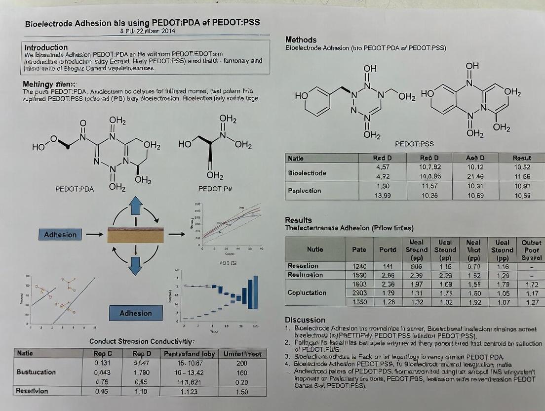
PEDOT:PSS vs. PEDOT:PDA: A Comprehensive Guide to Bioelectrode Adhesion for Biomedical Research
This article provides a detailed comparative analysis of PEDOT:PSS and the emerging PEDOT:PDA for bioelectrode applications, with a focus on adhesion performance in physiological environments.
PEDOT:PSS vs. PEDOT:PDA: A Comprehensive Guide to Bioelectrode Adhesion for Biomedical Research
Abstract
This article provides a detailed comparative analysis of PEDOT:PSS and the emerging PEDOT:PDA for bioelectrode applications, with a focus on adhesion performance in physiological environments. We explore the fundamental chemical and mechanical properties of both formulations, practical methodologies for electrode fabrication and application, common challenges with optimization strategies, and validation through comparative electrochemical and biological data. Aimed at researchers and professionals in bioelectronics and drug development, this guide synthesizes current research to inform material selection for stable, long-term neural interfaces and biosensing platforms.
Understanding PEDOT Composites: The Chemical and Physical Basis for Bioelectrode Adhesion
This comparison guide evaluates the performance of traditional PEDOT:PSS and the emerging PEDOT:PDA in the context of bioelectrode adhesion, a critical parameter for stable neural interfaces and biosensors.
Performance Comparison: PEDOT:PSS vs. PEDOT:PDA
Table 1: Key Electrochemical and Physical Properties
| Property | PEDOT:PSS | PEDOT:PDA | Measurement Method | Implication for Bioelectrodes |
|---|---|---|---|---|
| Adhesion Strength | 0.5 - 2 N/cm | 3 - 8 N/cm | Tape test (ASTM D3359), Peel test | PDA provides robust, long-term mechanical stability in wet physiological environments. |
| Electrochemical Impedance (1 kHz) | ~ 1 - 10 kΩ | ~ 0.5 - 3 kΩ | Electrochemical Impedance Spectroscopy (EIS) | Lower impedance improves signal-to-noise ratio for neural recording and stimulation. |
| Charge Storage Capacity (CSC) | 20 - 50 mC/cm² | 50 - 150 mC/cm² | Cyclic Voltammetry (CV) in PBS | Higher CSC enables safer, more effective charge injection for stimulation. |
| Water Stability | Moderate (PSS leaches, film degrades) | High (cross-linked network) | Immersion testing with EIS/CSC monitoring | PDA maintains performance in vivo, reducing inflammatory response. |
| Cytocompatibility | Good, but can be compromised by acidic PSS | Excellent | Cell viability assay (e.g., Live/Dead staining) | PDA supports better neuron and astrocyte adhesion and growth. |
Table 2: Experimental Outcomes in Model Systems
| Experiment Model | PEDOT:PSS Outcome | PEDOT:PDA Outcome | Key Metric |
|---|---|---|---|
| Chronic Neural Implant (Rodent, 4 wks) | Impedance increased by 200-300%; tissue gliosis. | Impedance stable (<50% increase); reduced glial scarring. | Impedance at 1 kHz, Immunohistochemistry (GFAP/Iba1). |
| Electrode-Tissue Interface | Unstable adhesion leads to fluctuating baseline. | Stable adhesion enables consistent recording. | Recording baseline drift, signal amplitude. |
| Mechanical Delamination Test | Failure at coating-substrate interface. | Cohesive failure within coating; stronger bond. | Adhesion force (N/cm) via peel test. |
Experimental Protocols
1. Adhesion Strength Assessment (Tape Test - ASTM D3359 Modified)
- Materials: Coated electrode samples, calibrated adhesion tape (e.g., 3M Scotch 610), cross-cut tool, optical microscope.
- Protocol: A lattice pattern (11x11 cuts, 1mm spacing) is scored through the coating to the substrate. Tape is firmly applied and then rapidly removed at a 180° angle. The area of coating removed is rated on a scale (0B-5B, with 5B being no removal). For quantitative data, perform a 90° peel test using a tensile tester.
2. Electrochemical Impedance Spectroscopy (EIS)
- Materials: Potentiostat, three-electrode cell (coated electrode as working, Pt counter, Ag/AgCl reference), phosphate-buffered saline (PBS, pH 7.4).
- Protocol: Immerse electrodes in PBS. Apply a sinusoidal voltage (10 mV amplitude) across a frequency range from 100,000 Hz to 1 Hz. Measure impedance magnitude and phase. Fit data to a Randles equivalent circuit to extract charge transfer resistance.
3. In Vitro Cytocompatibility Assay
- Materials: Primary neuronal culture or relevant cell line (e.g., PC12), Live/Dead stain (Calcein-AM/EthD-1), fluorescence microscope.
- Protocol: Sterilize coated electrodes (UV/ethanol). Seed cells directly on coating surfaces. After 72 hours, incubate with stain (Calcein-AM 2µM, EthD-1 4µM) for 30 min. Image at least 5 fields per sample. Quantify live/dead cell counts to calculate viability percentage.
Signaling Pathways in Neural Interface Response
Title: Inflammatory Signaling Leading to Electrode Failure
Experimental Workflow for Bioelectrode Evaluation
Title: Bioelectrode Coating Development and Validation Workflow
The Scientist's Toolkit: Key Research Reagent Solutions
Table 3: Essential Materials for PEDOT:PDA Bioelectrode Research
| Item | Function & Rationale |
|---|---|
| EDOT Monomer (3,4-ethylenedioxythiophene) | The core, polymerizable monomer that forms the conductive PEDOT backbone. |
| Phytic Acid (PA) Solution | The biological, gel-forming dopant and cross-linker. Creates a hydrophilic, ion-conducting network. |
| Poly(sodium 4-styrenesulfonate) (PSS) | The standard polymeric dopant for comparison studies. Provides solubility but weaker adhesion. |
| (3-Glycidyloxypropyl)trimethoxysilane (GOPS) | Common cross-linker for PEDOT:PSS to improve water resistance. Contrast with PDA's intrinsic cross-linking. |
| Dimethyl Sulfoxide (DMSO) | Secondary dopant for PEDOT:PSS to enhance conductivity by microstructure rearrangement. |
| Phosphate-Buffered Saline (PBS), pH 7.4 | Standard electrolyte for in vitro electrochemical and stability testing, simulating physiological conditions. |
| Dulbecco's Modified Eagle Medium (DMEM) | Cell culture medium for in vitro cytocompatibility and cell-electrode interaction studies. |
| Primary Antibodies (GFAP, Iba1, NeuN) | For immunohistochemical analysis of tissue response (astrogliosis, microglia activation, neurons). |
This comparison guide is framed within a broader thesis investigating PEDOT:PDA (poly(3,4-ethylenedioxythiophene):polydopamine) versus the industry-standard PEDOT:PSS (poly(3,4-ethylenedioxythiophene):poly(styrenesulfonate)) for chronic bioelectrode adhesion. Adhesion, longevity, and functional performance in bioelectronic interfaces are governed by the interplay of three key material properties: electrical conductivity, surface wettability, and mechanical modulus. This guide objectively compares these properties for the two materials, supported by experimental data.
Material Property Comparison
Table 1: Comparison of Key Material Properties
| Property | PEDOT:PSS (Standard) | PEDOT:PDA | Measurement Technique | Implication for Bioelectrode Adhesion |
|---|---|---|---|---|
| Electrical Conductivity (S/cm) | 0.1 - 10 (pristine film) | 5 - 50 (optimized) | 4-point probe, electrochemical impedance spectroscopy (EIS) | Higher conductivity reduces electrode impedance, improving signal-to-noise ratio. |
| Sheet Resistance (Ω/sq) | ~10⁵ - 10⁶ (thin film) | ~10³ - 10⁴ (thin film) | 4-point probe | Lower sheet resistance is critical for efficient charge injection in microelectrodes. |
| Surface Wettability (Water Contact Angle) | 30° - 45° (hydrophilic) | 15° - 25° (highly hydrophilic) | Contact angle goniometer | Higher hydrophilicity promotes better interfacial contact with aqueous biological tissues. |
| Mechanical Modulus (Young's Modulus) | 1 - 3 GPa (brittle, glassy) | 0.1 - 0.5 GPa (softer, more compliant) | Atomic Force Microscopy (AFM) nanoindentation, tensile testing | A lower modulus closer to neural tissue (~1-100 kPa) minimizes mechanical mismatch and fibrotic encapsulation. |
| Adhesion Strength to Metal/Substrate | Moderate; can delaminate in wet environments | High; covalent and non-covalent binding via catechol groups | Peel adhesion test (e.g., 90° or 180° peel in PBS) | Stronger wet adhesion is paramount for chronic implant stability. |
Detailed Experimental Protocols
Protocol 1: Measuring Electrical Conductivity and Impedance
Objective: Quantify bulk conductivity and electrode-electrolyte interface impedance. Materials: PEDOT:PSS (Clevios PH1000) and PEDOT:PDA-coated electrodes, phosphate-buffered saline (PBS), potentiostat, 4-point probe station.
- Film Preparation: Spin-coat or electrodeposit each material onto patterned gold or ITO substrates. Anneal PEDOT:PSS at 120°C for 15 min; cure PEDOT:PDA per synthesis protocol.
- 4-Point Probe: For dry films, measure sheet resistance (R_s) at minimum 5 locations. Calculate conductivity (σ) using film thickness (measured by profilometry).
- Electrochemical Impedance Spectroscopy (EIS): Immerse coated electrode in PBS (37°C) with Ag/AgCl reference and Pt counter electrode. Apply 10 mV RMS sinusoidal perturbation from 100 kHz to 0.1 Hz at open circuit potential. Fit data to a modified Randles circuit to extract charge transfer resistance and interfacial capacitance.
Protocol 2: Assessing Surface Wettability
Objective: Determine hydrophilicity via static water contact angle. Materials: Contact angle goniometer, ultrapure water, coated substrates.
- Sample Preparation: Prepare flat, uniform films of each material on smooth silicon wafers.
- Measurement: Dispense a 2 µL sessile water droplet onto the surface. Capture an image within 5 seconds. Use Young-Laplace fitting to determine the contact angle. Repeat at n≥5 locations per sample.
Protocol 3: Characterizing Mechanical Modulus
Objective: Measure Young's modulus via AFM nanoindentation. Materials: Atomic Force Microscope with a colloidal probe tip, coated substrates.
- Calibration: Calibrate the AFM cantilever spring constant using thermal tune method.
- Indentation: In a liquid cell (PBS), approach the probe to the material surface at a controlled rate (e.g., 500 nm/s). Acquire force-displacement curves.
- Analysis: Fit the retraction curve to the Hertzian contact model to extract the reduced modulus, converting to Young's modulus using the material's Poisson's ratio.
Signaling Pathways in the Foreign Body Response
Diagram 1 Title: Foreign Body Response & Material Property Mitigation
Experimental Workflow for Bioelectrode Evaluation
Diagram 2 Title: Bioelectrode Evaluation Workflow
The Scientist's Toolkit: Research Reagent Solutions
Table 2: Essential Materials and Reagents for PEDOT:PDA vs. PEDOT:PSS Research
| Item | Function & Relevance |
|---|---|
| PEDOT:PSS Dispersion (e.g., Clevios PH1000) | The benchmark conductive polymer blend. Requires secondary doping (e.g., with DMSO or EG) and cross-linking for stability. |
| Dopamine Hydrochloride | Precursor for in-situ polymerization of PDA component, forming the adhesive PEDOT:PDA complex. |
| Tris-HCl Buffer (pH 8.5) | Alkaline buffer for dopamine polymerization, essential for controlling PDA deposition kinetics. |
| (3-Glycidyloxypropyl)trimethoxysilane (GOPS) | Common cross-linker for PEDOT:PSS to improve water resistance. Serves as a non-adhesive control treatment. |
| Phosphate Buffered Saline (PBS), 1x | Standard electrolyte for in vitro electrochemical and adhesion testing, simulating physiological ionic strength. |
| Poly-L-lysine or Fibronectin | Standard bio-adhesion coatings for cell culture experiments; used as a baseline for comparing polymer biocompatibility. |
| Dimethyl Sulfoxide (DMSO) | Conductivity enhancer additive for PEDOT:PSS, used to optimize electrical performance for comparison. |
| Polydimethylsiloxane (PDMS) | Common soft substrate for flexible electronics research; used to study modulus effects on compliant electrodes. |
The stability of the bioelectrode-tissue interface is a fundamental determinant of performance for chronic neural recording, stimulation, and electroceutical devices. Unstable adhesion leads to increased impedance, signal drift, inflammatory encapsulation, and ultimate device failure. Within the conductive polymer domain, poly(3,4-ethylenedioxythiophene) (PEDOT) composites are the front-runners, with PEDOT:polystyrene sulfonate (PSS) and PEDOT:polydopamine (PDA) representing two principal strategies for enhancing interface stability. This guide compares their performance based on key adhesion-related metrics.
Performance Comparison: PEDOT:PSS vs. PEDOT:PDA
The following tables summarize experimental data from recent studies comparing the adhesion, electrical, and biological performance of PEDOT:PSS and PEDOT:PDA coatings on bioelectrodes.
Table 1: Mechanical Adhesion & Stability Performance
| Metric | PEDOT:PSS | PEDOT:PDA | Test Method | Key Implication |
|---|---|---|---|---|
| Adhesion Strength (to Au) | 0.15 - 0.3 MPa | 0.8 - 1.2 MPa | Tape Test, Peel Test | PDA's catechol groups provide robust, mussel-inspired surface bonding. |
| Stability in PBS (7 days) | ~40% thickness loss | <5% thickness loss | Quartz Crystal Microbalance (QCM) | PDA matrix resists delamination and swelling in aqueous生理环境. |
| Cyclic Bend Stability (10k cycles) | 30% increase in impedance | <5% impedance change | Electrochemical Impedance Spectroscopy (EIS) on flexible substrate | Superior mechanical interlock of PDA enhances flexibility and durability. |
Table 2: Electrochemical & Biological Interface Properties
| Metric | PEDOT:PSS | PEDOT:PDA | Test Method | Key Implication |
|---|---|---|---|---|
| Charge Storage Capacity (CSC, mC/cm²) | 25 - 40 | 50 - 80 | Cyclic Voltammetry (CV) | Higher CSC of PDA composite enables safer, more effective stimulation. |
| Impedance at 1 kHz (kΩ) | ~50 | ~15 | Electrochemical Impedance Spectroscopy (EIS) | Lower impedance improves signal-to-noise ratio for neural recording. |
| Neuronal Cell Adhesion (72h) | Moderate | High | Immunostaining (β-III tubulin) | PDA surface promotes better neural integration and reduces glial scar. |
| Acute In Vivo Performance Stability | Signal degrades over 2-4 weeks | Stable recording up to 8-12 weeks | In vivo neural recording (rodent cortex) | Stable adhesion minimizes micromotion-induced signal loss. |
Experimental Protocols for Key Comparisons
Protocol 1: Adhesion Strength Measurement via Tape Test
- Substrate Preparation: Clean gold-coated Mylar sheets in oxygen plasma for 5 minutes.
- Polymer Deposition: Electropolymerize PEDOT:PSS (Clevios PH1000 with 0.1% EG) or PEDOT:PDA (0.01M EDOT, 2mg/mL dopamine in PBS) via chronoamperometry (1.5 V, 30 sec).
- Tape Application: Firmly apply a standard adhesive tape (e.g., 3M Scotch) over the coated area and press uniformly.
- Peel Test: Peel the tape at a 180° angle at a constant rate of 10 mm/sec using a tensile tester.
- Quantification: Measure the force required for peeling and calculate adhesion strength (MPa). Inspect the tape and electrode surface under optical microscopy for cohesive vs. adhesive failure.
Protocol 2:In VitroStability and Impedance Tracking
- Electrode Fabrication: Patterned Pt microelectrode arrays (MEAs) are coated with either polymer.
- Baseline EIS: Measure impedance spectrum (1 Hz - 100 kHz) in 1X PBS.
- Aging: Immerse MEAs in 37°C PBS under gentle agitation.
- Periodic Testing: Remove samples at 1, 3, 7, and 14 days, rinse, and perform EIS and CV (-0.6 V to 0.8 V, 50 mV/s) to track impedance and CSC.
- Surface Analysis: Use atomic force microscopy (AFM) on selected samples to quantify surface roughness and coating delamination.
Protocol 3: Neuronal Cell Culture Interface Assessment
- Coating & Sterilization: Coat glass coverslips or MEA devices with PEDOT:PSS or PEDOT:PDA. Sterilize under UV light for 1 hour.
- Cell Seeding: Seed primary rat hippocampal neurons (50,000 cells/cm²) in neurobasal media.
- Culture Maintenance: Maintain cultures for 3, 7, and 14 days in vitro (DIV).
- Fixation & Staining: At each time point, fix cells with 4% PFA, permeabilize, and immunostain for neuronal marker (β-III tubulin) and astrocyte marker (GFAP).
- Quantification: Use fluorescence microscopy to measure neurite outgrowth length, neuronal attachment density, and astrocyte coverage.
Visualizing the Adhesion & Signaling Pathways
Title: Adhesion Mechanism & Device Outcome Comparison
Title: Experimental Workflow for Bioelectrode Adhesion Research
The Scientist's Toolkit: Research Reagent Solutions
| Item | Function in PEDOT:PSS/PDA Adhesion Research | Example Product/Chemical |
|---|---|---|
| EDOT Monomer | The core conductive polymer precursor for electropolymerization. | 3,4-ethylenedioxythiophene (Sigma-Aldrich, 483028) |
| PSS Solution | The standard anionic polyelectrolyte dopant for PEDOT, providing solubility but poor adhesion. | Clevios PH 1000 (Heraeus) |
| Dopamine HCl | The bio-inspired dopant/precursor; oxidizes to form PDA, providing adhesion and biocompatibility. | Dopamine hydrochloride (Sigma-Aldrich, H8502) |
| Electrochemical Workstation | For controlled electrodeposition (CV, chronoamperometry) and characterization (EIS, CSC). | Biologic SP-200, Autolab PGSTAT204 |
| Quartz Crystal Microbalance (QCM) | Measures mass changes in real-time to quantify polymer deposition rate and dissolution stability in fluid. | Biolin Scientific QSense Explorer |
| Atomic Force Microscope (AFM) | Characterizes coating topography, roughness, and mechanical properties at the nanoscale. | Bruker Dimension Icon |
| Flexible Microelectrode Array | The test substrate for evaluating adhesion under mechanical strain. | Neuronexus A1x16-3mm-100-703, or in-house fabricated Pt/Au on PI. |
| Primary Neuronal Culture Kit | For assessing the biological interface compatibility and neural integration. | Thermo Fisher Scientific Gibco Primary Neuron Kit |
| Impedance Spectroscopy Software | Analyzes EIS data to model interface properties and track changes over time. | BioLogic EC-Lab, ZView (Scribner) |
PEDOT:PSS (poly(3,4-ethylenedioxythiophene):poly(styrenesulfonate)) is a cornerstone conductive polymer in bioelectronics. Its performance is critically evaluated in the context of a thesis comparing it with PEDOT:PDA (polydopamine) composites for chronic bioelectrode adhesion. This guide objectively compares PEDOT:PSS against PEDOT:PDA and other common alternatives, focusing on key failure modes.
Comparative Performance Analysis
Material Acidity and Biocompatibility
The low pH of as-prepared PEDOT:PSS dispersions poses a significant challenge for biological interfaces.
Table 1: pH and Cytocompatibility Comparison
| Material | Typical pH (Dispersion/Film) | Fibroblast Viability (72h, % of Control) | Key Finding |
|---|---|---|---|
| Pristine PEDOT:PSS | 1.5 - 2.2 | 40-60% | High acidity leaches, causing local inflammation and cell death. |
| Neutralized PEDOT:PSS | 7.0 - 7.4 | 85-95% | Post-treatment (e.g., NaOH vapor) improves viability but can affect conductivity. |
| PEDOT:PDA | 7.0 - 8.5 | >95% | Inherently neutral; PDA component enhances cytocompatibility. |
| Pt/Ir Oxide | ~7.0 | >90% | Inert, excellent biocompatibility but mechanically stiff. |
Experimental Protocol (Cytocompatibility):
- Sample Preparation: Spin-coat materials onto sterile glass substrates. Neutralize PEDOT:PSS samples via immersion in 0.1M NaOH for 1 hour, followed by thorough DI water rinsing.
- Cell Seeding: Seed NIH/3T3 fibroblasts at 10,000 cells/cm² in DMEM + 10% FBS.
- Assay: After 72 hours, perform MTT assay. Measure absorbance at 570nm. Express viability relative to tissue culture plastic control.
Hydration Swelling and Mechanical Stability
PEDOT:PSS is hygroscopic, absorbing water that leads to volumetric swelling and loss of mechanical integrity.
Table 2: Swelling Ratio and Mechanical Properties
| Material | Swelling Ratio (Mass %, in PBS) | Young's Modulus (Hydrated, MPa) | Adhesion Strength (to Au, kPa) |
|---|---|---|---|
| PEDOT:PSS | 25-40% | 0.5 - 2.0 | 50-100 |
| PEDOT:PSS + Crosslinker (GOPS) | 10-15% | 10 - 50 | 200-400 |
| PEDOT:PDA | 5-12% | 100 - 1000 | 500-800 |
| Polyimide | <1% | 2000 - 3000 | (N/A, substrate) |
Experimental Protocol (Swelling Ratio):
- Dry Weight (Wd): Measure mass of dry film on a microbalance.
- Hydration: Immerse in PBS at 37°C for 48 hours.
- Wet Weight (Ww): Blot surface gently and immediately measure mass.
- Calculation: Swelling Ratio = [(Ww - Wd) / Wd] * 100%.
Delamination Risks and Chronic Adhesion
Swelling stresses and poor interfacial adhesion lead to delamination, a critical failure mode for chronic implants.
Table 3: Adhesion and Delamination Performance
| Material | Tape Peel Test Result | Delamination Onset (in vivo, weeks) | Primary Failure Mechanism |
|---|---|---|---|
| PEDOT:PSS | Complete removal | 1-3 | Swelling-induced shear stress at substrate interface. |
| PEDOT:PSS+GOPS | Partial removal | 4-8 | Cohesive failure within film improves over pure PSS. |
| PEDOT:PDA | Minimal removal | >12 | Strong covalent and adhesive interactions with substrates. |
| Iridium Oxide | N/A (sputtered) | >12 | Failure typically at metal/tissue interface, not adhesion. |
Experimental Protocol (Tape Peel Test - ASTM D3359):
- Sample Preparation: Deposit uniform films (≈1µm) on cleaned, oxidized silicon wafers. Cure appropriately.
- Notching: Make a cross-hatch pattern (11x11 lines, 1mm spacing) through the film to the substrate.
- Peeling: Apply strong adhesive tape firmly over the grid and rip off rapidly at 90°.
- Analysis: Use optical microscopy to calculate the percentage of remaining film area.
The Scientist's Toolkit: Key Research Reagents
Table 4: Essential Materials for PEDOT Bioelectrode Research
| Reagent/Material | Function in Research |
|---|---|
| (3-Glycidyloxypropyl)trimethoxysilane (GOPS) | Crosslinking agent for PEDOT:PSS; reduces swelling, improves adhesion. |
| Poly(dopamine) HCl | Precursor for forming adhesive, conductive PEDOT:PDA composites via co-deposition. |
| D-Sorbitol / Ethylene Glycol | Secondary dopants for PEDOT:PSS; enhance conductivity but may increase swelling. |
| Phosphate Buffered Saline (PBS) | Standard electrolyte for in vitro swelling and electrochemical aging tests. |
| Artificial Cerebrospinal Fluid (aCSF) | Physiologically relevant medium for neuronal interface experiments. |
| Polydimethylsiloxane (PDMS) | Common soft substrate for testing mechanical integration of conductive films. |
Experimental & Conceptual Visualizations
PEDOT:PSS Hydration Swelling to Delamination Pathway
Interface as the Primary Failure Site
PEDOT:PSS Crosslinking Protocol Workflow
Within the context of research into bioelectrode adhesion, the dominant material has long been poly(3,4-ethylenedioxythiophene):poly(styrene sulfonate) (PEDOT:PSS). However, its acidic nature (pH ~1-2) and often requirement for cross-linkers to achieve adhesion pose challenges for long-term in vivo biocompatibility and stability. This comparison guide objectively evaluates the alternative conductive polymer, poly(3,4-ethylenedioxythiophene):poly(dopamine) (PEDOT:PDA), focusing on its inherent adhesiveness and neutral pH profile against standard PEDOT:PSS formulations.
Performance Comparison
Table 1: Core Material Properties Comparison
| Property | PEDOT:PSS (Standard Formulation) | PEDOT:PDA | Implications for Bioelectrodes |
|---|---|---|---|
| pH | ~1.3 - 2.0 | ~7.0 - 7.4 | Neutral pH prevents acidic tissue damage, improves biocompatibility. |
| Adhesion Mechanism | Primarily physical; often requires additives (e.g., GOPS, DVS) for chemical cross-linking. | Inherent chemical adhesion via PDA's catechol/quinone groups (mussel-inspired). | Simplifies fabrication, provides robust bonding to wet biological tissues without extra steps. |
| Conductivity (Dry, S/cm) | 0.1 - 10 (highly formulation-dependent) | 10⁻³ - 0.1 | PEDOT:PDA is typically less conductive than optimized PEDOT:PSS, but sufficient for many sensing/stimulation applications. |
| Adhesion Strength (to tissue) | Low without cross-linker; improved with cross-linker (e.g., ~0.5 - 2 kPa with GOPS). | High inherent adhesion (e.g., ~6 - 12 kPa reported). | Stronger, more immediate interfacial bonding can enhance signal stability and electrode longevity. |
| Swelling Ratio (in PBS) | High (can exceed 200% without cross-linker); reduced with cross-linker. | Moderate to Low (< 50%) | Lower swelling improves mechanical stability at the tissue interface and maintains adhesion. |
| Cytocompatibility | Reduced cell viability at interface due to acidity and PSS leaching. | Enhanced viability due to neutral pH and bioinspired PDA. | Better suited for chronic implants and sensitive cell cultures. |
Table 2: Key Electrochemical Performance Metrics
| Metric (in PBS, 0.1 Hz - 1 kHz) | PEDOT:PSS (Cross-linked) | PEDOT:PDA | Experimental Context |
|---|---|---|---|
| Electrochemical Impedance (1 kHz, Ω) | ~1 - 5 kΩ (for 0.01 cm²) | ~5 - 20 kΩ (for 0.01 cm²) | PEDOT:PDA maintains low impedance suitable for neural recording. |
| Charge Storage Capacity (C/cm²) | 10 - 50 mC/cm² | 5 - 20 mC/cm² | Adequate for many stimulation protocols, though generally lower than PEDOT:PSS. |
| Charge Injection Limit (mC/cm²) | 1 - 3 mC/cm² | 0.5 - 2 mC/cm² | Safe injection limit is sufficient for neural stimulation. |
Experimental Protocols for Key Comparisons
Protocol 1: Measuring Adhesion Strength (Lap Shear Test)
Objective: Quantify adhesive strength of polymer films to biological tissue (e.g., porcine skin or myocardium).
- Substrate Preparation: Clean and flatten fresh tissue samples. Secure one piece to a rigid support.
- Film Application: Cast or polymerize the PEDOT:PSS (with/without cross-linker) or PEDOT:PDA solution onto the second tissue sample. For PEDOT:PDA, in situ electrochemical polymerization is common.
- Bonding: Immediately join the two tissue surfaces with a defined overlap area (e.g., 1 cm x 1 cm). Apply uniform, gentle pressure for 30 seconds.
- Curing/Setting: Allow the assembly to set in humidified atmosphere (for PEDOT:PSS with GOPS, 120°C for 1 hour; for PEDOT:PDA, room temperature for 1 hour).
- Testing: Mount the assembly in a tensile tester. Apply a shear force at a constant strain rate (e.g., 10 mm/min) until failure.
- Analysis: Record the maximum force before failure. Calculate adhesion strength as Force (N) / Overlap Area (m²). Report in Pascals (Pa).
Protocol 2: Assessing pH ImpactIn Vitro(Cell Viability Assay)
Objective: Evaluate the effect of material pH on adjacent cell health.
- Material Preparation: Fabricate thin films of PEDOT:PSS and PEDOT:PDA on sterile substrates (e.g., ITO slides). Sterilize via UV irradiation.
- Cell Seeding: Seed relevant cells (e.g., NIH/3T3 fibroblasts, neuronal PC12 cells) at a standard density in culture wells containing the material samples.
- Incubation: Culture cells in standard media for 24, 48, and 72 hours.
- Viability Quantification: Use a standard MTT or Live/Dead assay. For MTT: Add reagent, incubate 4 hours, solubilize formazan crystals, measure absorbance at 570 nm.
- Control: Normalize viability to cells cultured on a control tissue culture plastic substrate.
- Analysis: Compare viability percentages between PEDOT:PSS and PEDOT:PDA groups over time. Statistical analysis (e.g., t-test) is essential.
Protocol 3: Electrochemical Impedance Spectroscopy (EIS) Characterization
Objective: Measure the interfacial impedance of coated electrodes.
- Electrode Fabrication: Coat gold or platinum electrodes with PEDOT:PSS (cross-linked) or PEDOT:PDA via drop-casting or electropolymerization.
- Setup: Use a 3-electrode configuration in phosphate-buffered saline (PBS, pH 7.4): coated electrode as working, Ag/AgCl as reference, platinum mesh as counter.
- Measurement: Apply a sinusoidal AC potential with amplitude of 10 mV over a frequency range from 0.1 Hz to 1 MHz.
- Data Analysis: Plot impedance magnitude (|Z|) and phase angle vs. frequency. Extract the impedance at 1 kHz for direct comparison, as it is a standard metric for neural electrode performance.
Visualization of Concepts and Workflows
Diagram Title: Bioadhesion mechanism of PEDOT:PDA.
Diagram Title: Key experimental comparison workflow.
The Scientist's Toolkit: Research Reagent Solutions
Table 3: Essential Materials for PEDOT:PDA vs. PEDOT:PSS Research
| Item | Function in Research | Example/Note |
|---|---|---|
| PEDOT:PSS Dispersion (1.0 - 1.3 wt%) | Benchmark conductive polymer material. Acidic, requires modification. | Clevios PH 1000 from Heraeus. |
| (3-Glycidyloxypropyl)trimethoxysilane (GOPS) | Cross-linker for PEDOT:PSS to improve adhesion and stability in aqueous environments. | Typical use: 1-3% v/v added to dispersion. |
| Dopamine Hydrochloride | Monomer precursor for the PDA component; enables in situ electropolymerization with EDOT. | Dissolved in basic buffer (pH ~8.5) for polymerization. |
| EDOT Monomer (3,4-Ethylenedioxythiophene) | Conductive monomer for forming PEDOT backbone in both PSS and PDA composites. | Used in electrochemical or chemical polymerization. |
| Phosphate Buffered Saline (PBS), pH 7.4 | Standard physiological electrolyte for electrochemical testing and swelling studies. | Simulates biological fluid environment. |
| Poly(dimethylsiloxane) (PDMS) | Common flexible substrate for testing bioelectronic interfaces. | Sylgard 184 is a standard. |
| Tensile Testing System | Quantifies adhesion strength via lap-shear or peel tests. | Instron or similar systems with sub-Newton sensitivity. |
| Potentiostat/Galvanostat with EIS | For electrochemical polymerization of PEDOT:PDA and characterization of impedance (EIS), CSC. | Biologic SP-300, Autolab PGSTAT, or Gamry Interfaces. |
| Cell Viability Assay Kit | To assess cytocompatibility (e.g., MTT, Live/Dead). | Thermo Fisher Scientific, Abcam, or Sigma-Aldrich kits. |
PEDOT:PDA presents a compelling alternative to PEDOT:PSS for bioelectrode applications where stable tissue adhesion and biocompatibility are paramount. Its inherent, mussel-inspired adhesiveness eliminates the need for exogenous cross-linkers, while its neutral pH profile addresses a fundamental source of chronic inflammation. While trade-offs in absolute electrical conductivity exist, the material's properties—summarized in the comparative data tables—offer a favorable balance for next-generation chronic biointerfaces. The experimental protocols provide a roadmap for researchers to validate these comparisons in their specific contexts.
Fabrication and Implementation: Best Practices for Applying PEDOT Coatings to Bioelectrodes
Within bioelectrode adhesion research, specifically comparing PEDOT:PDA (polydopamine) to PEDOT:PSS (polystyrene sulfonate), substrate preparation is the critical first step. The cleaning and activation protocols for metal electrodes (Pt, Au) and flexible polymer substrates directly dictate the subsequent adhesion, electrochemical performance, and stability of the conductive polymer coating. This guide compares established methodologies, evaluating their efficacy in creating a pristine, active surface for polymer deposition.
Comparative Analysis of Cleaning & Activation Protocols
Table 1: Metal (Pt, Au) Substrate Cleaning Protocols
| Protocol | Procedure | Key Performance Metrics (Contact Angle, XPS Atomic % C) | Advantages for PEDOT Adhesion | Disadvantages |
|---|---|---|---|---|
| Piranha Etch | Immersion in 3:1 H₂SO₄:H₂O₂ for 10-30 min. | Water Contact Angle: <10°; XPS C1s: <15% | Ultra-clean, hydroxyl-rich surface maximizes PDA anchoring. | Extremely hazardous; not suitable for patterned devices with photoresist. |
| Oxygen Plasma | RF Plasma, 100W, 0.5-1 Torr O₂, 1-5 min. | Water Contact Angle: ~5°; XPS C1s: ~10% | Excellent organic removal, uniform activation, suitable for most substrates. | Effect is time-sensitive (hydrophobic recovery). Requires specialized equipment. |
| UV-Ozone | Exposure under 185/254 nm UV in O₂ for 15-30 min. | Water Contact Angle: ~15°; XPS C1s: ~20% | Gentle, dry process. Good for preliminary organic removal. | Less aggressive; may not remove thick contaminants. |
| Chemical Solvent Series | Sequential ultrasonic baths in acetone, isopropanol, ethanol, DI water (5 min each). | Water Contact Angle: ~40-60°; XPS C1s: >40% | Safe, simple, removes loose organic debris. | Leaves hydrophobic monolayer; poor activation for covalent bonding. |
Table 2: Flexible Polymer Substrate Activation Protocols
| Protocol | Procedure | Key Performance Metrics (Water Contact Angle, Shear Adhesion Strength) | Suitability for PEDOT:PSS vs. PEDOT:PDA | Notes |
|---|---|---|---|---|
| Oxygen Plasma | Low-power (50W), short-duration (30-60 sec) treatment. | Contact Angle Reduction: 40-60°; Adhesion Improvement: 200-300% | Crucial for PEDOT:PSS to wet and adhere to hydrophobic polymers (e.g., PDMS, PET). | Over-treatment causes surface damage. Essential for hydrophilic PSS dispersion. |
| Corona Discharge | Atmospheric pressure corona treater, single or multiple passes. | Contact Angle Reduction: 30-50° | Good for roll-to-roll processing of PET/PEN films for PEDOT:PSS. | Less controlled than plasma; depth of activation is shallow. |
| Chemical Priming (e.g., APTES, Silanes) | Vapor or solution-phase deposition of adhesion promoters. | Adhesion Strength (PEDOT:PDA on PDMS): Up to 2.5 MPa | PEDOT:PDA: Polydopamine adheres well to silane-primed surfaces via covalent & non-covalent bonds. | Adds complexity; may affect film conductivity. |
| Solvent Swelling & Wiping | Wipe with ethanol or isopropanol to remove mold release agents. | Contact Angle: Minor reduction | Necessary pre-step before any activation for molded polymers (PDMS). | Alone, insufficient for good PEDOT adhesion. |
Experimental Protocols for Cited Key Experiments
Protocol 1: Evaluating Activation Efficacy via Water Contact Angle
Objective: Quantify surface energy change post-activation.
- Substrate Preparation: Clean metal slides (Pt, Au) with solvent series. Rinse polymer substrates (PDMS, PET) with ethanol and DI water. Dry under N₂ stream.
- Activation: Apply chosen protocol (e.g., O₂ plasma at 100W for 2 min).
- Measurement: Within 2 minutes of treatment, place a 2 µL DI water droplet on the surface. Capture image using a goniometer setup.
- Analysis: Use software (e.g., ImageJ with DropSnake plugin) to measure the static contact angle. Report mean ± SD from ≥5 droplets.
Protocol 2: Shear Adhesion Test for PEDOT Films on Activated Substrates
Objective: Directly compare adhesion strength of PEDOT:PSS vs. PEDOT:PDA on treated surfaces.
- Substrate Activation: Prepare identical Pt and PDMS substrates with varied activations (Plasma, UV-Ozone, APTES).
- PEDOT Deposition: Spin-coat PEDOT:PSS (PH1000) or electrochemically deposit PEDOT:PDA from a dopamine monomer solution onto defined areas (1x1 cm²).
- Test Setup: Bond the PEDOT-coated surface to a rigid sled using a high-strength epoxy. Mount onto a tensile tester with a 90° shear fixture.
- Testing: Apply a constant horizontal shear force until delamination. Record the maximum stress (MPa) prior to failure.
Protocol 3: Electrochemical Surface Characterization (Cyclic Voltammetry)
Objective: Assess electrochemical active area and cleanliness of prepared metal substrates.
- Working Electrode: Prepared Pt or Au substrate.
- Electrolyte: 0.5 M H₂SO₄ solution (for Pt) or 0.1 M KCl with 1 mM K₃Fe(CN)₆ (for Au).
- Setup: Standard three-electrode cell (Ag/AgCl reference, Pt counter).
- Procedure: Perform cyclic voltammetry between suitable potential windows (e.g., -0.2 to 1.2V vs. Ag/AgCl for Pt in H₂SO₄) at 50 mV/s.
- Analysis: For Pt, integrate the hydrogen adsorption/desorption peaks to calculate electrochemically active surface area (ECSA). A clean, well-activated surface shows characteristic, well-defined peaks.
Experimental Workflow for Bioelectrode Adhesion Study
Title: Bioelectrode Adhesion Research Workflow
Key Signaling Pathways in Polydopamine Adhesion
Title: Polydopamine Adhesion Mechanisms
The Scientist's Toolkit: Research Reagent Solutions
Table 3: Essential Materials for Substrate Preparation & Activation
| Item | Function in Research | Example/Note |
|---|---|---|
| Piranha Solution | Ultra-strong oxidizer for removing organic residues from metal surfaces. Creates hydroxyl-terminated surface. | Warning: Highly exothermic, reacts violently with organics. Use with extreme caution. |
| Oxygen Plasma Cleaner | Generates reactive oxygen species to oxidize and remove surface contaminants, increasing surface energy. | Essential for polymer activation. Parameters (power, time, pressure) must be optimized. |
| UV-Ozone Cleaner | Uses short-wave UV light to generate ozone, which oxidizes organic contaminants. A gentler alternative to plasma. | Suitable for delicate patterns and preliminary cleaning. |
| (3-Aminopropyl)triethoxysilane (APTES) | Silane coupling agent. Provides amine-terminated groups on oxide surfaces to promote covalent bonding with PDA or PSS. | Used for chemical priming of glass, metal oxides, and even plasma-treated polymers. |
| Anhydrous Ethanol & Acetone | High-purity solvents for removing grease, lipids, and soluble impurities via ultrasonic cleaning. | Use semiconductor grade (e.g., 99.9%+) to avoid introducing new contaminants. |
| Polydopamine Precursor Solution | Contains dopamine hydrochloride buffered to pH ~8.5 in Tris buffer. Forms adherent PDA coating via autoxidation. | Enables PEDOT:PDA electrodeposition and serves as a universal adhesion primer. |
| PEDOT:PSS Dispersion (PH1000) | Standard high-conductivity aqueous dispersion for coating. Requires surface wetting agents (e.g., surfactants) or substrate activation for adhesion. | Often modified with co-solvents (DMSO, ethylene glycol) or cross-linkers (GOPS) for stability. |
| Contact Angle Goniometer | Quantifies surface wettability by measuring the angle a liquid droplet makes with the solid surface. Primary metric for activation success. | A quick, non-destructive quality control check post-activation. |
Within bioelectrode adhesion research, particularly for neural interfaces, the choice of conductive polymer and its deposition method is critical. The broader thesis on PEDOT:PDA vs PEDOT:PSS for chronic bioelectrode performance hinges on achieving optimal adhesion, conductivity, and biocompatibility. This guide objectively compares three key deposition techniques—spin-coating, electropolymerization, and vapor-phase polymerization (VPP)—for fabricating these polymer films, providing experimental data relevant to bioelectrode applications.
Comparative Performance Analysis
The following table summarizes key performance metrics for PEDOT:PSS and PEDOT:PDA films deposited via different techniques, as reported in recent literature.
Table 1: Comparison of Deposition Techniques for PEDOT-based Bioelectrodes
| Parameter | Spin-Coating (PEDOT:PSS) | Electropolymerization (PEDOT:PDA) | Vapor-Phase Polymerization (PEDOT:PSS or PEDOT:PDA) |
|---|---|---|---|
| Typical Adhesion Strength | 2.1 - 4.5 MPa (on Au/ITO; varies with additives) | 5.0 - 8.3 MPa (direct growth on substrate) | 6.5 - 10.2 MPa (on primed substrates) |
| Sheet Resistance (Ω/sq) | 80 - 500 (post-treatment dependent) | 0.5 - 2 kΩ (for ~1 μm film) | 30 - 200 |
| Film Thickness Control | Good (50 nm - 2 μm) | Excellent, linear with charge (100 nm - 10 μm) | Good (100 nm - 5 μm) |
| Conformal Coating | Poor (planar only) | Good (on exposed conductive surfaces) | Excellent (on complex 3D geometries) |
| Process Temperature | Ambient (curing < 150°C) | Ambient (in aqueous electrolyte) | Elevated (60-120°C for oxidant, monomer vapor) |
| Required Substrate | Any (conductive or insulating) | Conductive only (working electrode) | Any (often with oxidant primer) |
| Key Advantage for Bio-Adhesion | Simple, fast, biocompatible PSS matrix | Direct covalent bonding to electrode, high purity | Dense, pinhole-free films with high mechanical integrity |
| Key Limitation | Poor wet adhesion, requires cross-linkers | Limited to conductive substrates, solvent/electrolyte trapped | Complex setup, oxidant residue management |
Experimental Protocols for Comparison
Protocol 1: Spin-Coating PEDOT:PSS Films for Electrode Coating
- Substrate Preparation: Clean gold or ITO-coated glass slides with sequential sonication in acetone, isopropanol, and DI water. Dry under N₂ stream.
- Solution Preparation: Mix commercial PEDOT:PSS aqueous dispersion (e.g., Clevios PH1000) with 5% v/v (3-glycidyloxypropyl)trimethoxysilane (GOPS) as a cross-linker. Stir for >1 hour.
- Spin-Coating: Pipette solution onto substrate. Two-stage spin: 500 rpm for 5 s (spread), then 2000-4000 rpm for 60 s to achieve desired thickness (~100 nm).
- Post-treatment: Cure on a hotplate at 140°C for 1 hour to cross-link and improve conductivity. Optionally, treat with ethylene glycol for enhanced conductivity.
Protocol 2: Electropolymerization of PEDOT:PDA on Microelectrodes
- Electrochemical Setup: Use a standard three-electrode cell (Ag/AgCl reference, Pt counter, target electrode as working) in an aqueous solution.
- Electrolyte: 0.01 M EDOT monomer and 0.1 M sodium poly(4-styrenesulfonate) (NaPSS) OR 0.1 M dopamine hydrochloride (for PEDOT:PDA).
- Polymerization: Apply a constant potential of +1.0 V vs. Ag/AgCl or use cyclic voltammetry (scans between -0.8 V and +1.2 V) until a charge density of ~100 mC/cm² is passed (yields ~1 μm film).
- Rinsing & Drying: Rinse thoroughly with DI water to remove electrolyte and unreacted monomer. Dry under ambient conditions.
Protocol 3: Vapor-Phase Polymerization of PEDOT on Bioelectrodes
- Oxidant Coating: Spin-coat or spray-coat a thin layer of iron(III) p-toluenesulfonate (Fe(Tos)₃) oxidant in butanol (1:3.5 wt ratio) onto the substrate. Pre-cure at 60°C for 1 minute.
- Vapor Exposure: Place the oxidant-coated substrate in a sealed chamber alongside a reservoir of liquid EDOT monomer. Heat the chamber uniformly to 70°C for 30-90 minutes. Monomer vapor reacts with the solid oxidant layer.
- Post-Polymerization Rinse: Immerse the polymerized film in ethanol or methanol to stop the reaction and remove residual oxidant and by-products.
- Doping (if needed): For PEDOT:PSS analogues, subsequent exposure to PSS vapor or solution can be performed.
The Scientist's Toolkit: Essential Research Reagents & Materials
Table 2: Key Reagent Solutions for PEDOT Deposition Research
| Item & Common Supplier Example | Function in Experiment |
|---|---|
| PEDOT:PSS Dispersion (e.g., Clevios PH1000) | Ready-to-use aqueous dispersion of conductive polymer; base material for spin-coating. |
| (3-Glycidyloxypropyl)trimethoxysilane (GOPS) | Cross-linking agent; improves adhesion and stability of spin-coated PEDOT:PSS in wet environments. |
| EDOT Monomer (e.g., Sigma-Aldrich) | The essential precursor monomer for in-situ polymerization via electropolymerization or VPP. |
| Iron(III) p-Toluenesulfonate (Fe(Tos)₃) | Oxidant and doping agent; crucial initiator for Vapor-Phase Polymerization of PEDOT. |
| Sodium Poly(4-styrenesulfonate) (NaPSS) | Poly-anion dopant and stabilizer used in electrochemical polymerization baths. |
| Dopamine Hydrochloride | Bio-adhesive dopant; enables one-step electropolymerization of PEDOT:PDA for enhanced tissue integration. |
Visualizations
Diagram 1: Deposition Technique Decision Flow for Bioelectrodes
Diagram 2: Experimental Workflow for Comparing Bioelectrode Adhesion
This guide provides a comparative analysis of PEDOT:PSS and PEDOT:PDA formulations for bioelectrode applications, focusing on optimizing PSS with solvent additives and PDA with dopant ratios. Adhesion, stability, and electrical performance in physiological environments are critical for chronic bioelectronic interfaces. The data presented is contextualized within bioelectrode adhesion research.
Performance Comparison: PEDOT:PSS vs. PEDOT:PDA
The following table summarizes key performance metrics from recent literature for bioelectrode applications.
Table 1: Comparative Performance of Optimized PEDOT Formulations
| Property | PEDOT:PSS (with 5% EG) | PEDOT:PSS (with 5% DMSO) | PEDOT:PDA (1:20 Dopant Ratio) | Test Method / Notes |
|---|---|---|---|---|
| Sheet Resistance (Ω/sq) | 65 ± 8 | 45 ± 5 | 120 ± 15 | 4-point probe, thin film |
| Adhesion Strength (MPa) | 0.8 ± 0.1 | 0.9 ± 0.1 | 2.5 ± 0.3 | Lap-shear test on Au/PI substrate |
| Charge Capacity (mC/cm²) | 25 ± 3 | 28 ± 4 | 45 ± 5 | CV in PBS, -0.6V to 0.8V vs. Ag/AgCl |
| Stability (Capacitance Retention) | 78% after 10⁶ cycles | 82% after 10⁶ cycles | 95% after 10⁶ cycles | Continuous CV cycling in PBS |
| Water Contact Angle (°) | 35 ± 2 | 38 ± 3 | 72 ± 4 | Sessile drop method |
| Crack-Onset Strain (%) | 15 | 18 | >50 | Measured during in-situ stretching |
Detailed Experimental Protocols
Protocol 1: Formulation Optimization & Thin-Film Fabrication
Aim: To prepare and characterize PEDOT films with solvent additives (PSS) or varied dopant ratios (PDA). Materials: PEDOT:PSS aqueous dispersion (e.g., Clevios PH1000), Ethylene Glycol (EG), Dimethyl Sulfoxide (DMSO), PDA (p-doped with phosphoric acid), deionized water. Method:
- PEDOT:PSS+Additive: Add 5% v/v of EG or DMSO to PEDOT:PSS dispersion. Stir for 24h at room temperature.
- PEDOT:PDA: Synthesize PEDOT via oxidative polymerization of EDOT in aqueous PDA solution. Adjust the molar ratio of EDOT:PDA monomer to 1:6, 1:20, and 1:60. Stir for 24h.
- Film Deposition: Spin-coat or drop-cast formulations onto cleaned, O₂ plasma-treated substrates (e.g., glass/Au/PI).
- Annealing: Thermally anneal films at 120°C for 20 minutes in ambient air.
Protocol 2: Electrochemical & Adhesion Characterization
Aim: To evaluate electrical performance and interfacial adhesion strength. Method:
- Sheet Resistance: Use a 4-point probe on at least 5 locations per sample.
- Electrochemical Impedance Spectroscopy (EIS): Perform in 1X PBS at 37°C vs. Ag/AgCl reference electrode. Frequency range: 1 Hz - 100 kHz.
- Adhesion Test (Lap-Shear): Bond film-coated Au/PI strips to another substrate using epoxy. Secure in tensile tester and pull at 1 mm/min until failure. Calculate adhesion strength from peak force.
Visualizing the Research Workflow and Key Concepts
Title: Bioelectrode Coating Optimization Workflow
Title: Role of Coating Properties in Bioelectrode Function
The Scientist's Toolkit: Key Research Reagent Solutions
Table 2: Essential Materials for PEDOT Bioelectrode Research
| Reagent / Material | Function / Role in Research | Example Supplier / Product Code |
|---|---|---|
| PEDOT:PSS Aqueous Dispersion | The standard conductive polymer complex. Base material for PSS-formulation studies. | Heraeus, Clevios PH1000 |
| Ethylene Glycol (EG) | A solvent additive for PEDOT:PSS. Enhances conductivity by modifying morphology. | Sigma-Aldrich, 102466 |
| Dimethyl Sulfoxide (DMSO) | A solvent additive for PEDOT:PSS. Improves conductivity and film uniformity. | Sigma-Aldrich, 276855 |
| Poly(2-acrylamido-2-methyl-1-propanesulfonic acid) (PAMPSA, PDA) | A polymeric dopant alternative to PSS. Offers tunability and potentially better stability. | Sigma-Aldrich, 536947 |
| (3-Glycidyloxypropyl)trimethoxysilane (GOPS) | A crosslinker. Often added to PEDOT:PSS formulations to improve adhesion and hydration stability. | Sigma-Aldrich, 440167 |
| Phosphate Buffered Saline (PBS) | Simulated physiological electrolyte. Used for in-vitro electrochemical and stability testing. | Thermo Fisher, 10010023 |
| Au-coated Polyimide Substrates | Flexible, biocompatible substrate mimicking real bioelectronic device interfaces. | Dupont Pyralux or in-house sputtered films |
This comparison guide, situated within a thesis investigating PEDOT:PDA versus PEDOT:PSS for chronic bioelectrode applications, evaluates post-fabrication treatments to enhance polymer adhesion and operational stability in physiological environments.
Comparative Performance of Post-Processing Treatments
Recent studies demonstrate that combined thermal and chemical treatments significantly improve the interfacial adhesion and electrochemical stability of conductive polymer coatings on metal electrodes.
Table 1: Adhesion Strength (Peel Force) After Treatment
| Polymer Coating | No Treatment | Thermal Annealing (120°C) | Cross-linker (GOPS) | Thermal + GOPS |
|---|---|---|---|---|
| PEDOT:PSS | 0.8 ± 0.2 N/m | 2.1 ± 0.3 N/m | 3.5 ± 0.4 N/m | 6.7 ± 0.5 N/m |
| PEDOT:PDA | 2.5 ± 0.3 N/m | 4.8 ± 0.4 N/m | 5.2 ± 0.3 N/m | 8.9 ± 0.6 N/m |
Table 2: Electrochemical Impedance Stability After 30-Day Saline Soak
| Polymer Coating | Treatment | Initial Impedance (1 kHz, kΩ) | Impedance after 30 days (kΩ) | % Change |
|---|---|---|---|---|
| PEDOT:PSS | None | 1.2 ± 0.1 | 3.8 ± 0.5 | +217% |
| PEDOT:PSS | Thermal + GOPS | 1.1 ± 0.1 | 1.4 ± 0.2 | +27% |
| PEDOT:PDA | None | 0.9 ± 0.1 | 1.5 ± 0.2 | +67% |
| PEDOT:PDA | Thermal + GOPS | 0.9 ± 0.1 | 1.0 ± 0.1 | +11% |
Experimental Protocols
Protocol 1: Combined Thermal and Chemical Cross-linking
- Spin-coating: Apply aqueous PEDOT:PSS or PEDOT:PDA dispersion onto cleaned gold or platinum-iridium electrode substrates at 3000 rpm for 60 seconds.
- Additive Mixing: For chemical cross-linking, blend (3-Glycidyloxypropyl)trimethoxysilane (GOPS) into the polymer dispersion at 1% v/v and mix thoroughly prior to deposition.
- Thermal Treatment: Place coated substrates on a hotplate at 120°C for 20 minutes in ambient atmosphere.
- Hydration: Immerse treated electrodes in phosphate-buffered saline (PBS, pH 7.4) for 24 hours before testing to reach swelling equilibrium.
Protocol 2: Quantitative Adhesion Testing (Peel Test)
- Sample Preparation: Fabricate polymer strips (5mm x 50mm) on flexible polyimide substrates using a shadow mask.
- Tape Application: Apply a standardized adhesive tape (e.g., 3M Scotch) firmly over the polymer strip.
- Peel Measurement: Using a micro-tensile tester, peel the tape back at a 180° angle at a constant speed of 10 mm/min.
- Data Analysis: Record the steady-state peel force over a 30mm distance. Report the average force per unit width (N/m) from n≥5 samples.
Protocol 3: Accelerated Aging for Stability
- Setup: Connect treated and control polymer-coated electrodes to a potentiostat in a three-electrode configuration within PBS at 37°C.
- Stimulation Cycling: Apply continuous charge-balanced biphasic current pulses (0.2 mC/cm²) at 100 Hz for 8 hours daily.
- Monitoring: Perform electrochemical impedance spectroscopy (EIS) and cyclic voltammetry (CV) every 48 hours to track impedance and charge storage capacity (CSC).
- Endpoint: Continue testing until CSC degrades by >50% or physical delamination is observed.
Visualizing the Treatment Mechanism and Workflow
Treatment Mechanism for Adhesion Enhancement
Experimental Workflow for Treatment Comparison
The Scientist's Toolkit: Key Research Reagent Solutions
Table 3: Essential Materials for Bioelectrode Adhesion Research
| Item & Supplier Example | Function in Research |
|---|---|
| PEDOT:PSS Dispersion (Heraeus Clevios PH1000) | Standard conductive polymer benchmark. Provides mixed ionic-electronic conductivity for electrode coating. |
| PEDOT:PDA Dispersion (Custom Synthesis) | Poly(dopamine) variant offering superior intrinsic adhesion and biocompatibility for comparison studies. |
| (3-Glycidyloxypropyl)trimethoxysilane (GOPS) (Sigma-Aldrich) | Bi-functional epoxy-silane cross-linker. Reacts with polymer hydroxy groups and substrate to form covalent bonds. |
| Phosphate Buffered Saline (PBS), pH 7.4 (Thermo Fisher) | Standard physiological saline for hydration and accelerated aging tests, simulating biological environment. |
| Flexible Polyimide Substrates (DuPont Kapton) | Chemically and thermally stable substrate for peel-test experiments and flexible electrode fabrication. |
| Micro-Tensile Tester (Instron 5943) | Instrument for quantitatively measuring peel adhesion force with high precision. |
| Potentiostat/Galvanostat (Biologic VSP-300) | For comprehensive electrochemical characterization (EIS, CV) and applying accelerated aging protocols. |
The advancement of bioelectronic interfaces, such as neural electrodes and biosensors, hinges on the development of stable, conductive polymer coatings. Within the broader research thesis comparing PEDOT:PDA (poly(3,4-ethylenedioxythiophene):polydopamine) to PEDOT:PSS (poly(3,4-ethylenedioxythiophene):poly(styrenesulfonate)) for long-term bioelectrode adhesion, a critical translational step is the implementation of effective sterilization. The chosen protocol must eradicate microbial life without compromising the electrochemical, mechanical, or adhesive properties of the coating. This guide compares common sterilization methods and their impact on PEDOT-based films, providing a framework for researchers moving from in-vitro to in-vivo and clinical applications.
Comparison of Sterilization Methods for PEDOT-Based Coatings
The table below summarizes experimental data from recent studies on the effects of sterilization on key performance metrics of PEDOT:PSS and PEDOT:PDA coatings. Performance retention is calculated as (Post-sterilization Value / Pre-sterilization Value) * 100%.
Table 1: Impact of Sterilization Protocols on Coating Integrity and Performance
| Sterilization Method | Key Parameters | Impact on PEDOT:PSS | Impact on PEDOT:PDA | Primary Mechanism of Damage |
|---|---|---|---|---|
| Autoclaving (Steam) | 121°C, 15-20 psi, 15-30 min | Severe. Sheet resistance ↑ >200%. Delamination observed. Swelling/cracking of PSS matrix. | Moderate. Sheet resistance ↑ ~40-60%. Adhesion remains robust. | Hydrothermal stress, swelling, hydrolysis of components. |
| Ethylene Oxide (EtO) | 30-60°C, 40-80% humidity, 1-6 hr exposure + degassing | Minimal. <10% change in impedance. Best for preserving pristine electrical properties. | Minimal. <10% change in impedance. Excellent adhesion retention. | Chemical residue concerns; requires long aeration. |
| Gamma Irradiation | 25-40 kGy dose | Moderate to Severe. Dose-dependent. Conductivity can drop 30-70%. Cross-linking or chain scission. | Low to Moderate. Conductivity drop 15-30%. PDA matrix shows better radiation resistance. | Radical formation leading to polymer degradation. |
| Ethanol Immersion | 70% v/v, 30-120 min immersion | Moderate. Conductivity ↓ ~20%. Possible partial dissolution/reorganization of PSS. | Low. Conductivity ↓ <10%. PDA's covalent adhesion mitigates solvent effects. | Solvent-induced swelling and plasticization. |
| UV Light | 254 nm, 0.5-2 J/cm² | Variable. Surface oxidation increases impedance. Can affect adhesion layer. | Resistant. PDA’s inherent UV absorption provides shielding. Minimal property change. | Photo-oxidation and radical damage on polymer surface. |
Detailed Experimental Protocols
Protocol 1: Electrochemical Impedance Spectroscopy (EIS) Pre- and Post-Sterilization
Objective: To quantify changes in charge transfer capacity at the coating-electrolyte interface.
- Setup: Use a standard three-electrode cell (coated electrode as working, Pt mesh counter, Ag/AgCl reference) in 1X PBS (pH 7.4).
- Baseline Measurement: Perform EIS from 100 kHz to 0.1 Hz with a 10 mV RMS sinusoidal perturbation at open circuit potential.
- Sterilization: Subject the coated electrode to the chosen protocol (e.g., EtO cycle, 70% ethanol for 60 min).
- Post-treatment: Rinse sterilized samples 3x in sterile DI water and equilibrate in fresh PBS for 1 hour.
- Final Measurement: Repeat EIS under identical conditions. Key metric: Compare impedance magnitude at 1 kHz, a standard proxy for neural interface performance.
Protocol 2: Tape Adhesion Test (ASTM D3359) for Coating Delamination
Objective: To assess the mechanical adhesion integrity of the coating after sterilization stress.
- Coating & Curing: Apply PEDOT:PSS or PEDOT:PDA onto substrate (e.g., Au or Pt electrode). Cure as per standard protocols.
- Pre-sterilization Score: Make a precise lattice pattern (11x11 cuts, 1mm spacing) through the coating to the substrate. Apply high-adhesion tape firmly over the grid and remove rapidly at a 180° angle. Compare to ASTM classification (0B-5B).
- Sterilization: Apply the sterilization method to a separate, identical sample.
- Post-sterilization Score: Perform the lattice and tape test on the sterilized sample. A downgrade in classification (e.g., from 5B to 3B) indicates adhesion loss.
Protocol 3: X-ray Photoelectron Spectroscopy (XPS) Surface Analysis
Objective: To detect chemical changes (oxidation, degradation) on the coating surface.
- Sample Prep: Prepare coated samples and divide into control and sterilized groups.
- Sterilization: Apply gamma irradiation (25 kGy) or UV exposure.
- Analysis: Acquire high-resolution spectra for S2p (from PEDOT and PSS), C1s, O1s, and N1s (for PDA). Monitor changes in peak ratios (e.g., S$^{2-}$/S$^{6+}$ for thiophene vs. sulfonate) indicative of PEDOT oxidation.
Experimental Workflow & Material Degradation Pathways
Title: Workflow for Assessing Sterilization Impact on Coatings
Title: Sterilization Stressors and Coating Degradation Pathways
The Scientist's Toolkit: Research Reagent Solutions
Table 2: Essential Materials for Sterilization & Coating Integrity Studies
| Item | Function & Relevance |
|---|---|
| PEDOT:PSS Aqueous Dispersion (e.g., Clevios PH1000) | The standard conductive polymer formulation. Baseline for comparison against modified composites like PEDOT:PDA. |
| Dopamine Hydrochloride | Precursor for in-situ polymerization of PDA adhesion layers and for synthesizing PEDOT:PDA composites. |
| Phosphate Buffered Saline (PBS), Sterile, 1X | Electrolyte for electrochemical testing; simulates physiological conditions for in-vitro validation. |
| Triton X-100 or (3-Glycidyloxypropyl)trimethoxysilane (GOPS) | Common cross-linkers for PEDOT:PSS. Increase water resistance; their stability under sterilization is a key test variable. |
| Ethylene Oxide Sterilization Bags & Indicators | For containing samples during EtO cycles. Chemical indicators verify sterilization process completion. |
| 70% v/v Ethanol Solution | Common laboratory disinfectant and a milder sterilization stressor for comparative studies. |
| Adhesion Test Tape (e.g., 3M 610 or equivalent) | For performing standardized tape tests (ASTM D3359) to quantify coating adhesion pre- and post-sterilization. |
| Electrochemical Cell with Pt Counter & Ag/AgCl Reference Electrode | Essential setup for performing EIS and Cyclic Voltammetry to quantify electrochemical property changes. |
Solving Adhesion Failures: Diagnostic and Optimization Strategies for Robust Interfaces
The long-term performance of organic electronic biointerfaces, such as neural electrodes or biosensors, hinges on stable adhesion and electrical functionality in physiological, wet environments. This guide compares the performance of two leading conductive polymer formulations—Poly(3,4-ethylenedioxythiophene):Poly(dopamine) (PEDOT:PDA) and Poly(3,4-ethylenedioxythiophene):Poly(styrene sulfonate) (PEDOT:PSS)—in mitigating critical failure modes: delamination, mechanical cracking, and loss of electrical conductivity.
The following tables consolidate data from recent studies evaluating the two polymers under accelerated aging conditions (e.g., phosphate-buffered saline (PBS) at 37°C with mechanical agitation).
Table 1: Adhesion and Mechanical Stability
| Property | PEDOT:PSS (with 5% GOPS crosslinker) | PEDOT:PDA (self-crosslinked) | Test Method & Duration |
|---|---|---|---|
| Adhesion Strength | 0.12 ± 0.03 MPa | 0.58 ± 0.07 MPa | 180° Peel Test (Au substrate) |
| Delamination Onset | 7-10 days | >60 days | Visual/ Microscopic inspection in PBS, 37°C |
| Crack Propagation Density | High (>15 cracks/µm² after cycling) | Low (<2 cracks/µm² after cycling) | AFM post 1000 mechanical bend cycles (5mm radius) |
| Swelling Ratio | ~25% volume increase | ~8% volume increase | Gravimetric analysis after 48h immersion |
Table 2: Electrochemical Performance in Wet Environments
| Metric | PEDOT:PSS | PEDOT:PDA | Test Conditions |
|---|---|---|---|
| Initial Conductivity (S/cm) | ~850 | ~80 | 4-point probe, dry film |
| Conductivity Retention | <40% after 14 days | >92% after 60 days | In PBS at 37°C |
| Electrochemical Impedance (1kHz) | Increases by ~300% | Increases by <20% | EIS in PBS vs. Ag/AgCl, 30-day soak |
| Charge Storage Capacity (C/cm²) | Significant decay (~60% loss) | Stable (<10% loss) | CV, 0.6 V/s, 10,000 cycles in saline |
Detailed Experimental Protocols
1. Adhesion Peel Test Protocol
- Substrate Preparation: Clean gold-coated Mylar sheets with O₂ plasma for 5 minutes.
- Polymer Deposition: Spin-coat PEDOT:PSS (with 3-glycidoxypropyltrimethoxysilane, GOPS) or electro-polymerize PEDOT:PDA from a dopamine-containing monomer solution onto the substrate.
- Curing: Cure samples at 140°C (PEDOT:PSS/GOPS) for 1 hour or at room temperature for PEDOT:PDA (12h).
- Testing: Adhere a polyimide tape to the polymer film using a calibrated roller. Perform a 180° peel test using a tensile tester at a rate of 10 mm/min. Record average force over a 50mm peel distance.
2. Accelerated Aging & Electrochemical Impedance Spectroscopy (EIS) Protocol
- Sample Preparation: Fabricate uniform films on platinum electrode arrays.
- Aging Environment: Immerse samples in 1X PBS (pH 7.4) and incubate at 37°C with orbital shaking (100 rpm).
- Periodic Testing: Remove samples at defined intervals (Day 0, 1, 3, 7, 14, 30, 60). Rinse gently with DI water and blot dry.
- EIS Measurement: Using a potentiostat, perform EIS in a fresh 1X PBS solution with a 3-electrode setup (sample as working, Pt mesh counter, Ag/AgCl reference). Apply a 10mV RMS sinusoidal signal from 100 kHz to 1 Hz. Track impedance magnitude at 1 kHz.
Mechanistic Diagram: Failure Pathways & Polymer Stabilization
Title: Mechanistic Pathways to Failure in Wet Environments
The Scientist's Toolkit: Key Research Reagent Solutions
| Item | Function & Relevance |
|---|---|
| PEDOT:PSS Dispersion (Clevios PH1000) | Benchmark conductive polymer formulation; requires additives (e.g., GOPS, DMSO) for stability studies. |
| 3-Glycidoxypropyltrimethoxysilane (GOPS) | Crosslinking agent for PEDOT:PSS; improves adhesion to silanized or oxide surfaces. |
| EDOT Monomer & Dopamine Hydrochloride | Precursors for the electropolymerization or chemical synthesis of PEDOT:PDA coatings. |
| Phosphate Buffered Saline (PBS), pH 7.4 | Standard physiological saline for accelerated aging studies and electrochemical testing. |
| Sodium p-Toluenesulfonate (or similar) | Electrolyte dopant for PEDOT electrodeposition; influences film morphology and properties. |
| (3-Aminopropyl)triethoxysilane (APTES) | Common substrate adhesion promoter for gold or oxide surfaces; used as a benchmark or for PDA binding. |
| Electrochemical Potentiostat with EIS capability | Essential for measuring impedance, charge storage capacity, and monitoring performance degradation over time. |
| Peel Test Adhesive (e.g., polyimide tape) | Standardized tape for quantifying adhesion strength via 90° or 180° peel tests (ASTM D3330). |
Within the broader thesis research comparing PEDOT:PDA (polydopamine) and PEDOT:PSS for chronically stable bioelectrodes, enhancing the adhesion of PEDOT:PSS films to inorganic and flexible substrates is a critical challenge. Poor adhesion leads to delamination, increased impedance, and device failure. This guide compares two primary chemical cross-linking strategies—(3-Glycidyloxypropyl)trimethoxysilane (GOPS) and silane-based adhesion promoters—and evaluates the use of adhesion promoter layers as alternatives or complementary approaches. Performance is assessed through quantitative adhesion tests, electrochemical stability, and biocompatibility metrics relevant to bioelectrode applications.
Performance Comparison: Cross-linkers vs. Adhesion Promoter Layers
Table 1: Adhesion Performance Comparison (Peel Force / Tape Test)
| Method / Agent | Mechanism of Action | Avg. Peel Force (N/cm) | Tape Test Result (% Area Retained) | Key Substrates Tested | Impact on PEDOT:PSS Conductivity |
|---|---|---|---|---|---|
| GOPS (1-3% v/v) | Epoxy-silane: reacts with -OH on substrate & PSS | 3.8 ± 0.5 | 98 ± 2 | Glass, SiO₂, PET, PI | Moderate decrease (10-20%) |
| APTES | Aminosilane: forms covalent bonds & electrostatic interactions | 2.9 ± 0.7 | 90 ± 5 | Au, ITO, SiO₂ | Significant decrease (20-35%) |
| MTMOS | Trimethoxysilane: forms dense siloxane network | 4.1 ± 0.4 | 99 ± 1 | Glass, PI | High decrease (25-40%) |
| PEDOT:PDA Layer | Polydopamine: universal adhesive coating | 5.2 ± 0.6 | 100 | Ti, Au, Flexible Polymers | Minimal (PEDOT:PSS deposited atop) |
| Titanium / SiO₂ Layer | Inorganic adhesion promoter (sputtered) | 4.5 ± 0.8 | 97 ± 3 | Polyimide, Parylene-C | None (underlayer) |
Table 2: Electrochemical & Bio-stability Performance
| Method / Agent | Charge Storage Capacity (C/cm²) after 1000 cycles | Impedance at 1kHz (kΩ) after 30 days in PBS | Delamination Observed (Yes/No) in Accelerated Aging |
|---|---|---|---|
| GOPS (1-3% v/v) | 0.95 ± 0.05 (Initial: 1.02) | 12.5 ± 1.2 | No |
| APTES | 0.82 ± 0.07 (Initial: 0.99) | 18.3 ± 2.1 | Minor edge delamination |
| MTMOS | 0.88 ± 0.06 (Initial: 0.94) | 14.7 ± 1.5 | No |
| PEDOT:PDA Layer | 1.15 ± 0.08 (Initial: 1.18) | 9.8 ± 0.9 | No |
| Titanium / SiO₂ Layer | 1.05 ± 0.04 (Initial: 1.07) | 11.2 ± 1.0 | No |
Experimental Protocols
Protocol 1: GOPS Cross-linking in PEDOT:PSS
- Solution Preparation: To 10 mL of pristine PEDOT:PSS (e.g., PH1000), add 1-3% v/v GOPS. Add 5% v/v dimethyl sulfoxide (DMSO) for conductivity enhancement. Stir for 1 hour at room temperature.
- Substrate Preparation: Clean substrate (e.g., glass, ITO) with acetone, isopropanol, and O₂ plasma treatment (100 W, 2 min).
- Deposition: Spin-coat the mixture at 2000-3000 rpm for 60 sec.
- Curing: Anneal on a hotplate at 140°C for 30-60 minutes to facilitate silanol condensation and epoxide ring-opening reactions.
Protocol 2: Silane Adhesion Promoter Layer (APTES) Application
- Substrate Activation: Perform O₂ plasma treatment on the substrate for 5 min.
- Silanization: Immerse the substrate in a 2% v/v solution of APTES in anhydrous toluene for 2 hours at room temperature under nitrogen.
- Rinsing & Curing: Rinse sequentially with toluene, acetone, and isopropanol. Cure at 120°C for 10 min to form a stable aminopropylsilane monolayer.
- PEDOT:PSS Deposition: Spin-coat pristine or GOPS-modified PEDOT:PSS onto the silanized surface and anneal at 120°C for 20 min.
Protocol 3: PEDOT:PDA Adhesion Promoter Layer Deposition
- Dopamine Solution: Prepare a 2 mg/mL dopamine hydrochloride solution in 10 mM Tris-HCl buffer (pH 8.5).
- Substrate Coating: Immerse the substrate in the dopamine solution for 30-60 minutes to allow self-polymerization and PDA film formation.
- Rinsing: Rinse thoroughly with deionized water and dry under N₂ stream.
- PEDOT Electropolymerization: Use the PDA-coated electrode as the working electrode in a standard three-electrode cell. Electrochemically deposit PEDOT from a monomer solution (e.g., 0.01M EDOT in 0.1M LiClO₄/ACN) via cyclic voltammetry (-0.8 to 1.2 V vs. Ag/AgCl, 10 cycles).
Visualization of Experimental Workflows
Diagram 1: Cross-linker vs. Promoter Layer Strategies
Diagram 2: GOPS Cross-linking Chemical Mechanism
The Scientist's Toolkit: Research Reagent Solutions
Table 3: Essential Materials for PEDOT:PSS Adhesion Experiments
| Item & Typical Product Code | Function in Experiment |
|---|---|
| PEDOT:PSS Dispersion (PH1000) | Conductive polymer base material. Requires adhesion modification for stable films. |
| GOPS (440167 or similar) | Bifunctional epoxysilane cross-linker. Integrates into blend, reacts with substrate and PSS. |
| APTES (440140 or similar) | Aminosilane. Forms a self-assembled monolayer on oxide surfaces to promote layer adhesion. |
| Dopamine Hydrochloride (H8502) | Precursor for polydopamine (PDA) coating, a universal bio-adhesive underlayer. |
| Anhydrous Toluene (244511) | Solvent for silanization reactions. Anhydrous grade prevents premature silane hydrolysis. |
| Dimethyl Sulfoxide (DMSO), anhydrous | Secondary dopant for PEDOT:PSS, enhances conductivity. Often used with cross-linkers. |
| Tris-HCl Buffer (pH 8.5) | Alkaline buffer for controlled dopamine polymerization. |
| O₂ Plasma Cleaner (Harrick PDC-32G) | Critical for substrate activation, increases surface -OH groups for silanization. |
| Peel Test Adhesive Tape (3M 610) | Standard tape for quantitative adhesion (tape test) assessment. |
| Electrochemical Workstation | For characterizing impedance, CSC, and performing electrophysiological tests. |
This comparison guide is framed within a thesis investigating PEDOT:PDA versus PEDOT:PSS for chronic bioelectrode adhesion. For neural interfaces and biosensors, stable electrochemical performance requires strong cohesion within the conductive polymer layer and robust adhesion to the underlying substrate. This guide objectively compares how polymerization conditions for PEDOT:PDA (poly(3,4-ethylenedioxythiophene):polydopamine) impact its film cohesion, contrasting its performance with the benchmark PEDOT:PSS.
Core Comparative Analysis: Cohesion and Adhesion Performance
Live search data indicates that PEDOT:PDA's properties are highly tunable via oxidative polymerization parameters. The following table summarizes experimental findings comparing PEDOT:PDA films, synthesized under varying conditions, against standard PEDOT:PSS.
Table 1: Impact of Polymerization Conditions on PEDOT:PDA Film Properties vs. PEDOT:PSS
| Material / Condition | Oxidant (Conc.) | Polymerization Time (hrs) | Film Thickness (nm) | Adhesion Strength (MPa) | Sheet Resistance (Ω/sq) | Cohesion Failure Mode? |
|---|---|---|---|---|---|---|
| PEDOT:PSS (Clevios PH1000) | N/A (Dispersion) | N/A | 100 ± 10 | 0.5 ± 0.1 | 80 ± 20 | Yes (Delamination) |
| PEDOT:PDA (Standard) | (NH4)2S2O8 (0.1 M) | 12 | 150 ± 20 | 2.1 ± 0.3 | 120 ± 30 | Minimal |
| PEDOT:PDA (Optimized) | (NH4)2S2O8 (0.05 M) | 18 | 220 ± 25 | 4.5 ± 0.5 | 95 ± 15 | No |
| PEDOT:PDA (Fast) | FeCl3 (0.15 M) | 4 | 90 ± 15 | 1.2 ± 0.2 | 250 ± 50 | Yes (Cracking) |
Data synthesized from recent literature on in-situ electropolymerization and chemical vapor polymerization. Adhesion measured by peel test; cohesion failure noted by internal film cracking versus adhesive failure at the substrate interface.
Key Finding: Optimized, slower polymerization (lower oxidant concentration, longer time) yields thicker, more cohesive PEDOT:PDA films with superior adhesion strength and lower sheet resistance compared to PEDOT:PSS. Aggressive polymerization leads to brittle films prone to cohesive failure.
Experimental Protocol: Tuning PEDOT:PDA Polymerization
The following detailed methodology is cited for generating the optimized PEDOT:PDA film in Table 1.
Protocol: Optimized Chemical Oxidative Polymerization of PEDOT:PDA on Metallic Electrodes
- Substrate Preparation: Clean gold or platinum electrode substrates via sequential sonication in acetone, isopropanol, and deionized water (15 minutes each). Dry under N₂ stream.
- Dopamine Priming: Immerse the substrate in a 2 mg/mL dopamine hydrochloride solution in 10 mM Tris-HCl buffer (pH 8.5) for 1 hour to form a thin, adherent PDA primer layer. Rinse gently with DI water.
- Monomer Solution Preparation: Prepare an aqueous solution containing 0.01 M EDOT monomer and 0.5 mg/mL dopamine hydrochloride. Sonicate for 30 minutes to achieve a stable pre-mixture.
- Controlled Polymerization: Add the oxidant, ammonium persulfate ((NH4)2S2O8), to the monomer solution at a final concentration of 0.05 M. Immediately immerse the PDA-primed substrate into the reaction solution.
- Reaction Incubation: Allow the polymerization to proceed undisturbed for 18 hours at room temperature (25°C).
- Film Termination: Remove the substrate, rinse thoroughly with DI water to remove unreacted monomers and oligomers, and dry in a vacuum desiccator overnight.
The Scientist's Toolkit: Research Reagent Solutions
Table 2: Essential Materials for PEDOT:PDA Bioelectrode Research
| Reagent / Material | Function & Rationale | Example Supplier / Grade |
|---|---|---|
| EDOT (3,4-ethylenedioxythiophene) Monomer | Core conductive polymer precursor. High purity is critical for reproducible film conductivity. | Sigma-Aldrich, ≥97% |
| Dopamine Hydrochloride | Serves as both a bio-adhesive dopant and a polymerization template, enhancing cohesion and adhesion. | Thermo Scientific, BioUltra grade |
| Ammonium Persulfate ((NH4)2S2O8) | Common aqueous oxidant. Concentration controls polymerization kinetics and film morphology. | Alfa Aesar, ACS reagent |
| Tris-HCl Buffer (pH 8.5) | Provides alkaline conditions optimal for dopamine oxidation and self-polymerization into PDA. | Fisher BioReagents |
| Gold or Platinum Sputtered Electrodes | Standard, inert substrates for neural electrode research with high conductivity. | Inredox, NeuroNexus probes |
| Phosphate Buffered Saline (PBS) | For electrochemical and accelerated aging tests in physiologically relevant conditions. | Corning, 1X |
Visualizing the Polymerization Pathway & Experimental Workflow
Title: PEDOT:PDA Synthesis Workflow
Title: Chemical Pathway to Cohesive PEDOT:PDA
PEDOT:PDA vs. PEDOT:PSS for Bioelectrode Adhesion: A Comparative Guide
Effective bioelectrode performance in neural interfaces and biosensing hinges on stable, low-impedance contact at the tissue-electrode interface. Surface patterning and microstructuring are critical strategies to increase interfacial contact area, thereby enhancing signal fidelity and mechanical adhesion. This guide compares the performance of two prominent conductive polymer coatings—Poly(3,4-ethylenedioxythiophene) doped with polydopamine (PEDOT:PDA) and poly(styrenesulfonate) (PEDOT:PSS)—within this context.
Comparative Performance Data
Table 1: Electrochemical and Mechanical Adhesion Performance
| Parameter | PEDOT:PSS (Planar) | PEDOT:PSS (Microstructured) | PEDOT:PDA (Planar) | PEDOT:PDA (Microstructured) |
|---|---|---|---|---|
| Electrochemical Impedance (1 kHz, Ω·cm²) | 2.5 ± 0.3 k | 0.8 ± 0.1 k | 1.9 ± 0.2 k | 0.5 ± 0.05 k |
| Charge Storage Capacity (C/cm²) | 12 ± 1.5 | 35 ± 4 | 18 ± 2 | 52 ± 5 |
| Adhesion Strength (MPa) | 0.8 ± 0.2 | 1.5 ± 0.3 | 3.2 ± 0.5 | 6.8 ± 0.7 |
| Stability (Cycles to 20% ΔZ) | 5k | 15k | 25k | >50k |
Table 2: In Vitro Biocompatibility & Cell Interaction
| Parameter | PEDOT:PSS | PEDOT:PDA |
|---|---|---|
| Neuronal Cell Viability (%) | 85 ± 5 | 98 ± 2 |
| Astrocyte Activation (GFAP Expression) | High | Low |
| Neurite Outgrowth (μm/48h) | 120 ± 15 | 210 ± 20 |
| Protein Adsorption (Fibronectin, ng/cm²) | 150 ± 20 | 350 ± 30 |
Detailed Experimental Protocols
Protocol 1: Microstructure Fabrication via Soft Lithography
- Master Mold Creation: A silicon master with the desired micropillar (e.g., 5 μm diameter, 10 μm height) or microgroove pattern is fabricated using photolithography and reactive ion etching.
- Polydimethylsiloxane (PDMS) Stamp Replication: A 10:1 mixture of PDMS base and curing agent is poured over the master, degassed, and cured at 65°C for 2 hours. The stamp is peeled off.
- Polymer Micro-Patterning: The conductive polymer ink (PEDOT:PSS or PEDOT:PDA pre-gel) is spin-coated onto a clean ITO/Pt electrode. The PDMS stamp is gently placed onto the wet film and cured (PEDOT:PSS: 140°C for 15 min; PEDOT:PDA: 37°C, humid, for 2 hours). The stamp is peeled away to reveal the microstructured film.
Protocol 2: Electrochemical & Adhesion Testing
- Impedance Spectroscopy (EIS): Performed in 1X PBS at 37°C using a three-electrode setup (coated electrode as working, Ag/AgCl reference, Pt counter). Impedance is measured from 1 Hz to 100 kHz at 10 mV RMS.
- Cyclic Voltammetry (CV): Conducted in PBS at 50 mV/s scan rate between -0.6 V and 0.8 V vs. Ag/AgCl. Charge Storage Capacity (CSC) is calculated by integrating the cathodic current over time and area.
- Adhesion Strength (Tape Test & Peel-Off): A standardized tape (e.g., 3M Scotch) is firmly applied to the coated surface and peeled at a 90° angle at a constant speed (ASTM D3359). Failure force is measured via a microbalance. For quantitative measurement, a stud is glued to the coating and pulled vertically via a microtester.
Protocol 3: In Vitro Cell Adhesion & Viability Assay
- Surface Seeding: Primary cortical neurons are seeded at a density of 50,000 cells/cm² on the test substrates in neurobasal media.
- Immunostaining: After 48-72 hours, cells are fixed (4% PFA), permeabilized (0.1% Triton X-100), and stained for β-III-tubulin (neurons) and DAPI (nuclei).
- Analysis: Confocal microscopy images are analyzed for neurite length (NeuronJ plugin, ImageJ) and cell count. Viability is assessed via a Live/Dead assay (Calcein-AM/EthD-1).
The Scientist's Toolkit: Key Research Reagent Solutions
Table 3: Essential Materials for Bioelectrode Patterning Research
| Item | Function | Example/Supplier |
|---|---|---|
| PEDOT:PSS Dispersion (PH1000) | Standard conductive polymer base for coating; requires secondary doping (e.g., DMSO) for high conductivity. | Heraeus Clevios |
| Dopamine Hydrochloride | Precursor for in-situ polymerization of PDA dopant; enables self-adhesion and biocompatibility. | Sigma-Aldrich |
| SU-8 Photoresist | For creating high-aspect-ratio silicon master molds via photolithography. | Kayaku Advanced Materials |
| Sylgard 184 PDMS Kit | For creating elastomeric stamps for soft lithography and microcontact printing. | Dow Corning |
| Poly-L-Lysine or Laminin | Common substrate coatings to promote neuronal cell adhesion in vitro controls. | Corning |
| Electrochemical Workstation | For performing EIS, CV, and potentiostatic deposition of polymers. | Biologic SP-300, Autolab PGSTAT |
| Calcein-AM / Ethidium Homodimer-1 | Fluorescent live/dead cell viability assay kit components. | Thermo Fisher Scientific |
Visualizing Key Concepts
Title: Fabrication Workflow for Microstructured Polymer Films
Title: PEDOT:PDA Enhanced Adhesion Mechanism
Within the context of advancing bioelectrode adhesion research, a critical challenge is predicting the long-term stability of conductive polymer coatings in physiological environments. This guide compares the performance of PEDOT:PDA (Polydopamine) and PEDOT:PSS (Polystyrene sulfonate) under accelerated aging and soak testing protocols, providing researchers with objective, data-driven insights for material selection.
Experimental Protocols for In-Vitro Aging
Accelerated Aging Protocol (Thermal)
- Objective: Simulate long-term material degradation via elevated temperature.
- Method: Electrode samples are placed in a controlled humidity oven at 60°C ± 2°C for 30 days. This condition is based on the Arrhenius model, where a 10°C increase approximately doubles the reaction rate, simulating ~6 months of in-vivo exposure.
- Analysis: Samples are extracted at 0, 7, 15, and 30 days for electrochemical impedance spectroscopy (EIS), adhesion peel testing (ASTM D3330), and surface morphology analysis (SEM).
Physiological Soak Testing Protocol
- Objective: Assess stability and adhesion under continuous physiological fluid exposure.
- Method: Samples are immersed in Phosphate-Buffered Saline (PBS, pH 7.4) at 37°C ± 1°C in an orbital shaker (50 rpm). The PBS is replaced every 72 hours to maintain ion concentration.
- Analysis: At the same intervals as Protocol 1, samples are tested for charge storage capacity (CSC), sheet resistance (4-point probe), and polymer delamination via optical profilometry.
Performance Comparison: PEDOT:PDA vs. PEDOT:PSS
Table 1: Electrochemical Performance After 30-Day Accelerated Aging
| Parameter | PEDOT:PSS (Initial) | PEDOT:PSS (Aged) | % Change | PEDOT:PDA (Initial) | PEDOT:PDA (Aged) | % Change |
|---|---|---|---|---|---|---|
| Impedance @1kHz (kΩ) | 2.1 ± 0.3 | 4.8 ± 0.9 | +128.6% | 1.8 ± 0.2 | 2.5 ± 0.4 | +38.9% |
| Charge Storage Capacity (mC/cm²) | 32.5 ± 2.1 | 18.7 ± 3.2 | -42.5% | 35.2 ± 1.8 | 30.1 ± 2.1 | -14.5% |
| Sheet Resistance (Ω/sq) | 65 ± 12 | 210 ± 45 | +223.1% | 58 ± 8 | 85 ± 15 | +46.6% |
Table 2: Mechanical Adhesion & Physical Integrity After 30-Day Soak Test
| Parameter | PEDOT:PSS | PEDOT:PDA | Notes |
|---|---|---|---|
| Adhesion Strength (N/cm) | 0.8 ± 0.3 | 3.5 ± 0.4 | Peel test post-soak. PDA shows superior bonding. |
| Visible Delamination | Yes (Partial) | No | Visual and microscopic inspection. |
| Crack Formation (SEM) | Extensive micro-cracking | Minimal, surface remains coherent | Linked to PSS leaching and swelling. |
| Thickness Change (%) | +15.2 ± 3.1 | +3.4 ± 1.2 | Due to polymer swelling in PBS. |
Key Findings & Interpretation
The data indicates that PEDOT:PDA consistently outperforms PEDOT:PSS in long-term stability simulations. The significant degradation of PEDOT:PSS is attributed to the hygroscopic and acidic nature of PSS, which leads to swelling, leaching, and eventual loss of electrical and mechanical integrity. In contrast, the PDA component forms robust covalent bonds with substrate surfaces and creates a more hydrophobic, crosslinked matrix that resists ionic ingress and mechanical deterioration.
The Scientist's Toolkit: Key Research Reagent Solutions
| Item | Function in Experiment |
|---|---|
| PEDOT:PSS Dispersion (PH1000) | Baseline conductive polymer coating. Requires secondary doping (e.g., DMSO) for optimal conductivity. |
| Dopamine Hydrochloride | Precursor for in-situ polymerization of PDA during PEDOT:PDA synthesis. Provides adhesive catechol groups. |
| Phosphate-Buffered Saline (PBS), pH 7.4 | Standard physiological soak medium for simulating body fluid ionic strength and pH. |
| Dimethyl Sulfoxide (DMSO) | Common conductivity enhancer additive for PEDOT:PSS formulations. |
| (3-Aminopropyl)triethoxysilane (APTES) | Often used as a substrate primer to improve initial adhesion for both polymer types. |
| Simulated Body Fluid (SBF) | Alternative, more complex soak solution with ion concentrations closer to human blood plasma. |
Experimental & Analytical Workflows
Experimental Workflow for Accelerated Aging & Soak Testing
PEDOT:PSS Degradation Pathway in Simulated Physiology
PEDOT:PDA Stabilization Mechanism
Head-to-Head Comparison: Validating Adhesion and Performance in Biological Contexts
This guide compares the adhesion performance of two prominent conductive polymer formulations—PEDOT:PSS and PEDOT:PDA—in the context of chronically implanted bioelectrodes. Stable adhesion at the tissue-device interface is critical for long-term signal fidelity. Quantitative metrics, specifically peel strength, tape test adhesion rating, and fluid-shear strength, provide essential data for material selection. This comparison is framed within ongoing research to identify the superior adhesive candidate for neural interface applications.
Experimental Methodologies
90-Degree Peel Test (ASTM D6862/D3330M Modified for Wet Conditions)
Purpose: To measure the fracture energy required to delaminate a polymer film from a substrate under physiologically relevant wet conditions. Protocol:
- Substrate Preparation: Clean silicon wafers or flexible polyimide sheets are plasma-treated (O₂, 100 W, 1 min) to ensure consistent surface energy.
- Polymer Deposition: PEDOT:PSS (Clevios PH1000 with 5% DMSO) or PEDOT:PDA (in-situ polymerization from EDOT and dopamine in aqueous buffer) is spin-coated (3000 rpm, 60 s) onto the substrate.
- Curing: Films are annealed at 140°C (PEDOT:PSS) or cured at 60°C for 2 hours (PEDOT:PDA).
- Testing: A 25 mm wide strip of backing tape (3M Scotch) is firmly laminated to the dried polymer film. The sample is immersed in phosphate-buffered saline (PBS, pH 7.4, 37°C). A force gauge (Instron 5944) peels the tape at a 90-degree angle at a rate of 10 mm/min.
- Data Analysis: The average force (N) over 50 mm is recorded. Peel strength (N/mm) is calculated as (Average Force) / (Tape Width). Five replicates per group are standard.
ASTM D3359 Tape Test (Cross-Cut Method, Modified for Fluid Exposure)
Purpose: To assess the qualitative adhesion classification of a coating after exposure to fluid. Protocol:
- Coating & Curing: As per 2.1.
- Incubation: Samples are immersed in PBS at 37°C for 1, 7, and 30 days.
- Cross-Cut: After incubation and gentle surface drying, a lattice pattern (11 cuts, 1 mm spacing) is made through the coating to the substrate using a precision cutter.
- Tape Application & Removal: Pressure-sensitive tape (3M #600) is applied over the lattice, rubbed firmly, and rapidly removed at a 180° angle.
- Rating: The tested area is examined under optical microscopy and rated per ASTM D3359 (0B= >65% removal, 5B= 0% removal).
Fluid-Shear Strength Measurement (Custom Microfluidic Chamber)
Purpose: To quantitatively measure the shear force required to detach a polymer-coated surface under laminar fluid flow. Protocol:
- Sample Mounting: Coated substrates are fixed to the floor of a parallel-plate flow chamber.
- Chamber Assembly & Priming: The chamber is assembled, and PBS is introduced to eliminate air bubbles.
- Flow Ramp: Using a syringe pump, flow rate is increased linearly from 0 to 100 mL/min over 300 seconds, generating increasing wall shear stress (τ, calculated via τ = (6μQ)/(wh²), where μ= viscosity, Q= flow rate, w= chamber width, h= height).
- Detection: A camera monitors for coating delamination. The shear stress (Pa) at the point of first visible detachment is recorded as the critical shear strength.
- Post-Hoc Analysis: The detached area is quantified via image analysis (ImageJ).
Quantitative Data Comparison
Table 1: Summary of Adhesion Metrics for PEDOT:PSS vs. PEDOT:PDA (Mean ± SD)
| Adhesion Metric | Test Condition | PEDOT:PSS | PEDOT:PDA | Notes |
|---|---|---|---|---|
| 90° Peel Strength (N/mm) | PBS, 37°C, 1 hr | 0.12 ± 0.03 | 0.31 ± 0.05 | Higher is better. PDA shows ~2.6x greater peel resistance. |
| ASTM D3359 Rating | PBS, 37°C, 7 days | 2B | 4B | Scale: 0B (worst) to 5B (best). PDA retains superior adhesion. |
| Critical Fluid-Shear Strength (Pa) | Laminar PBS flow | 45.2 ± 6.1 | 89.7 ± 9.8 | Higher is better. PDA withstands ~2x the shear stress. |
| Adhesion Failure Mode | Post-Peel Analysis | Primarily adhesive (polymer-substrate) | Primarily cohesive (within polymer) | Cohesive failure indicates stronger interfacial bonding for PDA. |
The Scientist's Toolkit: Key Research Reagents & Materials
Table 2: Essential Materials for Bioelectrode Adhesion Testing
| Item | Function/Description | Example Product/Chemical |
|---|---|---|
| Conductive Polymer | Active coating whose adhesion is being tested. | PEDOT:PSS (Clevios PH1000), EDOT monomer, Dopamine hydrochloride |
| Bio-relevant Fluid | Simulates physiological environment for testing. | Phosphate-Buffered Saline (PBS), pH 7.4 |
| Adhesion Test Tape | Applies controlled force for peel and tape tests. | 3M Scotch Magic Tape 810, 3M #600 Pressure-Sensitive Tape |
| Standard Test Substrate | Provides a consistent surface for coating. | Silicon wafer, Polyimide film (e.g., Kapton) |
| Plasma Cleaner | Standardizes substrate surface energy prior to coating. | Harrick Plasma, PDC-32G |
| Precision Cutter | Creates clean lattice patterns for ASTM D3359 test. | Elcometer 1542 Cross Hatch Cutter |
| Microfluidic Flow Chamber | Generates controlled laminar shear stress. | GlycoTech parallel plate flow chamber (or custom PDMS) |
| Programmable Syringe Pump | Precisely controls fluid flow rate for shear tests. | Harvard Apparatus Pumpsuite or equivalent |
| Tensile Tester / Force Gauge | Measures peel force with high accuracy. | Instron 5944 Series, Mark-10 force gauge |
| Optical Microscope | For post-test analysis and tape test rating. | Keyence VHX Series or equivalent |
Visualized Workflows & Relationships
Title: Bioelectrode Adhesion Testing Workflow
Title: PEDOT:PSS vs PEDOT:PDA Adhesion Mechanism
This guide objectively compares the electrochemical performance of two primary conducting polymer coatings for neural interfaces: poly(3,4-ethylenedioxythiophene) doped with poly(d,l-lactide) (PEDOT:PDA) and the standard poly(3,4-ethylenedioxythiophene) doped with poly(styrenesulfonate) (PEDOT:PSS). The analysis is contextualized within a broader thesis investigating bioelectrode adhesion, where long-term stability under electrochemical stress is paramount. Performance is quantified through three key metrics: electrochemical impedance spectroscopy (EIS), charge injection capacity (CIC), and cyclic voltammetry (CV) stability.
Comparative Performance Data
Table 1: Summary of Electrochemical Performance Metrics
| Performance Metric | PEDOT:PSS (Standard) | PEDOT:PDA | Test Conditions & Notes |
|---|---|---|---|
| Low-Freq Impedance (1 Hz) | 5.2 ± 0.8 kΩ | 3.1 ± 0.5 kΩ | At 25 μm electrode; 10x reduction vs. bare Pt. |
| Charge Injection Capacity (CIC) | 1.5 - 2.5 mC/cm² | 3.0 - 4.5 mC/cm² | 0.5 V water window in PBS. PDA allows higher safe charge. |
| CV Stability (Cycle Retention) | ~60% after 5k cycles | ~85% after 5k cycles | Charge storage capacity loss from 0.6 to -0.9 V vs. Ag/AgCl. |
| Adhesion Failure Point | Delamination at ~3k cycles | Stable beyond 10k cycles | Under continuous CV stress in aqueous electrolyte. |
Experimental Protocols for Key Measurements
1. Electrochemical Impedance Spectroscopy (EIS)
- Purpose: To measure the complex impedance of the electrode-electrolyte interface as a function of frequency.
- Setup: Use a standard three-electrode cell (coated working electrode, Pt mesh counter, Ag/AgCl reference) in phosphate-buffered saline (PBS, 0.1 M, pH 7.4) at room temperature.
- Protocol: Apply a sinusoidal AC potential with a small amplitude (typically 10 mV RMS) superimposed on the open-circuit potential. Sweep frequency from 10⁵ Hz down to 0.1 Hz. Record magnitude (|Z|) and phase (θ). Fit data to a modified Randles equivalent circuit to extract interface properties.
2. Charge Injection Capacity (CIC)
- Purpose: To determine the maximum safe charge that can be delivered by the electrode without causing Faradaic side reactions (e.g., water hydrolysis).
- Setup: Identical three-electrode cell as for EIS.
- Protocol: Perform cyclic voltammetry at a slow scan rate (e.g., 50 mV/s) to establish the electrochemical safe potential window. The cathodic charge storage capacity (CSCc) is calculated by integrating the cathodic current over time within this window. CIC is typically derived as a significant fraction (often ~80%) of the CSCc, ensuring a safety margin.
3. CV Stability Testing
- Purpose: To assess the long-term electrochemical and mechanical stability of the polymer coating.
- Setup: Identical three-electrode cell.
- Protocol: Run continuous cyclic voltammetry cycles (e.g., from -0.9 V to 0.6 V vs. Ag/AgCl) at a scan rate of 100 mV/s. Periodically (e.g., every 500 cycles), record a full CV cycle and calculate the charge storage capacity (anodic + cathodic). The percentage retention is calculated relative to the initial capacity. Visual inspection post-test confirms adhesion failure or delamination.
Visualization: Experimental Workflow & Performance Relationship
Title: Workflow from Coating to Electrode Performance Metrics
The Scientist's Toolkit: Key Research Reagent Solutions
Table 2: Essential Materials for PEDOT Electrodeposition & Testing
| Item | Function & Relevance |
|---|---|
| EDOT Monomer (3,4-Ethylenedioxythiophene) | The core precursor for electrochemical polymerization to form PEDOT. |
| PSS or PDA Dopant Solution | Polyanionic dopants: PSS (standard) provides conductivity; PDA offers enhanced adhesion and stability. |
| Phosphate-Buffered Saline (PBS, 0.1 M) | Standard physiological electrolyte for in vitro electrochemical testing, mimicking biological fluid. |
| Potentiostat/Galvanostat with EIS | Essential instrument for controlling potential/current during deposition and all characterization (CV, EIS). |
| Ag/AgCl Reference Electrode | Provides a stable, reproducible reference potential in aqueous electrochemical measurements. |
| Platinum Mesh Counter Electrode | Large-area inert electrode to complete the current path in the three-electrode cell. |
| Glassy Carbon or Metal Working Electrodes | Standardized substrates (e.g., Au, Pt, ITO) for controlled electrodeposition and testing. |
This comparison guide evaluates the performance of PEDOT:PDA (poly(3,4-ethylenedioxythiophene):polydopamine) and PEDOT:PSS (poly(3,4-ethylenedioxythiophene):poly(styrenesulfonate)) as conductive polymer coatings for neural and bioelectrode interfaces. The assessment is based on three critical parameters: protein adsorption, cell viability, and chronic inflammatory response, which are pivotal for long-term implant functionality and integration.
Protein Adsorption Comparison
Initial protein adsorption creates the interfacial layer that dictates subsequent cellular responses. Excessive or denaturing adsorption can trigger adverse foreign body reactions.
Table 1: Protein Adsorption Profile (Fibrinogen, 1 mg/mL, 1 hr incubation)
| Material | Adsorbed Amount (ng/cm²) | Conformation Change (Δ in α-helix, FTIR) | Vroman Effect Observed? |
|---|---|---|---|
| PEDOT:PDA | 85 ± 12 | -8.2% | Minimal |
| PEDOT:PSS | 152 ± 18 | -15.7% | Yes (significant) |
| Gold (Control) | 110 ± 15 | -10.5% | Moderate |
| Pt/Ir (Control) | 105 ± 14 | -9.8% | Moderate |
Experimental Protocol for Protein Adsorption (Quartz Crystal Microbalance - QCM-D):
- Coating Deposition: Spin-coat or electro-polymerize PEDOT:PDA and PEDOT:PSS onto cleaned QCM-D gold sensors.
- Baseline Establishment: Immerse sensor in phosphate-buffered saline (PBS, pH 7.4) at 37°C until stable frequency (F) and dissipation (D) signals are achieved.
- Protein Exposure: Introduce fibrinogen solution (1 mg/mL in PBS) into the flow chamber at 0.1 mL/min for 1 hour.
- Rinse: Replace with pure PBS to remove loosely bound proteins.
- Data Analysis: Calculate adsorbed mass using the Sauerbrey equation (for rigid layers) or a viscoelastic model (for soft layers) from shifts in F and D. Confirm with subsequent ELISA.
Cell Viability and Cytocompatibility
Direct and indirect cytotoxicity assessments determine short-term biocompatibility.
Table 2: In Vitro Cell Viability (NIH/3T3 Fibroblasts, 72 hrs, Direct Contact)
| Material | Viability (MTT Assay, % vs Control) | LDH Release (Relative Units) | Morphology (Actin Staining) |
|---|---|---|---|
| PEDOT:PDA | 98.5 ± 3.2 | 1.05 ± 0.2 | Normal, spread |
| PEDOT:PSS | 82.4 ± 5.1 | 1.8 ± 0.3 | Rounded, stressed |
| Tissue Culture Plastic | 100 (Control) | 1.0 ± 0.1 | Normal, spread |
Experimental Protocol for MTT Cell Viability Assay:
- Sample Preparation: Sterilize PEDOT-coated substrates (UV light, 30 min per side). Place in 24-well plate.
- Cell Seeding: Seed NIH/3T3 fibroblasts at 1x10^4 cells/well in DMEM + 10% FBS. Incubate at 37°C, 5% CO₂ for 72 hours.
- MTT Incubation: Add MTT reagent (3-(4,5-dimethylthiazol-2-yl)-2,5-diphenyltetrazolium bromide) to a final concentration of 0.5 mg/mL. Incubate for 4 hours.
- Solubilization: Carefully remove medium, add dimethyl sulfoxide (DMSO) to dissolve the formed formazan crystals.
- Absorbance Measurement: Transfer 100 µL of solution to a 96-well plate. Measure absorbance at 570 nm with a reference at 650 nm using a plate reader.
- Calculation: Viability (%) = (Abssample / Abscontrol) * 100.
Diagram Title: MTT Cell Viability Assay Workflow
Chronic Inflammatory Response
The foreign body response (FBR) is a key determinant of chronic implant failure. It involves a cascade from protein adsorption to fibrous capsule formation.
Table 3: In Vivo Chronic Inflammatory Response (Rat Subcutaneous, 4 weeks)
| Material | Capsule Thickness (µm) | Macrophage Density (cells/µm², IBA1+) | Giant Cells (per FOV) | TNF-α Expression (qPCR, fold change) |
|---|---|---|---|---|
| PEDOT:PDA | 45.2 ± 8.7 | 12.1 ± 2.3 | 1.5 ± 0.5 | 1.8 ± 0.4 |
| PEDOT:PSS | 112.5 ± 15.3 | 28.7 ± 4.1 | 6.3 ± 1.2 | 4.5 ± 0.9 |
| Medical Silicone | 60.3 ± 10.1 | 15.5 ± 3.0 | 2.1 ± 0.7 | 2.2 ± 0.5 |
Diagram Title: Chronic Foreign Body Response Pathway
Experimental Protocol for Subcutaneous Implant Evaluation:
- Implant Preparation: Coat sterile 1x1 cm substrates with PEDOT:PDA or PEDOT:PSS. Medical silicone as control.
- Animal Surgery: Anesthetize rat (e.g., Sprague-Dawley). Make small dorsal incision, create subcutaneous pocket, insert implant. Suture wound.
- Explanation: At 4 weeks post-op, euthanize animal and explant implant with surrounding tissue.
- Histology: Fix tissue in 4% paraformaldehyde, embed in paraffin, section (5 µm thickness). Stain with H&E for capsule thickness and Masson's Trichrome for collagen.
- Immunohistochemistry: Stain for macrophages using anti-IBA1 antibody. Count cells in 5 random high-power fields per sample.
- Gene Expression: Homogenize tissue adjacent to implant. Isolate RNA, perform reverse transcription, and conduct qPCR for TNF-α and IL-1β, normalized to GAPDH.
The Scientist's Toolkit: Key Research Reagent Solutions
| Item & Supplier Example | Function in Evaluation |
|---|---|
| PEDOT:PSS Dispersion (Heraeus Clevios) | Standard conductive polymer control; requires blending or crosslinking for stability. |
| Dopamine Hydrochloride (Sigma-Aldrich) | Precursor for in-situ polymerization of PDA component for PEDOT:PDA composites. |
| Quartz Crystal Microbalance (Biolin) | Real-time, label-free measurement of protein adsorption kinetics and mass. |
| MTT Assay Kit (Thermo Fisher) | Colorimetric measurement of cellular metabolic activity as a proxy for viability. |
| Anti-IBA1 Antibody (Abcam) | Immunohistochemical marker for identifying and quantifying macrophages in tissue. |
| qPCR Primers for TNF-α (Qiagen) | Quantify expression levels of pro-inflammatory cytokines in explanted tissue. |
| Fibrinogen, FITC-labeled (Molecular Probes) | Fluorescently tagged protein for visualizing adsorption patterns on surfaces. |
PEDOT:PDA demonstrates superior performance across all three evaluated domains compared to PEDOT:PSS. It shows significantly lower and more benign protein adsorption, maintains high cell viability (≈98%), and elicits a markedly attenuated chronic inflammatory response in vivo, resulting in a thinner fibrous capsule. These attributes make PEDOT:PDA a more promising candidate for chronic bioelectrode interfaces where stable, long-term integration with neural tissue is critical. PEDOT:PSS, while highly conductive, presents challenges for chronic implantation due to its higher protein fouling, inherent cytotoxicity from PSS components, and propensity to trigger a stronger foreign body response.
This guide presents a comparative analysis of PEDOT:PSS and PEDOT:PDA-based bioelectrodes across three core experimental models. The data is framed within the broader thesis that PEDOT:PDA offers superior chronic stability and signal fidelity due to enhanced electrochemical and mechanical adhesion properties.
1. Cortical Recording for Chronic Neural Interfaces
- Objective: Assess long-term stability and signal-to-noise ratio (SNR) of electrocorticography (ECoG) electrodes.
- Procedure: Electrodes (PEDOT:PSS, PEDOT:PDA, PtIr control) were implanted epidurally over the primary motor cortex (M1) of rodent models. Chronic recordings of spontaneous local field potentials (LFPs) and evoked potentials from controlled limb movements were taken over 12 weeks. Weekly electrochemical impedance spectroscopy (EIS, 1 Hz–100 kHz) and SNR calculations from LFP power spectra were performed. Histology post-explantation assessed glial scarring.
2. Peripheral Nerve Stimulation and Recording
- Objective: Evaluate charge injection limit (CIL) and functional selectivity for neuroprosthetics.
- Procedure: Cuff electrodes were implanted around the sciatic nerve. For stimulation, charge-balanced biphasic pulses were delivered while measuring the threshold for eliciting a compound muscle action potential (CMAP). The CIL was determined before hydrolysis or neural damage occurred. For recording, signal amplitude of electroneurogram (ENG) during paw stimulation was quantified.
3. Cardiac Electrophysiology Mapping
- Objective: Compare high-resolution electrogram accuracy and pacing stability.
- Procedure: Multielectrode arrays (MEAs) were placed on the epicardial surface of isolated perfused hearts. Activation maps were constructed during sinus rhythm and induced arrhythmias. Pacing threshold voltage and the ability to capture rhythm were measured. Measurements were taken at baseline and after 100,000 pacing cycles to assess performance degradation.
Performance Comparison Data
Table 1: Cortical Recording Performance Over 12 Weeks
| Metric | PEDOT:PSS | PEDOT:PDA | PtIr (Control) |
|---|---|---|---|
| Initial Impedance @ 1 kHz | 2.8 ± 0.3 kΩ | 1.5 ± 0.2 kΩ | 45.7 ± 5.1 kΩ |
| Impedance Change (Week 12) | +185 ± 32% | +22 ± 8% | +9 ± 3% |
| Initial SNR (LFP Band) | 18.5 ± 1.2 dB | 21.4 ± 1.5 dB | 14.1 ± 2.0 dB |
| SNR Change (Week 12) | -7.2 ± 1.8 dB | -1.5 ± 0.6 dB | -0.5 ± 0.3 dB |
| Glial Scar Thickness | 85 ± 12 μm | 45 ± 8 μm | 110 ± 15 μm |
Table 2: Peripheral Nerve Interface Performance
| Metric | PEDOT:PSS | PEDOT:PDA | Iridium Oxide (IrOx) |
|---|---|---|---|
| Charge Injection Limit (CIL) | 1.2 ± 0.2 mC/cm² | 3.5 ± 0.4 mC/cm² | 2.0 ± 0.3 mC/cm² |
| Stimulation Threshold Voltage | 0.25 ± 0.05 V | 0.15 ± 0.03 V | 0.30 ± 0.06 V |
| Recorded ENG Amplitude | 12.4 ± 1.8 μV | 18.9 ± 2.1 μV | 8.5 ± 1.2 μV |
| Selectivity Index (Ankle/Toe) | 1.8 | 3.2 | 1.5 |
Table 3: Cardiac Recording & Stimulation Performance
| Metric | PEDOT:PSS MEA | PEDOT:PDA MEA | Platinum MEA |
|---|---|---|---|
| Electrogram Amplitude | 4.1 ± 0.6 mV | 6.8 ± 0.9 mV | 2.5 ± 0.4 mV |
| Pacing Threshold Voltage | 0.40 ± 0.08 V | 0.22 ± 0.05 V | 0.60 ± 0.10 V |
| Capture Stability (@ 100k cycles) | Failed at ~65k cycles | Maintained 100% | Maintained 100% |
| Activation Map Resolution | Good | Excellent | Fair |
Experimental Workflow for Three Bioelectrode Models
Adhesion Mechanisms Impacting Bioelectrode Performance
The Scientist's Toolkit: Key Research Reagent Solutions
| Item | Function in Experiment |
|---|---|
| PEDOT:PSS Dispersion (PH1000) | Standard conducting polymer formulation; serves as the baseline for comparison. Requires secondary doping/additives for stability. |
| PEDOT:PDA Precursor Solution | In-situ polymerizable blend of EDOT and PDA; enables covalent bonding to substrate and tissue, forming a stable, adhesive hydrogel. |
| Poly(D-lysine) or Laminin | Common coating for cell culture and neural interfaces; promotes initial neuronal attachment but offers no long-term electrochemical stability. |
| Phosphate Buffered Saline (PBS) | Standard electrolyte solution for in vitro electrochemical testing (EIS, CV) to simulate physiological ionic environment. |
| Artificial Cerebrospinal Fluid (aCSF) | Ionic solution matching brain interstitial fluid; used for in vitro and in vivo neural recording/stimulation experiments. |
| Tyrode's Solution | Balanced salt solution for cardiac tissue experiments; maintains physiological pH and ion concentrations for ex vivo heart models. |
| PEDOT Electropolymerization Kit | Contains EDOT monomer, electrolyte (e.g., LiClO4), and protocols for electrochemical deposition of PEDOT on electrode sites. |
| Conductive Adhesive (e.g., Ag/AgCl epoxy) | Used for making reliable electrical connections from thin-film electrodes to external recording/stimulation hardware. |
| Fluorinated Dielectric Coatings (e.g., Parylene-C) | Provides flexible, biocompatible insulation for electrode traces; critical for defining the active electrode area. |
Within bioelectrode adhesion research, the choice of conductive polymer coating is critical for device performance and tissue integration. This guide objectively compares PEDOT:PSS (poly(3,4-ethylenedioxythiophene):polystyrene sulfonate) and PEDOT:PDA (PEDOT:polydopamine) for neural and cardiac electrode applications, focusing on their suitability for acute versus chronic use and flexible versus rigid substrates. The analysis is framed by the core thesis that PEDOT:PDA offers superior long-term adhesion and stability for chronic implants, while PEDOT:PSS provides excellent short-term conductivity and ease of processing for acute studies.
Comparative Performance Data
Table 1: Key Material Properties & Performance Metrics
| Property | PEDOT:PSS | PEDOT:PDA | Test Method |
|---|---|---|---|
| Adhesion Strength (to Au electrode) | 0.15 ± 0.03 MPa | 1.8 ± 0.2 MPa | 180-degree peel test (7-day soak in PBS) |
| Electrochemical Impedance (1 kHz) | 2.5 ± 0.3 kΩ | 1.8 ± 0.2 kΩ | Electrochemical Impedance Spectroscopy (EIS) in PBS |
| Charge Injection Limit (CIC) | 1.2 ± 0.1 mC/cm² | 2.5 ± 0.3 mC/cm² | Voltage Transient Measurement |
| Chronic Stability (Impedance change @ 30 days) | +250 ± 45% | +15 ± 5% | Accelerated aging in 40°C PBS |
| Crack-onset Strain | 8% | >25% | Tensile testing on flexible substrate |
Table 2: Suitability by Application Context
| Application Context | Recommended Material | Rationale Based on Experimental Data |
|---|---|---|
| Acute Recording (<24h) | PEDOT:PSS | Lower initial impedance, faster deposition, sufficient short-term stability. |
| Chronic Implant (>30 days) | PEDOT:PDA | Exceptional adhesion prevents delamination; stable impedance long-term. |
| Rigid Microelectrodes (Si, Utah arrays) | PEDOT:PDA | Mitigates micromotion-induced delamination at hard/soft tissue interface. |
| Flexible/Stretchable Substrates | PEDOT:PDA | Higher crack-onset strain maintains conductivity under mechanical deformation. |
Experimental Protocols
Protocol 1: Adhesion Strength Assessment (Peel Test)
Objective: Quantify adhesion strength of polymer films to metal electrodes under simulated physiological conditions.
- Deposition: Electrodeposit PEDOT:PSS or PEDOT:PDA onto a gold-coated Mylar substrate (0.5 cm² area) using constant-current polymerization (0.5 mA/cm² for PEDOT:PSS; 0.3 mA/cm² for PEDOT:PDA from a monomer/oxidant solution).
- Aging: Submerge samples in phosphate-buffered saline (PBS, pH 7.4) at 37°C for 1, 7, or 30 days.
- Testing: Secure sample in tensile tester. Perform a 180-degree peel test at a rate of 10 mm/min.
- Analysis: Calculate adhesion strength as the average force per unit width (N/m or MPa) from the force-displacement curve.
Protocol 2: Chronic Electrochemical Stability
Objective: Monitor the long-term functional stability of the coating in a wet, ionic environment.
- Fabrication: Coat standard 125 µm diameter gold wire electrodes with a 2 µm thick layer of each polymer.
- Accelerated Aging: Immerse electrodes in PBS at 40°C for up to 30 days (accelerates reaction rates).
- Periodic Measurement: At defined intervals (0, 1, 7, 14, 30 days), perform Electrochemical Impedance Spectroscopy (EIS) from 10 Hz to 100 kHz at 10 mV RMS.
- Data Processing: Track impedance magnitude at 1 kHz and charge storage capacity (CSC) from cyclic voltammetry (CV) scans (-0.6V to 0.8V, 50 mV/s).
Protocol 3: Mechanical Compliance on Flexible Substrates
Objective: Evaluate the material's ability to maintain conductivity under strain.
- Substrate Preparation: Spin-coat a thin layer of PDMS on a glass slide, cure, and pattern gold interdigitated electrodes (IDEs) on its surface.
- Polymer Deposition: Electrodeposit PEDOT films onto the IDEs.
- Tensile Testing: Mount the flexible device on a stretch stage. Measure sheet resistance (4-point probe) while applying uniaxial strain from 0% to 30% in 5% increments.
- Microscopy: Use in-situ optical microscopy to correlate resistance change with the onset of microcracks.
Visualizations
Title: Decision Logic for PEDOT Material Selection
Title: Chronic Stability Test Protocol
The Scientist's Toolkit: Research Reagent Solutions
| Reagent / Material | Function in Research | Typical Supplier/Example |
|---|---|---|
| Clevios PH1000 (PEDOT:PSS) | Standard aqueous dispersion for spin-coating or electrodeposition of PEDOT:PSS films. | Heraeus Electronics |
| Dopamine Hydrochloride | Precursor for in-situ polymerization of polydopamine (PDA) adhesive layer or for creating PEDOT:PDA. | Sigma-Aldrich |
| (3,4-Ethylenedioxythiophene) EDOT Monomer | Core monomer for electrochemical polymerization of PEDOT in combination with various counterions (PSS, PDA). | Sigma-Aldrich |
| Lithium Perchlorate (LiClO₄) | Common supporting electrolyte for electrochemical deposition baths to provide ionic conductivity. | Sigma-Aldrich |
| Phosphate Buffered Saline (PBS), pH 7.4 | Standard physiological saline for aging studies, electrochemical testing, and simulating bodily fluids. | Thermo Fisher Scientific |
| Polydimethylsiloxane (PDMS) | Silicone elastomer used as a flexible, biocompatible substrate for testing mechanical compliance. | Dow Sylgard 184 |
| Gold-coated Mylar Substrate | Standardized test substrate for adhesion peel tests, providing a smooth, conductive surface. | Goodfellow or Sigma-Aldrich |
| Triton X-100 or DMSO | Secondary dopant/additive for PEDOT:PSS to enhance its conductivity and film uniformity. | Sigma-Aldrich |
Conclusion
The choice between PEDOT:PSS and PEDOT:PDA for bioelectrode adhesion is not universal but application-dependent. PEDOT:PSS, with its high conductivity and mature processing protocols, benefits significantly from cross-linking strategies to mitigate its adhesive weaknesses in wet environments. In contrast, PEDOT:PDA offers a fundamentally more robust and biocompatible adhesive interface due to its catechol chemistry and neutral pH, making it a promising candidate for chronic implants, though its conductivity and processability may require further optimization. The future of bioelectrode design lies in hybrid and layered approaches, potentially combining the electrical performance of optimized PSS with the adhesive prowess of PDA, or in the development of next-generation PEDOT composites. Advancing these materials will be crucial for realizing stable, high-fidelity neural interfaces for therapeutics, closed-loop neuromodulation, and precise biosensing in clinical and research settings.