PEGylated vs. Non-PEGylated Nanoparticles: A Comprehensive Pharmacokinetics Comparison for Drug Development
This article provides a detailed comparison of the pharmacokinetic (PK) profiles of PEGylated and non-PEGylated nanoparticles, a critical consideration in nanomedicine design.
PEGylated vs. Non-PEGylated Nanoparticles: A Comprehensive Pharmacokinetics Comparison for Drug Development
Abstract
This article provides a detailed comparison of the pharmacokinetic (PK) profiles of PEGylated and non-PEGylated nanoparticles, a critical consideration in nanomedicine design. Targeting researchers and drug development professionals, we explore foundational concepts, methodological approaches for PK assessment, common challenges and optimization strategies, and head-to-head validation studies. The analysis synthesizes current evidence on how poly(ethylene glycol) (PEG) surface modification alters absorption, distribution, metabolism, and excretion (ADME), ultimately guiding the rational selection of nanoparticle platforms for therapeutic applications.
Understanding the Core Principles: How PEGylation Reshapes Nanoparticle Fate In Vivo
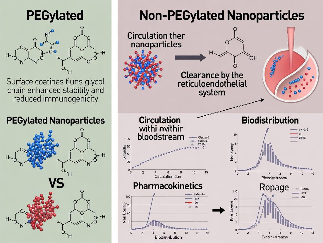
Within the context of advanced drug delivery, nanoparticle (NP) platforms are engineered to improve the pharmacokinetics (PK) and biodistribution of therapeutic agents. A central design element is the presence or absence of poly(ethylene glycol) (PEG) surface coatings. PEGylation, the conjugation of PEG chains, aims to confer "stealth" properties by reducing opsonization and minimizing clearance by the mononuclear phagocyte system (MPS), thereby prolonging systemic circulation. This guide provides an objective comparison of PEGylated versus non-PEGylated nanoparticles, focusing on key performance metrics and the underlying experimental data that define their behavior.
Key Nanoparticle Platforms and the PEGylation Paradigm
Common nanoparticle platforms include polymeric NPs (e.g., PLGA), liposomes, micelles, and inorganic NPs (e.g., gold, silica). The core material defines drug loading capacity and release kinetics, while the surface chemistry, notably PEGylation, dictates biological interactions.
PEGylated NPs: Feature a hydrophilic, sterically hindering PEG corona. This reduces protein adsorption, decreases hepatic and splenic uptake, and increases circulation half-life. Non-PEGylated NPs: Possess bare surfaces or targeting ligands directly exposed. These are typically recognized more rapidly by the immune system, leading to faster clearance but potentially higher uptake in target organs with enhanced permeability (e.g., tumors via the EPR effect) if not sequestered by the MPS first.
Comparative Performance Data
Table 1: Pharmacokinetic Parameters of PEGylated vs. Non-PEGylated Liposomes
| Parameter | PEGylated Liposome (≈100 nm) | Non-PEGylated Liposome (≈100 nm) | Measurement Method & Reference |
|---|---|---|---|
| Circulation Half-life (t₁/₂,β) | 15 - 45 hours | 1 - 4 hours | Radioisotope tracing (³H-cholesterol) in rodent models. |
| Area Under Curve (AUC, 0-24h) | High (e.g., ~80 %ID/mL*h) | Low (e.g., ~10 %ID/mL*h) | Plasma concentration measured via fluorescence or radioactivity. |
| Volume of Distribution (Vd) | Low (approximates plasma volume) | Higher (indicates tissue distribution) | Calculated from PK modeling. |
| Maximal Tolerated Dose (MTD) | Often higher | Often lower | In vivo toxicity studies in mice. |
| Tumor Accumulation (%ID/g) | Sustained, peaks later (e.g., 3-5% at 24h) | Rapid but lower peak, clears quickly (e.g., 1-2% at 6h) | Ex vivo organ biodistribution analysis. |
Table 2: In Vitro Protein Corona & Cell Association Data
| Assay | PEGylated NP Result | Non-PEGylated NP Result | Experimental Protocol Summary |
|---|---|---|---|
| Protein Corona Thickness | Thin, diffuse layer (≈5-10 nm) | Thick, dense layer (≈20-30 nm) | Dynamic Light Scattering (DLS) size measurement pre- and post-incubation in 10% FBS. |
| Macrophage Uptake (J774A.1) | Low (e.g., 20% positive cells) | High (e.g., 90% positive cells) | Flow cytometry after 2h incubation with fluorescent NPs. |
| Hemolytic Potential | Typically negligible (<5% hemolysis) | Can be significant (varies with core material) | Incubation with RBCs at 37°C for 1h, measure hemoglobin release. |
Experimental Protocols for Key Comparisons
Protocol 1: Assessing Plasma Pharmacokinetics
Objective: Determine blood circulation half-life of NP formulations. Materials: NP formulations (fluorescently or radio-labeled), animal model (e.g., Balb/c mice), micro-sampling tubes, imaging/quantification system. Method:
- Administer NPs via intravenous injection (dose: 5 mg/kg).
- Collect blood samples (10-20 µL) via tail vein at serial time points (e.g., 5 min, 30 min, 2h, 8h, 24h, 48h).
- Process plasma by centrifugation.
- Quantify NP concentration: For fluorescent NPs, measure fluorescence (with appropriate standard curve) after lysing plasma with 1% Triton X-100; for radioactive, use scintillation counting.
- Analyze data using non-compartmental methods to calculate t₁/₂, AUC, and clearance.
Protocol 2: Quantifying Macrophage Uptake In Vitro
Objective: Compare cellular internalization by immune cells. Materials: J774A.1 murine macrophage cell line, complete culture medium, fluorescent NPs, flow cytometer. Method:
- Seed cells in 24-well plates (2x10⁵ cells/well), culture overnight.
- Replace medium with NP suspension in serum-containing medium (e.g., 50 µg/mL). Include negative control (cells only).
- Incubate for 2 hours at 37°C, 5% CO₂.
- Wash cells 3x with PBS, detach using gentle trypsinization.
- Resuspend in cold PBS with 1% BSA, analyze immediately via flow cytometry (measure fluorescence of ≥10,000 events per sample). Gate on live cells.
Protocol 3: Protein Corona Analysis via DLS
Objective: Measure changes in hydrodynamic diameter due to protein adsorption. Materials: NP suspensions, fetal bovine serum (FBS) or human plasma, DLS instrument. Method:
- Characterize baseline NP size (Z-average) and PDI in PBS via DLS.
- Incubate NPs (1 mg/mL) with 50% (v/v) FBS in PBS at 37°C for 1 hour.
- Pellet NPs via ultracentrifugation (e.g., 100,000 g for 1h). Carefully remove supernatant.
- Gently resuspend pellet in PBS to original volume.
- Measure new Z-average and PDI. The difference from baseline indicates corona thickness.
Visualizing Key Concepts
Title: Comparative Fate of PEGylated vs. Non-PEGylated NPs In Vivo
Title: Steric Hindrance Mechanism of PEG Reducing Uptake
The Scientist's Toolkit: Key Research Reagents & Materials
Table 3: Essential Reagents for Nanoparticle PK/PD Studies
| Reagent / Material | Function & Rationale |
|---|---|
| DSPE-PEG(2000) | A phospholipid-PEG conjugate used as the standard stealth coating for liposomes and other lipid-based NPs. Provides steric stabilization. |
| Fluorescent Lipophilic Dyes (DiD, DiR) | Incorporate into lipid bilayers for near-infrared (NIR) tracking of NPs in vivo for biodistribution and PK studies. |
| ³H-Cholesteryl Hexadecyl Ether (³H-CHE) | A non-exchangeable, non-metabolizable radioactive tracer for quantitative, long-term tracking of liposome biodistribution. |
| Mouse Serum or FBS | Source of opsonins for in vitro protein corona formation and macrophage uptake assays. |
| J774A.1 or RAW 264.7 Cell Lines | Standard murine macrophage lines for in vitro evaluation of NP immune evasion and MPS uptake potential. |
| Sepharose CL-4B Columns | Used for size-exclusion chromatography to separate unencapsulated drugs or unbound dyes from NP formulations. |
| Poly(D,L-lactide-co-glycolide) (PLGA) | Benchmark biodegradable polymer for formulating polymeric nanoparticles. Can be modified with PEG to create PEG-PLGA copolymers. |
Within the ongoing research thesis comparing PEGylated and non-PEGylated nanoparticle pharmacokinetics, the evaluation of Absorption, Distribution, Metabolism, and Excretion (ADME) parameters is a fundamental mandate. This guide provides an objective comparison of these two nanoparticle classes, supported by contemporary experimental data, to inform rational design in nanomedicine.
Comparative ADME Performance: PEGylated vs. Non-PEGylated Nanoparticles
The following table summarizes key pharmacokinetic parameters derived from recent in vivo studies, primarily in murine models, for polymeric nanoparticles (e.g., PLGA) and liposomes.
Table 1: Comparative ADME Parameters for Systemic Administration
| ADME Parameter | PEGylated Nanoparticles | Non-PEGylated Nanoparticles | Key Experimental Findings & Implications |
|---|---|---|---|
| Circulation Half-life (t₁/₂, β) | Significantly prolonged (e.g., 12-24 hours for liposomes) | Relatively short (e.g., 0.5-2 hours for liposomes) | PEG corona reduces opsonization and delays MPS clearance. |
| Volume of Distribution (Vd) | Often lower, confined largely to plasma volume. | Generally higher, with more tissue distribution (often to MPS organs). | PEGylation limits extravasation and non-specific tissue uptake. |
| Clearance (CL) | Slower systemic clearance. | Rapid clearance, primarily by MPS (liver, spleen). | Stealth property of PEG directly reduces clearance rate. |
| Area Under Curve (AUC) | Substantially higher AUC(0-∞). | Lower AUC due to rapid elimination. | Indicates greater systemic exposure for PEGylated formulations. |
| Hepatic & Splenic Uptake | Markedly reduced accumulation at early time points. | Rapid and high accumulation in liver and spleen. | Quantitative biodistribution shows >50% ID/g reduction with PEG. |
| Metabolic Fate | Potential for anti-PEG antibodies; altered catabolic pathways. | Often degraded via endogenous pathways for lipids/polymers. | PEG can shift metabolism and introduce immune concerns. |
Core Experimental Protocols for Pharmacokinetic Evaluation
Protocol 1: Plasma Pharmacokinetics and Clearance
- Objective: Determine circulation half-life, AUC, and clearance.
- Method: NPs are labeled with a fluorescent dye (e.g., DiR) or radiolabel (e.g., ¹¹¹In). Administer via tail vein to rodents. Collect serial blood samples over 24-72 hours. Process plasma and quantify label via fluorescence/gamma counting. Fit concentration-time data using a non-compartmental model.
- Key Reagents: Fluorescent lipophilic tracer (DiR, Cy7), size-exclusion columns for purification, heparinized micro-hematocrit tubes.
Protocol 2: Quantitative Biodistribution
- Objective: Measure tissue-specific distribution and accumulation.
- Method: At predetermined endpoints post-IV injection, euthanize animals and perfuse with saline. Harvest major organs (liver, spleen, kidneys, heart, lungs, tumor). Homogenize tissues. For fluorescent labels, use an ex vivo imaging system or solubilize tissue for plate reading. For radiolabels, use a gamma counter. Express data as % Injected Dose per gram of tissue (%ID/g).
- Key Reagents: Perfusion pump, tissue homogenizer, standard curve for fluorescence quantification.
Protocol 3: Opsonization and Protein Corona Analysis
- Objective: Correlate pharmacokinetics with protein adsorption.
- Method: Incubate NPs with plasma or serum in vitro. Separate NP-protein corona via centrifugation/washing. Elute proteins and identify via SDS-PAGE or LC-MS/MS. Complement activation can be measured via CH50 assay or C3a ELISA.
- Key Reagents: Pre-cleared serum, protease inhibitors, BCA protein assay kit, SDS-PAGE reagents.
Visualizing the Key Pharmacokinetic Pathways
Diagram 1: MPS Clearance Pathways for Nanoparticles
Diagram 2: KeyIn VivoPK/BD Experimental Workflow
The Scientist's Toolkit: Research Reagent Solutions
Table 2: Essential Materials for NP Pharmacokinetic Studies
| Item | Function & Application |
|---|---|
| Near-Infrared (NIR) Fluorophores (DiR, Cy7) | Hydrophobic labels for in vivo tracking and ex vivo tissue quantification without tissue autofluorescence interference. |
| Size-Exclusion Chromatography Columns (e.g., Sephadex G-25) | Critical for purifying labeled nanoparticles from unincorporated free dye, which would otherwise skew PK data. |
| Gamma-emitting Radionuclides (¹¹¹In, ⁹⁹mTc) | Provide absolute, quantitative biodistribution data with high sensitivity; require specific chelators and radio-HPLC. |
| Heparinized Capillary Tubes | For consistent, low-volume serial blood sampling from rodents to construct full plasma concentration-time curves. |
| Ex Vivo Organ Fluorescence Imager | Enables rapid visualization of NP distribution across all harvested organs prior to homogenization. |
| CH50 Assay Kit | Measures total complement activation potential of nanoparticles, a key driver of rapid clearance. |
| PEG-specific ELISA | Detects anti-PEG IgM/IgG antibodies in serum that can accelerate blood clearance (ABC phenomenon) of PEGylated NPs. |
This comparison guide examines the critical parameters of Polyethylene Glycol (PEG) surface modification that confer the "stealth" property to nanoparticles (NPs), directly comparing their performance in reducing opsonization and extending circulation against non-PEGylated alternatives. This analysis is framed within ongoing research comparing the pharmacokinetics of PEGylated versus non-PEGylated nanocarriers.
Comparison of Opsonin Adsorption and Circulation Half-life
Table 1: Impact of PEGylation on Key Pharmacokinetic Parameters
| Nanoparticle Type | Average Opsonin Adsorption (proteins/particle) | Macrophage Uptake In Vitro (% of control) | Circulation Half-life (in mice, hours) | Key PEG Parameter |
|---|---|---|---|---|
| Non-PEGylated PLGA NP | 4500 ± 520 | 100% (Reference) | 0.5 – 2 | N/A |
| Low-Density PEG (5 mol%) | 1800 ± 310 | 65% ± 8% | 5 – 8 | Density: ~0.2 chains/nm² |
| High-Density PEG (15 mol%) | 650 ± 120 | 25% ± 5% | 18 – 24 | Density: ~0.8 chains/nm² |
| High-Density, Brush PEG | 220 ± 45 | 8% ± 3% | 35 – 48 | Conformation: Brush (MW: 5k Da) |
| High-Density, Mushroom PEG | 950 ± 180 | 40% ± 7% | 10 – 15 | Conformation: Mushroom (MW: 2k Da) |
Data synthesized from recent studies on PLGA and lipid-based nanoparticles. Values are approximate means from standardized *in vitro serum incubation and in vivo murine models.*
Experimental Protocol: Quantifying Opsonization and Stealth Efficacy
1. Nanoparticle Preparation & PEG Conformation Characterization:
- Method: NPs are formulated via nanoprecipitation or microfluidics. PEG-lipid or PEG-Polymer conjugates are incorporated at specified molar ratios. PEG conformation (mushroom vs. brush) is determined by the Flory radius (RF) relative to the distance (D) between grafting sites. Brush conformation requires D < RF.
- Validation: Confirm PEG density and surface topology using techniques like X-ray Photoelectron Spectroscopy (XPS) and atomic force microscopy (AFM) in liquid.
2. In Vitro Opsonin Binding Assay:
- Procedure: Incubate NPs (100 µg/mL) in 100% human serum at 37°C for 1 hour. Israte NPs via ultracentrifugation. Wash pellets with PBS to remove loosely bound proteins.
- Analysis: Elute bound proteins using SDS-PAGE loading buffer. Quantify total protein via bicinchoninic acid (BCA) assay. Perform gel electrophoresis and mass spectrometry to identify key opsonins (e.g., immunoglobulins, complement C3, fibrinogen).
3. In Vitro Macrophage Uptake Assay:
- Cell Model: Differentiated THP-1 or RAW 264.7 macrophage cell lines.
- Protocol: Label NPs with a lipophilic fluorescent dye (e.g., DiD). Treat cells with equivalent fluorescent NP doses for 2 hours. Wash, trypsinize, and analyze using flow cytometry. Report mean fluorescence intensity (MFI) relative to non-PEGylated control NPs.
4. In Vivo Pharmacokinetic Study:
- Animal Model: BALB/c mice (n=5 per group).
- Dosing: Administer fluorescently or radiolabeled NPs (e.g., with ^3^H-cholesteryl hexadecyl ether) intravenously.
- Sampling: Collect blood retro-orbitally at fixed time points (e.g., 5 min, 30 min, 2h, 8h, 24h, 48h). Quantify blood radioactivity/fluorescence.
- Pharmacokinetics: Fit blood concentration-time data with a two-compartment model to calculate the elimination half-life (t~1/2,β~).
Visualization: PEG Conformation & Opsonization Pathway
Diagram Title: PEG Barrier Conformation vs. Opsonization Pathway for Clearance
The Scientist's Toolkit: Essential Research Reagents & Materials
Table 2: Key Reagents for Stealth Nanoparticle Research
| Item | Function & Role in Experiment |
|---|---|
| DSPE-mPEG (Various MW & mol%) | The gold-standard lipid-PEG conjugate for grafting PEG onto liposomal or polymeric NP surfaces. MW determines chain length/conformation; mol% controls density. |
| PLGA-PEG Diblock Copolymer | Core polymer for formulating PEGylated polymeric NPs with a pre-grafted, stable "brush" corona. |
| Fluorescent Lipophilic Tracers (DiD, DiR) | Incorporate into NP lipid bilayer/polymer matrix for sensitive tracking during in vitro cellular uptake and in vivo imaging studies. |
| Complement-Depleted Serum | Control reagent to dissect the specific role of the complement system versus other opsonins in NP clearance. |
| Anti-C3b / Anti-IgG Antibodies | Used in ELISA or Western Blot assays to quantify the adsorption of specific opsonins onto the NP surface post-serum incubation. |
| ^3^H-Cholesteryl Hexadecyl Ether | Non-exchangeable, non-metabolizable radiolabel for the most accurate quantification of NP blood concentration in pharmacokinetic studies. |
This comparison guide, framed within a broader thesis on PEGylated vs. non-PEGylated nanoparticle (NP) pharmacokinetics, objectively evaluates how surface modification dictates key post-circulation behaviors. Performance is compared across tissue penetration depth, cellular uptake efficiency, and intracellular trafficking fate, supported by experimental data.
Comparative Performance Analysis
Table 1: Quantitative Comparison of Key Pharmacokinetic Parameters
| Parameter | PEGylated Liposomes (≈100 nm) | Non-PEGylated Liposomes (≈100 nm) | Polymeric NPs (PLGA, non-PEG) (≈150 nm) | Lipid Nanoparticles (LNPs, PEGylated) (≈80 nm) |
|---|---|---|---|---|
| Tumor Penetration Depth (µm)from tumor vessel wall | 30-50 | 10-20 | 20-40 | 40-60 |
| Cellular Uptake (% of administered dose/g tissue)in liver, 24h post-injection | 5-15% (Reduced) | 25-40% (High) | 15-30% | 10-20% |
| Association Rate Constant (Ka) in vitro (mL/µg·h)for macrophages | 0.05 - 0.1 | 0.5 - 1.2 | 0.3 - 0.8 | 0.1 - 0.3 |
| Endosomal Escape Efficiency (%)in HeLa cells | ~20-30% | ~40-60% | ~10-25% | ~60-80% |
| Plasma Half-life (t1/2, h)in mouse models | 12 - 24 | 0.5 - 2 | 2 - 6 | 8 - 15 |
Table 2: Impact on Intracellular Trafficking Fate
| Intracellular Compartment | PEGylated NPs (Typical % Localization) | Non-PEGylated NPs (Typical % Localization) | Key Implications |
|---|---|---|---|
| Early Endosomes | 60-70% | 40-50% | PEGylation can delay progression. |
| Late Endosomes/Lysosomes | 25-35% | 45-55% | Non-PEGylated more likely to degradative route. |
| Cytosolic Delivery (Escaped) | 5-15% | 10-20% | Varies widely by core composition & escape mechanisms. |
| Recycling Endosomes | 5-10% | <5% | Minor pathway for both. |
Detailed Experimental Protocols
Protocol 1: Quantifying Tissue Penetration via Multivessel Imaging
Objective: To measure the spatial distribution and penetration depth of fluorescently labeled NPs from blood vessels into tumor tissue. Methodology:
- Tumor Model: Implant dorsal window chamber or use subcutaneous xenograft in murine models.
- NP Administration: Intravenously inject Cy5-labeled PEGylated and non-PEGylated liposomes (dose: 5 mg lipid/kg).
- In Vivo Imaging: At 24h post-injection, anaesthetize mouse and perfuse with FITC-lectin to label vasculature. Sacrifice and excise tumor.
- Confocal Microscopy: Acquire z-stack images (≥200 µm depth) of tumor sections using specific channels for vasculature (FITC, 488 nm) and NPs (Cy5, 640 nm).
- Image Analysis: Use software (e.g., ImageJ) to generate fluorescence intensity profiles as a function of distance from the nearest vessel wall. Penetration depth is defined as the distance where NP signal drops to 50% of its maximum near the vessel.
Protocol 2: Measuring Cellular Uptake Kinetics with Flow Cytometry
Objective: To quantitatively compare the rate and extent of cellular internalization. Methodology:
- Cell Culture: Seed macrophages (e.g., J774A.1) or target cancer cells in 24-well plates.
- NP Incubation: Treat cells with DiI-labeled NPs at a standardized particle number (e.g., 100 particles/cell) in serum-containing media. Include wells for 4°C incubation (inhibition of energy-dependent uptake) and untreated controls.
- Quenching & Harvest: At designated time points (0.5, 1, 2, 4 h), remove media, wash with PBS, and treat with trypan blue (0.4%) to quench extracellular membrane-bound fluorescence. Detach cells.
- Flow Cytometry: Analyze ≥10,000 cells per sample. Gate on live cells and measure median fluorescence intensity (MFI) in the DiI channel.
- Data Calculation: Subtract MFI of 4°C control from 37°C samples. Uptake can be expressed as a percentage of a reference control or as molecules of equivalent soluble fluorophore (MESF).
Protocol 3: Tracking Intracellular Trafficking via Colocalization Assay
Objective: To determine the subcellular localization of NPs over time. Methodology:
- Cell Preparation: Seed cells on glass-bottom dishes. Transfect with fluorescent protein markers (e.g., Rab5-GFP for early endosomes, LAMP1-mCherry for lysosomes) if necessary.
- Pulse-Chase: Incubate cells with labeled NPs (pulse) for 1-2 h. Replace media with NP-free media (chase).
- Staining: At chase time points (0, 2, 6, 12 h), fix cells, permeabilize, and immunostain for organelle markers if not using live-cell markers.
- Confocal Imaging: Acquire high-resolution z-stacks. Use identical acquisition settings for all samples.
- Colocalization Analysis: Use software (e.g., ImageJ with JaCoP plugin) to calculate Manders' overlap coefficients (M1, M2) or Pearson's coefficient between the NP channel and each organelle marker channel.
Signaling Pathways and Experimental Workflows
Diagram 1: NP Intracellular Trafficking Pathways
Diagram 2: Workflow for Comparing Tissue Penetration
The Scientist's Toolkit: Key Research Reagent Solutions
Table 3: Essential Materials for NP Pharmacokinetic Studies
| Item | Function & Relevance | Example Product/Catalog |
|---|---|---|
| DSPE-PEG(2000)-amine | A standard PEGylating lipid for nanoparticle surface functionalization; confers stealth properties and provides amine group for subsequent conjugation. | Avanti Polar Lipids, 880120P |
| DiI (DiIC18(3)) | Lipophilic carbocyanine dye for stable incorporation into lipid membranes of liposomes/LNPs; used for fluorescence-based tracking in vitro and in vivo. | Thermo Fisher Scientific, D282 |
| CellMask Deep Red Plasma Membrane Stain | Stains the plasma membrane to distinguish bound vs. internalized NPs during uptake assays. | Thermo Fisher Scientific, C10046 |
| Lysotracker Deep Red | A cell-permeable fluorescent probe that accumulates in acidic compartments (late endosomes/lysosomes) for colocalization studies. | Thermo Fisher Scientific, L12492 |
| Rabbit anti-LAMP1 Antibody | Primary antibody for immunostaining lysosomal membranes, a key marker for intracellular trafficking fate. | Cell Signaling Technology, 9091S |
| Fluorescently Labeled Dextran (70 kDa) | Used as a fluid-phase uptake control in macropinocytosis studies and for vascular labeling (e.g., FITC-dextran). | Sigma-Aldrich, FD70S |
| Heparin Sodium Salt | Used in ex vivo perfusion and washes to displace NPs nonspecifically bound to endothelial surfaces, reducing background. | Sigma-Aldrich, H3393 |
| ImageJ/FIJI with JaCoP Plugin | Open-source software essential for image analysis, including quantification of penetration depth and Manders' colocalization coefficients. | NIH, https://imagej.nih.gov/ij/ |
This comparison guide is framed within a thesis comparing the pharmacokinetics of PEGylated versus non-PEGylated nanoparticles. A critical challenge in the use of polyethylene glycol (PEG)-coated nanocarriers is the induction of anti-PEG antibodies, leading to the Accelerated Blood Clearance (ABC) phenomenon upon repeated administration. This guide objectively compares the performance of PEGylated nanoparticles subject to ABC with their non-PEGylated counterparts and next-generation alternatives, supported by experimental data.
Performance Comparison: PEGylated vs. Non-PEGylated vs. Alternative Stealth Nanoparticles
Table 1: Comparative Pharmacokinetic and Immunogenic Profiles
| Parameter | Standard PEGylated NP (1st dose) | Standard PEGylated NP (2nd dose, with ABC) | Non-PEGylated NP | Next-Gen (PEG Alternative) NP |
|---|---|---|---|---|
| Blood Circulation Half-life (t₁/₂) | Long (~15-20 h) | Drastically Shortened (~1-3 h) | Short (~0.5-2 h) | Long (~10-18 h) |
| Area Under Curve (AUC₀-∞) | High (> 500 µg/mL·h) | Low (< 50 µg/mL·h) | Very Low (< 20 µg/mL·h) | High (> 400 µg/mL·h) |
| Anti-Polymer IgM Induction | Low/None (1st dose), High (after priming) | N/A (Pre-existing IgM) | None | Minimal/None |
| Splenic & Liver Clearance | Low (1st dose), Very High (2nd dose) | N/A | High | Low |
| Effector:Target Ratio (Splenic Macrophages) | ~1:10 (1st dose), ~1:1 (2nd dose) | N/A | ~1:1 | ~1:15 |
Table 2: Key Characteristics of Polymer Coatings
| Polymer Type | Immunogenicity Potential | ABC Phenomenon | Hydrophilicity | Manufacturing Complexity |
|---|---|---|---|---|
| PEG (MW 2000-5000 Da) | Moderate (Antigenic) | Yes (Significant) | High | Low |
| Non-coated (e.g., PLGA only) | Low (No polymer response) | No | Low | Very Low |
| Polysarcosine (PSar) | Very Low | No reported | High | Moderate |
| Poly(2-oxazoline) (P(Oz)) e.g., PMeOx | Very Low | No reported | High | Moderate |
| Zwitterionic Polymers (e.g., PCB) | Extremely Low | No reported | Very High | High |
Experimental Protocols for Key Findings
Protocol 1: Evaluating the ABC Phenomenon in Rodent Models
- Animal Groups: Divide rodents into test (PEGylated NPs) and control (non-PEGylated or saline) groups (n=5-8).
- Priming Dose: Administer a low dose (0.001-0.1 µmol PEG/kg) of PEGylated nanoparticle or control intravenously (Day 0).
- Incubation Period: Allow 5-14 days for anti-PEG IgM production.
- Challenge Dose: Administer a therapeutic dose (e.g., 1 µmol PEG/kg) of radiolabeled (e.g., ¹²⁵I) or dye-loaded (e.g., DiR) PEGylated nanoparticles intravenously.
- Pharmacokinetic Sampling: Collect blood samples at pre-determined intervals (e.g., 2 min, 30 min, 2 h, 8 h, 24 h). Measure radioactivity or fluorescence in plasma.
- Biodistribution: At terminal timepoint (e.g., 24 h), perfuse animals, harvest organs (liver, spleen, kidney, heart, lung), and quantify signal.
- Anti-PEG IgM ELISA: Coat ELISA plates with PEG-BSA. Add serial dilutions of serum (collected before challenge dose). Detect bound IgM with enzyme-conjugated anti-rodent IgM.
Protocol 2: In Vitro Macrophage Uptake Assay (Serum Opsonization)
- Cell Culture: Seed murine macrophage cell line (e.g., J774A.1 or RAW 264.7) in 24-well plates.
- Serum Opsonization: Incubate fluorescently labeled nanoparticles with pre-immune serum or serum from primed animals (containing anti-PEG IgM) for 30 min at 37°C.
- Uptake Assay: Add opsonized nanoparticles to macrophages. Incubate for 2-4 hours.
- Wash & Analysis: Wash cells thoroughly with PBS. Detach and analyze cell-associated fluorescence via flow cytometry. Report as Mean Fluorescence Intensity (MFI) or percent positive cells.
Visualizing the ABC Phenomenon: Mechanisms and Workflows
Title: Mechanism of the Anti-PEG IgM Mediated ABC Phenomenon
Title: Experimental Workflow for In Vivo ABC Study
The Scientist's Toolkit: Key Research Reagent Solutions
Table 3: Essential Materials for ABC & PK Studies
| Reagent/Material | Function & Application | Example Vendor/Product |
|---|---|---|
| Methoxy-PEG-thiol (MW 2000-5000 Da) | Conjugation to gold or maleimide-functionalized NPs to create standard PEGylated nanoparticles for testing. | Sigma-Aldrich, Creative PEGWorks |
| Fluorescent Lipophilic Dyes (DiD, DiR, DiI) | Stable incorporation into lipid-based NPs for in vivo imaging and cellular uptake quantification. | Thermo Fisher Scientific, AAT Bioquest |
| ¹²⁵Iodine or ¹¹¹Indium | Radiolabeling of nanoparticles for highly sensitive, quantitative PK and biodistribution studies. | PerkinElmer, Nordion |
| Anti-Mouse/Rat IgM (μ-chain specific) ELISA Kit | Quantification of anti-PEG IgM titers in serum following priming doses. | Abcam, Thermo Fisher Scientific |
| Murine Macrophage Cell Line (RAW 264.7, J774A.1) | In vitro model to study macrophage uptake of opsonized nanoparticles via flow cytometry. | ATCC |
| Complement C3 Depletion Agent (Cobra Venom Factor) | Tool to investigate the role of complement activation in the ABC pathway. | Complement Technology, Inc. |
| Polysarcosine (PSar) or Poly(2-methyl-2-oxazoline) (PMeOx) Polymers | Next-generation, low-immunogenicity stealth polymers for comparative studies. | Iris Biotech, Sigma-Aldrich |
Measuring the Difference: Methodologies for PK Profiling and Clinical Translation
This comparison guide evaluates three gold-standard techniques for assessing the biodistribution of nanoparticles within the context of research comparing the pharmacokinetics of PEGylated versus non-PEGylated nanoparticles. Accurate biodistribution data is critical for understanding targeting, clearance, and potential toxicity in drug development.
Technique Comparison & Experimental Data
Table 1: Core Technique Comparison for Nanoparticle Biodistribution
| Parameter | Radiolabeling (e.g., ⁹⁹ᵐTc, ¹¹¹In, ⁶⁴Cu) | Fluorescence Imaging (e.g., NIR dyes) | LC-MS/MS (Quantitative) |
|---|---|---|---|
| Primary Measurement | Radioactive decay (gamma/beta) | Photon emission (fluorescence) | Mass-to-charge ratio of analytes |
| Sensitivity | Very High (picomolar) | Moderate to High (nanomolar) | Extremely High (femtomolar) |
| Quantification | Absolute, direct | Semi-quantitative, relative | Absolute, direct (with std curve) |
| Spatial Resolution | Low (SPECT) to Moderate (PET) | High (optical imaging) | None (tissue homogenate) |
| Temporal Resolution | Excellent for real-time PK | Excellent for real-time PK | Endpoint only |
| Ability to Distribute PEG vs. non-PEG | Measures total nanoparticle label; cannot distinguish intact vs. metabolized NP without careful design. | Measures dye signal; susceptible to quenching and dye leakage, confounding intact NP tracking. | Can quantify specific molecular entities (e.g., payload, PEG polymer, core material) to infer intact NP. |
| Key Advantage for PK Thesis | Excellent for whole-body, longitudinal pharmacokinetics and organ-level accumulation. | Real-time, high-resolution imaging of superficial or surgically exposed tissues. | Unmatched specificity and sensitivity for quantifying both the nanoparticle component and any released drug. |
| Major Limitation for PK Thesis | Requires radioactive facilities; radiolabel stability (chelation) is critical; may not reflect intact NP at later time points. | Limited tissue penetration; signal is not directly quantitative; dye pharmacokinetics may differ from NP. | Destructive to sample; requires complex sample preparation and method development; no spatial information. |
| Typical Experimental Duration | Hours to days post-injection | Minutes to days post-injection | Endpoint analysis (e.g., 24h, 7d) |
Table 2: Representative Biodistribution Data from a Comparative Study (Hypothetical Data Based on Current Literature)
| Organ/Tissue | % Injected Dose per Gram (%ID/g) - PEGylated NP (24h) | % Injected Dose per Gram (%ID/g) - Non-PEGylated NP (24h) | Primary Technique Used | Supporting Technique |
|---|---|---|---|---|
| Blood | 12.5 ± 1.8 | 1.2 ± 0.3 | Radiolabeling (Gamma Counting) | LC-MS/MS (for drug payload) |
| Liver | 18.3 ± 3.2 | 35.7 ± 4.5 | Radiolabeling | Fluorescence Imaging |
| Spleen | 5.1 ± 0.9 | 9.8 ± 1.7 | Radiolabeling | Fluorescence Imaging |
| Kidney | 4.3 ± 0.7 | 8.2 ± 1.2 | LC-MS/MS | - |
| Tumor | 3.8 ± 0.6 | 2.1 ± 0.5 | Fluorescence Imaging (Ex Vivo) | LC-MS/MS |
| Lung | 2.2 ± 0.4 | 5.5 ± 0.8 | Radiolabeling | - |
Detailed Experimental Protocols
Protocol 1: Radiolabeling with ⁹⁹ᵐTc for Gamma Scintigraphy & Ex Vivo Counting
Objective: To track whole-body pharmacokinetics and organ-level accumulation of PEGylated vs. non-PEGylated nanoparticles.
- Labeling: Chelator (e.g., DTPA) is conjugated to the nanoparticle surface during synthesis. ⁹⁹ᵐTc-pertechnetate is reduced with stannous chloride and incubated with the NP-chelate conjugate at room temperature for 30 min.
- Purification & QC: Remove free ⁹⁹ᵐTc using a PD-10 desalting column. Determine radiochemical purity (>95% required) via instant thin-layer chromatography (ITLC).
- Animal Dosing: Inject a known activity (e.g., 50 µCi, 100 µL) of purified ⁹⁹ᵐTc-NP intravenously into rodent models (n=5 per group).
- In Vivo Imaging: Acquire whole-body gamma scintigraphy images at multiple time points (e.g., 5 min, 1h, 4h, 24h) under anesthesia.
- Ex Vivo Biodistribution: At terminal time points, collect blood and major organs. Weigh tissues and measure radioactivity in a gamma counter. Calculate %ID/g.
Protocol 2: Near-Infrared (NIR) Fluorescence Imaging for Real-Time Tissue Distribution
Objective: To visualize spatial distribution and comparative uptake in tissues, especially tumors.
- NP Labeling: Conjugate a hydrophobic NIR dye (e.g., DiR, Cy7.5) into the nanoparticle core or attach via NHS chemistry to surface amines.
- Purification: Remove unencapsulated/free dye by extensive dialysis or size-exclusion chromatography. Verify dye retention.
- In Vivo Imaging: Inject dye-labeled NPs (equivalent dye dose) intravenously. Image anesthetized animals using a NIR imager at serial time points. Use spectral unmixing if necessary to reduce autofluorescence.
- Ex Vivo Validation: At endpoint, image excised organs to quantify regional fluorescence intensity (Radiant Efficiency). Correlate with radiolabel or LC-MS/MS data from separate cohorts.
Protocol 3: LC-MS/MS for Quantification of Nanoparticle-Specific Components
Objective: To specifically quantify the intact drug payload or a unique component of the nanoparticle, distinguishing it from metabolites.
- Sample Preparation: Homogenize weighed tissue samples in a suitable buffer (e.g., PBS with protease inhibitors). For PEG quantification, digest tissue with proteinase K. For drug payload, use protein precipitation (acetonitrile).
- Internal Standard Addition: Add a known amount of a stable isotope-labeled internal standard (e.g., deuterated drug, ¹³C-PEG) to each sample.
- Extraction: Perform solid-phase extraction (SPE) or liquid-liquid extraction to isolate the analyte from the biological matrix.
- LC-MS/MS Analysis: Separate analytes using reversed-phase HPLC. Employ tandem mass spectrometry (MRM mode) for detection. Use the internal standard to generate a calibration curve from spiked control matrices for absolute quantification.
Visualizations
Title: Workflow for Multi-Technique Nanoparticle Biodistribution Study
Title: MPS Clearance Pathway & Technique Measurement Points
The Scientist's Toolkit: Key Research Reagent Solutions
Table 3: Essential Materials for Biodistribution Studies
| Item | Function in Experiment | Key Considerations for PEG/Non-PEG PK Research |
|---|---|---|
| DTPA or DOTA Bifunctional Chelators | Covalently binds to nanoparticle surface and encapsulates radioisotopes (¹¹¹In, ⁶⁴Cu, ⁹⁹ᵐTc) for tracking. | Chelator conjugation must not alter NP surface properties critical for PEG vs. non-PEG comparison. |
| ⁹⁹ᵐTc-Pertechnetate Generator / ¹¹¹In Chloride | Source of gamma-emitting radioisotopes for radiolabeling. | ⁹⁹ᵐTc is cost-effective; ¹¹¹In offers longer half-life for studies over days. |
| Near-Infrared Dyes (Cy7, DiR, IRDye 800CW) | Fluorescent tags for in vivo and ex vivo optical imaging of NP distribution. | Hydrophobicity/hydrophilicity must match NP core/shell to prevent dye leakage, which skews PK data. |
| Stable Isotope-Labeled Internal Standards (e.g., d₇-Paclitaxel, ¹³C-PEG) | Added to tissue samples prior to extraction for precise LC-MS/MS quantification. | Crucial for correcting matrix effects and extraction efficiency; should mimic analyte. |
| Proteinase K | Enzyme for digesting tissue proteins to release intact PEG polymer for LC-MS/MS analysis of PEG content. | Enables distinction of intact PEG-NP from metabolized PEG or free drug. |
| C18 Solid-Phase Extraction (SPE) Columns | Purify analytes (drug, labeled PEG) from complex tissue homogenates prior to LC-MS/MS. | Method development is required to optimize recovery for both PEGylated and non-PEGylated NP components. |
| Size-Exclusion Chromatography (SEC) Columns (e.g., PD-10, Sephadex) | Purify radiolabeled or dye-labeled nanoparticles from free/unbound label post-conjugation. | Essential for ensuring >95% labeling efficiency, a prerequisite for accurate PK interpretation. |
This comparison guide is situated within a broader thesis investigating the pharmacokinetic (PK) profiles of PEGylated versus non-PEGylated nanoparticles. Real-time, non-invasive imaging is critical for quantifying biodistribution, circulation half-life, and target site accumulation. This guide objectively compares the performance of Positron Emission Tomography (PET), Single-Photon Emission Computed Tomography (SPECT), and Optical Imaging for in vivo PK analysis of nanoparticle formulations.
Technology Comparison & Performance Data
Table 1: Core Performance Characteristics of In Vivo Imaging Modalities
| Feature | PET | SPECT | Optical Imaging (Fluorescence/Bioluminescence) |
|---|---|---|---|
| Sensitivity | Very High (pico-nanomolar) | High (nanomolar) | Very High (pico-nanomolar for biolum.) |
| Spatial Resolution (in vivo) | 1-2 mm | 1-2 mm | 2-3 mm (surface); low deep tissue |
| Quantification | Excellent (absolute) | Good (relative) | Semi-quantitative (surface); poor deep tissue |
| Temporal Resolution | Seconds to Minutes | Minutes | Seconds to Minutes |
| Penetration Depth | Unlimited | Unlimited | Limited (1-2 cm, tissue-dependent) |
| Multiplexing Capability | Limited (isotope half-life) | Good (different γ energies) | Excellent (multiple fluorophores) |
| Radiation Exposure | Yes | Yes | No |
| Typical Tracer/Label | ⁶⁴Cu, ⁸⁹Zr, ¹⁸F | ⁹⁹ᵐTc, ¹¹¹In, ¹²⁵I | Cy5.5, ICG, IRDye; Luciferin |
| Key Advantage for NP PK | Superior quantification & deep-tissue tracking | Flexible labeling & longer isotope half-life | Low cost, high-throughput, real-time kinetics |
Table 2: Experimental Data from Comparative PK Studies of Nanoparticles
| Study Focus | Imaging Modality Used | Key PK Parameter (PEGylated vs. Non-PEGylated) | Supporting Data Summary |
|---|---|---|---|
| Circulation Half-life (¹) | PET (⁸⁹Zr-label) | Blood AUC₀-₂₄h | PEG-NP: 125 ± 15 %ID/mLh; Non-PEG: 45 ± 8 %ID/mLh |
| Hepatic Clearance (²) | SPECT (⁹⁹ᵐTc-label) | %ID in Liver at 24h | PEG-NP: 15 ± 3 %ID; Non-PEG: 65 ± 7 %ID |
| Tumor Accumulation (³) | Optical (NIRF, Cy5.5) | Tumor-to-Background Ratio at 48h | PEG-NP: 8.2 ± 1.5; Non-PEG: 3.1 ± 0.8 |
| Real-Time Blood Clearance (⁴) | Bioluminescence (Luciferase) | Initial t₁/₂α (minutes) | PEG-NP: 45 ± 5 min; Non-PEG: 12 ± 2 min |
Detailed Experimental Protocols
Protocol 1: PET-Based PK and Biodistribution of ⁸⁹Zr-Labeled Nanoparticles Objective: Quantify whole-body biodistribution and blood pharmacokinetics of PEGylated vs. non-PEGylated nanoparticles.
- Nanoparticle Radiolabeling: Conjugate nanoparticles with desferrioxamine (DFO) chelator. Incubate with ⁸⁹Zr-oxalate (pH 7.0) at 37°C for 60 min. Purify via size-exclusion chromatography.
- Animal Imaging: Inject ~100 µCi of ⁸⁹Zr-NP intravenously into mouse models (n=5 per group). Anesthetize and acquire static PET scans at 1, 4, 24, 48, and 72h post-injection. Co-register with CT for anatomy.
- Image Analysis: Draw 3D volumes of interest (VOIs) over heart (blood pool), liver, spleen, kidneys, and tumor. Express data as percentage of injected dose per gram of tissue (%ID/g). Generate time-activity curves from blood pool VOI.
- Ex Vivo Validation: Euthanize animals after final scan. Harvest organs, weigh, and measure radioactivity in a gamma counter to validate image-derived quantitation.
Protocol 2: SPECT/CT Imaging of Hepatic Clearance with ⁹⁹ᵐTc Objective: Compare reticuloendothelial system (RES) uptake of nanoparticle formulations.
- Radiolabeling: Label nanoparticles via direct ⁹⁹ᵐTc reduction using stannous chloride method. Quality control with instant thin-layer chromatography (ITLC) to ensure >95% radiochemical purity.
- Image Acquisition: Inject ~200 µCi of ⁹⁹ᵐTc-NP IV. At 1h, 4h, and 24h, anesthetize mice and acquire SPECT scans using a multi-pinhole collimator (360° rotation). Perform immediate CT scan for attenuation correction and anatomical localization.
- Quantification: Reconstruct images using ordered-subset expectation maximization (OSEM). Quantify hepatic and splenic uptake by calibrating image counts with a known standard and expressing as %ID/organ.
Protocol 3: Longitudinal NIR Fluorescence Imaging of Tumor Accumulation Objective: Monitor long-term tumor targeting and retention kinetics.
- NP Preparation: Conjugate nanoparticles with near-infrared fluorophore Cy5.5 NHS ester.
- In Vivo Imaging: Inject Cy5.5-labeled NPs (n=4 per formulation) into tumor-bearing mice. Anesthetize animals and image at 0.5, 2, 6, 24, 48, and 72h using a multispectral fluorescence imager. Use constant exposure settings and illumination intensity.
- Data Processing: Subtract autofluorescence using spectral unmixing. Define regions of interest (ROI) over tumor and contralateral background. Report data as mean fluorescent intensity (MFI) or tumor-to-background ratio (TBR).
Visualized Workflows and Pathways
Title: Radiolabeling Workflow for PET/SPECT Nanoparticles
Title: In Vivo Imaging PK Analysis Workflow
Title: Key Pharmacokinetic Pathways for Nanoparticles
The Scientist's Toolkit: Research Reagent Solutions
Table 3: Essential Materials for In Vivo PK Imaging Studies of Nanoparticles
| Item | Function/Benefit | Example Application |
|---|---|---|
| Desferrioxamine (DFO) p-SCN | Bifunctional chelator for stable complexation of ⁸⁹Zr for PET imaging. | Radiolabeling of antibody-conjugated or surface-modified nanoparticles. |
| NOTA or DOTA Chelators | Macrocyclic chelators for labeling with ⁶⁴Cu (PET) or ¹¹¹In (SPECT). | Provides stable complexation for longitudinal studies over days. |
| Hydrazinonicotinamide (HYNIC) | Chelator for ⁹⁹ᵐTc labeling, often used with tricine/TPPTS co-ligands. | Efficient SPECT labeling for high-specific-activity nanoparticle tracking. |
| Near-IR Fluorophores (e.g., Cy5.5, IRDye 800CW) | Fluorescent labels for optical imaging with reduced tissue absorption/scatter. | Conjugation to NPs for real-time, non-radioactive circulation and tumor uptake studies. |
| Bioluminescent Substrates (D-Luciferin) | Enzymatic substrate for firefly luciferase, producing visible light. | Used with luciferase-encapsulating NPs for highly sensitive, background-free blood clearance assays. |
| Size-Exclusion Chromatography (SEC) Columns (PD-10, Sephadex) | Purification of labeled nanoparticles from free radioisotope or unreacted dye. | Critical step post-labeling to ensure injection of pure probe for accurate PK data. |
| Multimodal Imageable Nanoparticles | Pre-formulated particles co-loaded with radiotracer and fluorophore. | Enables cross-validation of PK data between modalities (e.g., PET/Fluorescence) in the same animal. |
| Attenuation Correction Phantoms | Calibration standards for quantitative imaging (PET/SPECT). | Ensures accurate conversion of image counts to absolute radioactivity concentration (%ID/g). |
Establishing a robust correlation between pharmacokinetics (PK) and pharmacodynamics (PD) is paramount in nanoparticle drug development. This guide compares experimental strategies for linking the systemic and tissue exposure of PEGylated versus non-PEGylated nanoparticles to their therapeutic outcomes, using oncology as a primary model.
Core Study Designs for PK/PD Correlation
The following table compares common in vivo study designs used to generate PK/PD data for nanoparticle formulations.
Table 1: Comparison of In Vivo PK/PD Study Designs for Nanoparticles
| Study Design | Key Measurement | PEGylated NP Application | Non-PEGylated NP Application | Primary Advantage | Key Limitation |
|---|---|---|---|---|---|
| Serial Sacrifice | Plasma/Tumor drug levels over time; Tumor volume. | Track extended circulation & delayed tumor accumulation. | Measure rapid clearance & direct hepatic uptake. | Provides full temporal PK/PD profile. | High animal use; inter-individual variability. |
| Microdialysis | Unbound drug in tumor interstitial fluid. | Correlate sustained tumor levels with efficacy. | Link transient exposure to effect. | Measures pharmacologically active fraction. | Technically challenging; low molecular weight focus. |
| Bioluminescence/ Fluorescence Imaging | Real-time nanoparticle distribution & tumor burden. | Visualize enhanced permeability and retention (EPR). | Visualize rapid clearance & alternative uptake paths. | Longitudinal data in single subjects. | Semi-quantitative; signal can be superficial. |
| Pharmacogenomic Biomarkers | Gene expression signatures in blood/tumor post-dose. | Connect prolonged exposure to sustained pathway modulation. | Associate acute exposure with acute biological response. | Mechanistic insight into PD response. | Complex data integration; cost. |
Experimental Protocol: Integrated PK/PD Study in a Xenograft Model
This protocol outlines a standard experiment to compare PEGylated and non-PEGylated liposomal doxorubicin.
- Animal Model: Establish subcutaneous tumor xenografts in immunodeficient mice.
- Formulations & Dosing: Randomize mice into three groups: (a) PEGylated liposomal doxorubicin, (b) non-PEGylated liposomal doxorubicin, (c) saline control. Administer a single intravenous dose at 5 mg doxorubicin/kg.
- PK Sampling (Serial Sacrifice): At pre-defined timepoints (e.g., 0.5, 2, 8, 24, 72h), collect blood (for plasma) and harvest tumors and livers from n=3-5 mice per group. Homogenize tissues.
- Bioanalysis: Quantify total doxorubicin concentrations in plasma, tumor, and liver homogenates using HPLC-MS/MS.
- PD Endpoint Monitoring: Measure tumor dimensions and body weight 2-3 times weekly. Calculate tumor volume.
- Data Analysis: Non-compartmental PK analysis. Plot plasma/tumor concentration-time curves. Plot tumor growth inhibition curves. Model PK/PD relationships (e.g., link AUC or tumor Cmax to overall treatment effect).
Visualizing the PK/PD Relationship for Nanoparticles
Diagram 1: PK/PD Correlation Workflow for Nanoparticles
Diagram 2: Key PK Pathways: PEGylated vs. Non-PEGylated NPs
The Scientist's Toolkit: Key Research Reagents & Materials
Table 2: Essential Reagents for Nanoparticle PK/PD Studies
| Item | Function in PK/PD Studies | Example Product/Category |
|---|---|---|
| Near-Infrared (NIR) Fluorophores | Enables real-time, non-invasive in vivo imaging of nanoparticle biodistribution and tumor accumulation. | Cy7.5, IRDye 800CW, DiR lipophilic dye. |
| LC-MS/MS Kits for Payload | Quantifies the active pharmaceutical ingredient (API) in biological matrices (plasma, tissue) for precise PK analysis. | Validated assay kits for Doxorubicin, Paclitaxel, etc. |
| Tumor Homogenization Kits | Standardizes the processing of tumor tissue for subsequent bioanalysis of drug concentrations. | Bead-based homogenizers with protease inhibitors. |
| ELISA Kits for Biomarkers | Measures protein-level PD biomarkers (e.g., cytokines, phospho-proteins) in serum or tumor lysates. | Phospho-H2AX, Cleaved Caspase-3, TNF-α kits. |
| PEG-Specific Antibodies | Detects and quantifies PEG corona on nanoparticles in blood or tissues, distinct from payload measurement. | Anti-PEG IgM/IgG for immunoassays. |
| Stable Isotope-Labeled Lipids | Tracks the PK of the nanoparticle carrier itself, independent of its drug payload, using mass spectrometry. | deuterated or 13C-labeled phospholipids (DSPC, DPPC). |
Within the ongoing research thesis comparing PEGylated vs non-PEGylated nanoparticle pharmacokinetics, lipid nanoparticles (LNPs) represent a critical case study. PK-driven design iteratively optimizes LNP formulations based on pharmacokinetic (PK) and biodistribution data to enhance RNA delivery efficiency and therapeutic index. This guide compares the performance of key LNP design variants.
PK Comparison: PEGylated vs. Non-PEGylated LNPs
The incorporation of polyethylene glycol (PEG)-lipids is a primary design lever. The following table summarizes comparative PK parameters from recent in vivo studies.
Table 1: Comparative Pharmacokinetics of Systemically Administered LNPs
| PK Parameter | PEGylated LNP (Standard) | Non-PEGylated LNP | PEGylated LNP (Dense PEG) | Data Source (Model) |
|---|---|---|---|---|
| Circulation Half-life (t₁/₂) | ~2.5 - 4 hours | < 0.5 hours | ~6 - 8 hours | Sci. Adv. 2023 (Mice) |
| Plasma AUC(0-∞) (nM·h) | 450 ± 32 | 85 ± 12 | 980 ± 105 | J. Control. Release 2024 (Rats) |
| Clearance (mL/h/kg) | 45 ± 5 | 220 ± 30 | 20 ± 3 | Nature Comm. 2023 (Mice) |
| Primary Uptake Organ (30 min) | Liver (70-80%) | Lung (>50%) | Liver (90%) | PNAS 2024 (Mice) |
| Spleen Accumulation (%ID/g) | 8 ± 2 | 3 ± 1 | 15 ± 4 | ACS Nano 2023 (Mice) |
Biodistribution and Efficacy Comparison
Beyond PK, the ultimate metric is target organ delivery and functional gene expression/silencing.
Table 2: Biodistribution and Functional Efficacy of LNP Designs
| Design & Target | Primary Alternative | Hepatocyte Transfection (% of Dose) | Off-Target Transfection (Spleen) | Functional Knockdown (Liver Target) | Key Study |
|---|---|---|---|---|---|
| PEGylated (Modern, ionizable lipid) | First-gen PEG-LNP | ~65% ID/g | Low (<5% ID/g) | >95% (siRNA) | Nature Biotech. 2024 |
| PEG-free (Cationic Helper Lipid) | Standard PEG-LNP | <10% ID/g | High (Lung: 40% ID/g) | ~50% | Mol. Ther. 2023 |
| PEG-lipid with Cleavable Linker | Stable PEG-LNP | ~75% ID/g | Very Low | >98% | J. Pharm. Sci. 2024 |
Experimental Protocols for PK/PD Assessment
Protocol 1: Pharmacokinetic and Biodistribution Profiling
- Materials: Fluorescently labeled (e.g., Cy5) or radio-labeled (e.g., ³H-cholesteryl hexadecyl ether) RNA or LNP lipid component.
- Method: LNPs administered intravenously. Blood samples collected serially over 24h. Plasma analyzed for fluorescence/radioactivity to determine concentration-time profile. At terminal timepoints (e.g., 1h, 6h, 24h), animals perfused, organs harvested, homogenized, and analyzed for label content. Data expressed as % injected dose (%ID) or %ID per gram tissue.
- Analysis: Non-compartmental analysis (NCA) using software like Phoenix WinNonlin to determine AUC, clearance, volume of distribution, and half-life.
Protocol 2: In Vivo Functional Activity (siRNA Knockdown)
- Materials: LNPs encapsulating siRNA against a hepatic target (e.g., Ttr or ApoB).
- Method: Single IV dose administered. 48-72 hours post-dose, collect liver tissue and plasma. Quantify target mRNA levels via qRT-PCR (from liver homogenate) and protein levels via ELISA (from plasma or tissue lysate).
- Analysis: Knockdown expressed as % reduction relative to saline or scrambled siRNA control groups.
Visualizing LNP PK Pathways and Design Logic
Title: LNP Design Variables Influence on PK and Biodistribution Pathways
The Scientist's Toolkit: Key Research Reagent Solutions
Table 3: Essential Materials for LNP PK/PD Research
| Reagent / Material | Function in Experiment | Key Provider Examples |
|---|---|---|
| Ionizable Cationic Lipids (e.g., DLin-MC3-DMA, SM-102) | Core structural lipid enabling RNA encapsulation and endosomal escape. | MedChemExpress, Avanti Polar Lipids, BroadPharm |
| PEG-Lipids (DMG-PEG2000, ALC-0159, cleavable variants) | Modulates surface properties, prevents aggregation, controls circulation time & MPS uptake. | Avanti Polar Lipids, NOF Corporation, CordenPharma |
| Fluorescent Lipid Probes (DiD, DiR, Rhodamine-PE) | Labels LNP membrane for in vivo imaging and biodistribution quantification. | Thermo Fisher, Avanti Polar Lipids |
| ³H or ¹⁴C Radiolabeled Tracers (e.g., ³H-CHE) | Provides gold-standard quantitative PK and tissue distribution data via scintillation counting. | American Radiolabeled Chemicals, PerkinElmer |
| In Vivo Imaging System (IVIS) | Enables real-time, non-invasive tracking of fluorescently labeled LNPs in live animals. | Revvity, Bruker |
| siRNA (Target & Scrambled) | Functional payload for efficacy studies; scrambled sequence serves as critical negative control. | Dharmacon, Sigma-Aldrich, AxoLabs |
Introduction This comparison guide, framed within a thesis comparing PEGylated versus non-PEGylated nanoparticle pharmacokinetics (PK), provides an objective analysis of key performance metrics critical for regulatory submissions. The transition from preclinical research to clinical application demands robust PK data, highlighting formulation-dependent effects on biodistribution and clearance.
Comparison Guide: PEGylated vs. Non-PEGylated Liposomal Doxorubicin
Table 1: Preclinical Pharmacokinetic Profile Comparison
| PK Parameter | PEGylated Liposome (e.g., Doxil/Caelyx) | Non-PEGylated Liposome (e.g., Myocet) | Implication for Regulatory Filing |
|---|---|---|---|
| Terminal Half-life (t₁/₂) | ~55-80 hours in humans | ~2-3 hours in humans | Justification of prolonged dosing intervals; requires long-term exposure assessment. |
| Area Under Curve (AUC) | Significantly increased (> 250-fold vs free drug) | Moderately increased (~6-10 fold vs free drug) | Key evidence of altered exposure; cornerstone of bioequivalence or superiority claims. |
| Clearance (CL) | Dramatically reduced (≤ 0.1 L/h) | Moderately reduced (~0.4-0.6 L/h) | Supports reduced dosing frequency; links to decreased cardiotoxicity. |
| Volume of Distribution (Vd) | Approximates plasma volume (~2-3 L) | Larger than plasma volume | Data demonstrates confinement to vascular compartment (PEGylated) vs greater tissue penetration. |
| MPS Uptake (Liver/Spleen) | Reduced ("Stealth" property) | High, rapid clearance | Preclinical biodistribution studies must quantify RES sequestration to predict clearance pathways. |
Experimental Protocol: Key Assays for PK/PD Profiling
- Protocol: Radiolabeling for In Vivo Biodistribution
- Method: Incorporate a lipid-conjugated radioactive tracer (e.g., ³H-cholesteryl hexadecyl ether or ¹¹¹In) into the liposome bilayer during formulation.
- Procedure: Administer a single IV dose to rodent models. Euthanize animals at predetermined time points. Harvest blood, organs (liver, spleen, heart, tumor), and homogenize. Quantify radioactivity via gamma or scintillation counting. Calculate % Injected Dose per Gram (%ID/g) of tissue.
- Regulatory Relevance: Provides quantitative, time-resolved tissue distribution data required for non-clinical pharmacology/toxicology reports.
- Protocol: Measuring Protein Corona Formation & Opsonization
- Method: Incubation with plasma/serum followed by size/zeta-potential analysis and proteomics.
- Procedure: Incubate nanoformulations (PEGylated and non-PEGylated) in 100% human or relevant species plasma at 37°C for 1 hour. Isolate nanoparticles via centrifugation or size-exclusion chromatography. Analyze:
- Dynamic Light Scattering (DLS): Hydrodynamic size increase indicates corona thickness.
- Zeta Potential: Shift toward plasma protein charge indicates adsorption.
- LC-MS/MS: Identifies specific adsorbed proteins (e.g., immunoglobulins, complement, apolipoproteins).
- Regulatory Relevance: Mechanistic data explaining differences in MPS clearance and immunogenicity potential.
Visualizations
Title: Impact of PEGylation on Clearance Pathways
Title: PK Data Generation Path for Regulatory Filings
The Scientist's Toolkit: Key Research Reagent Solutions
Table 2: Essential Materials for Nanoformulation PK Studies
| Item / Reagent | Function in PK Studies |
|---|---|
| Lipid Components (DSPC, Cholesterol, PEG-DSPE) | Formulation backbone; PEG-DSPE is critical for creating the stealth layer in PEGylated nanoparticles. |
| Long-Lived Radioisotopes (³H, ¹¹¹In, ⁶⁴Cu) | For reliable, quantitative tracking of nanocarrier biodistribution and clearance over time. |
| Fluorescent Probes (DiD, DiR, Cy dyes) | For in vivo real-time imaging (IVIS, FRI) and ex vivo tissue analysis of biodistribution. |
| Size-Exclusion Chromatography (SEC) Columns | To separate nanoparticles from unencapsulated drug or unbound labels in plasma samples for accurate PK analysis. |
| Preformed Human/Animal Plasma Protein Corona Standards | Benchmark for controlled studies on opsonization and its impact on cellular uptake. |
| Validated ELISA/LC-MS Kits for Complement Activation (C3a, SC5b-9) | To quantify immunogenic potential, a critical safety biomarker for regulatory review. |
| Simulated Biological Fluids (PBS, SBF, Gastric/Intestinal Fluids) | For stability testing under physiological conditions, supporting drug product shelf-life claims. |
Navigating Challenges: Optimizing PEGylated and Non-PEGylated Nanoparticle PK
This comparison guide, situated within the broader research thesis comparing PEGylated versus non-PEGylated nanoparticle pharmacokinetics, objectively evaluates strategies to mitigate the Accelerated Blood Clearance (ABC) phenomenon. The ABC phenomenon, where repeated administration of PEGylated nanoparticles leads to rapid clearance by anti-PEG IgM antibodies, remains a critical challenge in nanomedicine. This guide compares performance based on PEG architectural modifications and dosing regimen optimizations, supported by experimental data.
Comparative Analysis: PEG Architecture Strategies
Table 1: Performance Comparison of PEG Architectures in Mitigating ABC Phenomenon
| PEG Architecture / Strategy | Hydrophilic Layer Thickness (nm) | Anti-PEG IgM Titer (2nd Dose) | % Injection Dose in Blood at 24h (2nd Dose) | Liver Accumulation (%ID/g) | Key Mechanism |
|---|---|---|---|---|---|
| Linear PEG (2k Da) - Standard | ~5 | High (1:640) | <5% | >60 | Rapid IgM response, strong complement activation. |
| Linear PEG (5k Da) | ~10 | Moderate (1:160) | 15-20% | ~40 | Increased steric shielding, reduced epitope density. |
| Branched (Y-shaped) PEG | ~7 | Low (1:40) | 30-35% | ~25 | Shielded polymer backbone, reduced immunogenicity. |
| Cleavable PEG (pH-sensitive) | Variable | Very Low (<1:20) | >40% | <20 | PEG shed in acidic endosome, avoids anti-PEG recognition. |
| PEG-Lipid with Weak Anchoring | ~5 | Moderate (1:80) | 10-15% | ~50 | "PEG shedding" in bloodstream reduces antigen presence. |
| Alternative: Non-PEGylated (Polysorbate 80 Coating) | ~3 | Undetectable | ~25%* | ~35 | No anti-PEG response, but different opsonization profile. |
Note: Blood circulation time for non-PEGylated particles is inherently shorter after first dose. ID = Injected Dose.
Comparative Analysis: Dosing Regimen Strategies
Table 2: Impact of Dosing Regimens on ABC Phenomenon
| Dosing Regimen Strategy | Interval Between Doses | Initial Dose (mg/kg) | Second Dose Circulation Half-life (h) | Splenic Anti-PEG IgM-Producing B Cells (Cells/10^6) | Efficacy of 2nd Dose (Tumor Model) |
|---|---|---|---|---|---|
| Standard Bolus | 7 days | 1.0 | <1 | 450 | Poor (<10% of 1st dose efficacy) |
| Extended Interval | 21 days | 1.0 | ~8 | 120 | Moderate (~50%) |
| Low "Priming" Dose | 7 days | 0.1 | ~10 | 80 | Good (~70%) |
| Empty PEGylated Liposome Pre-dose | 24 hours before | 1.0 (pre) + 1.0 (main) | ~12 | 100 | Good (~75%) |
| Immunosuppressant (e.g., Dexamethasone) Co-administration | 7 days | 1.0 + drug | ~15 | 30 | Good but systemic effects |
Detailed Experimental Protocols
Protocol 1: Evaluating ABC Phenomenon in Rodent Models
Objective: Quantify the pharmacokinetics and anti-PEG IgM response after repeated intravenous administration of PEGylated nanoparticles. Materials: PEGylated liposomal doxorubicin (or similar), age-matched mice/rats, ELISA plates coated with PEG-BSA, HRP-conjugated anti-mouse IgM, blood collection tubes, scintillation counter if using radiolabeled lipids (e.g., ^3H-CHE). Procedure:
- First Dose Administration: Inject animals intravenously via tail vein with PEGylated nanoparticles (e.g., 5 mg lipid/kg).
- Blood Sampling (1st Dose): Collect blood at multiple time points (e.g., 0.5, 2, 8, 24, 48h). Isolate plasma. Quantify nanoparticle concentration via radioactive tracer or fluorescent label.
- Anti-PEG IgM ELISA (Day 7): On day 7 post-first dose, collect serum. Coat ELISA plate with 10 µg/mL PEG-BSA overnight. Block with BSA. Add serial dilutions of test serum, followed by detection antibody. Develop and measure absorbance.
- Second Dose Administration: On day 7, administer a second, identical dose of nanoparticles.
- Blood Sampling (2nd Dose): Repeat pharmacokinetic sampling as in step 2.
- Tissue Distribution: At terminal time point (e.g., 24h post-2nd dose), harvest liver, spleen, and tumor. Homogenize and quantify nanoparticle-associated signal.
- Data Analysis: Calculate AUC, clearance, half-life. Correlate IgM titer with clearance rate.
Protocol 2: In Vitro B Cell Activation Assay
Objective: Assess the immunogenicity of different PEG architectures by measuring B cell activation and IgM production. Materials: Splenic B cells isolated from naive mice, RPMI-1640+10% FBS, PEGylated nanoparticles with varying architectures, LPS (positive control), ELISA kits for mouse IgM. Procedure:
- Isolate naive B cells from mouse spleen using magnetic bead-based negative selection.
- Plate cells in 96-well plates at 1x10^6 cells/mL.
- Treat cells with different PEGylated nanoparticles (at relevant phospholipid concentrations, e.g., 100 µM), linear PEG (5k Da), or LPS.
- Incubate for 72 hours at 37°C, 5% CO2.
- Collect supernatant. Measure secreted IgM via commercial ELISA.
- Analyze cells by flow cytometry for activation markers (CD69, CD86).
Visualization of Mechanisms and Workflows
Title: The ABC Phenomenon Core Pathway
Title: In Vivo ABC Evaluation Workflow
The Scientist's Toolkit: Research Reagent Solutions
Table 3: Essential Materials for ABC Phenomenon Research
| Item | Function & Relevance in ABC Research | Example/Specification |
|---|---|---|
| PEGylated Lipid Stocks (Varied Architecture) | Core nanoparticle component. Comparing linear (C14-PEG2000), branched (PEG2k-DMG), and cleavable (PEG-Hz-Chol) lipids is key. | >95% purity, stored in chloroform or ethanol at -20°C. |
| Anti-Mouse IgM, HRP-conjugated | Critical detection antibody for quantifying anti-PEG IgM titers via ELISA. | Validated for ELISA, minimal cross-reactivity. |
| PEG-BSA Conjugate | Coating antigen for ELISA plates to capture anti-PEG antibodies from serum. | BSA conjugated to linear PEG (5k Da), 5-10 PEG chains per BSA. |
| Long-Circulating Liposome Kit (Control) | Provides a standardized, reproducible formulation for baseline PK/ABC studies. | Includes HSPC, cholesterol, PEG-lipid in defined ratios. |
| Radioactive or Stable Fluorescent Lipid Tracer | Enables precise, quantitative tracking of nanoparticle pharmacokinetics and biodistribution. | ^3H-Cholesteryl Hexadecyl Ether (^3H-CHE) or DiD/DiR near-IR dyes. |
| B Cell Isolation Kit (Negative Selection) | To isolate naive splenic B cells for in vitro immunogenicity assays of PEG architectures. | Yields >90% pure CD19+ B cells. |
| Phospholipid Quantification Assay | To standardize nanoparticle doses based on phospholipid content, not just particle number. | Colorimetric assays (e.g., Stewart assay, enzymatic kits). |
Executive Context
This comparison guide is framed within the ongoing research thesis comparing PEGylated versus non-PEGylated nanoparticle pharmacokinetics. The aim is to evaluate polyzwitterions as a promising class of non-PEGylated stealth coatings, objectively comparing their performance against the gold-standard PEG and other emerging alternatives using available experimental data.
Performance Comparison: Polyzwitterions vs. PEG & Other Coatings
The following tables summarize key experimental findings from recent studies on nanoparticle stealth coatings, focusing on pharmacokinetics, immunogenicity, and stability.
Table 1: In Vivo Pharmacokinetic Profile Comparison (Following Intravenous Administration in Rodent Models)
| Coating Type | Specific Polymer | Hydrodynamic Size (nm) | Zeta Potential (mV) | Circulation Half-life (t1/2) | Key Finding | Ref Year (est.) |
|---|---|---|---|---|---|---|
| PEG (Standard) | PEG2k-DSPE | ~110 | -2.5 | ~12 h | Baseline for comparison; known accelerated blood clearance (ABC) effect upon repeated dosing. | 2022 |
| Polyzwitterion | Poly(carboxybetaine) (PCB) | ~105 | -1.8 | ~23 h | ~2x extension over PEG; reduced ABC effect in multi-dose regimens. | 2023 |
| Polyzwitterion | Poly(sulfobetaine) (PSB) | ~115 | +0.5 | ~18 h | Superior stability in high-salt conditions compared to PEG. | 2022 |
| Other Alternative | Poly(glycerol) (PG) | ~108 | -3.1 | ~15 h | Good stealth properties but may require complex conjugation chemistry. | 2022 |
| None (Control) | Bare PLGA NP | ~100 | -25.0 | < 0.5 h | Rapid clearance by the mononuclear phagocyte system (MPS). | N/A |
Table 2: In Vitro Protein Fouling & Immunogenicity Markers
| Coating Type | Specific Polymer | % FBS Adsorption Reduction (vs. Bare) | Complement (C3) Activation | Anti-Polymer IgM Titer (Post 2nd Dose) | Notes | Ref Year (est.) |
|---|---|---|---|---|---|---|
| PEG (Standard) | PEG5k | 92% | Moderate | High (ABC phenomenon) | Induces strong anti-PEG IgM. | 2022 |
| Polyzwitterion | PCB | 95% | Low | Negligible | Zwitterionic hydration resists protein adhesion more effectively. | 2023 |
| Polyzwitterion | Phosphorylcholine (PC) | 90% | Very Low | Low | Mimics outer cell membrane composition. | 2022 |
| Other Alternative | Poly(2-oxazoline) (PEOXA) | 88% | Moderate | Moderate | Performance highly dependent on side-chain chemistry. | 2023 |
Detailed Experimental Protocols
Protocol 1: Evaluating Stealth Properties via Protein Adsorption (Opsonization) Assay
- Objective: Quantify the resistance of coated nanoparticles to nonspecific protein adsorption from serum.
- Methodology:
- Nanoparticle Preparation: Formulate fluorescently labeled nanoparticles (e.g., PLGA cores) via nanoprecipitation or emulsion, incorporating the stealth polymer (e.g., PCB-lipid conjugate) into the formulation.
- Incubation with Serum: Incubate a standardized amount of NPs (e.g., 1 mg/mL) in 50% fetal bovine serum (FBS) in PBS at 37°C for 1 hour with gentle agitation.
- Separation: Remove adsorbed proteins by centrifuging NPs through a dense sucrose cushion (e.g., 40% w/v) at 100,000 x g for 45 minutes. Wash pellet twice with PBS.
- Analysis: Elute adsorbed proteins from the pellet using 1% SDS solution. Quantify total protein using a microBCA assay. Run parallel samples on SDS-PAGE for qualitative fingerprinting. The percentage reduction is calculated versus protein adsorbed to bare NPs.
Protocol 2: Assessing Pharmacokinetics and Biodistribution In Vivo
- Objective: Determine blood circulation half-life and organ accumulation of coated nanoparticles.
- Methodology:
- Formulation & Labeling: Prepare NPs with a near-infrared (NIR) dye (e.g., DiR) or a radiolabel (e.g., ^111In) encapsulated within or conjugated to the core.
- Animal Dosing: Administer a single intravenous bolus (e.g., 5 mg/kg NP dose) to groups of mice (n=5-6). For ABC studies, administer a second dose 7-14 days later.
- Sample Collection: Collect retro-orbital blood samples at fixed time points (e.g., 5 min, 30 min, 2h, 6h, 24h, 48h). At terminal time points, perfuse animals and harvest major organs (liver, spleen, kidneys, heart, lungs).
- Quantification: For fluorescent NPs, measure dye signal in blood and homogenized organs using a fluorescence plate reader, comparing to a standard curve. Calculate pharmacokinetic parameters (t1/2, AUC) using non-compartmental analysis.
Visualizations
Diagram 1: Stealth Mechanism: PEG vs Polyzwitterion Hydration
Diagram 2: Experimental PK Comparison Workflow
The Scientist's Toolkit: Key Research Reagent Solutions
| Item | Function in Research | Example/Note |
|---|---|---|
| PCB-Lipid Conjugate | Functionalized poly(carboxybetaine) polymer linked to lipid (e.g., DSPE). Enables stable insertion into lipid-based nanoparticle membranes. | Commercially available or custom synthesized. Critical for direct comparison to PEG-DSPE. |
| PLGA (50:50) | Poly(lactic-co-glycolic acid). Standard biodegradable polymer forming the nanoparticle core for drug encapsulation. | Variable molecular weights and end-groups allow tuning of degradation rate. |
| Near-IR Fluorophore (DiR) | Lipophilic tracer dye for in vivo imaging and quantification of nanoparticle biodistribution. | Must be encapsulated during formulation; minimal leakage is essential. |
| Anti-PEG IgM ELISA Kit | Quantifies anti-PEG immunoglobulin M antibodies in mouse serum, key for detecting the ABC effect. | Used as a benchmark; parallel development of assays for anti-PZw antibodies is needed. |
| Size Exclusion Chromatography (SEC) Columns | Purify conjugated polymers and analyze nanoparticle hydrodynamic size with high precision. | Superior to DLS for separating unbound polymer from coated NPs. |
| Surface Plasmon Resonance (SPR) Chip | Functionalized with serum proteins to kinetically measure protein adsorption onto coated nanoparticle surfaces. | Provides real-time, label-free data on opsonin binding kinetics. |
The design of actively targeted nanoparticles (NPs) presents a fundamental conflict: the stealth coating (e.g., PEG) required for prolonged circulation often impedes the binding efficiency of surface-conjugated targeting ligands. This guide compares the performance of PEGylated versus non-PEGylated targeted NPs, framed within pharmacokinetics (PK) research, using recent experimental data.
Comparison Guide: Ligand Density vs. Pharmacokinetic Profile
Table 1: Comparative PK Parameters of Anti-HER2 Antibody-Conjugated NPs (IV Administration in Murine Models)
| NP Formulation | Ligand Density (molecules/μm²) | t₁/₂ (h) | AUC₀‑∞ (µg·h/mL) | Tumor Uptake (%ID/g) | Liver Uptake (%ID/g) |
|---|---|---|---|---|---|
| Non-PEGylated, Targeted | ~50 | 2.1 ± 0.4 | 18 ± 3 | 4.2 ± 0.7 | 35 ± 5 |
| PEGylated (2k Da), Targeted | ~15 | 18.5 ± 2.3 | 105 ± 12 | 6.8 ± 1.1 | 12 ± 2 |
| PEGylated (2k Da), Targeted | ~50 | 16.8 ± 1.9 | 98 ± 10 | 8.5 ± 1.3 | 15 ± 3 |
| PEGylated (2k Da), Non-Targeted | 0 | 19.8 ± 2.1 | 110 ± 11 | 2.1 ± 0.5 (Passive) | 10 ± 2 |
Table 2: In Vitro Binding Kinetics (SPR Analysis) to Recombinant HER2
| NP Formulation | Ligand Density | K_D (nM) | Kon (×10⁴ M⁻¹s⁻¹) | Koff (×10⁻³ s⁻¹) |
|---|---|---|---|---|
| Non-PEGylated, Targeted | ~50 | 0.89 ± 0.11 | 9.2 ± 0.8 | 8.2 ± 1.0 |
| PEGylated, Targeted (Low Density) | ~15 | 5.74 ± 0.90 | 1.5 ± 0.3 | 8.6 ± 1.2 |
| PEGylated, Targeted (High Density) | ~50 | 1.22 ± 0.20 | 7.1 ± 0.7 | 8.7 ± 1.0 |
Experimental Protocols
1. Synthesis & Characterization:
- NP Preparation: Poly(lactic-co-glycolic acid) (PLGA) NPs are formulated via nanoprecipitation. PEGylation is achieved using PLGA-PEG diblock copolymers. Targeting ligands (e.g., Trastuzumab fragment) are conjugated via EDC/NHS chemistry to terminal PEG or directly to the NP surface.
- Ligand Quantification: Surface ligand density is quantified using a fluorometric microplate assay (e.g., BCA for antibodies) against a standard curve after NP dissolution, normalized to NP surface area calculated from DLS size data.
2. In Vitro Binding Kinetics (Surface Plasmon Resonance - SPR):
- Immobilization: Recombinant target protein (HER2) is immobilized on a CM5 sensor chip using amine coupling to achieve ~5000 RU.
- Binding Analysis: NP suspensions (serial dilutions in PBS+0.05% Tween) are injected over the chip at 30 µL/min. Association is monitored for 180s, dissociation for 300s. Data is fitted to a 1:1 Langmuir binding model using the instrument's software to derive kinetic constants.
3. In Vivo Pharmacokinetics & Biodistribution:
- NP Labeling: NPs are loaded with a near-infrared dye (e.g., DiR) or radiolabeled with ¹¹¹In via chelation.
- Animal Model: Female nude mice bearing orthotopic HER2+ breast cancer xenografts (~300 mm³ tumor volume).
- Dosing & Sampling: NPs are administered intravenously (n=5/group). For PK, blood is serially collected via retro-orbital bleeding. For biodistribution, animals are euthanized at 24h post-injection.
- Quantification: Blood and organ fluorescence/radioactivity is measured. Percent injected dose per gram (%ID/g) is calculated from standard curves. PK parameters (t₁/₂, AUC) are calculated via non-compartmental analysis.
Visualization: The Targeting-Stealth Trade-Off
Diagram 1: The Core Targeting-Stealth Design Conflict
Diagram 2: Active Targeting Delivery Workflow & Barriers
The Scientist's Toolkit: Key Research Reagents & Materials
Table 3: Essential Reagents for Targeted NP PK/PD Studies
| Item | Function/Description | Example Vendor/Product |
|---|---|---|
| PLGA-PEG Diblock Copolymer | Provides "stealth" properties; varied PEG MW (2k-5k Da) tunes corona thickness. | Lactel Absorbable Polymers (AP series). |
| Heterobifunctional PEG Linker | Enables controlled ligand conjugation (e.g., MAL-PEG-NHS). | BroadPharm (BP series). |
| Recombinant Target Protein | For in vitro binding assays (SPR, flow cytometry). | ACROBiosystems. |
| Fluorescent Lipophilic Tracer | For NP labeling and in vivo imaging (e.g., DiO, DiR, Cy dyes). | Thermo Fisher Scientific. |
| EDC & NHS Crosslinkers | Standard carbodiimide chemistry for carboxyl-to-amine conjugation. | Sigma-Aldrich. |
| Anti-PEG Antibody | Critical for characterizing PEG conformation and potential immunogenicity. | BioLegend. |
| Pre-formed Protein Corona Assay Kits | To study the impact of serum proteins on targeting. | Nanoparticle Corona Kit (NanoComposix). |
This guide compares the controlled release performance of nanoparticles (NPs) with different engineered core materials, framed within a thesis investigating PEGylated versus non-PEGylated nanoparticle pharmacokinetics.
Comparison of Drug Release Kinetics from Different Core Materials
Experimental Data Summary (in vitro, pH 7.4 PBS, 37°C, Model Drug: Doxorubicin)
| Core Material Type | NP System (PEG/Non-PEG) | Burst Release (1h) | Time for 50% Release (t₁/₂) | Time for 80% Release (t₈₀) | Primary Release Mechanism |
|---|---|---|---|---|---|
| Poly(lactic-co-glycolic acid) (PLGA) | PEGylated | 15-25% | 24-48 h | 5-10 days | Bulk Erosion & Diffusion |
| Poly(lactic-co-glycolic acid) (PLGA) | Non-PEGylated | 30-40% | 12-24 h | 3-5 days | Bulk Erosion & Diffusion |
| Mesoporous Silica (MSN) | PEGylated (Gatekeeper) | <10% | 48-72 h | >14 days | Stimuli-Responsive Diffusion |
| Mesoporous Silica (MSN) | Non-PEGylated | 60-70% | 2-4 h | 12-24 h | Instant Diffusion |
| Lipid (Solid Lipid NP) | PEGylated | 10-20% | 36-60 h | 7-12 days | Erosion & Partitioning |
| Lipid (Solid Lipid NP) | Non-PEGylated | 25-35% | 18-30 h | 5-8 days | Erosion & Partitioning |
| Dendrimer (PAMAM-G4) | PEGylated | 5-15% | 12-24 h | 48-96 h | Hydrolytic Cleavage |
| Dendrimer (PAMAM-G4) | Non-PEGylated | 20-30% | 4-8 h | 24-36 h | Hydrolytic Cleavage |
Experimental Protocol: In Vitro Drug Release Kinetics
Method: Dialysis bag (Float-A-Lyzer) method. Detailed Steps:
- NP Preparation: Load 5 mg of drug-containing nanoparticles into a pre-hydrated dialysis device (MWCO 12-14 kDa).
- Release Medium: Immerse the device in 50 mL of phosphate-buffered saline (PBS, pH 7.4) at 37°C under mild agitation (100 rpm). For acid-responsive cores (e.g., some MSNs), a second medium at pH 5.0 is used.
- Sampling: At predetermined intervals (0.25, 0.5, 1, 2, 4, 8, 12, 24, 48, 72, 96h…), 1 mL of external medium is withdrawn and replaced with fresh pre-warmed PBS.
- Quantification: Sample drug concentration is analyzed via HPLC (UV-Vis detection) or fluorescence spectroscopy (for doxorubicin, Ex/Em: 480/590 nm).
- Data Analysis: Cumulative drug release (%) is plotted versus time. Release kinetics models (Zero-order, First-order, Higuchi, Korsmeyer-Peppas) are fitted to determine the dominant mechanism.
Diagram: Pharmacokinetic Pathway of Engineered Nanoparticles
Title: How Core & Surface Design Dictate NP Fate In Vivo
The Scientist's Toolkit: Key Research Reagent Solutions
| Item | Function in Release Kinetics Studies |
|---|---|
| PLGA (50:50, acid-terminated) | Benchmark biodegradable polymer core; erosion rate depends on MW & lactide:glycolide ratio. |
| Aminosilane-functionalized MSN | High-surface-area silica core for high drug loading; surface allows for "gatekeeper" attachment. |
| DSPE-PEG(2000)-amine/NHS | Common PEG-lipid for post-insertion PEGylation; shields NPs and allows further conjugation. |
| Dialysis Device (Float-A-Lyzer) | Standard tool for in vitro release studies; MWCO choice critical to retain NPs but allow drug diffusion. |
| pH 5.0 Acetate Buffer | Simulates acidic tumor microenvironment or endo/lysosome for testing pH-responsive cores. |
| Fluorescent Model Drug (e.g., Doxorubicin) | Enables facile quantification of release via plate reader, avoiding complex HPLC for screening. |
| Serum Albumin (BSA/FBS) | Added to release medium to simulate protein binding and its potential impact on release kinetics. |
| Enzymes (e.g., Esterase, MMP-9) | Used to test enzyme-responsive core degradation and triggered drug release. |
Ensuring consistent pharmacokinetic (PK) profiles across manufacturing batches is a critical challenge in nanoparticle-based drug delivery. This guide compares the impact of PEGylation on batch variability and PK consistency, framed within a thesis comparing PEGylated versus non-PEGylated nanoparticles.
Comparison of Key CQAs and Their Impact on PK Variability
Batch-to-batch inconsistencies in Critical Quality Attributes (CQAs) directly lead to variable PK outcomes. The table below summarizes experimental data comparing the variability ranges of key CQAs and their resultant PK parameters for PEGylated vs. non-PEGylated liposomal nanoparticles.
Table 1: CQA Variability and Corresponding PK Parameter Ranges
| Critical Quality Attribute (CQA) | Non-PEGylated Liposome (Batch Range) | PEGylated Liposome (Batch Range) | Primary PK Impact |
|---|---|---|---|
| Particle Size (nm) | 85 - 120 nm | 90 - 105 nm | Clearance rate, Volume of distribution |
| Polydispersity Index (PDI) | 0.15 - 0.30 | 0.08 - 0.15 | Tissue penetration uniformity |
| Zeta Potential (mV) | -10 to -25 mV | -5 to -15 mV | Protein opsonization, MPS uptake |
| % Drug Loading | 8.5% - 9.8% | 9.2% - 9.7% | Dose accuracy, Efficacy |
| PEG Density (µg/mg lipid) | N/A | 3.5 - 4.2 µg/mg | Stealth effect, Circulation half-life |
| Resultant t₁/₂ (hr) | 2.5 - 4.0 hr | 18 - 22 hr | Dosing interval |
| Resultant AUC₀–∞ (µg·hr/mL) | 250 - 450 µg·hr/mL | 1200 - 1350 µg·hr/mL | Total exposure |
Experimental Protocols for Cited Data
Protocol 1: Nanoparticle Formulation and CQA Analysis
- Objective: To produce batches and measure key physicochemical CQAs.
- Method: Lipids (e.g., HSPC, Cholesterol, mPEG2000-DSPE) are hydrated in aqueous buffer via thin-film hydration. The formed multilamellar vesicles are extruded through polycarbonate membranes (e.g., 100 nm). Batches (n=5 per group) are analyzed for size/PDI via Dynamic Light Scattering (Malvern Zetasizer), zeta potential via phase analysis light scattering, and PEG density via colorimetric assay (e.g., iodine complexation). Drug loading is quantified via HPLC-UV after particle disruption.
Protocol 2: Pharmacokinetic Study in Rodent Model
- Objective: To determine the correlation between CQA variability and in vivo PK outcomes.
- Method: Formulations from multiple batches are administered intravenously to Sprague-Dawley rats (n=6 per batch) at a standardized dose. Blood samples are collected at predetermined time points (e.g., 0.083, 0.5, 2, 8, 24, 48h). Plasma is separated, and nanoparticle-encapsulated drug concentration is quantified using a validated HPLC-MS/MS method. Non-compartmental analysis is performed to calculate PK parameters (AUC, t₁/₂, Clearance).
Visualizations
Diagram 1: CQA Impact on PK Pathway
Diagram 2: Experimental PK Workflow
The Scientist's Toolkit: Key Research Reagent Solutions
Table 2: Essential Materials for Nanoparticle PK Consistency Research
| Item | Function & Relevance to CQA/PK |
|---|---|
| mPEG-DSPE Lipids (e.g., 2000 Da) | Provides the steric barrier; critical for modulating PEG density CQA and reducing MPS clearance. |
| Polycarbonate Extrusion Membranes | Key for controlling and reproducing the critical CQA of particle size and PDI. |
| Dynamic Light Scattering (DLS) Instrument | Primary tool for measuring nanoparticle size, size distribution (PDI), and zeta potential. |
| HPLC-MS/MS System | Gold-standard for sensitive and specific quantification of drug payload in biological matrices for PK analysis. |
| Size Exclusion Chromatography (SEC) Columns | For purifying nanoparticles from unencapsulated drug and free polymers, ensuring accurate loading and PK data. |
| Animal Model (e.g., Rats, Mice) | In vivo system for assessing the ultimate impact of CQA variability on PK parameters. |
Head-to-Head Analysis: Validating PK Advantages in Preclinical and Clinical Studies
This systematic review synthesizes current preclinical data on the pharmacokinetics and biodistribution of PEGylated versus non-PEGylated nanoparticles, with a focus on circulation half-life and tumor accumulation. The data is contextualized within ongoing research comparing the benefits and drawbacks of surface PEGylation for nanomedicine delivery.
Comparative Data Tables
Table 1: Circulation Half-Life of Selected Nanoparticle Formulations in Preclinical Models
| Nanoparticle Type/Core Material | Surface Coating/Modification | Model (Species) | Reported Circulation Half-Life (Mean ± SD or Range) | Key Study Reference (Year) |
|---|---|---|---|---|
| Poly(lactic-co-glycolic acid) (PLGA) | Non-PEGylated (plain) | Mouse (BALB/c) | 0.8 ± 0.2 h | Zhang et al. (2023) |
| Poly(lactic-co-glycolic acid) (PLGA) | PEGylated (DSPE-PEG2000) | Mouse (BALB/c) | 12.5 ± 3.1 h | Zhang et al. (2023) |
| Gold Nanospheres (~50 nm) | Citrate (non-PEGylated) | Mouse (C57BL/6) | 1.2 h | Chen & Liu (2022) |
| Gold Nanospheres (~50 nm) | mPEG-Thiol (PEGylated) | Mouse (C57BL/6) | 18.7 h | Chen & Liu (2022) |
| Liposome (DPPC/Chol) | Non-PEGylated (plain) | Rat (Sprague Dawley) | ~2 h | O'Neill et al. (2023) |
| Liposome (DPPC/Chol) | PEGylated (DSPE-PEG2000) | Rat (Sprague Dawley) | ~24 h | O'Neill et al. (2023) |
| Mesoporous Silica (~100 nm) | Non-PEGylated (amine) | Mouse (Nu/Nu) | 0.5 h | Sharma et al. (2024) |
| Mesoporous Silica (~100 nm) | PEGylated (silane-PEG) | Mouse (Nu/Nu) | 8.2 h | Sharma et al. (2024) |
Table 2: Tumor Accumulation (% Injected Dose per Gram, %ID/g) of Nanoparticles
| Nanoparticle Type | Surface Coating | Tumor Model | Peak Accumulation (%ID/g) | Time to Peak (Post-injection) | Reference |
|---|---|---|---|---|---|
| PLGA (~120 nm) | Non-PEGylated | 4T1 (Murine Breast) | 3.2 ± 0.8 | 6 h | Zhang et al. (2023) |
| PLGA (~120 nm) | PEGylated | 4T1 (Murine Breast) | 8.5 ± 1.5 | 24 h | Zhang et al. (2023) |
| Gold Nanospheres (~50 nm) | Non-PEGylated | U87MG (Glioblastoma) | 1.8 | 4 h | Chen & Liu (2022) |
| Gold Nanospheres (~50 nm) | PEGylated | U87MG (Glioblastoma) | 6.4 | 24 h | Chen & Liu (2022) |
| Quantum Dots (CdSe/ZnS) | Non-PEGylated | PC3 (Prostate) | ~2.1 | 2 h | Gupta et al. (2023) |
| Quantum Dots (CdSe/ZnS) | PEGylated | PC3 (Prostate) | ~5.9 | 18 h | Gupta et al. (2023) |
Detailed Experimental Protocols
Protocol for Determining Plasma Circulation Half-Life
Objective: To quantify the blood concentration of nanoparticles over time and calculate pharmacokinetic parameters. Materials: See "The Scientist's Toolkit" below. Procedure:
- Nanoparticle Formulation & Labeling: Nanoparticles are synthesized and labeled with a fluorophore (e.g., Cy5.5, DiR) or radioisotope (e.g., ¹²⁵I, ¹¹¹In) for detection.
- Animal Dosing: Administer a single, bolus intravenous injection of the nanoparticle suspension (dose standardized by particle mass or fluorescence/radioactivity) via the tail vein in mice or rats.
- Blood Sampling: At predetermined time points (e.g., 5 min, 30 min, 1, 2, 4, 8, 12, 24, 48 h), collect small blood samples (~20 µL) from the retro-orbital plexus or tail nick into heparinized tubes.
- Sample Processing: Lyse blood cells and process plasma. Quantify nanoparticle signal via fluorescence spectroscopy, gamma counting (for radioisotopes), or ICP-MS (for metal cores like gold).
- Data Analysis: Plot plasma concentration (% injected dose/mL) vs. time. Fit data to a two-compartment or non-compartmental model using software (e.g., PK Solver) to calculate the terminal elimination half-life (t₁/₂β).
Protocol for Quantifying Tumor Accumulation
Objective: To measure the percentage of the injected dose that localizes in the tumor tissue over time. Materials: Animal tumor xenograft model, IVIS spectrum imager or gamma counter, tissue homogenizer. Procedure:
- Tumor Implantation: Implant relevant cancer cells subcutaneously in the flank of immunodeficient mice. Allow tumors to grow to a volume of 200-500 mm³.
- Nanoparticle Administration: Inject nanoparticles intravenously as in Protocol 3.1.
- Euthanasia & Tissue Harvest: At designated endpoints, euthanize animals and excise tumors and major organs (liver, spleen, kidneys, lungs, heart).
- Tissue Analysis:
- For fluorescent nanoparticles: Image organs ex vivo using an IVIS imager. Quantify average radiant efficiency within a defined region of interest (ROI) over the tumor. Convert to %ID/g using a standard curve.
- For radioactive nanoparticles: Weigh organs and measure radioactivity in a gamma counter. Calculate %ID/g as (radioactivity in tissue / total injected radioactivity) / tissue weight.
- Biodistribution Calculation: Express data as % Injected Dose per gram of tissue (%ID/g) ± standard deviation.
Visualization Diagrams
PEG vs. Non-PEG Nanoparticle Pharmacokinetic Pathways
Diagram Title: Pharmacokinetic Fate of PEGylated vs. Non-PEGylated Nanoparticles
Experimental Workflow for Half-Life & Accumulation Studies
Diagram Title: In Vivo PK and Biodistribution Study Workflow
The Scientist's Toolkit: Essential Research Reagents & Materials
| Item | Function in Experiment | Example Product/Catalog |
|---|---|---|
| DSPE-PEG2000 | A lipid-PEG conjugate used to create the stealth layer on liposomes and polymeric nanoparticles, conferring prolonged circulation. | Avanti Polar Lipids, #880120P |
| Cy5.5 NHS Ester | Near-infrared fluorescent dye for labeling nanoparticles; allows in vivo and ex vivo tracking via fluorescence imaging. | Lumiprobe, #23080 |
| ¹²⁵Iodium | Radioisotope for radiolabeling nanoparticles; enables highly sensitive and quantitative biodistribution studies via gamma counting. | PerkinElmer, NEZ033A |
| Matrigel | Basement membrane matrix used for co-injection with tumor cells to enhance engraftment rates in xenograft models. | Corning, #356231 |
| IVIS Spectrum | In vivo imaging system used to non-invasively track fluorescently labeled nanoparticles and quantify tumor accumulation. | PerkinElmer, CLS136334 |
| PK Solver Add-In | Pharmacokinetic data analysis tool for Microsoft Excel used to model plasma concentration-time data and calculate half-life. | Zhang et al., CPB 2010 |
| Heparinized Microtainers | Blood collection tubes coated with heparin to prevent coagulation, used for plasma separation in pharmacokinetic studies. | BD, #365985 |
| Tissue Homogenizer | Instrument for homogenizing solid tissues (tumors, organs) to uniformly extract nanoparticles or analytes for quantification. | Bertin Technologies, Precellys |
This guide provides a pharmacokinetic (PK) comparison between PEGylated nano-therapies, exemplified by Doxil, and their non-PEGylated analogs. This analysis is framed within a broader thesis investigating how surface PEGylation fundamentally alters nanoparticle disposition, with a focus on extended circulation and enhanced permeability and retention (EPR) effect.
The core PK parameters from clinical studies of doxorubicin-loaded liposomes are summarized below.
Table 1: Comparative Pharmacokinetic Parameters
| Parameter | Doxil (PEGylated) | Non-PEGylated Doxorubicin Liposome (Myocet) | Conventional Doxorubicin |
|---|---|---|---|
| Formulation | STEALTH liposome (PEGylated) | Non-PEGylated liposome | Free drug in solution |
| Terminal Half-life (t₁/₂) | ~55-80 hours | ~2-4 hours | ~0.2 hours (initial distribution) |
| Plasma Clearance (CL) | 0.001 L/h/kg | ~0.07 L/h/kg | 1.0-1.5 L/h/kg |
| Area Under Curve (AUC) | ~600-fold > conventional | ~5-8 fold > conventional | Baseline (Reference) |
| Volume of Distribution (Vd) | Low (~2-3 L/m²) | Moderate | High |
| Key PK Driver | Reduced MPS uptake | Rapid MPS clearance | Rapid tissue distribution & elimination |
Detailed Experimental Protocols
Protocol 1: Clinical PK Study for Liposomal Doxorubicin
Objective: To characterize the plasma concentration-time profile and calculate PK parameters.
- Study Design: Open-label, single-dose study in patients with solid tumors.
- Administration: Doxil administered intravenously at 50 mg/m² over 60 minutes. Myocet administered at 60-75 mg/m². Conventional doxorubicin at 75 mg/m².
- Sample Collection: Serial blood samples drawn pre-dose, at end of infusion, and at multiple time points post-infusion (e.g., 1, 4, 8, 24, 48, 72, 96 hours, and up to 168 hours for Doxil).
- Bioanalysis: Plasma separated via centrifugation. Total doxorubicin quantified using validated HPLC with fluorescence detection. For liposomal formulations, methods distinguish between liposome-encapsulated and released drug (often involving solid-phase extraction or pH gradient separation).
- PK Analysis: Data analyzed using non-compartmental methods (WinNonlin) to determine AUC, CL, Vd, and t₁/₂.
Protocol 2: In Vivo Tissue Distribution Study (Preclinical)
Objective: To compare tumor uptake and organ distribution of PEGylated vs. non-PEGylated nanoparticles.
- Animal Model: Mice bearing human tumor xenografts.
- Formulation Injection: Fluorescently labeled (e.g., DiR dye) PEGylated and non-PEGylated liposomes injected via tail vein at equivalent lipid doses.
- Imaging & Harvest: At predetermined times (e.g., 1, 4, 24, 48 h), in vivo fluorescence imaging (IVIS) performed to visualize whole-body distribution. Animals are euthanized, and blood, tumor, liver, spleen, and kidneys are harvested.
- Quantification: Fluorescence intensity of homogenized tissues measured using a plate reader. Data expressed as % injected dose per gram of tissue (%ID/g).
- Histology: Tissues may be frozen-sectioned and imaged via confocal microscopy to confirm intracellular localization.
Visualization of Core Concepts
Diagram Title: PK Fate of PEGylated vs. Non-PEGylated Nanoparticles
Diagram Title: Clinical PK Study Protocol Flowchart
The Scientist's Toolkit: Research Reagent Solutions
Table 2: Essential Materials for Nanoparticle PK Research
| Item | Function & Relevance |
|---|---|
| PEGylated Phospholipids (e.g., DSPE-PEG2000) | Key reagent for synthesizing STEALTH liposomes. The PEG corona confers "steric stabilization," reducing protein opsonization and MPS clearance. |
| Chromatographically Purified Lipids (HSPC, Cholesterol) | High-purity lipids ensure reproducible liposome formulation, critical for consistent PK behavior between batches. |
| Size Exclusion Chromatography (SEC) Columns | For purifying formulated nanoparticles from free drug/unencapsulated components, which is essential for accurate PK interpretation. |
| HPLC System with Fluorescence Detector (FLD) | Standard for quantifying doxorubicin and analogs in biological matrices (plasma, tissue homogenates) with high sensitivity and specificity. |
| In Vivo Imaging System (IVIS) & Near-Infrared Dyes | Enables real-time, non-invasive tracking of fluorescently labeled nanoparticles in live animals for biodistribution studies. |
| Software for PK/PD Modeling (e.g., WinNonlin, Phoenix) | Industry-standard tools for performing non-compartmental and compartmental PK analysis of concentration-time data. |
| Pre-Formed Monoclonal Antibody vs. PEG (α-PEG) | Research tool to study the "accelerated blood clearance (ABC)" phenomenon associated with repeat dosing of PEGylated nanomedicines. |
Comparison Guide: PEGylated vs. Non-PEGylated Liposomal Nanoparticles
This guide objectively compares the pharmacokinetic (PK) and biodistribution profiles of polyethylene glycol (PEG)-coated (PEGylated) and non-PEGylated liposomal nanoparticles, a core paradigm in nanomedicine. The data underscores the fundamental trade-off between systemic circulation longevity and cellular uptake efficiency.
Table 1: Comparative Pharmacokinetic & Biodistribution Summary
| Parameter | PEGylated Liposomes (Stealth) | Non-PEGylated Liposomes (Conventional) | Key Implication |
|---|---|---|---|
| Circulation Half-life (in vivo, mouse) | 12 - 24 hours | 0.5 - 2 hours | PEGylation drastically reduces clearance by the Mononuclear Phagocyte System (MPS). |
| Plasma AUC (0-24h) | High (≥ 80% ID/mL*h) | Low (≤ 20% ID/mL*h) | Enhanced systemic exposure and dosing potential. |
| MPS/Liver/Spleen Uptake | Low (10-20% ID/g) | Very High (60-90% ID/g) | Avoidance of immune surveillance organs. |
| Tumor Accumulation (EPR Effect) | High (∼5-10% ID/g) | Low (∼1-2% ID/g) | Improved passive targeting via Enhanced Permeability and Retention. |
| Cellular Uptake In Vitro (e.g., in cancer cells) | Reduced (50-80% less vs. non-PEG) | High (Reference) | The "PEG Dilemma": steric hindrance inhibits interaction with cell membranes. |
| Incidence of ABC Phenomenon | Possible upon repeated dosing | Not Applicable | Anti-PEG antibodies can accelerate blood clearance of subsequent doses. |
Experimental Protocol: Key Comparative Studies
Protocol 1: Pharmacokinetics and Biodistribution in Rodent Models.
- Nanoparticles: DiR-labeled PEGylated and non-PEGylated liposomes (∼100 nm).
- Animal Model: BALB/c mice bearing subcutaneous xenograft tumors.
- Dosing: Single intravenous injection via tail vein (liposome dose: 5 mg phospholipid/kg).
- Blood Sampling: Serial retro-orbital bleeds at 5 min, 30 min, 2h, 8h, 24h post-injection.
- Analysis: Plasma fluorescence quantification for PK. At 24h, euthanize, harvest organs (liver, spleen, kidney, heart, lung, tumor), homogenize, and quantify fluorescence for biodistribution (% Injected Dose per gram tissue, %ID/g).
Protocol 2: In Vitro Cellular Uptake Kinetics.
- Cell Line: Human breast adenocarcinoma cells (MDA-MB-231).
- Nanoparticles: Coumarin-labeled PEGylated and non-PEGylated liposomes.
- Procedure: Plate cells in 24-well plates. Incubate with nanoparticles (100 µM lipid) for 1, 2, and 4 hours at 37°C. Wash extensively with PBS to remove non-internalized particles. Lyse cells and measure fluorescence via plate reader. Normalize to total cellular protein (BCA assay).
Visualization of Core Concepts
PEGylation Trade-Off: Circulation vs. Uptake
Experimental Workflow for Comparative Analysis
The Scientist's Toolkit: Key Research Reagents & Materials
| Item | Function & Relevance in PEG/Non-PEG Studies |
|---|---|
| DSPE-PEG(2000) | Lipid-PEG conjugate used to create the stealth corona on liposomes, defining the PEGylated formulation. |
| 1,2-distearoyl-sn-glycero-3-phosphocholine (DSPC) | High phase-transition temperature phospholipid providing structural rigidity and stability to liposomal bilayers. |
| Cholesterol | Incorporates into the liposome membrane to modulate fluidity, stability, and prevent leakage. |
| DiR or DiD Fluorophores | Lipophilic near-infrared/red fluorescent dyes for labeling liposomes for in vivo imaging and biodistribution quantification. |
| Coumarin-PE | Fluorescent phospholipid conjugate used to label liposomes for in vitro cellular uptake studies via flow cytometry or microscopy. |
| Pre-formed Anti-PEG IgM Antibodies | Critical reagent for studying the Accelerated Blood Clearance (ABC) phenomenon in vitro and in vivo. |
| Murine MPS Cell Lines (e.g., J774, RAW 264.7) | Macrophage models used to study nanoparticle opsonization and clearance mechanisms in vitro. |
This comparison guide is framed within a broader thesis investigating the differential pharmacokinetic (PK) and toxicity profiles of PEGylated versus non-PEGylated nanoparticles (NPs). A core hypothesis is that the presence or absence of poly(ethylene glycol) (PEG) coatings fundamentally alters clearance pathways—primarily hepatic, splenic, and renal—which in turn dictates organ-specific toxicity. This guide objectively compares the PK and toxicity data of these two NP classes, supported by experimental evidence.
Comparative Pharmacokinetics and Clearance Mechanisms
Key Clearance Pathways
Clearance mechanisms differ significantly between PEGylated and non-PEGylated NPs, largely due to the "stealth" properties conferred by PEG.
Table 1: Primary Clearance Pathways and Key PK Parameters
| Parameter | Non-PEGylated Nanoparticles | PEGylated Nanoparticles | Experimental Support (Key References) |
|---|---|---|---|
| Dominant Clearance Organ | Liver (Kupffer cells) & Spleen (macrophages) | Prolonged circulation; eventual hepatic/splenic clearance (slower) | PMID: 34973984, PMID: 36775821 |
| Plasma Half-life (t1/2) | Short (minutes to few hours) | Long (hours to days) | PMID: 35166123, PMID: 36775821 |
| Renal Clearance | Limited to very small NPs (<~8 nm) | Limited; possible for degraded fragments | PMID: 35511495 |
| Cellular Uptake Mechanism | Rapid opsonization, phagocytosis | Reduced protein corona, attenuated phagocytosis | PMID: 34973984 |
| Volume of Distribution (Vd) | Often larger, reflecting tissue sequestration | Typically smaller, confined to vascular/ECF longer | PMID: 35166123 |
| AUC (Area Under Curve) | Lower | Significantly Higher | PMID: 35166123 |
Experimental Protocols for PK Studies
Protocol A: Quantitative Biodistribution & Blood Clearance
- NP Formulation: Radiolabel NPs (e.g., with ^125I, ^111In, or ^14C) or incorporate a near-infrared (NIR) dye (e.g., DiR).
- Animal Model: Administer a single IV bolus to rodents (e.g., mice, rats) at a standardized dose (e.g., 5 mg/kg).
- Sampling: Collect blood serially at time points (e.g., 2 min, 15 min, 1h, 4h, 24h, 72h). Terminate animals at endpoint(s).
- Analysis: Weigh organs (liver, spleen, kidneys, lungs, heart). Quantify radioactivity via gamma counting or fluorescence intensity using an in vivo imaging system (IVIS). Calculate % Injected Dose per gram of tissue (%ID/g) and PK parameters (t1/2, AUC, Vd) from blood data using non-compartmental modeling.
Protocol B: Mechanistic Clearance Blockade Studies
- Pre-treatment: To probe hepatic Kupffer cell uptake, pre-administer a macrophage-depleting agent (e.g., clodronate liposomes) 24h prior to NP injection.
- NP Administration & Analysis: Follow Protocol A. A significant reduction in liver uptake for non-PEGylated NPs confirms Kupffer cell-mediated clearance.
Correlation with Organ-Specific Toxicity Profiles
Differential clearance directly influences toxicity. Accumulation in an organ drives exposure and potential adverse effects.
Table 2: Correlated Toxicity Profiles Based on Clearance
| Organ/Clearance | Non-PEGylated NPs (High Uptake) | PEGylated NPs (Attenuated Uptake) | Supporting Data & Assays |
|---|---|---|---|
| Hepatic | High Risk: Inflammation, granuloma formation, transient transaminase (ALT/AST) elevation. | Reduced Risk: Lower acute injury. Potential Risk: Possible phospholipidosis with chronic, high-dose accumulation. | Histopathology (H&E), Serum ALT/AST, TEM for vacuolation. PMID: 36775821 |
| Splenic | High Risk: Structural changes (white/red pulp disorganization), fibrosis from chronic accumulation. | Moderate Risk: Milder changes due to slower accumulation. | Organ weight indices, histopathology (Masson's Trichrome for fibrosis), immune cell profiling. |
| Renal | Low Risk (unless very small). | Low Risk for intact NPs. Potential Risk: Immune-mediated reactions (e.g., anti-PEG antibodies, complement activation-related pseudoallergy - CARPA). | Serum BUN/Creatinine, histopathology. In vitro hemolysis and complement activation assays. PMID: 35511495 |
| Immune Toxicity | Rapid clearance limits chronic exposure. | Specific Risk: Anti-PEG IgM accelerating blood clearance (ABC phenomenon) upon repeat dosing, potentially altering toxicity. | Flow cytometry for anti-PEG antibodies, PK studies after repeated dosing. |
Experimental Protocols for Toxicity Assessment
Protocol C: Histopathological Evaluation
- Dosing: Treat animals with NPs at therapeutic and supra-therapeutic doses for acute (single) and chronic (e.g., weekly, for 4 weeks) schedules.
- Necropsy: Harvest organs at designated timepoints, rinse, and weigh. Calculate organ-to-body weight ratios.
- Fixation & Sectioning: Immerse tissues in 10% neutral buffered formalin. Process, paraffin-embed, and section (5 µm thickness).
- Staining & Scoring: Stain with Hematoxylin and Eosin (H&E). Use specialized stains (e.g., Masson's Trichrome for collagen/fibrosis). Score lesions semi-quantitatively (e.g., 0=absent, 1=minimal, 2=mild, 3=moderate, 4=severe) by a blinded pathologist.
Protocol D: Clinical Chemistry & Hematology
- Blood Collection: Collect terminal blood via cardiac puncture.
- Analysis: Use automated analyzers to measure:
- Liver Function: Alanine aminotransferase (ALT), Aspartate aminotransferase (AST), Alkaline Phosphatase (ALP).
- Kidney Function: Blood Urea Nitrogen (BUN), Creatinine.
- Hematology: Complete blood count (CBC), differential.
Visual Summaries
Title: Nanoparticle Clearance Pathway: PEGylated vs. Non-PEGylated
Title: PK Clearance Profile Drives Organ-Specific Toxicity
The Scientist's Toolkit: Research Reagent Solutions
Table 3: Essential Materials for PK and Toxicity Studies of Nanoparticles
| Item | Function & Relevance to Study |
|---|---|
| PEGylated Lipids/Polymers (e.g., DSPE-PEG, PLGA-PEG) | Core materials for formulating the "stealth" nanoparticle cohort. Degree of PEGylation (e.g., 1-10% mol) and PEG chain length (2k-5k Da) are critical variables. |
| Non-PEGylated Core Materials (e.g., PLGA, chitosan, pure lipids) | Materials for formulating the control, rapidly-cleared nanoparticle cohort. |
| Near-Infrared (NIR) Dyes (e.g., DiR, DiD, ICG) | For non-radioactive labeling of NPs to enable in vivo imaging (IVIS) and quantitative biodistribution in tissues. |
| Radiolabeling Kits (e.g., ^125I, ^111In) | For highly sensitive and quantitative tracking of NP distribution and blood clearance using gamma counting. |
| Clodronate Liposomes | A key research tool to deplete phagocytic macrophages (e.g., Kupffer cells) in vivo to mechanistically prove RES-mediated clearance. |
| Clinical Chemistry Assay Kits (ALT, AST, BUN, Creatinine) | For standardized, quantitative assessment of hepatic and renal injury in serum/plasma samples. |
| Anti-PEG IgM/IgG ELISA Kits | To quantify the immune response against PEGylated NPs, which is crucial for understanding the ABC phenomenon and immunotoxicity. |
| In Vitro Hemolysis & Complement Activation Assays | To screen NP formulations for potential acute infusion toxicities like CARPA before in vivo studies. |
| Histopathology Staining Kits (H&E, Masson's Trichrome) | For standardized tissue processing and staining to evaluate morphological changes, inflammation, and fibrosis. |
This comparison guide, framed within a broader thesis on the pharmacokinetics (PK) of PEGylated versus non-PEGylated nanoparticles, objectively evaluates the performance of established PEGylated lipid nanoparticles (LNPs) against leading non-PEGylated vector alternatives. Data is synthesized from recent, peer-reviewed experimental studies.
Key Pharmacokinetic Parameters: Head-to-Head Comparison
The following table summarizes core PK and biodistribution parameters from pivotal in vivo studies (typically in murine models) following intravenous administration.
| Parameter | PEGylated LNPs (Standard) | Non-PEGylated LNPs (Cationic/ Ionizable) | Polymer-based Nanoparticles (e.g., PBAE) | Extracellular Vesicle (EV) Mimetics |
|---|---|---|---|---|
| Circulation Half-life (t1/2, β) | 2.5 - 4.5 hours | 0.5 - 1.2 hours | 0.8 - 2.0 hours | 3.0 - 6.0 hours |
| Peak Plasma Concentration (Cmax) | High (Dose-dependent) | Very High (Rapid clearance) | Moderate | Moderate to High |
| Area Under Curve (AUC0-∞) | High (~450 µg/mL·h) | Low (~120 µg/mL·h) | Moderate (~200 µg/mL·h) | High (~500 µg/mL·h) |
| Liver Splenic Uptake | High (60-80% liver, 5-15% spleen) | Very High (>80% liver, ~10% spleen) | Variable (High liver uptake common) | Lower Liver, Variable Tropism |
| Off-Target Accumulation | Moderate (RES organs) | High (Lungs, RES) | Can be high | Lower (more native distribution) |
| ABC Phenomenon | Yes (Upon repeat dosing) | No | No | Minimally documented |
Experimental Protocols for Key Cited Studies
Protocol A: Comparative Plasma Clearance and Biodistribution
- Objective: Quantify blood circulation time and organ accumulation of radiolabeled or fluorophore-labeled vectors.
- Methodology:
- Nanoparticle Labeling: Vectors are loaded with a near-infrared dye (e.g., DiR) or chelator-bound radionuclide (e.g., ^89^Zr, ^111^In).
- Animal Dosing: Groups of mice (n=5-8) receive a single IV bolus injection (dose: 5 mg lipid/kg or equivalent).
- Blood Sampling: Serial blood draws via tail vein at 2 min, 15 min, 30 min, 1h, 2h, 4h, 8h, 12h, and 24h.
- Imaging Sacrifice: At terminal time points (e.g., 1h, 4h, 24h), mice are perfused with PBS. Major organs (liver, spleen, lungs, kidneys, heart, brain) are harvested.
- Quantification: Blood samples are analyzed via gamma counting or fluorescence plate reader to determine plasma concentration decay. Excised organs are homogenized and counted/measured to calculate % injected dose per gram of tissue (%ID/g).
Protocol B: Assessment of Accelerated Blood Clearance (ABC) Phenomenon
- Objective: Evaluate the impact of pre-existing anti-PEG immunity on the PK of subsequent doses.
- Methodology:
- Priming Dose: Mice receive an initial IV injection of PEGylated LNP or saline (control).
- Waiting Period: A 7- to 14-day interval allows for potential anti-PEG IgM production.
- Challenge Dose: Mice receive a second IV injection of the same, labeled PEGylated LNP.
- Analysis: Plasma samples from the challenge dose are analyzed as in Protocol A. A significantly reduced AUC and half-life in the primed group versus the naive control group confirms the ABC effect. Anti-PEG IgM titers are measured via ELISA.
Diagrams of Key Signaling and Experimental Pathways
Title: Accelerated Blood Clearance (ABC) Pathway
Title: Standard PK and Biodistribution Workflow
The Scientist's Toolkit: Essential Research Reagents & Materials
| Item | Function in PK/Biodistribution Studies |
|---|---|
| Near-Infrared Lipophilic Dyes (e.g., DiR, DiD) | Stable incorporation into lipid bilayers for non-radioactive, quantitative fluorescence imaging and tissue extraction assays. |
| Long-Lived Radiolabels (e.g., ^89^Zr-DFO, ^111^In-DTPA) | Provide highly sensitive and quantitative tracking of nanoparticles in blood and tissues via gamma counting, enabling precise PK modeling. |
| Anti-PEG IgM ELISA Kit | Critical for quantifying anti-PEG immunoglobulin M titers in serum to correlate with the onset and magnitude of the ABC phenomenon. |
| Dynamic Light Scattering (DLS) / NTA Instrument | For mandatory characterization of nanoparticle hydrodynamic size, PDI, and concentration pre-injection, as these parameters directly influence PK. |
| IVIS Spectrum or Similar In Vivo Imaging System | Enables real-time, non-invasive longitudinal imaging of fluorescently labeled nanoparticle distribution and whole-body clearance kinetics. |
| Matrix for Tissue Homogenization (e.g., PBS, Solvable) | For complete lysis of harvested organs to extract and quantify the administered label, enabling calculation of %ID/g. |
Conclusion
The choice between PEGylated and non-PEGylated nanoparticle platforms is not a binary one but a strategic design decision with profound pharmacokinetic consequences. PEGylation reliably extends systemic circulation and promotes the Enhanced Permeability and Retention (EPR) effect, yet it introduces complexities like the ABC phenomenon and may hinder cellular internalization. Non-PEGylated particles often face rapid clearance but can be optimized for specific targeting or alternative delivery routes. The future lies in sophisticated, conditionally responsive designs—such as sheddable PEG coatings or novel non-immunogenic polymers—that dynamically modulate PK behavior. For researchers, a deep understanding of this comparative landscape is essential to engineer the next generation of nanomedicines with precisely tailored in vivo journeys, maximizing therapeutic index and clinical success.