Polydopamine Coating for Membrane Hydrophilicity Enhancement: Mechanisms, Applications, and Biomedical Prospects
This article provides a comprehensive analysis of polydopamine (PDA) as a versatile surface modification tool for enhancing membrane hydrophilicity, a critical property in biomedical and drug development applications.
Polydopamine Coating for Membrane Hydrophilicity Enhancement: Mechanisms, Applications, and Biomedical Prospects
Abstract
This article provides a comprehensive analysis of polydopamine (PDA) as a versatile surface modification tool for enhancing membrane hydrophilicity, a critical property in biomedical and drug development applications. It explores the foundational chemistry and mechanisms behind PDA's strong adhesive and hydrophilic properties, detailing practical methodologies for coating application on various polymeric substrates. The content further addresses key challenges in process optimization and long-term stability, while validating performance through comparative analysis of hydrophilicity, antifouling resistance, and thermal stability against unmodified membranes. Aimed at researchers and drug development professionals, this review synthesizes current research to guide the implementation of PDA coatings in developing advanced membrane-based technologies.
The Science of Polydopamine: Unraveling the Chemistry Behind Superior Membrane Hydrophilicity
The remarkable ability of mussels to adhere to virtually any surface in wet, saline environments has inspired a transformative advancement in materials science. This biological adhesion is primarily mediated by mussel foot proteins (Mfps), which are rich in the post-translationally modified amino acid 3,4-dihydroxyphenylalanine (DOPA) [1]. The catechol functional groups within DOPA are crucial for robust interfacial binding. In 2007, researchers demonstrated that dopamine, an analog of L-Dopa, undergoes self-polymerization to form polydopamine (PDA), a versatile coating that mimics the adhesive properties of mussel proteins [2] [3]. This discovery unlocked a simple, one-step method for depositing thin, adherent films on a vast repertoire of material surfaces, overcoming the limitations of previous surface modification techniques that often required specific substrate chemistries or harsh conditions [2] [4].
In the context of membrane technology, particularly for water treatment and biomedical applications, surface hydrophilicity is a critical parameter that influences performance, fouling resistance, and longevity. PDA coatings have emerged as a powerful tool for enhancing membrane hydrophilicity, thereby improving water flux and antifouling properties [5]. This application note details the natural blueprint of mussel adhesion, the formation and deposition of PDA, and provides detailed protocols for its application in membrane surface engineering, specifically for hydrophilicity enhancement.
The Natural Blueprint: From Mussel Proteins to Polydopamine
Mussel Foot Proteins and their Adhesive Mechanisms
Mussels secrete a variety of Mfps to form a holdfast, or byssus, which anchors them to surfaces. Key adhesive proteins include Mfp-3, Mfp-5, and Mfp-6, which are located at the interface between the plaque and the substrate and contain a high mole percentage of DOPA (approximately 20-28 mol%) [1]. The catechol moiety of DOPA enables adhesion through multiple mechanisms:
- Complexation with Metal Ions: Catechols can form strong coordination bonds with metal oxides present on mineral surfaces.
- Hydrogen Bonding: The hydroxyl groups on catechol can form hydrogen bonds with organic and inorganic substrates.
- Cation-π Interactions: The aromatic ring of DOPA can interact with positively charged surfaces.
- Oxidative Cross-linking: DOPA can be oxidized to form quinones, which can undergo cross-linking reactions, strengthening the adhesive plaque [1] [6].
The versatility of these interactions allows mussels to adhere to diverse surfaces, from rocks to ship hulls.
Polydopamine as a Biomimetic Polymer
PDA is synthesized through the oxidative polymerization of dopamine in a weak alkaline aqueous solution (typically pH 8.5). While the precise molecular structure of PDA remains a subject of ongoing research, it is accepted that its formation involves both covalent polymerization and non-covalent self-assembly [3] [4]. The process begins with the oxidation of dopamine to dopaminequinone, followed by intramolecular cyclization, rearrangement, and further oxidation to form 5,6-dihydroxyindole (DHI) and its derivatives [3]. These species then polymerize and/or self-assemble into a dark brown, melanin-like material that retains the key catechol and amine functional groups of its biological counterpart.
The resulting PDA coating is universally adherent and provides a platform rich in functional groups, making it an ideal primer layer for secondary reactions or for directly modifying surface properties like hydrophilicity [2] [7].
Table 1: Key Functional Groups in Polydopamine and their Roles in Adhesion and Hydrophilicity
| Functional Group | Role in Adhesion | Role in Hydrophilicity Enhancement |
|---|---|---|
| Catechol (C₆H₄(OH)₂) | Forms coordination bonds with metal ions, hydrogen bonds, and undergoes π-interactions. | Increases surface energy and water interaction via hydrogen bonding. |
| Amino (-NH₂) | Contributes to electrostatic interactions and covalent grafting of molecules. | Can be protonated to create a positively charged, hydrophilic surface. |
| Quinone (C₆H₄O₂) | Formed from catechol oxidation; participates in Michael addition or Schiff base reactions. | Can be reduced back to catechol, maintaining a hydrophilic character. |
| Imine (-C=N-) | Results from reactions between quinones and amines; contributes to polymer structure. | Influences the electronic structure and polarity of the surface. |
Polydopamine for Membrane Hydrophilicity Enhancement
Mechanism of Hydrophilicity Improvement
The deposition of a PDA coating significantly alters the surface physicochemical properties of a membrane. The multitude of polar functional groups, particularly catechol and amine groups, on the PDA surface dramatically increases the surface energy and hydrophilicity [5] [3]. This is quantitatively measured by a decrease in the water contact angle. Studies have shown that a mere 30-second deposition of PDA can reduce the water contact angle of a polyethersulfone (PES) microfiltration membrane from 69.2° to 58.6°, with the angle decreasing further as coating time increases [5]. This enhanced hydrophilicity is crucial for water-based filtration processes as it promotes water permeation and reduces the adsorption of hydrophobic foulants.
Impact on Membrane Performance
The application of PDA coatings on membranes leads to tangible performance benefits, as demonstrated in recent studies:
Table 2: Impact of PDA Coating on Microfiltration Membrane Performance [5]
| Membrane Type (Pre-casting Time) | Mean Surface Pore Size (nm) | Coating Condition | Water Contact Angle (°) | Pure Water Flux (LMH/bar) | Sucrose Rejection (%) |
|---|---|---|---|---|---|
| M3 (3 seconds) | ~300 | Uncoated | 69.2 | 1977 | 37.4 |
| M3 (3 seconds) | ~300 | PDA-Coated (0.5 h) | 58.6 | 1152 | 58.3 |
| M60 (60 seconds) | ~300 | Uncoated | 75.4 | 1656 | 48.9 |
| M60 (60 seconds) | ~300 | PDA-Coated (0.5 h) | 64.5 | 976 | 71.3 |
| M15 (15 seconds) | ~450 | Uncoated | 73.3 | 3358 | 18.8 |
| M15 (15 seconds) | ~450 | PDA-Coated (0.5 h) | 65.4 | 1879 | 29.5 |
Data from this study reveals two key findings:
- Enhanced Rejection: On membranes with smaller surface pore sizes (~300 nm), the PDA coating acts as a hydraulic resistant layer, narrowing the pore entrances and significantly increasing sucrose rejection [5].
- Flux Reduction: The deposition of PDA inevitably leads to some degree of pore blocking or narrowing, resulting in a reduction of pure water flux. The trade-off between improved selectivity (rejection) and reduced permeability must be carefully balanced for the target application [5].
Furthermore, the hydrophilicity imparted by PDA improves antifouling performance. The hydrated layer formed on the hydrophilic surface acts as a barrier, repelling the adhesion of organic foulants, oils, and proteins [5] [8].
Experimental Protocols
Standard Protocol for PDA Coating on Flat-Sheet Membranes
This protocol describes the deposition of a PDA coating on a polymeric microfiltration membrane to enhance its surface hydrophilicity [5].
Materials:
- Dopamine hydrochloride (≥98.0%, Solarbio)
- TRIS-HCl buffer (Ultrapure grade, Solarbio), 10 mM, pH 8.5
- Polyethersulfone (PES) microfiltration membrane (or other polymeric membrane)
- Deionized water
- Sonicator bath
Equipment:
- Beaker or glass reaction vessel
- Magnetic stirrer and stir bar
- Laboratory oven (optional, for temperature control)
Procedure:
- Membrane Pre-treatment: Cut the pristine PES membrane into desired dimensions. Rinse thoroughly with deionized water and sonicate for 10 minutes to remove any preservatives or contaminants. Dry the membrane at room temperature.
- Solution Preparation: Dissolve dopamine hydrochloride in the 10 mM TRIS-HCl buffer (pH 8.5) to a final concentration of 2 mg/mL. Stir gently to ensure complete dissolution.
- Coating Process: Immerse the pre-treated membrane into the dopamine solution. Ensure the membrane is fully submerged and that no air bubbles are trapped on the surface.
- Polymerization: Allow the reaction to proceed for a predetermined time (e.g., 0.5 to 4 hours) at room temperature (25°C) with gentle agitation (e.g., 60 rpm). The solution will gradually darken, turning from colorless to dark brown.
- Termination and Washing: Carefully remove the membrane from the dopamine solution. Rinse it copiously with deionized water and sonicate for 5-10 minutes to remove any loosely adsorbed PDA particles or oligomers.
- Drying: Dry the coated membrane at room temperature or in a vacuum oven at 40°C for further characterization and use.
Notes:
- The thickness and density of the PDA coating can be controlled by varying the deposition time, dopamine concentration, and reaction temperature [5] [9].
- To minimize internal pore blockage, consider shorter coating times or lower dopamine concentrations if high water flux is a priority.
Protocol for Accelerated PDA Coating Using an Oxidant
The standard PDA deposition process can be slow. This protocol uses sodium periodate (NaIO₄) as an oxidant to significantly accelerate the polymerization rate and achieve a thicker, more hydrophilic film [2].
Materials:
- Dopamine hydrochloride
- TRIS-HCl buffer (10 mM, pH 8.5) or other suitable buffer
- Sodium periodate (NaIO₄)
- Deionized water
Procedure:
- Solution Preparation: Prepare a dopamine solution (2 mg/mL) in the TRIS-HCl buffer.
- Oxidant Addition: Add solid sodium periodate to the dopamine solution to a final concentration of 10 mM. Stir briefly to dissolve.
- Coating Process: Immediately immerse the substrate into the solution. The polymerization will proceed rapidly.
- Termination and Washing: After the desired coating time (typically 10 minutes to 2 hours), remove the substrate and wash thoroughly with deionized water.
Notes:
- PDA films formed with NaIO₄ as an oxidant exhibit faster growth and can reach a thickness close to 100 nm in about 2 hours. They also display markedly improved hydrophilicity compared to films formed using dissolved O₂ as the sole oxidant [2].
The Scientist's Toolkit: Essential Research Reagents
Table 3: Key Reagent Solutions for Polydopamine Coating Research
| Reagent / Material | Function / Role | Typical Usage & Notes |
|---|---|---|
| Dopamine Hydrochloride | Monomer precursor for PDA formation. | Typically used at 0.5-4 mg/mL in buffer. Protect from light and oxygen to prevent premature oxidation. |
| TRIS-HCl Buffer (pH 8.5) | Provides an alkaline environment for dopamine oxidation and self-polymerization. | 10 mM concentration is standard. Mimics the pH of seawater where mussels adhere. |
| Sodium Periodate (NaIO₄) | Strong oxidant to accelerate dopamine polymerization. | Allows for rapid film growth and can enhance film hydrophilicity [2]. |
| Ammonium Persulfate | Alternative strong oxidant for PDA synthesis. | Another common oxidant used to speed up the reaction kinetics [2]. |
| Polyethersulfone (PES) Membrane | A common hydrophobic polymer substrate for modification. | Shows significant hydrophilicity improvement after PDA coating [5]. |
| Sodium Hydroxide (NaOH) | Catalyst for PDA nanoparticle synthesis. | Used in ethanol-water mixtures to control the size of PDA nanospheres [9]. |
Visualization of Concepts and Workflows
Mussel Adhesion and Polydopamine Coating Mechanism
Diagram Title: From Mussel Adhesion to Biomimetic PDA Coating
Experimental Workflow for Membrane Hydrophilicity Enhancement
Diagram Title: PDA Hydrophilicity Enhancement Workflow
Polydopamine (PDA), a synthetic analogue of melanin inspired by mussel adhesion proteins, has emerged as a versatile platform for surface modification and functionalization across diverse scientific fields. Its significance is particularly pronounced in membrane science, where PDA coatings offer a robust and straightforward method for enhancing surface hydrophilicity. The inherent hydrophobic nature of many synthetic membranes, such as polyamide (PA) and polyvinyl chloride (PVC), leads to issues like fouling, reduced flux, and undesirable biological interactions in biomedical applications [10] [11]. PDA modification addresses these challenges by introducing a hydrophilic, adhesive, and chemically functional layer that can permanently alter surface properties without compromising the bulk material. This application note details the chemical mechanisms underlying dopamine polymerization and provides standardized protocols for applying these principles to membrane hydrophilicity enhancement, serving as a critical resource for researchers and drug development professionals working in this domain.
Chemical Mechanisms of Dopamine Polymerization
The formation of polydopamine is a complex process involving the oxidative self-polymerization of dopamine. The molecular structure of dopamine, featuring catechol and primary amine functional groups, is key to its behavior [12]. This process can proceed in a weakly alkaline environment (typically pH 8.5) even without additional oxidants, though the reaction rate can be significantly accelerated by their presence [13] [12].
The widely accepted mechanism, illustrated in Figure 1, begins with the oxidation of dopamine to dopaminequinone. This is followed by a series of intramolecular cyclization, oxidation, and isomerization steps leading to the formation of 5,6-dihydroxyindole (DHI) [12] [14]. Subsequent oxidation of DHI generates radical species that undergo random polymerization and cross-linking. The final PDA structure is not a simple linear polymer but is best described as a complex heterogeneous mixture of cross-linked oligomers, primarily comprising units of DHI and uncyclized dopamine, which form supramolecular aggregates through various covalent and non-covalent interactions [12] [15].
Figure 1: Pathway of Polydopamine Formation
The strong adhesion of PDA to virtually any substrate arises from multiple synergistic interactions. As shown in Figure 2, the catechol groups in the polymer can form hydrogen bonds with surface hydroxyls or amines, undergo coordination bonds with metal ions on or from the substrate, and participate in covalent bonding via Michael-type addition or Schiff base reactions with nucleophilic surface groups [12] [14] [11]. Furthermore, π-π stacking interactions between the indole rings in PDA and aromatic structures on the substrate surface, such as in polyester textiles or certain polymer membranes, contribute significantly to adhesion stability [12] [14]. This multifaceted adhesion mechanism ensures the formation of a stable, conformal coating that is resistant to delamination.
Figure 2: Adhesion Mechanisms of Polydopamine
Quantitative Data on PDA-Modified Surfaces
The impact of PDA modification on material properties can be quantitatively assessed through various metrics. Table 1 summarizes key performance data from recent studies on hydrophilicity enhancement, demonstrating the significant improvements achievable through PDA coating and co-deposition strategies.
Table 1: Hydrophilicity and Performance Enhancement via PDA Modification
| Substrate Material | Modification Method | Key Performance Metric | Result (Before → After Modification) | Reference |
|---|---|---|---|---|
| Polyvinyl Chloride (PVC) | Co-deposition with Hyperbranched Polylysine (HBPL) | Water Contact Angle | Significantly reduced to 43.2° | [11] |
| Polyamide (PA) Composite Membrane | Coating with GO-TiO₂ nanocomposite | Membrane Flux (Permeability) | Increased from 28% to 61% | [10] |
| Commercial PA Membrane (Filmtec TW30) | Coating with TiO₂ nanoparticles | Maintained Flux Ratio (Antifouling) | Clearly improved, especially under UV | [10] |
| Three Coal Types (Lignite, Bituminous, Anthracite) | PDA/Polyacrylamide (PAM) Co-deposition | Coal Quality Indicators (Moisture, Ash, etc.) | Changes < 1%, minimal impact on bulk properties | [13] |
| Textiles for Oil/Water Separation | PDA-based functionalization | Oil Permeation Flux / Separation Efficiency | Up to 4000 L m⁻² h⁻¹ / >99.9% | [12] |
Beyond hydrophilicity, PDA modification influences thermal and chemical stability, which is critical for membrane applications involving sterilization or harsh operational environments. Simultaneous thermal analysis of PDA/PAM-modified coal samples demonstrated that the hydrophilic modification had a negligible effect on the thermal behavior of the substrate, with changes in moisture, ash, volatile matter, and fixed carbon content all remaining within 1% [13]. Furthermore, the stability of PDA coatings under extreme pH conditions can be significantly enhanced. For example, the incorporation of hyperbranched polylysine (HBPL) with PDA drastically reduced the detachment of the coating from PVC surfaces when exposed to 1.0 M NaOH solutions compared to PDA alone [11].
Experimental Protocols
Standard Protocol for PDA Coating via Solution Immersion
This is the foundational method for depositing a PDA coating on a surface to enhance hydrophilicity.
4.1.1 Research Reagent Solutions
Table 2: Essential Reagents for Standard PDA Coating
| Reagent/Material | Function/Explanation | Typical Specification |
|---|---|---|
| Dopamine Hydrochloride | Monomer precursor for PDA film formation. | Purity ≥ 98% |
| Tris(hydroxymethyl)aminomethane (Tris) | Buffer to maintain a stable, weakly alkaline pH (8.5-8.8) for polymerization. | Analytical Grade |
| Hydrochloric Acid (HCl) or Sodium Hydroxide (NaOH) | To adjust the pH of the Tris-buffer solution to the target value. | 0.1 - 1.0 M solutions |
| Deionized (DI) Water | Solvent for all aqueous solutions. | High resistivity (e.g., >18 MΩ·cm) |
| Target Substrate (e.g., PA, PVC membrane) | The material to be modified. | Cleaned and cut to desired size |
4.1.2 Step-by-Step Procedure
- Solution Preparation: Prepare a 10 mM Tris-buffer solution (0.05 mol L⁻¹) using DI water. Adjust the pH to 8.5 using HCl or NaOH, verified with a calibrated pH meter [14] [11].
- Dopamine Addition: Add dopamine hydrochloride to the Tris-buffer to achieve a concentration of 2 mg/mL (≈10.5 mM). Stir briefly to ensure complete dissolution. Note: The solution will initially be clear but will gradually darken due to oxidation.
- Substrate Immersion: Immediately immerse the pre-cleaned substrate into the dopamine solution, ensuring it is fully submerged. Avoid overlapping or crowding of samples.
- Polymerization Reaction: Allow the reaction to proceed for a defined period (typically 4-24 hours) at room temperature (25-35°C) with constant, gentle agitation (e.g., on a rocking shaker or with magnetic stirring) [13] [14].
- Termination and Rinsing: After the desired coating time, remove the substrate from the solution. Rise it thoroughly with copious amounts of DI water and/or gently sonicate in DI water to remove any loosely adhered PDA particles.
- Drying: Dry the coated substrate under a stream of inert gas (e.g., N₂) or in a ambient air oven at a mild temperature (e.g., 40°C) for 8 hours [14].
Advanced Protocol: PDA/Polyacrylamide Co-deposition
This protocol describes a co-deposition strategy to create a highly hydrophilic surface on hydrophobic materials like coal for dust suppression, a concept transferable to hydrophobic polymer membranes [13].
4.2.1 Reagent Solutions
- Dopamine (DA) and Polyacrylamide (PAM): Serve as the primary coating materials.
- Copper Sulfate (CuSO₄) and Hydrogen Peroxide (H₂O₂, 30.0%): Act as triggering agents to accelerate the oxidation and self-polymerization rate of DA [13].
4.2.2 Step-by-Step Procedure
- Deposition Liquid Synthesis: Prepare an aqueous solution containing both Dopamine (DA) and Polyacrylamide (PAM).
- Reaction Triggering: Add Copper Sulfate (CuSO₄) and Hydrogen Peroxide (H₂O₂) to the DA/PAM solution to initiate and accelerate the polymerization and co-deposition process.
- Substrate Treatment: Immerse the substrate in the deposition liquid for a set duration to allow for the formation of the PDA/PAM composite coating.
- Rinsing and Drying: Remove the substrate, rinse with DI water, and dry. This method has been shown to significantly enhance the wetting effect on inherently hydrophobic surfaces [13].
Advanced Protocol: PDA with Hyperbranched Polylysine (HBPL)
This protocol significantly improves the stability of the hydrophilic coating, especially under alkaline conditions, which is a known weakness of pure PDA films [11].
4.3.1 Reagent Solutions
- Hyperbranched Polylysine (HBPL): Synthesized via melt phase polycondensation of L-lysine hydrochloride [11].
- Dopamine Hydrochloride and Tris-buffer: As in the standard protocol.
4.3.2 Step-by-Step Procedure (Simultaneous Co-deposition)
- HBPL Solution Preparation: Synthesize or acquire HBPL and prepare an aqueous solution.
- Mixed Solution Preparation: Prepare a Tris-buffer solution (pH 8.5) containing both dopamine hydrochloride and HBPL. A mass ratio of DA/HBPL of approximately 1:1 has been found to be effective [11].
- Co-deposition: Immerse the substrate in the DA/HBPL/Tris solution for 24 hours to allow for simultaneous deposition and cross-linking.
- Post-treatment: Remove, rinse, and dry the substrate as before. The resulting PVC-pDA/HBPL film demonstrates enhanced hydrophilicity (water contact angle of 43.2°) and superior stability in strong base compared to PVC-pDA alone [11].
The workflow for selecting and executing these protocols is summarized in Figure 3.
Figure 3: Experimental Protocol Selection
The Scientist's Toolkit
Table 3: Essential Research Reagent Solutions for PDA Hydrophilicity Enhancement
| Reagent / Material | Core Function | Application Notes & Rationale |
|---|---|---|
| Dopamine Hydrochloride | Fundamental monomer for PDA film formation. | Purity is critical for reproducible polymerization kinetics and film quality. |
| Tris-HCl Buffer (pH 8.5) | Provides optimal alkaline environment for auto-oxidation and self-polymerization. | The standard and most reliable buffer system for PDA deposition. |
| Copper Sulfate (CuSO₄) / Hydrogen Peroxide (H₂O₂) | Oxidant trigger system to accelerate polymerization. | Reduces coating time significantly; useful for rapid prototyping [13]. |
| Polyacrylamide (PAM) | Hydrophilic polymer for co-deposition. | Introduces additional water-absorbing groups, creating a super-hydrophilic surface [13]. |
| Hyperbranched Polylysine (HBPL) | Multifunctional macromer for co-deposition. | Enhances deposition mass, stability in alkaline environments, and provides amino groups for further functionalization [11]. |
| Polyvinyl Chloride (PVC) / Polyamide (PA) | Common hydrophobic substrate materials. | Representative model systems for demonstrating hydrophilicity enhancement in biomedical and filtration applications [10] [11]. |
The functional groups inherent to polydopamine (PDA)—catechol, amine, and quinone—are the fundamental drivers of its surface hydrophilicity. In the context of membrane modification, this hydrophilicity is paramount, as it directly enhances water permeability and imparts robust antifouling properties [16] [17]. The inspiration for PDA is drawn from mussel adhesive proteins, which are renowned for their exceptional adhesion in wet environments, a characteristic attributed to their high content of catecholic 3,4-dihydroxy-L-phenylalanine (DOPA) and amine-rich lysine residues [16]. This review delineates the specific role of each functional group in modulating hydrophilic behavior and provides detailed protocols for leveraging these properties in membrane surface engineering, forming a core chapter of a broader thesis on advancing membrane technology through bio-inspired coatings.
The Hydrophilic Triad: Structure and Function
The surface hydrophilicity of polydopamine is an emergent property resulting from the synergistic interaction of its constituent functional groups. The following table summarizes the distinct contributions of catechol, amine, and quinone units.
Table 1: Key Functional Groups in Polydopamine and Their Roles in Hydrophilicity
| Functional Group | Chemical Nature | Primary Role in Hydrophilicity | Additional Contributions |
|---|---|---|---|
| Catechol (3,4-dihydroxybenzene) | Two adjacent hydroxyl groups on an aromatic ring [16]. | Serves as a potent hydrogen bond donor and acceptor, strongly binding water molecules [17]. | Provides strong adhesion to substrates via metal coordination, hydrogen bonding, and π–π interactions [16] [12]. |
| Amine (Primary/Secondary) | Nitrogen-based groups (-NH₂, -NH-) [16]. | Enhances surface energy and hydrophilicity; can be protonated to form cationic, hydrophilic surfaces [17] [18]. | Critical for cohesion and film growth through amine-quinone interplay; enables secondary reactions with other molecules [19] [18]. |
| Quinone | Oxidized form of catechol (cyclic di-ketone) [19]. | Its electron-deficient nature increases surface polarity, enhancing affinity for polar water molecules [17]. | Acts as a reactive handle for Michael addition or Schiff base reactions with nucleophiles (e.g., thiols, amines) for functionalization [17] [19]. |
The interplay between these groups is dynamic. The alkaline environment used for PDA synthesis oxidizes catechol to quinone, and the amine groups are integral to the polymerization process, forming a complex, cross-linked network that is rich in hydrophilic moieties [16] [18]. The resulting surface is highly hydrated, which forms a physical and energetic barrier that prevents the adhesion of hydrophobic foulants like oils, proteins, and microbes [17].
Quantitative Impact on Membrane Performance
The introduction of PDA's hydrophilic functional groups onto membrane surfaces leads to measurable improvements in key performance metrics. The following table consolidates quantitative data from various studies on PDA-modified membranes.
Table 2: Quantitative Performance Metrics of PDA-Modified Membranes
| Membrane Substrate | Modification Type | Key Performance Change | Reference |
|---|---|---|---|
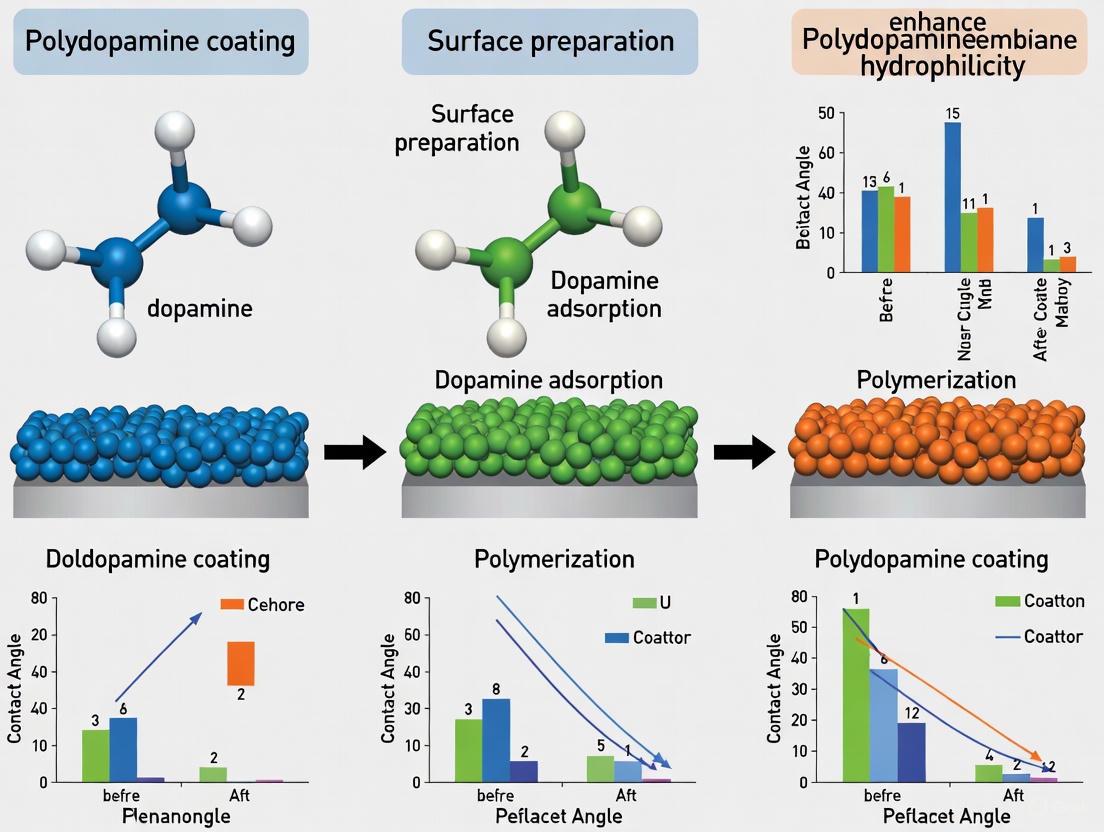
| Polyethersulfone (PES) Ultrafiltration | Two-step PDA modification (blending + coating) [20]. | Pure water flux increased from 15 to 50 L/m²·h (over 3x improvement) [20]. | [20] |
| Polyethersulfone (PES) | PDA coating as an adhesive layer for TiO₂ nanoparticles [5]. | Achieved ~82% rejection of Bovine Serum Albumin (BSA) [5]. | |
| Polypropylene Microfiltration (PPMM) | Co-deposition of PDA/PEI followed by TiO₂ embedding [21]. | Water flux surged from 605 to 5720 L m⁻² h⁻¹ (LMH) under 0.1 MPa [21]. | [21] |
| Microfiltration (MF) PES Membranes | PDA coating on membranes with controlled pore sizes [5]. | Coating on tighter pores (~300 nm) enhanced sucrose rejection without severe flux decline [5]. | |
| Polyethersulfone (PES) | Two-step PDA modification [20]. | Significant improvement in anti-fouling resistance against humic acid [20]. | [20] |
The relationship between surface chemistry and performance is further visualized below, illustrating how the fundamental properties of PDA's functional groups translate into measurable membrane enhancements.
Experimental Protocols for Membrane Hydrophilization
Standard Protocol for PDA Dip-Coating on Membrane Surfaces
This is the most prevalent method for applying a hydrophilic PDA coating to various membrane substrates [16] [17].
Research Reagent Solutions:
- Dopamine Hydrochloride: The precursor monomer for PDA formation.
- Tris-HCl Buffer (10 mM, pH 8.5): The standard alkaline buffer to trigger the oxidative self-polymerization of dopamine. Tris(hydroxymethyl)aminomethane is dissolved in purified water, and the pH is adjusted to 8.5 using HCl.
- Purified Water: Used for all solution preparation and subsequent rinsing steps.
Step-by-Step Procedure:
- Solution Preparation: Dissolve dopamine hydrochloride in the Tris-HCl buffer to a final concentration of 2 mg/mL [20]. The solution will appear clear initially but will gradually darken as polymerization begins.
- Substrate Immersion: Fully immerse the pre-cleaned and wetted membrane substrate into the freshly prepared dopamine solution. Ensure the entire surface is in contact with the solution, avoiding air bubbles.
- Reaction Incubation: Allow the reaction to proceed for a predetermined coating time, typically 4 to 24 hours, under constant, mild agitation (e.g., on a shaking platform) at room temperature [16] [20]. The coating thickness and surface roughness increase with time.
- Termination and Rinsing: After the coating period, remove the membrane from the solution and rinse it thoroughly with copious amounts of purified water. Gentle sonication in a water bath can be used to remove loosely adhered PDA particles [20].
- Drying and Storage: The modified membrane can be stored in purified water at 4°C or air-dried for further characterization and use.
Two-Step Modification for Enhanced Performance
This protocol involves blending dopamine into the membrane casting solution followed by a post-casting polymerization step, which can lead to a more uniform distribution of hydrophilic groups and improved flux [20].
Research Reagent Solutions:
- Polyethersulfone (PES): Base polymer for the membrane.
- N-methyl-2-pyrrolidone (NMP): Solvent for the dope solution.
- Dopamine Hydrochloride: Additive for blending.
- Tris-HCl Buffer (10 mM, pH 8.5): For the post-casting polymerization step.
Step-by-Step Procedure:
- Dope Solution Preparation: Prepare a standard PES dope solution in NMP (e.g., 17.5 wt%). Add dopamine hydrochloride to the dope solution and stir until completely dissolved. Dopamine concentrations of 0.5 to 4 wt% (relative to the total solution) have been effectively used [20].
- Membrane Casting: Cast the homogeneous dope solution onto a clean glass plate using a doctor blade and immediately immerse it into a purified water coagulation bath. This initiates phase separation, forming the solid membrane structure with embedded dopamine.
- Post-Casting Polymerization: Remove the nascent membrane from the water bath and immerse it in a Tris-HCl buffer solution (pH 8.5) for 5 to 36 hours [20]. This step polymerizes the embedded dopamine, forming a stable, hydrophilic PDA network within the membrane matrix.
- Post-treatment: Rinse the final membrane thoroughly with water to remove residual buffer salts and any unreacted compounds.
The experimental workflow for these two primary methods is summarized in the following diagram:
The Scientist's Toolkit: Key Reagents and Materials
Table 3: Essential Research Reagents for PDA-Based Hydrophilization
| Reagent/Material | Function/Description | Typical Usage in Protocols |
|---|---|---|
| Dopamine Hydrochloride | The essential monomer precursor that self-polymerizes to form polydopamine [16]. | Standard coating solution: 2 mg/mL in Tris buffer, pH 8.5 [20]. |
| Tris-HCl Buffer | A weak alkaline buffer (pH ~8.5) that dissolves dopamine and initiates its autoxidation and polymerization [16] [17]. | Used at 10 mM concentration as the standard solvent for dip-coating [20]. |
| Polyethersulfone (PES) | A common hydrophobic polymer used for manufacturing ultrafiltration and microfiltration membranes [5] [20]. | Serves as the primary substrate for modification; typically used at 15-20 wt% in dope solutions [20]. |
| N-methyl-2-pyrrolidone (NMP) | A polar aprotic solvent used to dissolve polymer resins like PES for membrane casting [20]. | The primary solvent in the dope solution for two-step modification protocols [20]. |
The hydrophilic character of polydopamine is not the result of a single functional group but a synergistic effect orchestrated by the catechol, amine, and quinone units. The catechol groups provide a powerful hydrogen-bonding capability, the amines increase surface energy and enable cross-linking, and the quinones enhance polarity and provide reactive sites for further customization. As demonstrated by the provided protocols and quantitative data, harnessing the chemistry of these groups allows researchers to reliably transform hydrophobic membrane surfaces into highly hydrophilic ones, leading to tangible gains in water flux and antifouling performance. This fundamental understanding is crucial for the rational design of next-generation, high-performance separation membranes.
Polydopamine (PDA), a bioinspired polymer mimicking mussel adhesion proteins, has emerged as a revolutionary surface modification tool in membrane technology. Since its introduction for surface functionalization in 2007, PDA has garnered significant scientific interest due to its exceptional universal adhesion properties, which enable robust coating formation on virtually any substrate surface [22] [23]. This adhesive capability arises from PDA's complex chemical structure rich in catechol, amine, and imine functional groups, which facilitate diverse interaction mechanisms including hydrogen bonding, metal coordination, π-π stacking, and covalent bonding [12] [22].
In the context of membrane modification, PDA coatings offer a versatile strategy for enhancing surface hydrophilicity, improving antifouling resistance, and introducing functional groups for further modification. The technology is particularly valuable for addressing the inherent hydrophobicity of common polymeric membrane materials such as polyethersulfone (PES), polyvinylidene fluoride (PVDF), polypropylene (PP), and polysulfone (PSf), which are prone to organic fouling during operation [24] [23]. This application note systematically examines PDA compatibility with these diverse membrane substrates, providing structured quantitative data, detailed experimental protocols, and practical guidance for researchers pursuing membrane hydrophilicity enhancement through PDA coating.
PDA Compatibility and Performance Across Membrane Substrates
Extensive research has demonstrated PDA's successful deposition on numerous polymeric membrane substrates, significantly altering their surface properties and separation performance. The following sections and comparative tables summarize key findings regarding PDA compatibility and its effects on different membrane materials.
Table 1: PDA Coating Compatibility and Performance on Different Membrane Substrates
| Membrane Substrate | Hydrophilicity Improvement (Contact Angle Reduction) | Key Demonstrated Benefits | Research Context & Notes |
|---|---|---|---|
| Polyethersulfone (PES) | ~20-30° reduction [24] | Enhanced hydrophilicity, improved antifouling properties, functionalization platform [21] [25] | Often modified via blending with PDA-coated nanoparticles (e.g., MoS₂@PDA) to improve compatibility and dye separation [25]. |
| Polyvinylidene Fluoride (PVDF) | ~20-30° reduction [24] | Improved antifouling properties, underwater superoleophobicity for oil/water separation [12] [24] | Successfully modified via one-step non-solvent induced phase separation (NIPS) with PDA [24]. |
| Polypropylene (PP) | Significant improvement demonstrated [23] | Excellent antifouling membranes achieved via PDA co-deposition [23] | Rapid deposition achieved using CuSO₄/H₂O₂ as an inducer (40 min) [23]. |
| Polysulfone (PSf) | High improvement potential [21] | Enhanced hydrophilicity and antifouling ability [21] | Recognized as a candidate for PDA modification, though specific contact angle data less cited than PES/PVDF. |
| Polyethylene (PE) | High improvement potential [23] | Effective surface hydrophilic modification [23] | Successfully modified via traditional dopamine deposition [23]. |
| Polytetrafluoroethylene (PTFE) | ~20-30° reduction [24] | Enhanced surface hydrophilicity [24] [23] | Notable for being modified despite its extreme hydrophobicity and chemical resistance. |
Table 2: Water Flux and Separation Performance Changes Post-PDA Modification
| Membrane Substrate | Pure Water Flux Change | Sepunction Performance | Application Context |
|---|---|---|---|
| PVDF Ultrafiltration | ~40% reduction reported after traditional PDA coating [24] | High separation efficiency (>99.9%) for oil/water mixtures [12] | Trade-off exists between hydrophilicity and permeability; blend membranes avoid pore blockage [24]. |
| PES Tight Ultrafiltration | 42.0 L m⁻² h⁻¹ bar⁻¹ (with MoS₂@PDA) [25] | Excellent dye rejection (98.17-99.88% for Janus Green B) [25] | MWCO can be finely tuned by adjusting MoS₂@PDA concentration [25]. |
| PP Microfiltration | Significant increase from 605 to 5720 LMH (with PDA/PEI & TiO₂) [21] | Enhanced performance in dynamic protein filtration [21] | Demonstrates performance enhancement possible with optimized PDA co-deposition. |
The universal adhesion of PDA stems from its multifaceted interaction mechanisms with substrate surfaces. As illustrated in the diagram below, these interactions include covalent bonding, metal coordination, hydrogen bonding, and π-π stacking, which collectively enable strong adhesion to diverse membrane materials.
Experimental Protocols for PDA Deposition on Membrane Substrates
Traditional Deposition Method
The conventional approach for PDA deposition involves simple immersion of membrane substrates in an alkaline dopamine solution under aerobic conditions [23].
Materials Required:
- Dopamine hydrochloride
- Tris(hydroxymethyl)aminomethane (Tris-base)
- Hydrochloric acid (HCl) for pH adjustment
- Deionized water
- Target membrane substrates (PES, PVDF, PP, PSf)
- Basic laboratory equipment: beakers, magnetic stirrer, pH meter
Step-by-Step Procedure:
- Solution Preparation: Prepare a 10 mM Tris-HCl buffer solution (pH ≈ 8.5) by dissolving Tris-base in deionized water and adjusting pH with HCl.
- Dopamine Solution: Dissolve dopamine hydrochloride in the Tris-Huffer at a concentration of 2 mg/mL under constant stirring.
- Membrane Pre-treatment: Cut membrane samples to desired size and pre-wet with ethanol/water if highly hydrophobic. Rinse thoroughly with deionized water.
- Deposition Process: Immerse membrane substrates completely in the dopamine solution. Ensure full surface exposure by separating stacked membranes.
- Reaction Conditions: Allow reaction to proceed for 24-48 hours at room temperature (25-35°C) with continuous gentle agitation to ensure oxygen supply.
- Post-treatment: Remove membranes from solution and rinse thoroughly with deionized water to remove loosely adhered PDA particles.
- Drying: Air-dry the modified membranes at room temperature or in a desiccator before characterization and use.
Key Considerations: This method produces homogeneous coatings but requires extended deposition times (24-48 hours). Coating thickness increases with deposition time and dopamine concentration [23].
Oxidant-Induced Rapid Deposition
To address the lengthy deposition time of traditional methods, oxidants can be incorporated to accelerate PDA polymerization.
Materials Required:
- Dopamine hydrochloride
- Copper sulfate (CuSO₄) and hydrogen peroxide (H₂O₂) as oxidation system
- Or alternative oxidants: ammonium persulfate, sodium periodate
- Deionized water
- Target membrane substrates
- Basic laboratory equipment
Step-by-Step Procedure:
- Oxidant Solution: Prepare an aqueous solution containing both dopamine hydrochloride (2 mg/mL) and CuSO₄/H₂O₂ (typically 0.01 M CuSO₄ and 0.1 M H₂O₂).
- Membrane Preparation: Cut and pre-wet membrane samples as described in Section 3.1.
- Deposition Process: Immerse membranes in the dopamine/oxidant solution at room temperature.
- Reaction Time: Allow reaction to proceed for significantly reduced time (40 minutes to 4 hours depending on desired coating thickness).
- Rinsing and Drying: Remove membranes, rinse thoroughly with deionized water, and air-dry.
Key Considerations: This method reduces deposition time from days to hours while maintaining coating quality. The added Cu²⁺ may impart additional antibacterial properties to the modified membranes [23].
One-Step Phase Inversion with PDA Integration
For PVDF and PES membranes, PDA can be incorporated directly during membrane fabrication via non-solvent induced phase separation (NIPS).
Materials Required:
- Polymer resin (PVDF or PES)
- Dopamine hydrochloride
- Solvent (typically N,N-Dimethylacetamide - DMAc)
- Sodium hypochlorite (NaClO) solution as oxidative coagulant
- Basic laboratory equipment: casting knife, glass plates, coagulation bath
Step-by-Step Procedure:
- Casting Solution: Dissolve PVDF or PES polymer (15-18 wt%) and dopamine hydrochloride (0.5-1.0 wt%) in DMAc solvent. Stir until completely dissolved.
- Membrane Casting: Cast the polymer-dopamine solution onto a non-woven fabric support using a casting knife with controlled gap thickness (200 μm).
- Phase Inversion: Immerse the cast film immediately into a coagulation bath containing dilute NaClO solution (0.5-1.0 g/L).
- PDA Formation: During phase separation, NaClO oxidizes dopamine to form PDA integrated within the membrane matrix.
- Membrane Post-treatment: Keep membranes in deionized water for 24 hours to remove residual solvent, then air-dry.
Key Considerations: This approach achieves uniform PDA distribution throughout membrane cross-section without causing pore blockage, effectively enhancing hydrophilicity while maintaining water flux [24].
The following workflow diagram illustrates the key methodological pathways for applying PDA coatings to membrane substrates, highlighting the procedural steps and decision points for each approach.
The Scientist's Toolkit: Essential Research Reagents
Table 3: Essential Reagents for PDA Membrane Modification Research
| Reagent/Chemical | Function/Application | Research Notes |
|---|---|---|
| Dopamine Hydrochloride | PDA precursor monomer | Storage: -20°C, protected from light and moisture; primary reagent for all PDA coating methods [24] [23]. |
| Tris(hydroxymethyl)aminomethane (Tris-base) | Buffer component (pH 8.5) for traditional deposition | Creates optimal alkaline environment for dopamine oxidation and polymerization [12] [23]. |
| Copper Sulfate (CuSO₄)/Hydrogen Peroxide (H₂O₂) | Oxidation system for rapid deposition | Significantly reduces coating time; Cu²⁺ may impart antibacterial properties [23]. |
| Sodium Hypochlorite (NaClO) | Oxidizing coagulant for one-step phase inversion | Enables PDA formation during membrane fabrication; concentration typically 0.5-1.0 g/L [24]. |
| Polyvinylpyrrolidone (PVP) | Pore-forming agent, co-deposition polymer | Often used in casting solutions; can be co-deposited with PDA to enhance anti-fouling properties [25] [26]. |
| Polyethyleneimine (PEI) | Co-deposition polymer for enhanced adhesion | Improves PDA adhesion and enables introduction of additional nanoparticles (e.g., TiO₂) [12] [21]. |
Polydopamine demonstrates remarkable compatibility with diverse membrane substrates including PES, PVDF, PP, and PSf, confirming its status as a universal modification platform for membrane hydrophilicity enhancement. The experimental protocols and data summarized in this application note provide researchers with practical guidance for implementing PDA coating technologies across various membrane systems. While deposition parameters require optimization for specific applications, the fundamental adhesion mechanisms remain consistent across different substrates, making PDA an exceptionally versatile tool for membrane surface engineering. Future research directions should focus on further reducing processing times, enhancing coating durability under harsh operational conditions, and exploring synergistic effects of PDA with complementary modification agents for advanced membrane functionality.
The Role of Surface Energy and Wettability in Hydrophilic Performance
Surface energy and wettability are fundamental interfacial properties that directly dictate the performance of materials in aqueous environments. In fields ranging from water treatment to biomedical implants, achieving and controlling hydrophilicity is a critical design objective. Hydrophilic surfaces, characterized by high surface energy and low water contact angles, promote water spreading, enhance permeation, and mitigate fouling. Polydopamine (PDA), a bioinspired polymer, has emerged as a versatile and effective coating for rendering diverse material surfaces hydrophilic. This application note details the quantitative effects of PDA coatings on surface wettability and provides standardized protocols for implementing and characterizing these modifications within a research context focused on membrane science.
Quantitative Data on PDA-Induced Hydrophilicity
The following tables consolidate experimental data from recent studies on PDA-mediated hydrophilic modification of various polymeric substrates.
Table 1: Water Contact Angle (WCA) Reduction on Polymer Surfaces via PDA Coating
| Substrate Material | Original WCA (°) | PDA-Modified WCA (°) | Modification Conditions | Citation |
|---|---|---|---|---|
| Poly(vinylidene fluoride) (PVDF) Membrane | 118.0 ± 1.5 | 53.0 ± 2.3 | 2 g/L DA, Tris-HCl pH 8.5 [27] | |
| Poly(vinyl chloride) (PVC) Film | ~85 (est.) | 43.2 | Co-deposition with HBPL (DA/HBPL mass ratio 1:1) [11] | |
| Poly(dimethylsiloxane) (PDMS) | ~110 (est.) | ~60 (est.) | 2 mg/mL DA, 10 mM Tris-HCl, pH 8.0, 6 hr [28] | |
| Polytetrafluoroethylene (PTFE) Membrane | ~140 (est.) | ~40 (after 200-cycle TiO₂ ALD on PDA primer) | PDA priming followed by Atomic Layer Deposition [29] |
Table 2: Impact of Post-Treatment and Coating Parameters on Hydrophilicity
| Factor Studied | Impact on Water Contact Angle (WCA) & Hydrophilicity | Citation |
|---|---|---|
| Thermal Post-Treatment (121°C for 24 hr on PDA-coated Ti) | Increased WCA vs. pristine PDA, but enhanced cell proliferation. Associated with increased surface quinone groups. [30] | |
| Dopamine Concentration (for PVDF modification) | Optimized at 1.65 g/L via RSM, achieving a WCA of 33.9°. [27] | |
| Co-deposition with Additives (Hyperbranched Polylysine - HBPL) | Significantly enhanced deposition and stability; greatest WCA reduction achieved at a DA/HBPL mass ratio of 1:1. [11] |
Experimental Protocols
Standard Protocol for PDA Coating of Polymeric Membranes
This protocol is adapted for modifying PVDF membranes for enhanced hydrophilicity in water treatment applications [27].
Research Reagent Solutions:
| Reagent/Solution | Function |
|---|---|
| Dopamine Hydrochloride | Monomer for PDA formation. |
| Tris-Hydroxymethyl Aminomethane (Tris-HCl) Buffer (10 mM, pH 8.5) | Provides alkaline environment for dopamine oxidation and polymerization. |
| Isopropyl Alcohol (IPA) | Pre-treatment solvent to remove impurities and wet membrane pores. |
| Deionized (DI) Water | Solvent and rinsing agent. |
Step-by-Step Workflow:
Membrane Pre-treatment:
- Immerse the pristine PVDF membrane in isopropyl alcohol for 2 hours to remove impurities and wet the pores.
- Subsequently, soak the membrane in DI water for 12 hours. Store the hydrated membrane until use.
Dopamine Solution Preparation:
- Prepare a 10 mM Tris-HCl buffer solution at pH 8.5.
- Dissolve dopamine hydrochloride in the Tris-HCl buffer to a concentration of 1.65 g/L (optimized value). Filter the solution if necessary.
PDA Deposition Coating:
- Immerse the pre-treated and wetted PVDF membrane in the freshly prepared dopamine solution.
- Place the container on an orbital shaker and agitate at 120 rpm for 4.5 hours at room temperature (25°C). The solution will gradually darken, turning from clear to brown or black.
Post-Coating Processing:
- Remove the membrane from the dopamine solution and rinse thoroughly with DI water to remove loosely adhered PDA particles.
- Dry the modified membrane in a vacuum drying oven.
- The resulting product is the PVDF/PDA membrane, ready for characterization and use.
Protocol for Co-deposition of PDA with Hyperbranched Polylysine (HBPL)
This protocol enhances the stability and hydrophilicity of PVC surfaces [11].
Workflow Diagram: Co-deposition of PDA and HBPL
Key Steps:
- Synthesize HBPL via melt phase polycondensation of L-lysine hydrochloride [11].
- Prepare a co-deposition solution containing both dopamine hydrochloride and HBPL at a mass ratio of approximately 1:1 in a Tris-HCl buffer (pH 8.5).
- Immerse the PVC substrate in the mixed solution to allow simultaneous deposition and cross-linking.
- Proceed with rinsing and drying as in the standard protocol. The resulting PVC-pDA/HBPL film exhibits superior hydrophilicity and coating stability compared to PDA alone.
Protocol for Contact Angle Measurement: Sessile Drop vs. Captive Bubble
Accurate measurement of wettability is critical. The captive bubble method is recommended for reliable results on hydrophilic membranes [31].
Diagram: Contact Angle Measurement Methods
Procedure for Captive Bubble Method [31]:
- Sample Preparation: Mount the membrane sample with the active layer facing down in a custom-made cell filled with ultrapure water.
- Bubble Formation: Use a microsyringe to introduce a small air bubble (typically 1-2 µL) onto the membrane surface from below.
- Image Capture & Analysis: Capture an image of the captive bubble immediately after formation. Measure the contact angle formed between the membrane surface and the bubble's tangent using image analysis software. Report the average of multiple measurements.
Mechanisms of Hydrophilicity Enhancement
PDA enhances surface hydrophilicity through a combination of chemical and physical mechanisms. The following diagram illustrates the intermolecular interactions and subsequent nucleation behavior that underpin this performance enhancement.
Diagram: Mechanisms of PDA-Induced Hydrophilicity and Mineralization
- Chemical Composition: The abundant presence of catechol, amine, and imine functional groups on the polymerized PDA surface acts as sites for strong hydrogen bonding with water molecules, significantly increasing the surface's affinity for water [11] [7].
- Physical Microstructure: PDA deposition often increases surface roughness, which can further amplify hydrophilicity by increasing the effective surface area available for water contact, as characterized by techniques like Atomic Force Microscopy (AFM) [11] [27].
- Impact on Mineralization: The charged groups on PDA (e.g., catechol) strongly interact with calcium ions (Ca²⁺) in solutions like simulated body fluid (SBF). This interaction changes the nucleation mode of calcium phosphate from a pure "islanding" (Volmer-Weber) mode on uncoated surfaces to a mixed "islanding" and planar (Stranski-Krastanov) mode on PDA-coated surfaces, leading to a more bonded and uniform mineral layer [32].
The Scientist's Toolkit
Table 3: Essential Research Reagent Solutions for PDA Coating
| Reagent / Material | Function / Role in Hydrophilic Modification |
|---|---|
| Dopamine Hydrochloride | The essential precursor monomer that undergoes oxidative self-polymerization to form the adherent polydopamine (PDA) coating. [11] [27] [28] |
| Tris-HCl Buffer (pH 8.5) | The standard alkaline buffer system that initiates and controls the oxidation and polymerization of dopamine. [27] [28] |
| Hyperbranched Polylysine (HBPL) | A polymeric additive that, when co-deposited with DA, enhances the deposition mass, stability, and hydrophilicity of the resulting coating via covalent and non-covalent interactions. [11] |
| Polyvinyl Alcohol (PVA) | A hydrophilic polymer often used in composite transition layers (e.g., with PDA and GO) to improve the adhesion and mechanical properties of subsequent coatings like hydroxyapatite. [33] |
| Graphene Oxide (GO) | A nanomaterial used in composite coatings; its oxygen-containing functional groups contribute to hydrophilicity and can be reinforced by PDA adhesion. [33] |
Practical Implementation: Coating Methods and Biomedical Applications of Polydopamine-Modified Membranes
In the broader context of research on polydopamine (PDA) coatings for membrane hydrophilicity enhancement, the dip-coating process stands out for its simplicity and effectiveness. This protocol details the optimization of two critical parameters—dopamine concentration and immersion time—to consistently produce high-performance PDA coatings on membrane substrates. PDA, a bioinspired polymer, can form uniform, adhesive coatings on virtually any substrate surface via a simple oxidative self-polymerization process in a weak alkaline aqueous environment [23] [34]. The primary goal of this hydrophilic modification is to mitigate membrane fouling by creating a highly hydrated layer that prevents the adhesion of organic contaminants, such as proteins, thereby improving flux and reducing cleaning frequency in applications like water treatment and biomedical separation [35] [23]. This document provides a standardized, optimized methodology for researchers and development professionals to achieve reproducible and effective PDA surface modifications.
Fundamental Principles of Polydopamine Coating
Polydopamine coating formation is inspired by the adhesive proteins found in mussels [34]. The process involves the oxidative self-polymerization of dopamine in a weak alkaline environment (typically pH = 8.5), leading to the formation of a thin, dark brown PDA film on the submerged substrate [23] [34]. The polymerization can proceed via two main pathways: covalent oxidative polymerization and physical self-assembly [36]. The former involves the oxidation of dopamine to dopaminequinone, followed by cyclization and polymerization to form 5,6-dihydroxyindole (DHI) units. The latter involves non-covalent interactions, such as hydrogen bonding and π-π stacking, among dopamine and its oxidized derivatives to form supramolecular assemblies [36]. The exceptional adhesive properties of PDA stem from the diverse interactions it can form with substrate surfaces, including hydrogen bonding, electrostatic interactions, π-π stacking, coordinative bonding, and covalent bonds [37]. Furthermore, the abundant catechol and amine functional groups on PDA provide active sites for further surface functionalization, allowing for the tailoring of membrane surface properties [36] [12].
Optimizing Coating Parameters: Key Data
The quality, thickness, and performance of the resulting PDA coating are highly dependent on the deposition conditions. The following tables summarize the effects and optimal ranges for key parameters, specifically dopamine hydrochloride (DA·HCl) concentration and immersion time, based on current literature.
Table 1: Effect of Dopamine Hydrochloride Concentration on Coating Properties
| DA·HCl Concentration (mg/mL) | Coating Characteristics | Impact on Membrane Hydrophilicity & Performance |
|---|---|---|
| 0.1 - 0.5 [12] | Thin, potentially uneven coating. | Moderate improvement in hydrophilicity; may be insufficient for long-term fouling resistance. |
| 1.0 - 2.0 [37] [35] | Standard Range: Robust and uniform films. Optimal for initiating hierarchical structures when combined with other agents (e.g., PAMAM) [37]. | Significant contact angle reduction; greatly enhanced protein fouling resistance and water flux [35]. |
| > 2.0 | Risk of excessive particle aggregation and pore blockage [35]. | Potential decline in pure water flux due to pore narrowing or sealing [35]. |
Table 2: Effect of Immersion Time on Coating Properties
| Immersion Time (Hours) | Coating Characteristics | Recommendation and Notes |
|---|---|---|
| 0.5 - 2 | Rapid initial growth; thin nanoscale film. | For a quick, base-layer coating. Film growth is most efficient in the first 2 hours [34]. |
| 4 - 6 | Increased thickness and surface coverage. | A balance between process time and coating robustness. |
| 12 - 24 [37] [35] [12] | Commonly Used Range: Ensures a thick, uniform, and stable coating. | Necessary for achieving superhydrophobic properties after post-modification [12]. Co-deposition with polymers like PEI can reduce required time [37]. |
| > 24 | Diminishing returns; possible instability from over-saturation [23] [34]. | Not typically recommended due to inefficiency and potential for heterogeneous coatings. |
Detailed Experimental Protocol
Reagent Setup and Solution Preparation
- Tris-HCl Buffer (10 mM, pH 8.5): Dissolve 1.57 g of tris(hydroxymethyl)aminomethane (TRIS, ≥99.8%) in 1 L of distilled water. Measure the pH with a calibrated pH meter. Adjust the pH to 8.5 ± 0.1 by adding drops of concentrated hydrochloric acid (HCl, 32%) under gentle stirring [34].
- Dopamine Hydrochloride Stock Solution (2 mg/mL): Weigh 200 mg of dopamine hydrochloride (DA·HCl, ≥98%) powder. Immediately before the coating process, dissolve the powder in 100 mL of the freshly prepared Tris-HCl buffer. Agitate gently until fully dissolved. Note: The solution will begin clear and gradually transition to light brown, indicating the start of polymerization.
Optimized Dip-Coating Procedure
- Substrate Pre-treatment: Cut the membrane substrate to the desired size. Clean the substrate by sonication in isopropyl alcohol (IPA) or ethanol for 10 minutes, followed by rinsing with copious amounts of distilled water to remove any organic residues [34]. Allow the substrate to air dry completely.
- Coating Initiation: Immerse the pre-treated substrate completely in the freshly prepared dopamine solution (2 mg/mL in Tris-Huffer, pH 8.5). Ensure the substrate is fully submerged and not trapped with air bubbles.
- Polymerization Reaction: Allow the reaction to proceed for a duration of 12-24 hours at room temperature with mild agitation (e.g., on a laboratory shaker or rocker). Agitation ensures uniform oxygen supply and prevents the settling of PDA aggregates on the membrane surface.
- Post-Coating Processing:
- Carefully remove the coated substrate from the solution using tweezers.
- Rinse the substrate thoroughly with distilled water to remove any loosely adhered PDA particles.
- Sonicate the coated membrane in distilled water for 10 minutes to further enhance coating uniformity and remove aggregates [34].
- Dry the modified membrane overnight at room temperature or in a vacuum oven at low temperature (e.g., 40°C).
Advanced Technique: O₂ Backflow for Ultrafiltration Membranes
For ultrafiltration (UF) membranes where pore penetration and narrowing are significant concerns, the following modified protocol is recommended to achieve a surface-selective coating [35]:
- Setup: Place the membrane in a specialized filtration cell. Introduce the aqueous dopamine solution from the active (selective) side of the membrane.
- Gas Supply: Simultaneously, supply pure oxygen (O₂) gas from the porous backside of the membrane. This creates a physical barrier that limits the penetration of dopamine monomers into the pore structure.
- Reaction: The O₂ backflow enhances the oxidation kinetics of dopamine at the membrane surface, promoting faster and more localized polymerization. This results in a PDA coating that primarily modifies the surface without severely compromising the membrane flux through pore blockage [35].
The Scientist's Toolkit: Essential Reagents and Materials
Table 3: Key Research Reagent Solutions
| Reagent / Material | Function in Protocol | Notes for Researchers |
|---|---|---|
| Dopamine Hydrochloride (DA·HCl) | The fundamental monomer for PDA film formation. | Critical: Hygroscopic and air-sensitive. Store in a desiccator at -20°C. Use immediately after weighing. |
| Tris(hydroxymethyl)aminomethane (TRIS) | Provides a weak alkaline buffer (pH 8.5) necessary for dopamine oxidation and self-polymerization [34]. | Ensure high purity (≥99.8%). Solution pH is critical and must be verified before adding dopamine. |
| Hydrochloric Acid (HCl, 32%) | Used for fine adjustment of the Tris buffer to the target pH of 8.5. | Handle with care using appropriate personal protective equipment (PPE) in a fume hood. |
| Polyamidoamine (PAMAM) Dendrimer | An additive for co-deposition to create thicker, more stable PDA coatings with increased surface roughness [37]. | A DA:PAMAM weight ratio of 1:1 has been shown to produce high-quality coatings [37]. |
| Oxygen (O₂) Gas Cylinder | Used in the advanced O₂ backflow technique to accelerate polymerization and prevent pore penetration in UF membranes [35]. | Provides a cleaner and faster alternative to chemical oxidants like CuSO₄/H₂O₂. |
Characterization and Performance Validation
To confirm the success of the dip-coating process, the following characterizations are recommended:
- Surface Hydrophilicity: Measure the static water contact angle. A successful coating will show a significant decrease (increase in hydrophilicity) compared to the unmodified membrane [35].
- Chemical Composition: Use X-ray Photoelectron Spectroscopy (XPS) to detect the presence of nitrogen (N), which is a key element in PDA and confirms surface deposition [35].
- Coating Morphology and Thickness: Analyze surface morphology using Atomic Force Microscopy (AFM) or Scanning Electron Microscopy (SEM). Determine coating thickness via ellipsometry or profilometry [34].
- Fouling Resistance Test: Conduct dynamic protein filtration tests using a model foulant like Bovine Serum Albumin (BSA). A successful PDA coating will demonstrate higher flux recovery ratios and lower irreversible fouling compared to the pristine membrane [35].
Achieving sustainable membrane hydrophilicity is a critical challenge in the design of advanced materials for biomedical and separation applications. Polydopamine (PDA), a bioinspired polymer mimicking mussel adhesion proteins, has emerged as a versatile tool for surface modification due to its strong adhesion properties and ability to enhance surface wettability [38]. Two principal methodological approaches—blending and surface polymerization—enable the integration of PDA and its composites onto various polymer substrates. The selection between these approaches significantly influences the structural integrity, functional performance, and operational stability of the modified membranes. This application note provides a detailed comparative analysis of these two strategies, supported by quantitative data and standardized experimental protocols, to guide researchers in selecting and implementing the optimal modification technique for specific research and development objectives.
Comparative Analysis of Modification Approaches
The fundamental distinction between blending and surface polymerization lies in the stage at which the modifier is introduced during membrane fabrication and modification.
Surface Polymerization is a post-fabrication technique where a pre-formed membrane is immersed in an aqueous dopamine solution. Under weak alkaline conditions (typically Tris-HCl buffer, pH = 8.5), dopamine undergoes oxidative self-polymerization, depositing a thin PDA coating onto the membrane surface and within its pores [39] [27] [38]. This method is renowned for its simplicity and substrate-independent adhesion, making it applicable to a wide range of materials including polyvinylidene fluoride (PVDF), polytetrafluoroethylene (PTFE), and polyvinyl chloride (PVC) [27] [11] [38]. A key advantage is the introduction of a rich surface chemistry that allows for further secondary reactions and functionalization [11] [38].
Blending involves the direct incorporation of PDA particles, its monomers, or co-deposition components into the polymer dope solution prior to membrane formation (e.g., via phase inversion) [11]. This method embeds the modifier within the membrane matrix, potentially leading to more uniform distribution and enhanced structural stability. Co-deposition blending, where dopamine is mixed with other polymers like hyperbranched polylysine (HBPL) in the coating solution, has been shown to significantly improve the deposition rate, stability, and final performance of the coating [11].
Table 1: Comparison of Blending and Surface Polymerization Approaches for Polydopamine Modification
| Feature | Surface Polymerization | Blending |
|---|---|---|
| Process Definition | Post-fabrication coating via immersion in dopamine solution [27] [38] | Pre-fabrication incorporation of modifier into polymer dope solution [11] |
| Key Mechanism | Oxidative self-polymerization and deposition on substrate surface [38] | Entrapment within the membrane matrix during formation |
| Typical Coating Thickness | Tunable nanoscale layers (e.g., ~18 nm in 15 min with microwave induction [38]) | Dependent on initial concentration in dope solution |
| Hydrophilicity Enhancement | Significant; contact angle reduction from >100° to ~53°–43° [27] [11] | Enhanced, with improved stability |
| Adhesion Stability | Good, though can be unstable in strong alkalis [11]; enhanced with co-deposition or oxidants [38] | Excellent, due to mechanical anchoring within the matrix |
| Experimental Scalability | Highly scalable for various shapes and sizes; simple equipment [38] | Scalable, tied to membrane fabrication process |
| Suitability for Secondary Reactions | Excellent; surface-rich catechol/amine groups allow for further grafting [11] [38] | Limited, as functional groups are less accessible |
Quantitative Performance Data
The efficacy of PDA modification is quantitatively assessed through metrics such as hydrophilicity (water contact angle), permeability (water flux), and antibacterial performance. The methodology, particularly the use of co-deposition in surface polymerization, directly impacts these outcomes.
Table 2: Quantitative Performance Metrics of Select PDA-Modified Membranes
| Substrate | Modification Approach & Details | Water Contact Angle (°) | Pure Water Flux | Key Performance Outcomes |
|---|---|---|---|---|
| PVDF [27] | Surface Polymerization (DA: 1.65 g/L, 4.5 h) | 69° → 33.9° | Higher than pristine membrane | Excellent antifouling ability; Enhanced biodiversity in MBR |
| PVC [11] | Surface Polymerization with Co-deposition (DA/HBPL mass ratio 1:1) | Reduced to 43.2° | Not Specified | Superior stability in strong acid/alkali vs. PDA alone |
| PTFE [39] | Surface Polymerization with ZnO-NPs (PDA as adhesive layer) | Not Specified | Superior fluid permeability | Robust antibacterial efficacy against E. faecalis and S. mutans |
| PLGA Nanoparticles [40] | Surface Polymerization (Prime-coating with PDA) | Not Applicable | Not Applicable | Functionalized with ligands; No cytotoxicity; Expected cellular interactions |
Detailed Experimental Protocols
Protocol 1: Surface Polymerization via Dip-Coating
This is the standard method for depositing a PDA coating on a pre-formed membrane [27] [38].
Materials:
- Dopamine hydrochloride
- Tris(hydroxymethyl)aminomethane (Tris)
- Hydrochloric acid (HCl)
- Deionized (DI) water
- Substrate membrane (e.g., PVDF, PTFE, PVC)
Procedure:
- Solution Preparation: Prepare a 10 mM Tris-HCl buffer solution (pH = 8.5) by dissolving Tris in DI water and adjusting the pH with HCl.
- Dopamine Solution: Dissolve dopamine hydrochloride in the Tris-HCl buffer to a concentration of 1.65 - 2.0 g/L [27]. The solution will appear clear initially.
- Substrate Pre-treatment: Immerse the substrate membrane in isopropyl alcohol for 2 hours to remove impurities, then soak in DI water for 12 hours [27].
- Polymerization Reaction: Immerse the pre-treated membrane in the dopamine solution. Keep the reaction vessel open to air and agitate on a shaker (e.g., 120 rpm) at room temperature for a specified duration (e.g., 4.5 hours [27]).
- Membrane Retrieval and Washing: After the reaction, remove the membrane from the solution and rinse thoroughly with DI water to remove any loosely adhered particles.
- Drying: Dry the modified membrane in a vacuum drying oven at room temperature or a specified low temperature (e.g., 25°C [27]) until constant weight is achieved.
Protocol 2: Co-deposition Blending for Enhanced Coating
This protocol details the simultaneous deposition of PDA with another polymer, such as hyperbranched polylysine (HBPL), to create a composite coating with enhanced properties [11].
Materials:
- Dopamine hydrochloride
- Hyperbranched polylysine (HBPL)
- Tris-HCl buffer (10 mM, pH = 8.5)
- DI water
- Substrate membrane (e.g., PVC)
Procedure:
- Coating Solution Preparation: Dissolve dopamine hydrochloride and HBPL in Tris-HCl buffer at a mass ratio of approximately 1:1 (DA/HBPL) [11].
- Substrate Immersion: Immerse the pre-cleaned substrate membrane into the co-deposition solution.
- Reaction and Deposition: Allow the reaction to proceed under agitation at room temperature for the desired coating time. The presence of HBPL accelerates deposition and increases the coating mass.
- Washing and Drying: Retrieve the membrane, rinse with DI water, and dry under vacuum.
Protocol 3: Characterization of Hydrophilicity and Permeability
Water Contact Angle Measurement: [27] [11]
- Use a contact angle goniometer for measurement.
- Cut the membrane sample into strips (approx. 5 cm x 1 cm).
- Place a deionized water droplet (~2 µL) on the membrane surface.
- Measure the static contact angle at at least five different locations on the sample surface and calculate the average value.
Pure Water Flux Measurement: [27]
- Assemble a membrane module with a known effective filtration area (e.g., 60.5 cm²).
- Pre-pressurize the membrane with deionized water at 0.10 MPa for 30 minutes to compact and wet it thoroughly.
- After the flux stabilizes, measure the volume of water permeated over a specific time under a constant applied pressure.
- Calculate the flux (J) using the formula: ( J = V / (A \times t) ), where ( V ) is the permeate volume (mL), ( A ) is the membrane area (cm²), and ( t ) is the collection time (hours).
Workflow and Performance Relationship Visualization
The following diagrams illustrate the procedural workflow for the two modification approaches and the logical relationship between process parameters and final membrane performance.
Diagram 1: Workflows for two modification approaches.
Diagram 2: Parameter effects on coating properties and performance.
The Scientist's Toolkit: Essential Research Reagents
Table 3: Key Reagents for Polydopamine-Based Membrane Modification
| Reagent/Material | Function/Application | Key Characteristics & Notes |
|---|---|---|
| Dopamine Hydrochloride [39] [27] [11] | Precursor for polydopamine coating. | Undergoes self-polymerization in weak alkaline conditions (pH ~8.5) to form adherent coatings. |
| Tris-HCl Buffer [39] [27] | Provides a stable alkaline environment (pH 8.5) for dopamine polymerization. | Standard concentration is 10 mM. |
| Hyperbranched Polylysine (HBPL) [11] | Co-deposition agent to enhance coating mass, stability, and hydrophilicity. | Used at ~1:1 mass ratio with dopamine; provides abundant amine groups. |
| Zinc Oxide Nanoparticles (ZnO-NPs) [39] | Functional nanomaterial for imparting antibacterial properties. | Adhered to membrane surface using PDA as an intermediate adhesive layer. |
| Polyvinylidene Fluoride (PVDF) Membrane [27] | Common hydrophobic substrate for modification. | Requires pre-treatment with alcohol to wet pores before PDA coating. |
| Ammonium Persulfate / Sodium Periodate [38] | Chemical oxidants to induce rapid dopamine polymerization. | Significantly reduces deposition time compared to air oxidation. |
Surface modification of polymeric membranes is a critical step in enhancing their performance for various separation and biomedical applications. Among the many coating materials, polydopamine (PDA) has emerged as a premier bio-inspired polymer for membrane surface engineering. Derived from the oxidative self-polymerization of dopamine, PDA exhibits exceptional adhesion properties across diverse substrates, high biocompatibility, and the ability to significantly improve membrane hydrophilicity and antifouling resistance [23]. Traditional immersion polymerization, while widely used, presents limitations including lengthy processing times, inconsistent coating morphology, and challenges in controlling deposition uniformity [41] [23].
To overcome these constraints, advanced deposition techniques such as spray coating and electropolymerization have been developed. These methods offer superior control over the PDA layer's thickness, uniformity, and structural properties, enabling more precise and efficient membrane modification [42] [41]. This article details the application notes and experimental protocols for these advanced deposition methods, providing researchers and development professionals with practical guidance for implementing these techniques in membrane hydrophilicity enhancement.
Table 1: Comparison of Polydopamine Deposition Methods for Membrane Modification
| Feature | Immersion Polymerization | Spray Coating | Electropolymerization |
|---|---|---|---|
| Process Principle | Spontaneous oxidative polymerization in solution [23] | Atomization and deposition of dopamine solution via nozzle [42] | Electrochemical oxidation of dopamine on a conductive substrate [41] [43] |
| Typical Coating Time | Several hours to days [41] [23] | Minutes to hours [42] | Minutes to hours (highly controllable) [41] |
| Coating Uniformity | Prone to PDA aggregation and rough surfaces [41] | High uniformity achievable with parameter optimization [42] | Can produce smooth, uniform films [41] |
| Key Advantages | Simple setup, universal adhesion [23] | Scalable, reduced material waste, suitable for patterned membranes [42] | Precise thickness control, rapid deposition, no chemical oxidants [41] [43] |
| Technical Challenges | Long duration, PDA aggregation, stability issues [41] [23] | Requires optimization of spray parameters [42] | Limited to conductive substrates [43] |
| Impact on Hydrophilicity | Significant improvement [23] | Enhanced hydrophilicity with improved flux [42] [41] | Enhances hydrophilicity [41] |
Spray Coating of Polydopamine
Principle and Application Advantages
Spray coating is a membrane fabrication and modification technique that involves atomizing a dopamine-containing solution and depositing it onto a substrate surface. This method stands out for its scalability and processing efficiency, using up to 50% less precursor material than traditional dip-coating without compromising performance [42]. The technique provides exceptional control over membrane thickness and morphology, which is crucial for producing uniform, defect-free, and ultrathin PDA layers [42]. Furthermore, spray coating can be adapted for patterning membrane surfaces, a feature shown to improve permeance by up to 50% by creating patterned selective layers without reducing pore size or porosity [42].
Detailed Experimental Protocol
Research Reagent Solutions
- Dopamine Hydrochloride Solution: Typically 2 mg/mL in Tris-HCl buffer (10 mM, pH 8.5). Functions as the PDA precursor [41].
- Tris-HCl Buffer (10 mM, pH 8.5): Provides the mildly alkaline environment necessary for dopamine oxidation and polymerization [41] [23].
- Polyethersulfone (PES) or Polyvinylidene Fluoride (PVDF) Membranes: Commonly used hydrophobic substrates for hydrophilic modification [41] [23].
Equipment Setup
- Spray Coating System: Consisting of an airbrush or ultrasonic spray nozzle, a solution reservoir, and a compressed air or nitrogen source.
- Motion Control System (Optional): A programmable stage to ensure consistent nozzle movement and coating uniformity.
- Fume Hood: To ensure safe handling of aerosols.
Step-by-Step Procedure
- Substrate Preparation: Clean the membrane substrate (e.g., PES UF membrane) with ethanol and deionized water to remove surface contaminants. Dry at room temperature.
- Dopamine Solution Preparation: Dissolve dopamine hydrochloride in Tris-HCl buffer to a concentration of 2 mg/mL. The solution should be used immediately after preparation to prevent premature polymerization.
- Spray Parameter Setup: Configure the spray system with the following typical parameters [42]:
- Nozzle-to-substrate distance: 15–25 cm
- Spray air pressure: 0.2–0.4 MPa
- Solution flow rate: 0.5–2.0 mL/min
- Coating Process: Fill the spray system reservoir with the dopamine solution. Initiate spraying, ensuring even coverage of the substrate surface. Multiple passes may be required to achieve the desired coating.
- Polymerization and Drying: After spraying, retain the coated membrane in a humid environment at room temperature for 30–60 minutes to allow for complete polymerization of the deposited dopamine. Rinse the resulting PDA-coated membrane thoroughly with deionized water to remove any unreacted monomers or loosely adhered particles. Air-dry before characterization [41].
Technical Considerations and Optimization
Achieving a consistent and uniform coating requires tight control of parameters such as spray angle, nozzle speed, and nozzle-to-substrate distance [42]. The surface morphology and performance of the sprayed PDA membrane are highly dependent on these parameters. Research indicates that electrosprayed PDA (ePDA) can produce a relatively smooth and uniform structure with enhanced chemical stability compared to immersion-coated PDA [41]. Furthermore, membranes fabricated via ePDA demonstrated higher water flux and improved rejection of organic dyes, highlighting the functional superiority of this advanced spraying technique [41].
Electropolymerization of Polydopamine
Principle and Application Advantages
Electropolymerization is a highly controlled method for depositing PDA films on conductive substrates through the application of an electrical potential. This technique initiates dopamine polymerization via electrochemical oxidation at the working electrode, bypassing the need for dissolved oxygen or chemical oxidants [43]. The process begins with the removal of electrons from the catechol groups in dopamine, forming dopamine-quinone, which subsequently undergoes intramolecular cyclization and cross-linking to form an insoluble PDA coating [43]. The primary advantage of this method is the precise control over coating thickness and morphology at the nanometer scale by adjusting electrochemical parameters such as applied potential, current density, and deposition time [41] [43].
Detailed Experimental Protocol
Research Reagent Solutions
- Dopamine Hydrochloride Solution: 0.5–2 mg/mL in a suitable electrolyte buffer (e.g., 10 mM PBS, pH 7.4). Serves as the monomeric building block.
- Phosphate Buffered Saline (PBS, 10 mM, pH 7.4): Functions as the electrolyte to support the electrochemical reaction.
- Conductive Substrate: Typically an indium tin oxide (ITO) electrode or a conductive membrane material.
Equipment Setup
- Electrochemical Workstation: Equipped with a standard three-electrode system.
- Working Electrode: The conductive substrate to be coated (e.g., ITO glass).
- Counter Electrode: A platinum wire or mesh.
- Reference Electrode: An Ag/AgCl or saturated calomel electrode (SCE).
Step-by-Step Procedure
- Substrate Preparation: Clean the conductive substrate (e.g., ITO glass) sequentially with acetone, ethanol, and deionized water in an ultrasonic bath for 15 minutes each. Dry under a stream of nitrogen gas.
- Electrolyte Preparation: Dissolve dopamine hydrochloride in the PBS buffer to achieve the desired concentration (e.g., 1 mg/mL). Degas the solution by bubbling with nitrogen for 10–15 minutes to remove dissolved oxygen, which can cause non-specific polymerization in the solution.
- Electrochemical Cell Assembly: Assemble the three-electrode system in the electrochemical cell containing the dopamine solution.
- Deposition Parameter Setup: Program the electrochemical workstation with the deposition parameters. A common method is chronoamperometry, which involves:
- Applying a constant potential of +0.4 V to +0.8 V (vs. Ag/AgCl) for a duration of 5–20 minutes [41].
- Polymerization Process: Initiate the electropolymerization process. A gradual increase in current is often observed, corresponding to the growth of the conductive PDA film on the working electrode.
- Post-Treatment: After deposition, carefully remove the coated substrate from the cell. Rinse it gently with copious amounts of deionized water to remove electrolyte residues and any non-adherent oligomers. Air-dry the ePDA-coated substrate in a clean environment.
Technical Considerations and Optimization
The properties of the ePDA film are profoundly influenced by the applied potential and deposition time. Higher potentials and longer times generally lead to thicker coatings. However, excessively high potentials can lead to over-oxidation and the formation of less adherent or more porous films. The electrolyte's pH and concentration also affect the polymerization kinetics and the resulting film's quality. Compared to traditional immersion polymerization, ePDA has been shown to produce coatings with enhanced chemical stability, demonstrating better resistance to alkaline conditions, organic solvents like DMF and DMSO, and reduced passive leakage when used in capsule-based drug delivery systems [41] [44].
Characterization and Performance Evaluation
Rigorous characterization is essential to validate the success of the deposition process and the enhanced functionality of the modified membranes.
Table 2: Key Performance Metrics of Spray-Coated and Electropolymerized PDA Membranes
| Characterization Method | Spray-Coated PDA Membrane | Electropolymerized PDA (ePDA) Membrane |
|---|---|---|
| Coating Thickness | Controllable via spray passes and solution concentration [42] | Highly controllable, nanometer-scale precision (e.g., 37 nm in 8 h) [41] |
| Surface Roughness | Can achieve relatively smooth and uniform structures [41] | Can produce smooth, uniform films [41] |
| Water Contact Angle | Significant reduction, indicating enhanced hydrophilicity [41] | Significant reduction, indicating enhanced hydrophilicity [41] |
| Water Flux | Higher flux reported for ePDA vs. iPDA [41] | Improved flux and rejection of dyes like Congo Red [41] |
| Chemical Stability | Enhanced stability in NaOH, DMF, and DMSO compared to iPDA [41] | Improved stability against acids, bases, and polar solvents [41] |
| Application Performance | Superior desalination performance, high permeance in patterned membranes [42] | Reduced passive leakage in drug capsules, potential for biosensing [41] [44] |
The Scientist's Toolkit: Essential Research Reagents
Table 3: Key Reagents and Materials for Advanced PDA Deposition
| Reagent/Material | Function/Application Note |
|---|---|
| Dopamine Hydrochloride | The essential monomer precursor for PDA formation. Must be stored dry and in the dark. Solutions should be prepared fresh and used immediately [41] [23]. |
| Tris-HCl Buffer (pH 8.5) | The standard alkaline buffer for traditional and spray-coating deposition. Provides the pH for oxidative polymerization initiated by atmospheric oxygen [41] [23]. |
| Phosphate Buffered Saline (PBS) | A common electrolyte for the electropolymerization process, providing the necessary ionic strength and a near-neutral pH [41] [43]. |
| Polyethersulfone (PES) Membrane | A widely used hydrophobic ultrafiltration membrane substrate, serving as a standard for modification studies [41]. |
| Indium Tin Oxide (ITO) Glass | A transparent conductive substrate, often used as the working electrode for the electropolymerization of PDA [41]. |
| Polycaprolactone (PCL) | A biodegradable polymer used to form the shell of double emulsion capsules, which can be effectively coated with PDA to reduce passive drug leakage [44]. |
Workflow and Decision Pathway
The following diagram illustrates the experimental workflow and logical decision process for selecting and implementing the appropriate advanced deposition method for a given research goal.
Advanced PDA Deposition Workflow
This workflow guides the selection of an appropriate advanced deposition method based on substrate properties and research objectives, highlighting the specific advantages of each technique for membrane modification.
Spray coating and electropolymerization represent significant advancements in the application of polydopamine for membrane surface engineering. These methods address critical limitations of traditional immersion techniques, offering researchers unparalleled control over the coating process and the final properties of the modified membranes. Spray coating stands out for its scalability and efficiency, making it suitable for industrial-scale applications, particularly where enhanced hydrophilicity and reduced fouling are desired [42] [41]. Electropolymerization, while requiring conductive substrates, provides exceptional precision for developing specialized coatings for advanced applications in drug delivery and sensing [41] [44]. By adopting these protocols, researchers and development professionals can leverage these advanced techniques to push the boundaries of membrane performance, contributing to more effective and reliable separation technologies and biomedical devices.
Creating Janus Membranes with Asymmetric Wettability for Specialized Applications
Application Notes
Janus membranes, characterized by their asymmetric wettability—typically a hydrophobic side and a hydrophilic side—have emerged as advanced functional materials for complex separation processes. Within the broader scope of a thesis on polydopamine (PDA) coating for membrane hydrophilicity enhancement, these membranes present a compelling application of bio-inspired surface chemistry to solve real-world challenges in water treatment and desalination.
Fundamental Principles and Applications
The core principle of a Janus membrane is its asymmetric surface chemistry, which creates a directional liquid transport phenomenon. This is typically achieved by constructing a hydrophobic layer and a hydrophilic layer on opposite sides of a substrate [45]. This architecture enables specialized functionalities that single-wettability membranes cannot achieve.
In membrane distillation (MD) for desalination, a Janus configuration with a thin hydrophilic layer on a hydrophobic substrate creates a hydration layer that repels oily substances and other foulants, thereby conferring anti-fouling properties [46]. Simultaneously, the underlying hydrophobic layer prevents the permeation of the liquid feed, allowing only water vapor to pass through [47]. This makes them particularly promising for treating challenging wastewaters like shale gas produced water (SGPW), which contains oils, surfactants, and other organic chemicals [46]. Beyond MD, Janus membranes are also engineered for the on-demand separation of oil-water mixtures and emulsions, with the asymmetric wettability allowing selective transport of either water or oil based on which side of the membrane is exposed to the feed [48] [49].
Performance Characteristics in Key Applications
The performance of Janus membranes is quantified by their separation efficiency, permeate flux, and long-term stability. The following tables summarize key performance metrics reported in recent studies for membrane distillation and oil-water separation applications.
Table 1: Performance of Janus Membranes in Membrane Distillation Desalination
| Membrane Type / Configuration | Feed Solution | Flux (kg m⁻² h⁻¹) | Salt Rejection (%) | Test Duration & Stability | Source |
|---|---|---|---|---|---|
| Multilayer PVDF/PEI with CuO | Highly saline water | 37.16 | 99.99 | 24 h stable operation | [50] |
| PSf-PDA (Pore-filling method) | 3.5 wt% NaCl | ~19.5* | ~99.99* | 72 h fouling resistance test | [47] |
| CTS/PFO-SiNPs on Omniphobic substrate | Real SGPW | Enhanced performance reported | High rejection maintained | Excellent anti-fouling & anti-wetting | [46] |
*Values estimated from graphical data in the source.
Table 2: Performance of Janus Membranes in Oil-Water Separation
| Membrane Type / Configuration | Separation Target | Separation Efficiency (%) | Flux (L m⁻² h⁻¹) | Source |
|---|---|---|---|---|
| Double-layer Janus Fabric (JF-72#) | Oil-water mixtures | >99% (after 30 cycles) | Maintained at high level | [48] |
| Double-layer Janus Fabric (JF-80#) | Oil-water mixtures | >99% (after 30 cycles) | Maintained at high level | [48] |
| Alumina Ceramic Membrane | Water-in-oil emulsion | Effective separation demonstrated | N/R | [49] |
N/R = Not explicitly reported in the provided context.
A critical challenge in fabricating Janus membranes is ensuring strong interfacial adhesion between the hydrophobic and hydrophilic layers to prevent delamination during long-term operation [47] [50]. Another key consideration is managing the intrusion of the hydrophilic coating solution into the pores of the hydrophobic substrate, which can compromise the membrane's wetting resistance [47]. The following protocols detail advanced methods to address these challenges.
Experimental Protocols
Protocol 1: Fabrication of a Janus Fabric Membrane for Oily Wastewater Separation
This protocol describes the creation of a Janus fabric membrane using a double-layer woven structure and gallic acid (GA)/polyethyleneimine (PEI) chemistry, providing a sustainable and cost-effective alternative to polydopamine.
Materials and Reagents
- Lyocell yarns (32 s/2) as the base substrate [48].
- Gallic Acid (GA) and Polyethyleneimine (PEI): For forming the stable, hydrophilic coating via Michael addition and Schiff base reactions [48].
- Polydimethylsiloxane (PDMS) and its curing agent: For creating the hydrophobic layer.
- Tris-HCl buffer solution (pH ≈ 8.5): To provide the weak alkaline environment necessary for the GA/PEI reaction [48].
- Organic solvents: Ethanol, toluene, petroleum ether, etc., for oil-water separation tests.
Step-by-Step Procedure
Preparation of Hydrophilic Yarns (GA/PEI@CL):
- Immerse pristine Lyocell yarns in a coating solution containing GA and PEI, dissolved in a Tris-HCl buffer solution (pH ≈ 8.5) [48].
- Allow the covalent cross-linking reaction to proceed. The phenolic hydroxyl groups of GA are oxidized to quinone structures, which then react with the amino groups of PEI [48].
- Remove the yarns and dry them. This results in hydrophilic yarns with micro- and nanoscale roughness.
Preparation of Hydrophobic Yarns (GA/PEI/PDMS@CL):
- Take the prepared GA/PEI@CL yarns and coat them with a solution of PDMS and curing agent [48].
- Cure the PDMS coating according to the manufacturer's instructions to create a stable hydrophobic layer on the yarns.
Weaving the Double-Layer Janus Fabric (JF) Membrane:
- Use a weaving loom to construct a double-layer fabric structure.
- Employ the prepared hydrophilic GA/PEI@CL yarns as the external warp and weft yarns (forming the hydrophilic side).
- Use the prepared hydrophobic GA/PEI/PDMS@CL yarns as the inner warp and weft yarns (forming the hydrophobic side) [48].
- Vary weaving parameters, such as warp density, to control the pore size and separation performance of the final JF membrane [48].
Separation Performance Testing:
- Conduct oil-water separation tests under gravity-driven conditions.
- Pour the oil-water mixture (e.g., isooctane-water) onto the hydrophilic side of the JF membrane.
- Collect the separated water in the permeate and measure the separation efficiency and flux [48].
The workflow for this fabrication process is summarized in the following diagram:
Protocol 2: Pore-Filling Method for Robust PSf-PDA Janus MD Membranes
This protocol outlines a novel pore-filling strategy to fabricate a polysulfone-polydopamine (PSf-PDA) Janus membrane for membrane distillation, specifically designed to prevent the intrusion of the hydrophilic PDA layer into the membrane pores.
Materials and Reagents
- Polysulfone (PSf) and N, N-dimethylformamide (DMF): For casting the hydrophobic substrate membrane [47].
- Polyethylene glycol (PEG 400): Used as a pore-forming agent [47].
- Dopamine hydrochloride: The precursor for the polydopamine hydrophilic coating.
- Tris-HCl buffer: To maintain the alkaline pH (8.5) for dopamine polymerization.
- Pore-filling materials: Ethanol, 2-propanol, glycerol, or acetone [47].
Step-by-Step Procedure
Fabrication of Hydrophobic PSf Substrate:
- Prepare a dope solution by dissolving PSf pellets and PEG in DMF [47].
- Cast the solution into a thin film using a doctor blade and immediately immerse it in a coagulation bath (e.g., water) for phase inversion.
- Rinse and dry the resulting porous PSf membrane.
Pore-Filling and PDA Deposition:
- Immerse the pristine PSf membrane in a selected pore-filling material (e.g., ethanol) for a specific duration [47].
- Prepare a dopamine solution (2 mg/mL) in Tris-HCl buffer (10 mM, pH 8.5).
- Transfer the pore-filled membrane into the dopamine solution. Allow the PDA coating to deposit on the membrane surface for a predetermined time (e.g., 4-12 hours) under continuous shaking [47].
- The filler material inside the pores prevents the deep penetration of the aqueous dopamine solution, confining the PDA layer predominantly to the surface.
Post-treatment and Characterization:
- Remove the membrane from the dopamine solution and rinse thoroughly with deionized water to remove any unreacted monomer or filler material.
- Dry the resulting PSf-PDA Janus membrane.
- Characterize the membrane by measuring its water contact angle to confirm asymmetric wettability and using SEM to examine the surface morphology [47].
Performance Evaluation in DCMD:
- Test the membrane in a direct contact membrane distillation (DCMD) setup.
- Use a feed solution of 3.5 wt% NaCl at an elevated temperature (e.g., 60 °C) and a cold distillate stream (e.g., 20 °C) [47].
- Monitor the permeate flux and conductivity over time to determine the salt rejection rate and membrane stability. For fouling resistance tests, add canola oil (1 g/L) to the feed solution [47].
The corresponding workflow is visualized below:
The Scientist's Toolkit: Research Reagent Solutions
Table 3: Essential Reagents for Fabricating Janus Membranes via Surface Modification
| Reagent / Material | Function / Role in Fabrication | Key Characteristics & Notes |
|---|---|---|
| Dopamine Hydrochloride | Self-polymerizes to form a thin, adherent polydopamine (PDA) hydrophilic coating on various substrates [47] [21]. | Mussel-inspired; universal adhesion; requires alkaline Tris-HCl buffer (pH ~8.5) for polymerization [21]. |
| Gallic Acid (GA) / Polyethyleneimine (PEI) | A cost-effective alternative to PDA; forms a stable hydrophilic coating via covalent cross-linking [48]. | GA is a plant-derived polyphenol; reacts with PEI's amino groups via Michael addition/Schiff base reaction [48]. |
| Polydimethylsiloxane (PDMS) | A widely used polymer for creating the hydrophobic layer of the Janus membrane [48]. | Provides low surface energy; typically applied with a curing agent; creates a durable hydrophobic surface. |
| Tris-HCl Buffer | Provides the weak alkaline environment (pH 8.5) necessary for the polymerization of dopamine and the GA/PEI reaction [48] [47]. | Crucial for controlling the reaction kinetics and ensuring a uniform coating. |
| Silica Nanoparticles (SiNPs) | Used to construct rough structures in the hydrophilic or omniphobic layer, enhancing hydrophilicity and underwater oleophobicity [46]. | Nanoparticles increase surface roughness and can be functionalized; used in spraying techniques. |
| Pore-Filling Materials (e.g., Ethanol) | Fill the pores of the hydrophobic substrate during hydrophilic coating to prevent PDA intrusion, mitigating pore wetting in MD [47]. | Ethanol identified as particularly effective; strategy enhances membrane performance and longevity. |
| Chitosan (CTS) | A natural polymer used to form a robust, hydrophilic top layer on omniphobic substrates [46]. | Biocompatible, cost-effective; offers mild chemical reactivity and excellent film-forming ability. |
Enhancing Drug Delivery Systems and Separation Processes through PDA Hydrophilicity
Polydopamine (PDA) has emerged as a transformative bioinspired material for enhancing interfacial interactions in biomedical and separation applications. This application note details the mechanisms through which PDA's hydrophilic properties improve drug delivery system stability and membrane separation efficiency. We provide validated experimental protocols and quantitative performance data demonstrating how PDA coatings reduce passive drug leakage by approximately 20% and enhance water flux in membrane processes by over 300%. The guidance presented enables researchers to leverage PDA's unique catechol-amine chemistry for optimizing surface modification across diverse substrates, with particular utility in controlled drug release and water treatment applications.
Polydopamine, the final oxidation product of dopamine, has attracted significant scientific interest due to its exceptional hydrophilic properties and universal adhesion capabilities. As a synthetic analogue of melanin, PDA exhibits excellent biocompatibility and can form conformal coatings on virtually any substrate surface through spontaneous oxidative polymerization under alkaline conditions [51] [2]. The presence of catechol, imine, and amine functional groups in its structure enables strong interfacial interactions through multiple mechanisms including hydrogen bonding, π-π interactions, and covalent bonding [52] [23]. This unique combination of properties makes PDA particularly valuable for enhancing drug delivery systems and separation processes where surface hydrophilicity plays a critical role in performance.
In drug delivery applications, PDA coatings address the challenge of unintended drug diffusion from carrier systems, while simultaneously enabling stimuli-responsive release through near-infrared (NIR) light irradiation or pH changes [51]. In separation science, PDA modification transforms inherently hydrophobic membranes into hydrophilic surfaces, significantly improving their antifouling properties and filtration efficiency [23] [53]. This application note provides detailed methodologies for implementing PDA coatings in both contexts, supported by quantitative performance data and mechanistic insights relevant to researchers and drug development professionals.
Fundamental Mechanisms of PDA Hydrophilicity
The hydrophilic character of PDA stems from its rich surface chemistry and specific structural features. Understanding these fundamental mechanisms is essential for optimizing PDA coatings for specific applications.
Chemical Basis of Hydrophilicity
PDA's hydrophilic properties primarily originate from its oxygen- and nitrogen-containing functional groups. The catechol groups exhibit strong hydrogen-bonding capability with water molecules, while the amine and imine groups contribute to surface energy modification that enhances wettability [23] [5]. When deposited on substrates, these functional groups create a hydration layer that reduces interfacial tension and facilitates water interaction. This effect is particularly valuable for modifying hydrophobic polymer surfaces used in drug delivery systems and separation membranes [53] [5].
Deposition and Adhesion Mechanisms
The universal adhesion of PDA occurs through multiple simultaneous interaction mechanisms. Catechol groups enable robust attachment to various surfaces through complexation, hydrogen bonding, and in some cases, covalent bonding [52]. This multi-mode adhesion allows PDA to form stable coatings on diverse materials including metals, polymers, ceramics, and semiconductors without requiring specialized surface pretreatment [23]. The deposition process involves oxidative polymerization of dopamine in alkaline conditions (typically pH 8.5), where dissolved oxygen acts as the primary oxidant, though chemical oxidants like sodium periodate or ammonium persulfate can accelerate the process [2] [23].
Table 1: Functional Groups in PDA and Their Roles in Hydrophilicity and Adhesion
| Functional Group | Role in Hydrophilicity | Role in Adhesion | Interaction Mechanisms |
|---|---|---|---|
| Catechol | Strong hydrogen bonding with water molecules | Primary adhesion mechanism | Hydrogen bonding, metal coordination, π-π interactions |
| Amine | Increases surface energy and wettability | Contributes to covalent bonding | Covalent binding, electrostatic interactions |
| Imine | Moderate hydrogen bonding capability | Stabilizes polymer structure | π-π interactions, hydrogen bonding |
Applications in Drug Delivery Systems
PDA coatings significantly enhance drug delivery systems by improving stability, enabling controlled release, and facilitating mucosal adhesion. The hydrophilic nature of PDA creates a protective barrier that reduces premature drug release while maintaining biocompatibility.
Enhanced Stability and Reduced Passive Leakage
A primary challenge in emulsion-based drug delivery is passive leakage of encapsulated compounds. PDA coatings effectively address this issue by forming an additional diffusion barrier on carrier surfaces. Recent studies with double emulsion capsules (DECs) demonstrate that PDA coatings reduce passive leakage by approximately 20% over eight days compared to uncoated systems [52]. This improvement stems from PDA's ability to form a conformal layer that decreases the effective porosity of the capsule shell while maintaining core integrity. The reduction in passive leakage enables more precise dosing and extends the effective shelf life of therapeutic formulations.
Controlled Release Mechanisms
PDA coatings enable spatially and temporally controlled drug release through two primary stimuli-responsive mechanisms:
- pH-Responsive Release: The amine groups in PDA undergo protonation in acidic environments (pH ~5.0), weakening chemical interactions between PDA and drug molecules. This enables targeted drug release in acidic tumor microenvironments or cellular compartments while maintaining stability during blood circulation (pH 7.4) [51].
- NIR-Triggered Release: PDA efficiently absorbs near-infrared light and converts it to localized heat through photothermal effects. This heating disrupts the structural integrity of the coating or carrier matrix, enabling on-demand drug release upon external light activation [51] [52]. This dual-stimuli responsiveness makes PDA-coated systems particularly valuable for cancer therapy applications where both microenvironment targeting and external control are desirable.
Table 2: Quantitative Performance of PDA-Coated Drug Delivery Systems
| System Parameter | Uncoated System | PDA-Coated System | Improvement | Experimental Conditions |
|---|---|---|---|---|
| Passive Leakage | Baseline | ~20% reduction | Significant | Measured over 8 days [52] |
| Cellular Uptake | Size-dependent | Enhanced and mechanism-modified | Moderate | HeLa cells, 4-24h incubation [51] |
| Drug Loading Capacity | Limited by stability | Increased via improved stability | Significant | Model drug: FITC-dextran [52] |
| Photothermal Response | None | Efficient NIR conversion | Enables new function | NIR laser irradiation [51] |
Mucosal Drug Delivery
PDA's adhesive properties facilitate enhanced mucosal drug delivery by promoting interaction with mucosal surfaces. The catechol groups in PDA mimic mucosal adhesion proteins, increasing residence time at application sites such as gastrointestinal, respiratory, and genitourinary tracts [54]. This prolonged contact time improves drug absorption and bioavailability, particularly for compounds with limited permeability. Additionally, PDA nanomaterials can be engineered to exhibit muco-penetrative properties, further enhancing their therapeutic efficacy for localized treatments [54].
Applications in Separation Processes
In separation science, PDA coatings transform membrane performance by enhancing hydrophilicity, improving antifouling properties, and enabling precise pore size control.
Membrane Hydrophilization
The inherent hydrophobicity of conventional membrane materials like polyethersulfone (PES), polyvinylidene fluoride (PVDF), and polypropylene leads to significant fouling issues during filtration processes. PDA coatings effectively mitigate this limitation by introducing hydrophilic functional groups that increase surface energy and water affinity. Research demonstrates that PDA-modified PES ultrafiltration membranes exhibit dramatically improved pure water flux, reaching 337 L·m⁻²·h⁻¹ compared to unmodified membranes [53]. This enhancement directly results from the improved wettability that reduces hydraulic resistance and facilitates water transport through membrane pores.
Antifouling Performance
PDA coatings significantly reduce membrane fouling by creating a hydration layer that impedes the adhesion of organic contaminants, proteins, and microorganisms. This effect is particularly evident in mixed matrix membranes incorporating PDA-modified fillers. For example, PES membranes functionalized with PDA@Ce-MOF exhibit flux recovery ratios (FRR) of approximately 87% after bovine serum albumin (BSA) filtration, compared to significantly lower recovery in unmodified membranes [53]. The improved FRR indicates reduced irreversible fouling, extending membrane lifespan and maintaining consistent performance over operational cycles.
Oil/Water Separation
PDA-based coatings enable highly efficient oil/water separation by enhancing surface wettability characteristics. When combined with other materials like TiO₂, PDA facilitates the creation of superhydrophobic-superoleophilic surfaces with water contact angles of 168.2° and oil contact angles approaching 0° [55]. These surfaces allow selective oil penetration while completely repelling water, achieving separation efficiencies exceeding 97% for oil/water mixtures and maintaining over 93.7% efficiency for water-in-oil emulsions even after 15 separation cycles [55]. The exceptional chemical stability and mechanical durability of these PDA-based coatings make them suitable for challenging separation environments, including industrial wastewater treatment and oil spill remediation.
Table 3: Performance Enhancement of PDA-Modified Separation Membranes
| Performance Metric | Unmodified Membrane | PDA-Modified Membrane | Improvement | Test Conditions |
|---|---|---|---|---|
| Pure Water Flux | Baseline | 337 L·m⁻²·h⁻¹ | Significant | PES UF membrane [53] |
| BSA Rejection | Variable, typically lower | 98% | Enhanced | UF process [53] |
| Flux Recovery Ratio | Often <60% | ~87% | Dramatic | After BSA filtration [53] |
| Oil/Water Separation | Inefficient | 97.2% efficiency | Enables application | After 15 cycles [55] |
Experimental Protocols
This section provides detailed methodologies for implementing PDA coatings in drug delivery and membrane separation applications, including specific conditions and quality control measures.
Protocol: PDA Coating of Double Emulsion Capsules for Drug Delivery
This protocol describes the application of PDA coatings to double emulsion capsules to reduce passive drug leakage and enable photothermally-triggered release [52].
Materials:
- Dopamine hydrochloride (≥98.0%)
- Tris-HCl buffer (10 mM, pH 8.5)
- Double emulsion capsules (pre-formed with drug load)
- Polycaprolactone (PCL) as polymer shell material
- FITC-dextran as drug model compound
Procedure:
- Generate monodisperse water-in-oil-in-water (W/O/W) double emulsions using a sequential co-flow capillary microfluidic system.
- Set flow rates to 0.15 mL/min (inner aqueous phase), 0.2 mL/min (middle polymer phase), and 1.0 mL/min (outer aqueous phase).
- Collect emulsions in a solution identical to the outer phase and allow solvent evaporation overnight at room temperature to form solid-shelled DECs.
- Prepare dopamine solution at 2 mg/mL in Tris-HCl buffer (pH 8.5).
- Immerse DECs in the dopamine solution at a ratio of 1:100 (solid:liquid).
- Agitate gently for 4-12 hours at room temperature to allow PDA deposition.
- Collect coated capsules by centrifugation at 3000 rpm for 5 minutes.
- Wash twice with deionized water to remove unreacted dopamine and PDA particles.
- Resuspend in appropriate storage buffer for future use.
Quality Control:
- Verify coating uniformity using scanning electron microscopy
- Confirm reduction in passive leakage by measuring fluorescence intensity in supernatant over 8 days
- Validate photothermal response by measuring drug release before and after NIR irradiation (808 nm, 1.5 W/cm² for 10 minutes)
Protocol: PDA Surface Modification of Separation Membranes
This protocol describes the PDA coating of polymeric microfiltration membranes to enhance hydrophilicity and fouling resistance [23] [5].
Materials:
- Polyethersulfone (PES) microfiltration membranes (0.1-1 μm pore size)
- Dopamine hydrochloride (≥98.0%)
- Tris-HCl buffer (10 mM, pH 8.5)
- Sodium hydroxide (for pH adjustment)
Procedure:
- Cut membrane samples to appropriate size (e.g., 10×10 cm).
- Pre-wet membranes in deionized water for 30 minutes.
- Prepare dopamine solution at 2 mg/mL in Tris-HCl buffer (pH 8.5).
- Adjust solution to pH 8.5 using sodium hydroxide if necessary.
- Place pre-wet membranes in dopamine solution completely submerged.
- Agitate gently using an orbital shaker (60-80 rpm) for 4-24 hours at room temperature.
- Remove membranes from the dopamine solution.
- Rinse thoroughly with deionized water to remove loosely adhered PDA particles.
- Dry coated membranes at room temperature for 24 hours or at 40°C for 2 hours.
Quality Control:
- Measure water contact angle before and after modification (target reduction >20°)
- Perform pure water flux tests to quantify permeability changes
- Conduct FTIR analysis to confirm presence of characteristic PDA peaks (O-H at 3434 cm⁻¹, N-H at 3434 cm⁻¹)
Optimization Notes:
- For faster deposition, add oxidants such as sodium periodate (10 mM) to reduce coating time to 40 minutes [23]
- Membrane surface pore size significantly affects PDA deposition; tighter surface pores (∼300 nm) result primarily in surface deposition, while larger pores (∼800 nm) allow internal modification [5]
Protocol: Induced Rapid Deposition of PDA
This protocol describes methods to accelerate PDA deposition, reducing processing time from hours to minutes while maintaining coating quality [23].
Materials:
- Dopamine hydrochloride
- Chemical oxidant (CuSO₄/H₂O₂, ammonium persulfate, or sodium periodate)
- Alternatively: microwave irradiation system or UV light source
Chemical Oxidation Method:
- Prepare dopamine solution at 2 mg/mL in deionized water.
- Add chemical oxidant (e.g., 10 mM sodium periodate).
- Immerse substrates immediately after oxidant addition.
- Agitate for 40 minutes at room temperature.
- Remove substrates and rinse thoroughly with deionized water.
Physical Induction Method (Microwave):
- Prepare dopamine solution at 2 mg/mL in deionized water.
- Place substrates in dopamine solution in a microwave-safe container.
- Apply microwave radiation (700 W) for 15 minutes.
- Remove substrates and rinse thoroughly with deionized water.
Quality Control:
- Measure coating thickness using ellipsometry or SEM (target 15-20 nm for most applications)
- Verify coating uniformity by visual inspection (should appear homogeneous without visible aggregates)
The Scientist's Toolkit: Essential Research Reagents
Table 4: Key Research Reagents for PDA-Based Hydrophilic Modification
| Reagent/Material | Function/Application | Usage Notes | Key References |
|---|---|---|---|
| Dopamine hydrochloride | PDA precursor | Dissolve in alkaline buffer (pH 8.5) for polymerization | [51] [2] [23] |
| Tris-HCl buffer | Provides alkaline environment for dopamine polymerization | Standard concentration: 10 mM, pH 8.5 | [52] [23] [5] |
| Chemical oxidants (NaIO₄, CuSO₄/H₂O₂, (NH₄)₂S₂O₈) | Accelerate dopamine polymerization | Reduce deposition time from hours to minutes | [2] [23] |
| Polyethersulfone (PES) membranes | Substrate for separation applications | Intrinsically hydrophobic, benefit significantly from PDA modification | [53] [5] |
| Polycaprolactone (PCL) | Polymer shell for drug delivery systems | Biocompatible with slow degradation rate | [52] |
Visualization of Processes and Workflows
PDA Application Mechanisms
PDA Coating Workflow
PDA hydrophilicity enhancement represents a versatile and effective strategy for improving performance in both drug delivery systems and separation processes. The catechol-amine chemistry of PDA enables universal adhesion and hydrophilic modification of diverse substrates without requiring complex surface pretreatment. In drug delivery, PDA coatings reduce passive leakage by approximately 20% and enable controlled release through pH and NIR responsiveness. In separation applications, PDA modification significantly enhances water flux, rejection rates, and antifouling properties. The protocols provided in this application note offer researchers validated methodologies for implementing PDA coatings, with specific guidance on parameter optimization for different application scenarios. As research in this field advances, PDA-based modifications continue to demonstrate significant potential for addressing interfacial challenges across biomedical and environmental applications.
Combating Biofouling and Improving Biocompatibility in Medical Devices
Biofouling and poor biocompatibility present significant challenges for medical devices, often leading to device failure, infection, and patient complications. Polydopamine (PDA), a bio-inspired polymer, has emerged as a versatile coating material to address these issues. Its unique properties, including excellent adhesion, hydrophilicity, and biocompatibility, make it particularly suitable for enhancing the surface properties of medical membranes and devices [22] [43]. This application note details standardized protocols and experimental data for implementing PDA coatings to combat biofouling and improve biocompatibility within a research framework focused on membrane hydrophilicity enhancement.
PDA is an artificial melanin-like biopolymer inspired by marine mussel adhesion proteins. Since its introduction in 2007, PDA has gained widespread recognition for its biocompatibility, excellent adhesion, biodegradability, antioxidant properties, and photothermal conversion capabilities [22]. These characteristics have enabled diverse applications across biology, coating surface modification, catalysis, environmental protection, sensing, and energy [22]. The material's versatility stems from its complex chemical structure containing multiple functional groups, including catechol, amine, and imine, which impart reactivity and strong adhesive properties [43].
Polydopamine Coating Mechanism and Properties
Chemical Foundation and Adhesion Mechanism
The exceptional adhesive properties of PDA originate from marine mussel adhesive proteins, which are rich in the amino acid dopamine. These proteins contain various catechol amino acids, such as dopamine (DA) and its derivatives, that enable robust bonding and crosslinking, facilitating effective underwater adhesion [22]. PDA mimics this natural adhesion mechanism through its catechol functional groups, which allow it to form strong bonds with virtually any substrate surface, irrespective of composition [56] [43].
The table below summarizes key properties of polydopamine relevant to medical device applications:
Table 1: Key Properties of Polydopamine for Medical Device Coatings
| Property | Description | Mechanism | Application Benefit |
|---|---|---|---|
| Adhesion | Strong surface binding | Catechol groups form covalent/non-covalent bonds | Universal coating applicability |
| Hydrophilicity | Water-attracting | Catechol and amine functional groups | Enhanced wettability; antifouling |
| Biocompatibility | Biological system compatibility | Melanin-like structure; non-toxic degradation | Reduced immune response; safe implantation |
| Photothermal Activity | Light-to-heat conversion | π-conjugated electronic structure | Remote-activated antimicrobial therapy |
| Functionalization | Secondary modification capability | Reactive quinones/amines for conjugation | Drug loading; surface tailoring |
Signaling Pathways and Mechanism of Action
The antifouling and biocompatibility enhancement mechanisms of PDA coatings operate through multiple pathways as illustrated in the following diagram:
Diagram 1: PDA Antifouling and Biocompatibility Mechanisms
PDA coatings enhance hydrophilicity through their catechol and amine functional groups, which create a hydration layer that reduces protein adsorption and cell adhesion [57] [5]. The photothermal properties of PDA enable near-infrared (NIR) light-triggered hyperthermia, causing bacterial membrane disruption [43]. Additionally, PDA's melanin-like structure and surface chemistry reduce immune activation and improve cell compatibility, contributing to overall biocompatibility [43] [56].
Experimental Protocols
Standard Polydopamine Coating Protocol for Polypropylene Filters
This protocol is adapted from successful hydrophilicity and thermal resistance enhancement of polypropylene filter yarn as documented by Karimi et al. [57]. The method describes PDA coating application prior to core wrapping of filter materials.
Table 2: Research Reagent Solutions for Basic PDA Coating
| Reagent/Material | Specification | Function | Notes |
|---|---|---|---|
| Dopamine Hydrochloride | ≥98.0% purity | PDA precursor monomer | Light-sensitive; store protected from light |
| Tris-HCl Buffer | 10 mM, pH 8.5 | Alkaline polymerization environment | Maintains optimal pH for oxidation |
| Polypropylene Substrate | Filter yarn or membrane | Coating substrate | Pretreatment may enhance adhesion |
| Deionized Water | High purity (>18 MΩ·cm) | Solvent preparation | Reduces ionic interference |
Coating Procedure:
Solution Preparation: Dissolve dopamine hydrochloride in Tris-HCl buffer (10 mM, pH 8.5) at a concentration of 2 mg/mL. The solution should appear clear and colorless initially.
Substrate Preparation: Clean polypropylene filter yarn thoroughly with ethanol and deionized water to remove surface contaminants. Allow to dry completely.
Immersion Coating: Immerse the polypropylene substrate in the dopamine solution ensuring complete coverage. Maintain the reaction at 25°C with constant gentle agitation.
Polymerization: Allow the reaction to proceed for 24 hours. The solution will gradually darken, turning brown to black as polymerization progresses.
Post-treatment: Remove the coated substrate and rinse thoroughly with deionized water to remove loosely adhered PDA particles.
Drying: Air-dry the coated filter at room temperature or in a vacuum desiccator.
Quality Control Assessment:
- FTIR Analysis: Confirm successful coating by identifying characteristic peaks at 3434 cm⁻¹ (O-H stretching), 3027 cm⁻¹ (intermolecular H-bonds), and 1600 cm⁻¹ (N-H bending) [57].
- SEM Imaging: Verify uniform coating morphology and complete substrate coverage without fiber adhesion.
- Water Adsorption Test: Coated samples typically exhibit approximately twice the water readsorption capacity compared to uncoated controls [57].
Polydopamine Nanoparticle Synthesis for Antimicrobial Applications
This protocol details the synthesis of PDA nanoparticles (PDA NPs) with enhanced antimicrobial properties, particularly valuable for infection-prone medical devices [43].
Table 3: Research Reagent Solutions for PDA Nanoparticle Synthesis
| Reagent/Material | Specification | Function | Notes |
|---|---|---|---|
| Dopamine Hydrochloride | Pharmaceutical grade | PDA NP precursor | Critical purity for biomedical use |
| Ammonium Hydroxide | ACS reagent, 28-30% NH₃ basis | Catalyst for polymerization | Controls NP size and morphology |
| Ethanol | Absolute, ≥99.5% | Co-solvent for size control | Affects nucleation and growth |
| Deionized Water | Filtered (0.22 μm) | Reaction medium | Prevents microbial contamination |
Nanoparticle Synthesis Procedure:
Solution Preparation: Prepare a water-ethanol mixture (3:1 ratio) in a clean reaction vessel. For each 100 mL of solution, add 100 mg of dopamine hydrochloride under stirring.
Basification: Add ammonium hydroxide dropwise to adjust the pH to 8.5 while maintaining vigorous stirring.
Polymerization: Continue stirring at room temperature for 24-48 hours. The solution will transition from clear to dark brown, indicating nanoparticle formation.
Purification: Centrifuge the PDA NP suspension at 15,000 × g for 20 minutes. Discard the supernatant and resuspend the pellet in deionized water. Repeat this washing process three times.
Characterization: Determine nanoparticle size distribution using dynamic light scattering and confirm morphology via scanning electron microscopy.
Electropolymerization of PEDOT:Polydopamine for Bioelectrodes
This advanced protocol describes the electropholymerization of PDA as a co-ion dopant for PEDOT, creating high-performance bioelectrode coatings with superior adhesion and biocompatibility [56].
Electropolymerization Procedure:
Electrolyte Preparation: Prepare an electrolyte solution containing 0.1 M EDOT and 2 mg/mL dopamine hydrochloride in phosphate-buffered saline (PBS, pH 7.2).
Electrode Setup: Configure a standard three-electrode system with the target substrate as the working electrode, platinum wire as the counter electrode, and Ag/AgCl as the reference electrode.
Potentiostatic Deposition: Apply a constant potential of 1.0 V versus Ag/AgCl reference electrode until a charge density of 50 mC/cm² is reached.
Post-processing: Rinse the coated electrode gently with deionized water to remove unreacted monomers.
Performance Validation:
- Evaluate adhesion through sonication tests (PEDOT:PDA demonstrates superior adhesion compared to PEDOT:PSS controls)
- Measure charge storage capacity (target: ~42 mC cm⁻²)
- Assess effective interface capacitance (target: ~17.8 mF cm⁻²) [56]
Performance Data and Applications
Quantitative Performance Enhancement
The table below summarizes experimental data demonstrating the performance enhancement achieved through PDA coating in various applications:
Table 4: Quantitative Performance Data of PDA Coatings
| Application | Parameter | Uncoated Control | PDA-Coated | Enhancement | Source |
|---|---|---|---|---|---|
| PP Filter Yarn | Water readsorption | Baseline | ~2x increase | 100% improvement | [57] |
| PP Filter Yarn | Thermal deformation time | Baseline | ~4x longer | 300% improvement | [57] |
| PES MF Membrane | Sucrose rejection | Variable by pore size | Significant improvement | Pore-size dependent | [5] |
| Bioelectrodes | Charge storage capacity | Reference | ~42 mC cm⁻² | Performance comparable to PEDOT:PSS | [56] |
| Bioelectrodes | Interface capacitance | Reference | ~17.8 mF cm⁻² | Enhanced signal transduction | [56] |
Membrane Pore Size Considerations in PDA Coating
Research indicates that membrane surface pore size significantly influences PDA deposition patterns and subsequent performance enhancements [5]. The following diagram illustrates the experimental workflow for optimizing PDA coating based on membrane characteristics:
Diagram 2: PDA Coating Optimization Workflow
For membranes with smaller surface pores (approximately <300 nm), PDA deposition occurs primarily on the membrane surface, potentially leading to enhanced rejection performance but possible flux reduction due to pore blocking [5]. Conversely, membranes with larger surface pores (approximately >500 nm) allow dopamine penetration into inner pores prior to polymerization, resulting in internal pore wall coating that enhances wettability without significant pore blocking [5]. This understanding enables researchers to tailor PDA coating strategies to specific membrane structures and application requirements.
Regulatory Considerations for Medical Devices
The biological evaluation of medical devices incorporating PDA coatings must align with the updated ISO 10993-1:2025 standard, which emphasizes integration within a comprehensive risk management framework [58]. Key considerations include:
- Risk Management Integration: Biological evaluation must be conducted as part of the overall risk management process, including identification of biological hazards, hazardous situations, and establishment of biological harms [58].
- Foreseeable Misuse Assessment: Evaluation must consider reasonably foreseeable misuse, such as use for longer than the intended period, resulting in longer duration of exposure [58].
- Exposure Duration Determination: Proper categorization of contact duration (limited, prolonged, or long-term) must account for multiple exposures and bioaccumulation potential [58].
For dental medical devices, ISO 7405:2025 provides specific biological test methods to be used in conjunction with the ISO 10993 series [59].
Polydopamine coatings represent a versatile and effective strategy for combating biofouling and improving biocompatibility in medical devices. The protocols and data presented in this application note provide researchers with standardized methodologies for implementing PDA coatings in various medical device contexts. The unique properties of PDA, including its universal adhesion, enhancement of hydrophilicity, antimicrobial capabilities, and excellent biocompatibility, make it particularly valuable for enhancing the performance and safety of medical membranes and implantable devices.
Future research directions should focus on optimizing PDA coating parameters for specific medical device applications, improving synthesis reproducibility, and conducting comprehensive long-term biocompatibility assessments to facilitate clinical translation. The integration of PDA coatings with other bioactive compounds, such as antimicrobial agents or anticoagulants, presents promising opportunities for developing next-generation medical devices with enhanced functionality and improved patient outcomes.
Overcoming Challenges: Strategies for Optimizing Polydopamine Coating Performance and Stability
Controlling Coating Thickness and Uniformity to Prevent Pore Blocking
In the pursuit of enhancing membrane hydrophilicity, polydopamine (PDA) has emerged as a premier bio-inspired coating material due to its exceptional adhesive properties and ability to impart sustained hydrophilic character. A central challenge in this application, however, lies in precisely controlling the PDA coating thickness and uniformity to prevent the blockage of membrane pores, which is critical for maintaining permeability and separation efficiency. This application note details validated protocols and strategies to achieve this control, framed within ongoing research for a doctoral thesis. The methods outlined are designed for researchers and scientists developing modified membranes for advanced separation processes, drug purification, and water treatment.
Core Challenges and Strategic Solutions
The deposition of PDA on porous substrates presents two primary challenges: uncontrolled intrusion of PDA into pore structures, leading to blockage and reduced flux, and delamination of thick or poorly adhered layers under operational stress [47] [60]. The following strategic solutions address these issues:
- Pore-Filling Method: A technique that involves pre-filling membrane pores with an inert, miscible fluid to physically limit the penetration of dopamine solution during the coating process, thereby confining the polymerization primarily to the surface [47].
- Substrate Surface Engineering: Controlling the surface pore size and morphology of the substrate membrane itself, as the kinetics and location of PDA deposition are highly dependent on the underlying pore architecture [5].
- Deposition Kinetics Control: Manipulating reaction parameters such as pH, oxidant use, and application of an electric field to steer the polymerization rate, which directly influences the thickness and morphology of the resulting PDA layer [61] [62].
The table below summarizes key findings from recent studies on controlling PDA deposition, highlighting the methods used and their impact on coating properties and final membrane performance.
Table 1: Strategies for Controlling PDA Coating on Porous Substrates
| Strategy | Specific Conditions | Key Findings on Coating | Impact on Membrane Performance | Source |
|---|---|---|---|---|
| Pore-Filling with Solvents | Fillers: Ethanol, 2-propanol, Glycerol, Acetone. Optimal: Ethanol. | Effective barrier against PDA intrusion; Alters surface roughness & deposition extent. | Ethanol as filler yielded highest flux (41.8 kg/m²h) and stable salt rejection (>99.9%) in DCMD; Superior fouling/oil-wetting resistance. | [47] |
| Substrate Pore Size Control | Pre-casting times of 3s (tight pores) vs. 15s (large pores). | Tight surface pores (~300 nm): Surface-sealed PDA layer. Large surface pores (~500 nm): PDA formed in inner pore walls. | Tight pores + PDA: High sucrose rejection (>90%), lower flux. Large pores + PDA: Low rejection (~40%), higher flux. | [5] |
| Electric Field-Induced Deposition (EPD) | Voltage: 20 V, pH: 3.0-8.5, Time: 3 min. | Rapid (minutes), uniform, dense coatings; Deposition occurs via ion migration and electrochemical oxidation. | Demonstrated for Ti implants; Method allows for tunable thickness and incorporation of drug-loaded nanocarriers. | [61] [63] |
| Oxidation Condition Control | NH₄OH conc.: 1.79%, Time: 1-120 h. | Smaller, uniform NPs at 24h (DH ≈ 154 nm, ζ-potential ≈ -41 mV); Progressive crosslinking & quinone formation over time. | Tunable photothermal performance; Critical for defining optical/thermal properties in biomedical apps. | [62] |
Detailed Experimental Protocols
Protocol: Pore-Filling Method for Hydrophilic-Hydrophobic Janus Membranes
This protocol is adapted from research on fabricating polysulfone (PSf)-PDA Janus membranes for membrane distillation, effective in preventing hydrophilic layer intrusion [47].
Research Reagent Solutions
| Reagent | Function |
|---|---|
| Polysulfone (PSf) Membrane | Hydrophobic substrate for PDA coating. |
| Dopamine Hydrochloride | Monomer for forming the polydopamine coating. |
| Tris-HCl Buffer (pH 8.5) | Alkaline environment for dopamine polymerization. |
| Ethanol (or 2-Propanol) | Pore-filling fluid to prevent PDA intrusion. |
| Sodium Hydroxide (NaOH) | For pH adjustment of the dopamine solution. |
Procedure:
- Membrane Pre-treatment: Cut the pristine PSf membrane to the desired size. Completely immerse the membrane in the selected pore-filling solvent (e.g., ethanol) for 1 hour to ensure all pores are filled.
- Dopamine Solution Preparation: Dissolve dopamine hydrochloride (2 mg/mL) in a 10 mM Tris-HCl buffer solution. Adjust the pH to 8.5 using NaOH.
- Coating Process: Remove the membrane from the pore-filling solvent and immediately immerse it into the freshly prepared dopamine solution. Ensure the entire membrane is submerged.
- Polymerization: Allow the reaction to proceed for a predetermined time (e.g., 4-6 hours) at room temperature with constant, gentle agitation.
- Post-treatment: After coating, remove the membrane from the dopamine solution and rinse thoroughly with deionized water to remove any unreacted monomer or loosely adhered PDA aggregates.
- Drying: Dry the modified membrane at room temperature overnight before performance testing.
Protocol: Controlled Oxidative Polymerization for PDA Nanoparticles
This protocol describes the synthesis of size-tunable PDA nanoparticles, which can be used as building blocks for surface coatings, offering an alternative to in-situ polymerization for pore blockage prevention [62].
Procedure:
- Solution Preparation: Mix 4 mL of ethanol with 9 mL of high-purity water in a suitable container.
- Alkalinization: Add ammonium hydroxide (NH₄OH) to the ethanol-water mixture to achieve a concentration of 1.79% (v/v). Stir the solution thoroughly.
- Monomer Addition: Add 50 mg of dopamine hydrochloride to the alkaline solution under continuous magnetic stirring.
- Polymerization Reaction: Allow the reaction to proceed for 24 hours at room temperature under continuous stirring. The solution will darken over time, indicating nanoparticle formation.
- Purification: After 24 hours, centrifuge the solution at 14,000 rpm for 30 minutes to pellet the PDA nanoparticles. Carefully decant the supernatant.
- Washing: Re-disperse the pellet in ultrapure water and repeat the centrifugation/washing cycle at least four times to remove all reaction residues.
- Storage: The purified PDA nanoparticles can be stored as a dispersion in water or lyophilized to obtain a dry powder for future use.
Visualization of Coating Strategies
The following workflow diagram illustrates the decision-making process for selecting the appropriate PDA coating strategy based on the substrate and desired outcome.
Concluding Remarks
Precise control over polydopamine coating is paramount for developing high-performance membranes where hydrophilicity and permeability must coexist. The pore-filling method stands out as a directly applicable and highly effective technique for porous substrates, while electric field-induced deposition offers a rapid and controllable alternative for conductive surfaces. Furthermore, a fundamental understanding that substrate pore architecture dictates PDA deposition is crucial for strategic membrane design. By adopting these protocols, researchers can systematically overcome the challenge of pore blocking, paving the way for reliable and efficient PDA-modified membranes in both industrial and pharmaceutical applications.
Polydopamine (PDA) coatings offer a versatile, biomimetic approach for enhancing the surface properties of membranes, with particular significance for improving hydrophilicity in water treatment applications [22] [12]. The exceptional adhesive properties of PDA, inspired by marine mussel adhesion proteins, allow it to form strong, uniform coatings on a wide range of substrate materials [22]. This application note details the critical optimization parameters—pH, temperature, and oxidant selection—that govern the polymerization kinetics of dopamine, thereby determining the structural, morphological, and functional properties of the resulting PDA coatings on membranes. Optimizing these parameters is essential for achieving reproducible coatings with tailored characteristics for specific membrane enhancement applications [64] [62].
Experimental Parameters and Quantitative Kinetics
The kinetics of polydopamine formation are predominantly influenced by reaction pH, temperature, and the oxidant system employed. These factors collectively determine the polymerization rate, final particle size, and coating morphology, which are critical for membrane hydrophilicity enhancement.
Table 1: Optimization of Polydopamine Synthesis Parameters and Their Effects
| Parameter | Tested Conditions | Observed Kinetic/Output Effect | Optimal Condition for Membrane Hydrophilicity |
|---|---|---|---|
| Temperature | 25°C, 40°C, 55°C [64] | Rate constant (k) increased from 2.38 × 10⁻⁴ to 5.10 × 10⁻⁴ with temperature rise (25-55°C) at 1.5 mmol TRIS [64] | Moderate temperature (35-55°C) for balanced kinetics and coating integrity [22] [12] |
| pH (via Buffer/Oxidant) | TRIS: pH 8.5, 8.98, 9.5 [64] | Higher pH (9.5) accelerates dopamine consumption and particle formation [64] | pH ~8.5 for uniform film formation and strong adhesion [22] [12] |
| Oxidant Type & Concentration | NH₄OH: 0.056% to 3.58% v/v [62]; TRIS: 1.5, 4.5, 7.5 mmol [64] | Higher oxidant concentration/number yields smaller, more uniform NPs (e.g., DH ≈ 154 nm at 1.79% NH₄OH, 24h) [62]; Higher TRIS accelerates reaction [64] | Controlled oxidant concentration (e.g., 1.5-4.5 mmol TRIS) for controlled growth and surface coverage [22] |
| Reaction Time | 1 to 120 hours [62] | Progression from catechol-rich (1-24 h) to quinone-dominated, crosslinked structure (>48 h); 24 h identified for uniform NPs [62] | 12-24 hours for a stable, functional coating layer [64] [12] |
Detailed Experimental Protocols
Standard Protocol for PDA Coating on Membrane Substrates
Principle: This protocol describes the in-situ deposition of a polydopamine coating onto a membrane surface via oxidative self-polymerization of dopamine under alkaline conditions using TRIS buffer, enhancing surface hydrophilicity [22] [64] [12].
Materials:
- Dopamine hydrochloride (DA·HCl)
- Tris(hydroxymethyl)aminomethane (TRIS), ≥99.8%
- Target membrane (e.g., polypropylene, polyester, or polyamide)
- Deionized water (18.2 MΩ·cm)
Procedure:
- Solution Preparation: Dissolve 300 mg of dopamine hydrochloride in 150 mL of deionized water (final concentration: ~13 mM) in a suitable container [64].
- Buffer Addition: Add TRIS to the dopamine solution to a final concentration of 10-30 mM (e.g., 1.5 mmol in 150 mL yields ~10 mM). This will adjust the pH to approximately 8.5, initiating the reaction [64] [12].
- Substrate Immersion: Immediately immerse the pre-cleaned and wetted membrane substrate into the reaction solution, ensuring it is fully submerged and no air bubbles are trapped on the surface.
- Polymerization Reaction: Allow the reaction to proceed for 12-24 hours at a controlled temperature of 25-55°C under constant, gentle agitation (e.g., using an orbital shaker) [64] [12].
- Termination and Washing: After the desired coating time, remove the membrane from the solution. Rinse thoroughly with copious amounts of deionized water to remove any loosely adhered PDA particles or unreacted monomers.
- Drying: Dry the coated membrane at ambient temperature or in an oven at a mild temperature (e.g., 40-60°C) for subsequent characterization and use.
Protocol for Kinetic Monitoring of Dopamine Polymerization
Principle: This procedure outlines the use of UV-Vis spectroscopy to monitor the consumption of dopamine monomer over time, allowing for the determination of polymerization kinetics under varying conditions [64].
Materials:
- Reaction mixture from Section 3.1 (without the membrane substrate)
- UV-Vis spectrophotometer with quartz cuvette
- Microcentrifuge
Procedure:
- Aliquot Collection: From the ongoing polymerization reaction, collect 1 mL aliquots at specific time intervals (e.g., 0, 1, 2.5, 5, 15, 45, 90, 180, 360, 600, and 1400 minutes) [64].
- Clarification: Centrifuge each aliquot immediately at high speed (e.g., 14,000 rpm) for 5-10 minutes to sediment any formed PDA particles.
- Dilution: Dilute the supernatant 20-fold with deionized water to ensure the absorbance reading is within the linear range of the instrument [64].
- Absorbance Measurement: Measure the UV-Vis absorbance of the diluted supernatant in a quartz cuvette at a wavelength of 280-284 nm, which corresponds to the absorption maximum of the dopamine monomer's aromatic ring [64].
- Data Analysis: Plot the absorbance at 280 nm against time. The decay of absorbance reflects the consumption of dopamine. The reaction rate constant (k) can be determined by fitting the data to an appropriate kinetic model (e.g., pseudo-first-order kinetics) [64].
Workflow and Parameter Relationships
The following diagram illustrates the logical sequence and interdependence of key decisions and steps in optimizing the polydopamine coating process for membrane modification.
Polydopamine Coating Optimization Flow
The Scientist's Toolkit: Key Research Reagents
Table 2: Essential Reagents for Polydopamine Coating Research
| Reagent | Function/Role in Polymerization | Key Considerations |
|---|---|---|
| Dopamine Hydrochloride (DA·HCl) | Monomer precursor for polydopamine formation [64] [62]. | Purity (≥98%) is critical for reproducible kinetics and coating quality. Store desiccated at -20°C protected from light and moisture. |
| Tris(hydroxymethyl)aminomethane (TRIS) | Common alkaline buffer/oxidant; controls initial pH (~8.5-9.5) and influences reaction kinetics and particle size [64]. | Concentration directly affects polymerization rate and final particle morphology. A common range is 10-50 mM. |
| Ammonium Hydroxide (NH₄OH) | Strong alkaline oxidant used for synthesizing well-defined PDA nanoparticles (NPs) [62]. | Concentration (e.g., 0.056%-3.58% v/v) and temperature critically control NP size and dispersity [62]. |
| Sodium Periodate (NaIO₄) | Strong chemical oxidant that can accelerate polymerization, even under mildly acidic or neutral conditions [12]. | Enables faster coating formation. The powerful oxidation potential requires careful control of concentration to prevent excessive degradation. |
| Oxygen (O₂) | Ubiquitous oxidant present in air-saturated aqueous solutions; drives the autoxidation pathway of dopamine [22] [12]. | Reaction rate can be influenced by O₂ concentration (e.g., stirring rate, headspace volume). |
| Polypropylene/Polyester Membrane | Representative hydrophobic substrate for PDA hydrophilicity enhancement studies [65] [12]. | Surface energy, porosity, and pre-cleaning method can affect initial PDA adhesion and coating uniformity. |
Preventing Delamination and Ensuring Long-Term Coating Adhesion
In the application of polydopamine (PDA) coatings to enhance membrane hydrophilicity, ensuring robust and durable adhesion to prevent interfacial delamination is a critical challenge for research and development. PDA, a bio-inspired polymer, is renowned for its strong adhesive properties and ability to form hydrophilic coatings on diverse substrates via simple oxidative self-polymerization [5]. Its molecular structure, rich in catechol and amine functional groups, is the foundation for its versatile binding capability [5] [66]. However, the long-term performance of PDA-modified membranes in filtration or drug delivery applications can be compromised by coating delamination, which is influenced by substrate properties, coating parameters, and operational conditions. This document outlines the core mechanisms of PDA adhesion, summarizes quantitative performance data, and provides detailed protocols for evaluating and ensuring long-term coating stability, specifically within the context of membrane hydrophilicity enhancement research.
Quantitative Data on PDA Coating Adhesion Performance
The effectiveness of a PDA coating in preventing delamination is quantifiable through various mechanical and performance tests. The following tables consolidate key findings from recent research, providing a reference for expected outcomes.
Table 1: Mechanical Performance Enhancement via PDA Surface Treatment
| Substrate Material | Application/Field | PDA Treatment Effect | Key Quantitative Improvement | Citation |
|---|---|---|---|---|
| Ti/CF/PEEK Hybrid Laminates | Fiber-Metal Laminates | Enhanced interfacial adhesion | 48.82% increase in Interlaminar Shear Strength (ILSS) | [67] |
| Polypropylene (PP) Filter Yarn | Thermal Resistant Filters | Improved thermal and mechanical properties | Deformation time under direct flame increased fourfold | [57] |
| Additively Manufactured Occlusal Veneers (Zirconia) | Dental Materials | Improved bond and fracture strength | Significant improvement in fracture resistance and bond strength (p<.05) | [68] |
| Additively Manufactured Occlusal Veneers (Ceramic-filled Resin) | Dental Materials | Improved bond and fracture strength | Significant improvement in fracture resistance and bond strength (p<.05) | [68] |
Table 2: Influence of Membrane Substrate Properties on PDA Coating Efficacy
| Substrate Property | Coating Regime | Impact on Coating & Membrane Performance | Citation |
|---|---|---|---|
| Large Surface Pores (≈500 nm) | PDA via Dopamine Polymerization | PDA forms a thick, dense layer on the surface, leading to significant pore blocking and water flux reduction. | [5] |
| Small Surface Pores (≈300 nm) | PDA via Dopamine Polymerization | Dopamine monomers penetrate inner pores prior to polymerization, enhancing inner wall wettability without severe blocking. | [5] |
| Hydrophobic Polymer (PCL) | Mineralization (CaP) without PDA | Mineral nucleation follows "islanding" mode (Volmer-Weber), resulting in a less bonded interface. | [69] |
| Hydrophobic Polymer (PCL) | Mineralization (CaP) with PDA priming | Mineral nucleation follows a mixed "islanding" and planar mode (Stranski–Krastanov), leading to a more bonded interface. | [69] |
Mechanisms of PDA-Enhanced Adhesion and Delamination Prevention
The exceptional adhesive strength of PDA, which underpins its ability to prevent delamination, originates from its unique chemistry and interaction with substrates.
Molecular Adhesion and Interfacial Bonding
The catechol groups in PDA undergo oxidation to quinones, which can form strong covalent bonds with nucleophilic groups (e.g., -NH₂, -SH) present on substrate surfaces [66]. Furthermore, PDA coatings provide abundant sites for secondary interactions, such as hydrogen bonding and metal-coordination complexes, which synergistically enhance adhesion across a wide range of materials [5] [67].
Nucleation and Interfacial Morphology
The initial nucleation behavior of a coating on a PDA-primed surface is critical for forming a robust interface. As demonstrated in mineralization studies, a PDA layer alters the nucleation mode of subsequent layers.
Substrate Pore Structure and Coating Formation
The surface morphology of the substrate, particularly pore size in membranes, dictates how a PDA coating forms, directly impacting adhesion and function. On membranes with small surface pores, dopamine monomers can penetrate the inner pore structure before polymerizing, creating a thin, conformal PDA layer that enhances wettability throughout the membrane bulk without significantly compromising permeability [5]. Conversely, on membranes with large surface pores, PDA tends to form a thicker, more massive layer on the surface, which can block pores and reduce flux but may provide a more robust platform for further functionalization [5].
Experimental Protocols for Adhesion Evaluation
To ensure the long-term adhesion of PDA coatings, standardized evaluation protocols are essential. Below are detailed methodologies for key tests.
Interlaminar Shear Strength (ILSS) Test
Objective: To quantify the adhesive strength between a PDA-coated substrate and a laminated composite layer. Materials: Universal mechanical tester, metal plates, high-strength adhesive, pressure-sensitive tape (per ASTM D5486), and sample specimens (e.g., 40 mm x 20 mm). Procedure:
- Fix the PDA-coated sample securely to a metal plate using a high-strength adhesive.
- Apply pressure-sensitive tape to the coated side of the sample with a defined pressure (e.g., 0.25 MPa) and holding time (e.g., 30 seconds).
- Mount the prepared sample in the mechanical tester.
- Perform a peel test at a constant cross-head speed (e.g., 5 mm/s) until complete delamination occurs.
- Record the peel force throughout the test. The ILSS is calculated from the average peel force per unit width of the sample [67].
- Post-test, examine the delaminated surfaces via SEM to analyze the failure mode (cohesive vs. adhesive).
Sonication Adhesion Test
Objective: To qualitatively and quantitatively assess the resistance of a coating to delamination under aggressive hydraulic stress. Materials: Sonicator (e.g., 120 W operating power), water bath, ICP-OES or similar analytical instrument, HNO₃ (70% w/v). Procedure:
- Weigh the PDA-coated sample and record the initial mass.
- Immerse the sample in a beaker of MilliQ water.
- Sonicate the sample for a fixed duration (e.g., 60 min) at a controlled temperature (e.g., 30°C).
- After sonication, carefully collect the supernatant, which contains any dislodged coating material.
- Digest the supernatant with concentrated HNO₃ and analyze via ICP-OES to quantify the amount of removed coating based on specific elements (e.g., calcium for mineral coatings, or iron from PDA-metal coordination) [69].
- Alternatively, dry and re-weigh the sonicated sample to determine mass loss.
Dynamic Adhesion under Filtration Stress
Objective: To evaluate the stability of a PDA coating and any anchored nanoparticles under crossflow filtration conditions. Materials: Crossflow filtration setup, feed solution, wiping cloth or soft sponge. Procedure:
- Install the PDA-coated membrane in the crossflow filtration cell.
- Subject the membrane to prolonged operation (e.g., >5 million chewing cycles analog or several hours of filtration) with periodic thermal cycling (e.g., between 5°C and 55°C) to simulate aging [68].
- Periodically, pause the system and perform a wiping test by gently wiping the membrane surface with a soft, wet cloth or sponge under standardized pressure.
- Analyze the wipe and the feed/permate solution for leached nanoparticles or polymer fragments using techniques like UV-Vis spectroscopy or total organic carbon (TOC) analysis [5].
- Post-test, characterize the membrane surface using FTIR or SEM to check for coating defects or delamination.
The Scientist's Toolkit: Essential Reagents and Materials
Table 3: Key Research Reagent Solutions for PDA Adhesion Studies
| Reagent/Material | Function in Protocol | Specific Example & Notes |
|---|---|---|
| Dopamine Hydrochloride | Precursor for PDA coating. | Purity: ≥98.0% (Solarbio). Typically dissolved in Tris-HCl buffer at 1-2 mg/mL concentration [5] [69]. |
| Tris-HCl Buffer (pH 8.5) | Provides alkaline environment for dopamine oxidative polymerization. | Ultrapure grade (Solarbio). The pH is critical for controlling polymerization kinetics [5]. |
| Polyethersulfone (PES) Membrane | A common hydrophobic substrate for modification. | Used as a model substrate to study the effect of pore size on PDA deposition [5]. |
| Polycaprolactone (PCL) Film | A model polymeric substrate for mineralization and interface studies. | Medical grade (e.g., Purasorb PC12, Mw 120 kDa) [69]. |
| Simulated Body Fluid (SBF) | Used for biomimetic mineralization to test PDA's efficacy as a priming layer. | 10x SBF concentration is often used to accelerate calcium phosphate deposition [69]. |
| Atomic Force Microscopy (AFM) | To characterize surface morphology, roughness, and measure adhesion forces at the nanoscale. | Used in tapping mode in liquid or air to obtain 3D topographical images without damaging the soft polymer surface [70] [71] [69]. |
Preventing delamination of polydopamine coatings is paramount for leveraging their full potential in membrane science. A multi-faceted approach—combining an understanding of the substrate's physical properties, optimizing the PDA deposition parameters, and employing rigorous mechanical and dynamic adhesion tests—is essential to develop coatings with superior longevity. The protocols and data outlined herein provide a foundation for researchers to systematically evaluate and enhance the adhesion of PDA coatings, thereby ensuring the reliability and performance of advanced functional membranes in demanding applications from water treatment to drug delivery.
Polydopamine (PDA) has emerged as a premier bio-inspired coating material for membrane surface modification, renowned for its exceptional hydrophilic enhancement capabilities and versatile adhesion properties [5] [21]. The catechol and amine functional groups inherent to PDA structure facilitate strong interfacial interactions, significantly improving membrane wettability and antifouling performance [5]. However, the long-term operational stability of PDA coatings under environmental stressors remains a critical concern for implementation in industrial membrane processes and biomedical applications. Ultraviolet (UV) radiation and thermal fluctuations can induce physicochemical transformations in PDA structure, potentially compromising coating integrity and hydrophilic functionality over time [60]. This application note systematically evaluates PDA performance degradation mechanisms under accelerated aging conditions and provides validated stabilization protocols to ensure reliable performance in membrane hydrophilicity enhancement applications.
Quantitative Analysis of PDA Stability Under Environmental Stressors
Long-Term Stability Assessment
Comprehensive investigation of PDA coatings deposited on various substrates reveals significant property evolution during extended environmental exposure. Research demonstrates that PDA maintains functional activity for at least four weeks under standard laboratory conditions, with measurable alterations in chemical composition and surface characteristics [60].
Table 1: Physicochemical Evolution of PDA Coatings Over Four-Week Aging Period
| Time Point | Chemical Structure (FTIR) | Surface Morphology (SEM) | Wettability (Contact Angle) | Metal Reduction Activity |
|---|---|---|---|---|
| Initial | Distinct catechol/quinone peaks | Uniform, continuous coating | Highly hydrophilic | Strong Ag⁺ reduction |
| Week 1 | Early quinone group oxidation | Minor surface roughening | Moderate hydrophilic increase | Maintained reduction capacity |
| Week 2 | Progressive quinone depletion | Visible structural features | Stabilized wetting properties | Slight activity decline |
| Week 4 | Significant quinone loss | Increased roughness/porosity | Variable by substrate | Substantial activity reduction |
Substrate dependence significantly influences PDA degradation kinetics, with variations observed across soda-lime glass, cellulose paper, and polycarbonate materials [60]. The substrate-dependent aging behavior underscores the critical importance of interfacial compatibility in designing durable PDA-modified membrane systems.
Thermal Stability Performance
PDA and its carbonized derivatives demonstrate remarkable thermal resilience in high-temperature applications, maintaining structural integrity and functionality under extreme conditions relevant to industrial membrane processing.
Table 2: Thermal Performance of PDA-Based Coatings
| Application Context | Temperature Regime | Performance Metrics | Structural Response |
|---|---|---|---|
| Carbon Fiber-Reinforced Ceramics | Up to 1800°C (post-carbonization) | Work of fracture: 19,082 ± 3458 J/m² | Transformation to layered carbon structure |
| Fused Deposition Modeling | Melt processing (~200°C) | Tensile strength: 44.5 MPa (21.92 MPa yield) | Enhanced fiber-matrix adhesion |
| Standard Membrane Operations | Below 80°C | Maintained hydrophilicity and flux | Gradual oxidation and cross-linking |
The carbonization pathway enables exceptional thermal stability, where PDA coatings transform into coherent carbon layers at 1200°C in inert atmosphere, facilitating application in high-temperature composite systems [72]. This thermal resilience translates to outstanding mechanical performance, with PDA-integrated composites demonstrating fracture toughness improvements exceeding 100% at elevated temperatures [72].
Experimental Protocols for PDA Stability Assessment
Accelerated UV Aging Protocol
Objective: Quantify PDA coating stability under controlled UV exposure to predict long-term performance in outdoor membrane applications.
Materials:
- Polydopamine-coated substrates (e.g., PES, PVDF membranes)
- UV chamber with wavelength control (UVA: 315-400 nm, UVB: 280-315 nm)
- FTIR spectrometer (for catechol/quinone quantification)
- Contact angle goniometer
- Atomic force microscope (surface topography)
Procedure:
- Baseline Characterization: Analyze pre-aged PDA coatings using FTIR to establish initial catechol/quinone ratio (characteristic peaks: catechol O-H ~3434 cm⁻¹, quinone C=O ~1600-1700 cm⁻¹) [60]
- UV Exposure Regimen:
- Mount PDA samples in UV chamber at controlled temperature (25°C) and humidity (50% RH)
- Apply continuous irradiation at intensity relevant to operational environment (e.g., 0.5-1.0 W/m² UVB)
- Remove samples at predetermined intervals (0, 24, 48, 96, 168 hours) for analysis
- Post-Exposure Analysis:
- FTIR spectral deconvolution to track quinone group oxidation kinetics
- Surface wettability assessment via sessile drop method
- Topographical mapping to identify UV-induced roughness development
- Filtration performance evaluation for membrane samples (flux and rejection)
Data Interpretation: Progressive quinone depletion correlates directly with hydrophilic functionality loss. Greater than 20% quinone content reduction typically indicates significant performance degradation requiring stabilization interventions [60].
Thermal Stress Testing Protocol
Objective: Evaluate PDA coating integrity under thermal cycling conditions simulating membrane cleaning and sterilization procedures.
Materials:
- PDA-functionalized membranes
- Precision oven with temperature programming
- Thermogravimetric analyzer (TGA)
- Mechanical testing instrumentation
- X-ray photoelectron spectroscopy (XPS) capability
Procedure:
- Initial Characterization:
- Document initial thickness, chemical composition, and mechanical properties
- Establish baseline hydrophilic performance via contact angle measurements
- Thermal Cycling:
- Program thermal cycles matching intended application parameters (e.g., 40-80°C for water treatment membranes)
- Implement extended dwell times at maximum temperature (e.g., 2-8 hours)
- Incorporate rapid cooling phases to simulate process shutdowns
- Continue cycling until performance metrics stabilize or degrade significantly
- High-Temperature Assessment (for specialty applications):
- For carbonized PDA applications, program controlled pyrolysis ramp (4°C/min) to 1200°C under inert atmosphere [72]
- Characterize resulting carbon layer structure and adhesion properties
- Post-Thermal Analysis:
- TGA to quantify residual mass and decomposition kinetics
- XPS for elemental composition and functional group preservation
- Mechanical integrity testing via peel/adhesion measurements
- Nanoscale structural evaluation using electron microscopy
Data Interpretation: Thermal stability thresholds are application-dependent. Standard polymeric membranes typically maintain functionality below 80°C, while carbonized PDA systems withstand extreme temperatures exceeding 1000°C [72].
Stabilization Strategies for Enhanced PDA Durability
Cross-Linking Enhancement
Molecular cross-linking represents a primary approach for mitigating PDA degradation under UV and thermal stress:
- Polyethylenimine (PEI) Co-deposition: Incorporate PEI during PDA polymerization to create amine-rich composite coatings with enhanced structural integrity. PEI facilitates improved substrate adhesion and internal cross-linking density, reducing oxidative susceptibility [21]
- Glutaraldehyde Vapor Treatment: Post-deposition cross-linking using glutaraldehyde vapor significantly improves coating cohesion without compromising hydrophilic character. Optimized treatment conditions (2-4 hours, 25°C) enhance UV resistance by 30-40% compared to native PDA [60]
- Metallic Ion Coordination: Exploit catechol-metal coordination chemistry to create robust coordination networks. Iron (III) or copper (II) ions introduced during or after deposition form stable complexes that reduce quinone oxidation rates under UV exposure [73]
Substrate Interface Optimization
Interfacial engineering critically influences PDA coating stability through several mechanisms:
- Surface Pre-activation: Plasma treatment or chemical functionalization of substrate surfaces prior to PDA deposition significantly improves coating adhesion and uniformity, directly impacting stress resistance [5]
- Controlled Pore Architecture: For membrane applications, substrate pore size optimization (300-500 nm ideal) ensures conformal PDA deposition without defect formation, enhancing coating longevity under filtration pressures and cleaning cycles [5]
- Multilayer Architecture: Implement nanoscale interlayers (e.g., SiO₂, TiO₂) between substrate and PDA coating to mitigate thermal expansion mismatch and provide additional UV screening effects [21]
Advanced Polymerization Techniques
Novel polymerization methodologies substantially improve PDA coating quality and intrinsic stability:
- Electric Field-Assisted Polymerization (EFAP): Application of controlled current (10-50 mA) during deposition accelerates polymerization kinetics, achieving coating thicknesses up to 1800 nm with enhanced density and reduced defect concentration [72]. EFAP-generated coatings demonstrate 3-5× improved thermal stability compared to conventional coatings
- Oxidant-Accelerated Polymerization: Incorporation of sodium periodate (NaIO₄) or copper sulfate (CuSO₄) catalysts significantly enhances deposition rate and coating cross-linking density, yielding materials with superior resistance to environmental degradation [72]
- UV-Assisted Polymerization: Controlled ultraviolet irradiation during deposition promotes catechol dehydrogenation and increases cross-linking efficiency, creating coatings with inherent UV stability for outdoor applications [72]
The Scientist's Toolkit: Essential Research Reagents
Table 3: Critical Reagents for PDA Stability Research
| Reagent/Material | Function/Application | Key Considerations |
|---|---|---|
| Dopamine Hydrochloride | PDA precursor monomer | Purity >98% recommended; store under inert atmosphere at -20°C |
| Tris-HCl Buffer (pH 8.5) | Alkaline polymerization medium | Fresh preparation critical; pH stability essential for reproducible kinetics |
| Polyethylenimine (PEI) | Co-deposition cross-linker | Molecular weight selection (10-70 kDa) influences penetration and stability |
| Sodium Periodate | Oxidation catalyst | Accelerates polymerization; concentration optimization required |
| Silver Nitrate | Coating activity validation | Reduces to metallic silver on active PDA surfaces |
| Polyethersulfone Membranes | Hydrophobicity modification substrate | Standardize surface pore size (0.1-1.0 μm) for comparable results |
The operational stability of polydopamine coatings under ultraviolet and thermal stress represents a multifaceted challenge demanding comprehensive material and processing strategies. Key findings indicate that unmodified PDA coatings experience progressive chemical transformation during extended environmental exposure, predominantly through quinone group oxidation and structural rearrangement [60]. However, implementation of targeted stabilization approaches – including molecular cross-linking, substrate interface optimization, and advanced polymerization techniques – enables significant durability enhancement while preserving essential hydrophilic character.
For membrane applications prioritizing long-term hydrophilicity, we recommend PEI-assisted co-deposition combined with electric field-assisted polymerization to achieve dense, cross-linked coatings with inherent resistance to UV degradation. In high-temperature environments, the controlled carbonization pathway transforms PDA into coherent, thermally stable interlayers maintaining functionality above 1000°C [72]. Through selective implementation of these stabilization protocols based on specific application requirements, PDA coatings can deliver reliable, durable performance across diverse membrane processing and separation applications.
Pore-Filling Techniques to Maintain Porosity While Enhancing Surface Hydrophilicity
The application of polydopamine (PDA) coatings to enhance the surface hydrophilicity of porous substrates represents a significant advancement in membrane science, particularly for biomedical and environmental separation processes. PDA, a bioinspired polymer, exhibits exceptional adhesive properties that allow it to form uniform coatings on a wide range of materials. This characteristic, combined with its inherent hydrophilicity and biocompatibility, has positioned PDA as a premier surface modification agent [74]. However, a fundamental challenge persists: the uncontrolled penetration and polymerization of dopamine within the porous architecture often leads to pore blockage, compromising the membrane's permeability and selectivity [47].
This application note addresses this critical challenge by detailing effective pore-filling techniques. These methodologies are designed to leverage the benefits of PDA surface modification—such as enhanced hydrophilicity, improved fouling resistance, and superior flux performance—while strategically preserving the essential porous structure of the substrate [47] [21]. The protocols outlined herein are framed within a broader thesis on optimizing PDA coatings for membrane applications, providing researchers with reproducible methods to achieve controlled surface hydrophilization without sacrificing porosity.
Core Principle: The Pore-Filling Strategy
The fundamental principle of the pore-filling strategy involves pre-filling membrane pores with an inert, sacrificial material prior to PDA deposition. This temporary filler acts as a physical barrier, preventing the infiltration of dopamine monomers and PDA oligomers into the pore matrix during the coating process. Consequently, PDA polymerization is confined predominantly to the membrane surface, creating a thin, hydrophilic layer while maintaining the open pore structure necessary for high permeability [47].
The efficacy of this technique was demonstrated in membrane distillation, where a filled-pore approach successfully prevented the intrusion of a hydrophilic PDA layer into a hydrophobic polysulfone (PSf) support. This resulted in a Janus membrane with asymmetric wettability, which exhibited enhanced performance in direct contact membrane distillation (DCMD) by mitigating pore wetting and fouling [47]. The selection of an appropriate filler material is therefore critical, as its physicochemical properties directly influence the final membrane characteristics, including wettability, surface roughness, and the extent of PDA deposition.
The performance of membranes modified using pore-filling techniques is quantified through key metrics such as contact angle, flux, and separation efficiency. The data below summarize findings from relevant studies.
Table 1: Performance Metrics of Pore-Filled PDA-Modified Membranes in Separation Applications
| Membrane Type | Application | Water Contact Angle (°) | Flux (L m⁻² h⁻¹) | Separation Efficiency (%) | Source |
|---|---|---|---|---|---|
| PSf-PDA (Ethanol filler) | DCMD (3.5 wt% NaCl) | Significant reduction (vs. pristine) | High sustained flux | High salt rejection | [47] |
| Superwetting Textiles | Oil/Water Separation | >150 (Superhydrophobic) or <10 (Superhydrophilic) | Up to 4179 | >99.32 | [12] [75] |
| PTFE-PDA (9-h coating) | DCMD with EfOM | N/A | Lowest flux reduction | N/A | [47] |
Table 2: Impact of Different Filler Materials on PSf-PDA Membrane Properties [47]
| Filler Material | Impact on PDA Penetration | Resulting Membrane Wettability | Surface Roughness |
|---|---|---|---|
| Ethanol | Minimal intrusion | Optimal hydrophilicity | Controlled |
| 2-Propanol | Moderate control | Moderate hydrophilicity | Moderate |
| Glycerol | Less effective | Less hydrophilic | Higher |
| Acetone | Less effective | Less hydrophilic | Higher |
Experimental Protocols
Protocol: Pore-Filling with Solvent-Based Fillers for Janus Membrane Fabrication
This protocol details the procedure for creating a hydrophilic-hydrophobic Janus membrane using a solvent-based pore-filling technique, as validated in membrane distillation studies [47].
4.1.1 Materials and Reagents
- Base membrane (e.g., Polysulfone (PSf) flat-sheet membrane).
- Dopamine hydrochloride.
- Tris-HCl buffer (10 mM, pH 8.5).
- Pore-filling solvent (e.g., Ethanol, 2-Propanol, Glycerol, Acetone). Analytical grade.
- Sodium hydroxide (NaOH) or Hydrochloric acid (HCl) for pH adjustment.
- Deionized water.
4.1.2 Equipment
- Laboratory oven or temperature-controlled water bath.
- Orbital shaker.
- Vacuum filtration setup.
- Analytical balance.
- pH meter.
- Beakers and glass containers.
4.1.3 Step-by-Step Procedure
- Membrane Pre-treatment: Cut the pristine PSf membrane to the desired size. Immerse it in deionized water for 30 minutes to wet the pores, then dry it at room temperature.
- Pore-Filling Step: Immerse the pre-treated membrane in the selected pore-filling solvent (e.g., ethanol) for 1 hour at room temperature with gentle agitation to ensure complete pore saturation.
- Dopamine Solution Preparation: Dissolve dopamine hydrochloride (2 mg/mL) in the Tris-HCl buffer (10 mM, pH 8.5). Adjust the pH to 8.5 using NaOH if necessary. The solution will initially be colorless and may turn pink, then brown over time.
- PDA Coating: Remove the membrane from the filler solvent and briefly blot excess solvent from the surface. Immediately immerse the filled membrane into the freshly prepared dopamine solution.
- Polymerization: Allow the polymerization to proceed for a predetermined time (e.g., 4-24 hours) on an orbital shaker at 35°C. The solution will darken to a deep brown/black color, indicating PDA formation.
- Post-treatment and Filler Removal: Carefully remove the coated membrane from the dopamine solution. Rinse thoroughly with deionized water to stop the polymerization and remove any loosely adhered PDA particles. The filler solvent is simultaneously flushed out during this rinsing step.
- Drying: Dry the modified membrane at room temperature before further characterization or use.
Protocol: Co-deposition of PDA with Nanomaterials
For enhanced mechanical strength and additional functionality, PDA can be co-deposited with nanomaterials like graphene oxide (GO) [21].
4.2.1 Materials and Reagents
- Base membrane.
- Dopamine hydrochloride.
- Tris-HCl buffer (10 mM, pH 8.5).
- Nanomaterial (e.g., Graphene Oxide, TiO₂ nanoparticles).
- Deionized water.
4.2.2 Procedure
- Dispersion Preparation: Disperse the nanomaterial in Tris-HCl buffer using sonication to create a homogeneous suspension.
- Co-deposition Solution: Add dopamine hydrochloride directly to the nanomaterial dispersion and mix thoroughly.
- Co-deposition: Immerse the base membrane into the co-deposition solution. Allow the reaction to proceed for a set duration (e.g., 4-24 hours) with constant shaking.
- Rinsing and Drying: Remove the membrane, rinse with deionized water, and dry. In this single-step process, the nanomaterials are incorporated directly into the growing PDA matrix, creating a composite layer on the membrane surface without the need for a separate filler.
The Scientist's Toolkit: Essential Research Reagents
Table 3: Key Reagents for Pore-Filling and PDA Modification Experiments
| Reagent/Material | Function/Description | Key Consideration |
|---|---|---|
| Dopamine Hydrochloride | Monomer for forming the hydrophilic polydopamine coating. | Use fresh, light-protected solutions; purity affects polymerization kinetics. |
| Tris-HCl Buffer (pH 8.5) | Provides the alkaline environment necessary for dopamine self-polymerization. | pH is critical; must be accurately buffered at 8.5 for consistent results. |
| Ethanol (Filler) | A sacrificial solvent that fills membrane pores to block PDA intrusion. | High purity ensures complete removal and minimal residue. |
| Polysulfone (PSf) Membrane | A common hydrophobic substrate for creating Janus membranes. | Surface properties and initial porosity can vary; standardize source. |
| Graphene Oxide (GO) Nanosheets | Nanomaterial for co-deposition with PDA to enhance hydrophilicity and mechanical strength. | Requires good dispersion in aqueous solution to prevent agglomeration. |
Visualizing the Workflow and Mechanism
The following diagrams illustrate the core pore-filling concept and the experimental workflow.
Diagram 1: Pore-Filling Technique Mechanism. This workflow shows the key steps of the pore-filling strategy: saturating the membrane with a solvent, applying the PDA coating, and removing the filler to yield a surface-modified, porous membrane [47].
Diagram 2: Pore-Filling Experimental Workflow. A simplified flowchart of the procedural steps for conducting a pore-filling experiment, from membrane preparation to final characterization [47].
The strategic implementation of pore-filling techniques is paramount for successfully integrating high-performance PDA coatings onto porous substrates. By enabling precise control over the location of PDA deposition, these methods effectively decouple surface hydrophilicity enhancement from the detrimental effects of pore blockage. The protocols and data summarized in this application note provide a validated framework for researchers to develop advanced functional membranes with tailored properties for demanding applications in drug development, water purification, and biomedical engineering. Future research directions should focus on optimizing filler materials for different membrane chemistries, scaling up the coating processes, and further exploring the long-term stability and performance of these modified membranes in real-world operational environments.
Balancing Hydrophilicity with Membrane Mechanical Strength and Permeability
In membrane-based separation processes, achieving an optimal balance between high hydrophilicity, robust mechanical strength, and high permeability remains a significant challenge. While hydrophilicity is crucial for enhancing water flux and imparting antifouling properties, it can often come at the expense of mechanical integrity and controlled permeability. Polydopamine (PDA), a bio-inspired polymer, has emerged as a versatile tool for modulating these properties. This application note details the role of PDA in fine-tuning this critical balance, providing structured experimental data, standardized protocols, and mechanistic insights to guide researchers in the field.
Fundamental Principles and Quantitative Performance
Polydopamine enhances membrane performance through several key mechanisms. Its catechol and amine functional groups drastically improve surface hydrophilicity, as measured by reduced water contact angles. Furthermore, it acts as an excellent interfacial compatibilizer, particularly between polymer matrices and inorganic nanofillers, strengthening the mechanical structure and mitigating defects. The following table summarizes the property enhancements achievable through PDA incorporation.
Table 1: Property Enhancement via Polydopamine Incorporation
| Property | Enhancement Mechanism | Reported Improvement/Value | Key Experimental Conditions |
|---|---|---|---|
| Hydrophilicity | Introduction of catechol/amine groups; improved surface energy [76] [21] | Significant increase in water flux; reduced contact angle | MOF-5@PDA MMMs; PDA/GO thin-film composites [76] |
| Mechanical Strength | Improved interfacial compatibility between polymer and fillers; cross-linking [76] [21] | Enhanced adhesion and stress transfer; reduced void defects | PES membranes with MOF-5@PDA [76] |
| Permeability/Flux | Synergy of hydrophilic surfaces and tuned pore morphology [76] [21] | ~10-20% higher separation efficiency for oil/water emulsions | MOF-5@PDA vs. unmodified MOF membranes [76] |
| Anti-Fouling Resistance | Hydrated surface layer creating a barrier against foulants [76] [77] [21] | Improved fouling resistance model based on XDLVO theory | Analysis of interactions with soybean oil, petroleum ether [76] |
| Controlled Permeability/Reduced Leakage | Formation of a dense, conformal coating acting as a diffusion barrier [44] | ~20% reduction in passive leakage over 8 days | PDA-coated Double Emulsion Capsules (DECs) [44] |
Experimental Protocols
Protocol 1: Fabrication of PDA-Incorporated Mixed Matrix Membranes (MMMs)
This protocol describes the synthesis of a MOF-5@PDA composite and its incorporation into a Polyethersulfone (PES) matrix to create a high-performance MMM for oil/water separation [76].
3.1.1 Research Reagent Solutions
Table 2: Essential Reagents for MMM Fabrication
| Reagent/Material | Function/Explanation |
|---|---|
| Dopamine Hydrochloride | Monomer for in-situ polymerization and formation of the adhesive PDA layer. |
| Tris-HCl Buffer (pH = 8.5) | Provides a mild alkaline environment necessary for the oxidative self-polymerization of dopamine. |
| MOF-5 (e.g., ZIF-8) | Metal-Organic Framework; provides high surface area, porosity, and inherent hydrophilicity. |
| Polyethersulfone (PES) | The primary polymer constituting the membrane matrix. |
| N, N-Dimethylformamide (DMF) | Solvent for the polymer and phase inversion process. |
| Zinc Nitrate Dihydrate & Terephthalic Acid | Common precursors for the synthesis of MOF-5 crystals. |
3.1.2 Step-by-Step Procedure
Synthesis of MOF-5@PDA Composite:
- Disperse pre-synthesized MOF-5 particles in a 2 mg/mL solution of dopamine hydrochloride in a 10 mM Tris-HCl buffer (pH 8.5) [76].
- Stir the mixture for 24 hours at room temperature to allow for the self-polymerization of dopamine and the formation of a uniform PDA coating on the MOF particles.
- Centrifuge the resulting MOF-5@PDA particles, wash with deionized water, and dry.
Casting of MMM:
- Prepare a dope solution by dissolving PES pellets in DMF (e.g., 18 wt% PES).
- Disperse a specific weight percentage (e.g., 0.5-1.5 wt%) of the synthesized MOF-5@PDA composite into the dope solution. Use ultrasonication to ensure homogeneous dispersion.
- Degas the casting solution to remove air bubbles.
- Cast the solution onto a clean glass plate using a casting knife with a controlled gate height (e.g., 200 µm).
- Immediately immerse the glass plate into a deionized water coagulation bath for phase inversion. The membrane will solidify and detach.
Post-Treatment:
- Rinse the fabricated membrane thoroughly with deionized water to remove residual solvent.
- Store the membrane in deionized water prior to testing.
Diagram 1: Workflow for MMM Fabrication
Protocol 2: One-Pot PDA Coating of Double Emulsion Capsules (DECs) for Reduced Leakage
This protocol outlines a microfluidics-based method for creating PDA-coated capsules that exhibit reduced passive leakage and on-demand drug release capabilities [44].
3.2.1 Research Reagent Solutions
Table 3: Essential Reagents for DEC Fabrication
| Reagent/Material | Function/Explanation |
|---|---|
| Polycaprolactone (PCL) | Biocompatible polymer forming the primary capsule shell; provides mechanical stability and controlled permeability. |
| Dichloromethane (DCM) | Organic solvent for dissolving PCL in the middle oil phase. |
| FITC-Dextran | Hydrophilic drug model molecule, encapsulated in the inner aqueous phase. |
| Polyvinyl Alcohol (PVA) | Surfactant, used in the outer aqueous phase to stabilize the double emulsion. |
| Dopamine Hydrochloride | Monomer for forming the secondary PDA coating layer. |
3.2.2 Step-by-Step Procedure
Microfluidic Generation of W/O/W Double Emulsions:
- Use a sequential co-flow capillary microfluidic device.
- Set the inner aqueous phase (e.g., 0.15 mL/min) containing your active ingredient or a model drug (e.g., FITC-Dextran).
- Set the middle phase (e.g., 0.2 mL/min) as a solution of PCL in DCM.
- Set the outer aqueous phase (e.g., 1.0 mL/min) as a PVA solution.
- Collect the formed monodisperse W/O/W double emulsions.
Solidification of DECs:
- Allow the collected emulsion to stand overnight at room temperature. This enables the evaporation of DCM from the middle phase, solidifying the PCL shell and forming stable DECs.
PDA Coating:
- Prepare a dopamine solution (e.g., 2 mg/mL) in a Tris-HCl buffer (pH 8.5).
- Immerse the solidified DECs in the dopamine solution under gentle agitation.
- Allow the reaction to proceed for a predetermined coating time (e.g., 4-24 hours) to form a conformal PDA layer on the DEC surface.
- Wash the resulting PDA-coated DECs to remove unreacted monomers.
Diagram 2: Workflow for DEC Fabrication and Release
Analytical and Characterization Methods
To validate the success of PDA modification and its impact on the critical balance of properties, the following characterizations are essential.
Table 4: Key Characterization Techniques
| Technique | Parameters Measured | Insight Gained |
|---|---|---|
| Fourier Transform Infrared (FTIR) Spectroscopy | Presence of characteristic peaks for PDA (e.g., 1285 cm⁻¹, 1355 cm⁻¹) [76] | Confirms successful polymerization and incorporation of PDA. |
| Contact Angle Goniometry | Water Contact Angle (WCA) | Direct measure of surface hydrophilicity/hydrophobicity. |
| Tensile Testing | Young's Modulus, Tensile Strength, Elongation at Break | Quantifies mechanical strength and flexibility. |
| Flux and Rejection Tests | Pure Water Flux, Solute/Separation Efficiency | Evaluates permeability and selectivity performance. |
| XDLVO Theory Analysis | Interfacial free energy between membrane and foulants [76] | Predicts and analyzes fouling potential and resistance. |
The strategic incorporation of polydopamine provides a powerful pathway to engineer membranes that successfully balance hydrophilicity, mechanical strength, and permeability. The protocols and data herein offer a reproducible framework for researchers to develop advanced membranes for demanding applications in water treatment and controlled drug delivery. Future work will focus on optimizing PDA deposition kinetics and exploring its synergy with a broader range of nanofillers to push the boundaries of membrane performance.
Performance Validation: Comparative Analysis of Polydopamine-Coated Membranes in Biomedical Contexts
Within the broader research on polydopamine (PDA) coating for membrane hydrophilicity enhancement, quantitative assessment of the resulting surface wettability is paramount. The water contact angle (WCA) serves as a primary, quantitative measure of wetting, defined as the angle formed at the three-phase boundary where a liquid, gas, and solid intersect [78]. Surfaces with a WCA lower than 90° are classified as hydrophilic, indicating a tendency for water to spread and adhere to the surface [79]. This application note details standardized protocols for WCA measurements and complementary absorption studies, providing a framework for evaluating the efficacy of polydopamine-based surface modifications aimed at enhancing hydrophilicity for applications in advanced separation materials and biomedical devices [13] [12].
Key Measurement Methodologies and Quantitative Data
The selection of an appropriate measurement technique is critical for accurate hydrophilicity assessment. The following sections summarize the primary methods, while Table 1 provides a comparative overview of their characteristics and applications relevant to PDA-coated membranes.
Table 1: Comparison of Contact Angle Measurement Methods
| Method | Principle | Measured Angle(s) | Typical Applications | Key Considerations |
|---|---|---|---|---|
| Sessile Drop [79] [80] | Optical analysis of a static droplet on a solid surface. | Static Contact Angle | Surface free energy calculations, quality control, treatment optimization [79]. | Suitable for relatively smooth, homogeneous surfaces; provides a quick wettability check. |
| Needle Method [79] [78] | Dynamic change of droplet volume via a needle. | Advancing (ACA) & Receding (RCA) Contact Angles | Study of wetting on smart surfaces, superhydrophobic surfaces [79]. | Quantifies contact angle hysteresis (θA - θR), which relates to surface heterogeneity. |
| Tilting Method [79] [78] | Tilting the substrate until the droplet moves. | Advancing & Receding Contact Angles, Roll-off Angle | Smart surfaces, measurement of roll-off angle [79]. | Directly measures droplet mobility and sliding angle. |
| Captive Bubble [79] [78] | Analysis of an air bubble attached to a submerged solid. | Static or Advancing/Receding Contact Angles | Highly hydrophilic surfaces, contact lenses, solid-liquid-liquid systems [79]. | Ideal for surfaces where a sessile water droplet would spread uncontrollably. |
| Wilhelmy Plate [79] [78] | Measurement of force exerted on a solid immersed in liquid. | Advancing & Receding Contact Angles (average) | Fibers, materials with uniform geometry and chemistry on all sides [79]. | Provides an average contact angle for the entire immersed perimeter. |
| Washburn Method [79] [78] | Capillary rise measurement in a packed powder column. | Contact Angle (for θ < 90°) | Powders, porous materials like PDA-coated particles [79]. | Requires prior determination of a material constant with a completely wetting liquid. |
Quantitative data from recent studies demonstrates the impact of PDA-based coatings. For instance, hydrophilic modification of coal surfaces via PDA/polyacrylamide (PAM) co-deposition significantly enhanced wettability for dust control, a direct result of reduced water contact angle [13]. Furthermore, the utility of these measurements extends to optimizing material performance. As shown in Table 2, PDA-coated textiles engineered for oil/water separation achieve either superhydrophobicity or superhydrophilicity/underwater superoleophobicity, with performance characterized by high separation efficiency and permeation flux [12].
Table 2: Performance of Selected PDA-Coated Superwetting Textiles for Oil/Water Separation
| Type of Fabric/Textile | Modification Materials | Separation Performance | Key Durability Features |
|---|---|---|---|
| Cotton fabric | PDA NPs, SiO₂, HMDS | Flux: 4000 L m⁻² h⁻¹, Efficiency: >99.9% [12] | Excellent mechanical stability, resistance to abrasion, UV, boiling water, and organic solvents [12]. |
| Blended fabric (35% cotton, 65% polyester) | PDA NPs, ODA | Separation Efficiency: 97.1% [12] | Resistance to wear and NaCl solution (3.5 wt%) [12]. |
| Cotton, polyester, wool fabric | PDA NPs, HDTMS | Not Specified | Self-healing ability, resistance to abrasion and washing (20 cycles) [12]. |
| Cotton | PDA NPs, PEI, Borax, TA | Separation Efficiency: >98% [12] | Resistance to high temperature, abrasion, washing; flame retardant [12]. |
Detailed Experimental Protocols
Protocol 1: Sessile Drop Contact Angle Measurement
This protocol for measuring the static contact angle is adapted from ASTM standards and Nature Protocols [80] [81]. It is suitable for flat, PDA-coated membranes.
1. Sample Preparation
- Cut the PDA-coated sample into strips of appropriate dimensions (e.g., 15 mm wide) [81].
- Clean the surface with suitable solvents (e.g., ethanol) using lint-free cloths to remove contaminants and airborne hydrocarbons. Avoid touching the measurement area with bare hands [81].
- For polymer-based substrates, remove static charges using an air ionizer to prevent erratic droplet behavior [81].
- Mount the sample on the goniometer stage, ensuring it lies flat. Allow it to equilibrate to ambient conditions (recommended: 23 ± 2°C) for at least 15 minutes before testing [81].
2. Equipment and Environment Setup
- Use a contact angle goniometer equipped with a high-resolution camera, adjustable sample stage, liquid dispensing system, and controlled light source [80] [81].
- Maintain room temperature at 23 ± 2°C and monitor relative humidity to minimize evaporation effects. Ensure the instrument is placed on a stable surface free from vibrations [81].
- Fill a clean syringe with high-purity deionized water (surface tension: 72.8 ± 0.5 mN/m at 20°C). Ensure no air bubbles are present in the syringe or needle [81].
3. Droplet Deposition and Image Capture
- Position the needle 1-2 mm above the sample surface. Form a pendant droplet of the target volume (typically 5-8 µL, e.g., 6 µL) at the needle tip [81].
- Slowly raise the sample stage until it contacts the bottom of the pendant droplet. Then, lower the stage carefully to deposit the droplet onto the surface [81].
- Begin timing immediately. Capture the droplet image within 10-15 seconds of deposition to minimize evaporation artifacts [81]. Ensure lighting provides clear droplet edge definition without shadows.
4. Data Analysis and Reporting
- Use the instrument's software to analyze the droplet image. The Young-Laplace equation fitting is commonly used to determine the contact angle accurately [80] [78].
- Verify that the software correctly identifies the droplet baseline and profile. Perform manual adjustment if necessary.
- Record the contact angle value, droplet volume, precise measurement time post-deposition, and environmental conditions.
- Repeat the measurement at least 5-10 times at different locations on the sample surface to ensure statistical reliability [81]. Report the mean value and standard deviation.
Protocol 2: Dynamic Contact Angle Measurement via Needle Method
This protocol measures the advancing (ACA) and receding (RCA) contact angles, providing insight into contact angle hysteresis.
1. Initial Setup
- Follow the sample preparation and equipment setup steps described in Protocol 1.
- With a small droplet deposited on the surface, keep the needle in contact with the droplet or positioned very close to it.
2. Advancing Contact Angle (ACA) Measurement
- Gradually increase the volume of the droplet by pumping more liquid at a slow, controlled rate (e.g., 0.2 µL/s) while recording the process with the camera [79] [78].
- As the droplet volume increases, the three-phase boundary will advance. The advancing contact angle is the value measured just as the baseline of the drop begins to expand [79].
3. Receding Contact Angle (RCA) Measurement
- After the ACA measurement, gradually decrease the droplet volume by sucking the liquid back into the syringe at a slow, controlled rate [79] [78].
- As the droplet retracts, measure the contact angle just as the baseline begins to withdraw. This is the receding contact angle [79].
- Calculate the contact angle hysteresis (CAH) as the difference between the advancing and receding angles: CAH = θA - θR.
Experimental Workflow for Coating and Characterization
The following diagram illustrates the logical workflow for preparing a polydopamine-coated membrane and conducting a comprehensive hydrophilicity assessment.
The Scientist's Toolkit: Research Reagent Solutions
Table 3: Essential Materials and Reagents for Hydrophilicity Studies on PDA Coatings
| Item | Function/Brief Explanation | Example Application / Rationale |
|---|---|---|
| High-Purity Deionized Water | Primary probe liquid for WCA. Verified surface tension (72.8 ± 0.5 mN/m at 20°C) is critical for accurate measurements and surface energy calculations [81]. | Standard wettability testing. |
| Dopamine Hydrochloride | Precursor for polydopamine (PDA) coating. Self-polymerizes under alkaline conditions to form a thin, adherent coating that enhances surface hydrophilicity and roughness [13] [12]. | Fabrication of hydrophilic coatings on membranes. |
| Tris-HCl Buffer (pH 8.5) | Provides a weakly alkaline environment (pH ~8.5) to facilitate the oxidative self-polymerization of dopamine without the need for strong oxidants [12]. | Standard PDA coating procedure. |
| Polyacrylamide (PAM) | Water-soluble polymer used in co-deposition with PDA. Contains hydrophilic amide groups that can further enhance the wettability and water retention of the modified surface [13]. | Hydrophilic modification for enhanced wetting. |
| Optical Tensiometer | Instrument for sessile drop and dynamic contact angle measurements. Comprises a camera, precision dispenser, sample stage, and light source [79] [78]. | Core instrument for WCA measurements. |
| Force Tensiometer | Instrument used for Wilhelmy plate method and Washburn method. Equipped with a highly sensitive balance to measure wetting forces [79] [78]. | Contact angle on fibers or powder coatings. |
| Picoliter Dispenser | Attachment for optical tensiometer that produces droplets with diameters ~30 µm. Allows measurement on very small or curved areas [78]. | WCA on patterned PDA coatings or micro-features. |
| Washburn Holder | A cylindrical holder with a porous frit used with a force tensiometer for packing powder samples to determine their contact angle via capillary rise [79] [78]. | Wettability of PDA-modified powders. |
Robust quantification of hydrophilicity through water contact angle measurements and absorption studies is a cornerstone of developing and optimizing polydopamine-coated membranes. By adhering to standardized protocols, researchers can reliably characterize the static and dynamic wetting properties of these advanced materials. The data generated is invaluable for correlating coating parameters with performance outcomes, ultimately guiding the rational design of highly efficient membranes for targeted applications in separation science, biomedicine, and environmental technology.
Within the scope of broader research on polydopamine (PDA) coatings for membrane hydrophilicity enhancement, evaluating antifouling performance is a critical step. This application note details standardized protocols for quantifying a coating's resistance to protein adsorption and bacterial adhesion, two primary indicators of fouling propensity. Protein adsorption constitutes the initial stage of biofilm formation, creating a conditioning film that facilitates subsequent microbial attachment [82]. Preventing this initial adhesion is a key strategy of antifouling coatings, as it avoids the complexities of killing microorganisms and prevents the formation of a protective layer of dead cells that can foster further contamination [83] [82]. This document provides researchers with detailed methodologies to reliably compare the performance of different antifouling coatings, such as PDA-modified surfaces, against established standards like poly(ethylene glycol) (PEG) and zwitterionic polymers.
Experimental Protocols for Antifouling Assessment
Quantitative Fabrication of Antifouling Coatings via Polydopamine Assistance
A universal method for fabricating antifouling coatings on inert surfaces involves the use of a PDA intermediate layer. This substrate-independent strategy allows for the covalent anchoring of hydrophilic polymers onto materials that are otherwise difficult to functionalize, such as plastics, metals, and ceramics [84].
- Key Mechanism: A thin PDA layer is first deposited onto the substrate via oxidative self-polymerization. This layer acts as a universal adhesive and provides reactive functional groups (e.g., catechols, amines) for subsequent chemical grafting [84] [85].
- Procedure:
- Substrate Preparation: Clean substrates (e.g., titanium disks, glass slides, SPR sensor chips) sequentially with acetone, ethanol, and deionized water, then dry in a nitrogen stream.
- PDA Deposition: Immerse substrates in a dopamine hydrochloride solution (2 mg/mL in 10 mM Tris-HCl buffer, pH 8.5). React for 4-24 hours at 25-37°C with constant shaking.
- Rinsing: Thoroughly rinse the obtained
cpTi-PDAspecimens with deionized water to remove loosely bound PDA and air-dry. - Polymer Grafting: Immerse the PDA-coated substrates in a solution of the desired antifouling polymer (e.g., PEG-COOH or a zwitterionic polymer like PMEN) along with coupling agents such as EDC/NHS to facilitate amidation reaction with the PDA layer.
- Characterization: Monitor coating thickness and mass deposition in real-time using Surface Plasmon Resonance (SPR), which offers a resolution of ≤0.01 nm for thickness and ≤0.1 ng/cm² for dry mass [84].
Protocol for Measuring Protein Adsorption
Protein adsorption tests evaluate the non-specific adsorption of model proteins like Bovine Serum Albumin (BSA) and Fibrinogen (Fg) onto the coated surfaces.
- Materials: Coated specimens, protein solution (e.g., 1 mg/mL BSA in PBS), PBS buffer, micro-BCA protein assay kit.
- Procedure:
- Incubation: Immerse each coated specimen in 1 mL of the protein solution and incubate at 37°C for 1 hour.
- Rinsing: Gently rinse the specimens with copious PBS to remove non-adsorbed protein.
- Elution: Place the specimens in 1 mL of a 1% (w/v) sodium dodecyl sulfate (SDS) solution and incubate at 37°C for 2 hours to elute the adsorbed proteins.
- Quantification: Use a micro-BCA assay to determine the protein concentration in the eluent. The amount of adsorbed protein is calculated against a standard curve and normalized to the surface area.
- Performance Benchmark: High-performance nonfouling surfaces typically exhibit nonspecific protein adsorption of less than 5 ng/cm² when exposed to blood plasma or serum [83].
Protocol for Evaluating Bacterial Adhesion Resistance
This protocol assesses a coating's ability to resist the initial attachment of bacteria, a critical step in preventing biofilm formation [82].
- Materials: Coated specimens, bacterial cultures (e.g., E. coli (Gram-negative) and S. aureus (Gram-positive)), nutrient broth (e.g., Brain Heart Infusion), PBS, crystal violet solution.
- Procedure - Colony Forming Unit (CFU) Assay:
- Culture Preparation: Grow bacterial strains to mid-log phase and dilute to a concentration of ~10⁶ CFU/mL in the appropriate medium.
- Incubation: Immerse sterile coated specimens in the bacterial suspension and incubate statically at 37°C for a set period (e.g., 2-4 hours).
- Rinsing: Gently rinse specimens with PBS to remove non-adhered cells.
- Detachment & Plating: Sonicate the specimens in PBS to detach adhered bacteria. Serially dilute the suspension and plate on agar plates. Incubate overnight at 37°C.
- Quantification: Count the resulting colonies and calculate the CFU/cm² on the specimen surface.
- Alternative Method - Live/Dead Staining and Microscopy: After incubation and rinsing, stain with a LIVE/DEAD BacLight bacterial viability kit. Visualize under a confocal laser scanning microscope to observe the density and distribution of adhered cells.
The following workflow diagram illustrates the key stages of fabricating and evaluating a PDA-based antifouling coating.
Performance Data and Comparative Analysis
Quantitative Comparison of Antifouling Polymers
The table below summarizes the reported performance of various antifouling coatings, highlighting the superior performance of zwitterionic polymers.
Table 1: Comparative Antifouling Performance of Polymer Coatings
| Polymer Coating | Key Mechanism(s) | Protein Adsorption | Bacterial Adhesion Reduction | Key Characteristics |
|---|---|---|---|---|
| Zwitterionic Polymers (e.g., PMPC, PSBMA) | Electrostatic-induced hydration layer; forms a strongly bound water barrier [84] [83]. | Achieves ultra-low fouling (<5 ng/cm²) [83]. | Up to 99% reduction vs. control; 40% CFU reduction in clear aligner resin [83] [86]. | Excellent performance; stability can be enhanced with PDA anchoring [84]. |
| Poly(ethylene glycol) (PEG) | "Steric repulsion" and "water barrier" from tightly bound water [84]. | Considered the historical gold standard [84] [83]. | Up to 99% suppression of E. coli and S. aureus [83]. | Performance highly dependent on chain density and conformation; can oxidize in vivo [84] [83]. |
| Polydopamine (PDA) Intrinsic | Adhesion, photothermal activity, and metal ion coordination [43] [85]. | Varies with formulation. | >95% reduction for Ag-PDA against S. aureus and E. coli [85]. | Highly versatile; serves as a platform for functionalization rather than a pure anti-adhesive [43]. |
| PDA + Metal Ions (e.g., Ag⁺, Zn²⁺, Cu²⁺) | Combination of PDA properties with sustained release of antibacterial metal ions [85]. | Not Primary Mechanism. | Varies by metal: Ag-PDA shows highest efficacy, Zn/Cu-PDA offer good activity with better biocompatibility [85]. | Multifunctional: Ag-PDA excels in bactericidal activity, while Zn-PDA shows superior corrosion resistance [85]. |
The Researcher's Toolkit: Essential Reagents and Materials
Table 2: Key Research Reagent Solutions for Antifouling Coating Development
| Item | Function/Description | Example Application |
|---|---|---|
| Dopamine Hydrochloride | Monomer for forming the universal adhesive PDA intermediate layer. | Initial coating on substrates to enable subsequent polymer grafting [84] [85]. |
| Tris-HCl Buffer (pH 8.5) | Mildly alkaline buffer to facilitate the oxidative self-polymerization of dopamine. | Used as the solvent for dopamine during PDA deposition [85] [87]. |
| Zwitterionic Polymer (e.g., PMEN) | Random copolymer bearing phosphorylcholine zwitterions for supreme antifouling. | Grafted onto PDA-coated surfaces to create a cell membrane-mimetic, non-fouling surface [84]. |
| PEG-COOH (MW 2000-5000) | Carboxyl-terminated Poly(ethylene glycol) for grafting onto PDA layers. | Covalently immobilized via amidation reaction on PDA pre-coated surfaces to create a PEGylated antifouling coating [84]. |
| EDC / NHS | Coupling agents (1-Ethyl-3-(3-dimethylaminopropyl)carbodiimide / N-Hydroxysuccinimide). | Activates carboxyl groups on polymers (e.g., PEG-COOH) for stable amide bond formation with amines on the PDA layer [84]. |
| Surface Plasmon Resonance (SPR) | Label-free, real-time optical technique for monitoring thin film deposition and protein adsorption. | Quantitatively fabricate coatings with controlled thickness and measure protein adsorption with high sensitivity (<0.1 ng/cm²) [84]. |
The following diagram illustrates the primary mechanisms by which different antifouling coatings function to resist protein adsorption and bacterial adhesion.
The protocols and data presented herein provide a framework for systematically evaluating the antifouling performance of PDA-enhanced coatings and other polymeric materials. Key conclusions for researchers include:
- Polymer Selection: Zwitterionic polymers currently demonstrate top-tier performance in preventing initial protein adsorption and bacterial adhesion. PEG remains a strong benchmark but requires careful control over grafting density.
- PDA's Versatile Role: While intrinsic PDA exhibits some antimicrobial properties, its primary value lies in its role as a universal adhesion promoter. It enables the robust immobilization of other, more potent antifouling polymers like zwitterions onto diverse substrates.
- Performance Validation: Always employ a combination of characterization methods. SPR is ideal for quantitative, real-time analysis of coating fabrication and protein adsorption, while CFU assays provide direct, biological evidence of anti-adhesion efficacy.
Integrating these standardized assessment protocols early in the development of PDA-based coatings for membrane modification will accelerate the creation of high-performance, durable antifouling surfaces for biomedical and industrial applications.
Polydopamine (PDA) coating has emerged as a versatile surface modification technique for enhancing the hydrophilicity of separation membranes. A significant extension of this research investigates the thermal stability and deformation resistance of these coatings under high-temperature conditions, which is critical for expanding their application into harsh industrial environments. This application note details protocols for evaluating the deformation resistance of PDA-coated membranes and related composite materials, providing researchers with standardized methodologies for assessing high-temperature performance.
The following tables summarize key quantitative findings from recent studies on PDA-based coatings and analogous stable systems under thermal and mechanical stress.
Table 1: Performance Enhancement of PDA-Coated Materials Under Thermal Stress
| Material System | Test Condition | Key Performance Metric | Result | Reference |
|---|---|---|---|---|
| PDA-coated Polypropylene (PP) filter yarn | Direct flame heating | Deformation time vs. uncoated sample | 4x longer | [57] |
| PDA-coated PP filter yarn | Ambient condition | Water re-adsorption capacity | ~2x increase | [57] |
| Carbonized PDA (C-PDA) on Carbon Fibers (Cf) | Pyrolysis at 1200°C | Coating structure | Well-bonded, layered, no cross-sticking | [72] |
| Cf/ZrB₂ Composite with C-PDA interphase | 1800°C | Work of fracture | 19,082 ± 3,458 J/m² | [72] |
Table 2: Comparative Shock and Deformation Resistance of Stable Alloys
| Material | Shock Condition | Structural Response | Damage Accumulation | Reference |
|---|---|---|---|---|
| Nanocrystalline Cu-3Ta alloy | Multiple shocks at 12 GPa | Nearly complete dislocation structure recovery | Minimal to none | [88] |
| Ultrafine-grained Cu (OFHC) | Single shock at 12 GPa | Extensive grain growth (~70x) & dense dislocation cells | Significant | [88] |
Experimental Protocols
Protocol: Electric Field-Assisted Polymerization (EFAP) for High-Thickness PDA Coating
This protocol overcomes the traditional limitations of slow deposition and low thickness of PDA coatings, enabling the creation of micro-scale layers suitable for high-temperature applications [72].
- Objective: To deposit a uniform, dense, and thick Polydopamine (PDA) coating on a substrate with high time efficiency.
- Materials:
- Dopamine hydrochloride (DA)
- Tris(hydroxymethyl)aminomethane (Tris buffer)
- Deionized water
- Substrate (e.g., carbon fibers, membrane)
- Direct current (DC) power supply
- Graphite plate electrodes
- Procedure:
- Solution Preparation: Prepare a fresh DA-Tris aqueous solution with a typical concentration ratio of 2:1 (e.g., 2 mg/mL DA to 1 mg/mL Tris) [72].
- Setup: Immerse the substrate in the solution and connect it to the positive electrode (anode). Place symmetric graphite negative electrodes (cathodes) at a fixed distance (e.g., 4 cm) [72].
- Deposition: Perform the EFAP process at room temperature. Apply a constant current; for example, a current of 0.05 A has been used to achieve a deposition rate of ~5589 nm/h [72].
- Control Thickness: Adjust the coating thickness from nano- to micro-scale by controlling parameters such as deposition time, applied current, and DA concentration. A thickness exceeding 1800 nm is achievable [72].
- Post-processing: Rinse the coated substrate with deionized water and dry at 80°C under vacuum [72].
Protocol: Carbonization of PDA Coating for High-Temperature Interphase
This protocol converts the organic PDA coating into a carbonaceous (C-PDA) interphase, providing thermal stability and functional properties for ceramic composites [72].
- Objective: To convert a Polydopamine (PDA) coating into a carbonized PDA (C-PDA) coating with a stable layered structure for extreme temperature applications.
- Materials:
- PDA-coated substrate (from Protocol 3.1)
- Tube furnace
- Inert gas (e.g., Argon)
- Procedure:
- Loading: Place the dried, PDA-coated substrate in a tube furnace.
- Carbonization: Heat the sample to 1200°C under a continuous argon flow (e.g., 160 mL/min). Maintain a slow heating rate (e.g., 4°C/min) to prevent structural damage and hold at the peak temperature for 1 hour [72].
- Cooling: Allow the sample to cool under an argon atmosphere.
- Outcome: The resulting C-PDA coating is well-bonded to the substrate, exhibits a layered structure, and shows no cross-sticking between adjacent fibers [72].
Protocol: Direct Evaluation of Thermal Deformation Resistance
This protocol provides a direct method to compare the thermal resilience of coated versus uncoated materials.
- Objective: To qualitatively and quantitatively assess the resistance of a coated material to direct thermal deformation.
- Materials:
- Coated sample (e.g., PDA-coated PP yarn)
- Uncoated control sample
- Heat source (e.g., direct flame)
- Timing device
- Procedure:
- Preparation: Mount samples of coated and uncoated material in a safe configuration.
- Testing: Simultaneously apply a direct flame to equivalent sections of both samples.
- Measurement: Record the time taken for each sample to show visible deformation (e.g., melting, shrinking). Coated polypropylene has been shown to resist deformation four times longer than its uncoated counterpart [57].
The Scientist's Toolkit: Research Reagent Solutions
Table 3: Essential Materials for PDA-Based Thermal Stability Research
| Reagent/Material | Function/Explanation | Example Specification |
|---|---|---|
| Dopamine Hydrochloride | Precursor monomer for polydopamine coating formation. | Purity > 98% [72] |
| Tris Buffer | Creates a mild alkaline environment (pH ~8.5) necessary for the oxidative self-polymerization of dopamine. | Purity > 98% [72] |
| Carbon Fibers (Cf) | A common high-temperature substrate or reinforcement material; ideal for evaluating coating performance in composites. | e.g., T700, sizing-removed [72] |
| Polypropylene (PP) Filter Yarn | A model hydrophobic polymer substrate for demonstrating hydrophilicity and thermal resistance enhancement via PDA coating. | Standard industrial grade [57] |
| Graphite Electrodes | Used as cathodes in the EFAP setup to complete the circuit for electric field-assisted deposition. | [72] |
Workflow Visualization
The following diagram illustrates the integrated experimental pathway from coating deposition to high-temperature application and evaluation.
Polydopamine (PDA) coating, inspired by the adhesive proteins of mussels, has emerged as a versatile surface modification strategy in materials science. Its exceptional adhesion properties, biocompatibility, and ability to enhance interfacial interactions make it a compelling subject for research, particularly in membrane technology. While a primary focus of PDA application has been on enhancing membrane hydrophilicity for water treatment and biomedical applications, the concurrent improvement of mechanical properties is critical for long-term performance and durability. This Application Note provides a systematic analysis of the tensile strength and durability enhancements imparted by PDA coatings across various substrates, supported by quantitative data and detailed experimental protocols. The findings presented herein aim to guide researchers and scientists in optimizing PDA-coated materials for demanding applications in drug development and separation technologies.
The following tables consolidate experimental data from recent studies on the mechanical performance of PDA-coated materials. The data demonstrate that PDA coatings significantly enhance tensile, flexural, and compressive strength, while also improving resistance to environmental degradation.
Table 1: Tensile, Flexural, and Compressive Strength of PDA-Coated Composites
| Material System | Coating/Modification | Tensile Yield Stress (MPa) | Ultimate Tensile Strength (MPa) | Flexural Strength (MPa) | Compression Strength (MPa) | Reference |
|---|---|---|---|---|---|---|
| rPLA/Kenaf Fiber (5 wt% KF) | Uncoated | 14.42 | 18.6 | - | ~37.6* | [89] |
| rPLA/Kenaf Fiber (5 wt% KF) | PDA-Coated | 21.92 | 44.5 | 54.7 | - | [89] |
| rPLA/Kenaf Fiber (20 wt% KF) | PDA-Coated | - | - | - | 65.4 | [89] |
| 3D-Printed Polyurethane (PU) | Uncoated | - | - | - | Baseline | [90] |
| 3D-Printed Polyurethane (PU) | PDA/Graphene Coating | - | - | - | Significantly Enhanced | [90] |
*Calculated approximate value based on the reported 74% improvement for the 20 wt% KF composite.
Table 2: Durability and Functional Performance of PDA-Modified Materials
| Material System | Coating/Modification | Durability & Functional Performance | Reference |
|---|---|---|---|
| Superwetting Textiles | PDA with ODA | Resistance to wear and 3.5 wt% NaCl solution. | [12] |
| Superwetting Textiles | PDA Microcapsules | Resistance to stretching, compression, friction, and mechanical washing; self-healing superhydrophobicity. | [12] |
| Superwetting Textiles | PDA NPs / PEI | Resistance to high-temperature degradation, abrasion, and washing; flame-retardant. | [12] |
| 3D-Printed PU Scaffolds | PDA/Graphene Coating | Enhanced UV resistance due to synergistic effect; PDA acts as a UV absorber. | [90] |
| Polydopamine Coating | PDA on various substrates | Coating stability and activity maintained over a four-week conditioning period. | [60] |
Experimental Protocols for Key Analyses
Protocol: Fabrication and Tensile Testing of PDA-Coated rPLA/Kenaf Fiber Composites
This protocol is adapted from studies on reinforcing biodegradable composites for fused deposition modeling (FDM) [89].
1. Materials and Equipment:
- Matrix: Recycled Polylactic Acid (rPLA).
- Reinforcement: Bast kenaf fibers (KF).
- Coating Precursor: Dopamine hydrochloride.
- Buffer: 10 mM Tris-HCl buffer, pH 8.5.
- Equipment: FDM 3D printer, melt compounder, universal testing machine.
2. Coating and Composite Fabrication: 1. Fiber Treatment: Produce biodegradable filaments by reinforcing rPLA with 5-20 wt% kenaf fibers. 2. PDA Coating: Coat the KF-reinforced filaments with dopamine hydrochloride prior to melt compounding. The coating is typically performed by immersing the filaments in a dopamine solution (2 mg/mL in Tris-HCl buffer, pH 8.5) under constant stirring for several hours. 3. Composite Production: Fabricate test specimens using FDM according to standardized models (e.g., ASTM D638 Type V dog bone specimens).
3. Mechanical Testing: 1. Tensile Test: Perform tensile tests using a universal testing machine. 2. Parameters: Use a constant crosshead speed (e.g., 1 mm/min) until failure. 3. Data Collection: Record tensile yield stress and ultimate tensile strength. Comparative analysis reveals that PDA treatment substantially enhances fiber–matrix adhesion, leading to significant improvements in tensile yield stress (up to 21.92 MPa) and ultimate tensile strength (44.5 MPa), far exceeding uncoated rPLA (14.42 MPa and 18.6 MPa, respectively) [89].
Protocol: Assessing UV Durability of PDA/Graphene-Coated 3D-Printed Polymers
This protocol outlines methods for evaluating the aging resistance of coated polymer scaffolds [90].
1. Materials and Equipment:
- Substrate: 3D-printed Polyurethane (PU) scaffolds.
- Coating Solutions: Dopamine hydrochloride in Tris buffer (pH 8.5); graphene dispersion.
- Equipment: Stereolithography 3D printer, UV aging chamber, universal testing machine.
2. Coating Procedure: 1. Scaffold Fabrication: Fabricate porous PU scaffolds via stereolithography based on a pre-designed model. 2. PDA Priming: Immerse the scaffolds in a dopamine HCl solution (e.g., 2 mg/mL in 25 mM Tris buffer, pH 8.5) and stir overnight. Rinse and dry thoroughly. 3. Graphene Deposition: For composite coatings, add graphene (e.g., 0.5 mg/mL) to the dopamine solution for a one-pot co-deposition process.
3. UV Durability Testing: 1. Exposure: Subject coated and uncoated scaffolds to controlled UV irradiation in a weathering chamber for set durations. 2. Mechanical Evaluation: Perform compression tests before and after UV exposure using an electronic universal testing machine at a constant compressive rate (e.g., 1 mm/s). 3. Analysis: Compare the retention of mechanical properties. PDA–graphene-modified scaffolds exhibit greater UV resistance over time, attributed to synergistic effects where PDA acts as a UV absorber and graphene enhances overall stability [90].
The Scientist's Toolkit: Research Reagent Solutions
Table 3: Essential Reagents for PDA Coating and Mechanical Characterization
| Reagent / Material | Function & Application in Research | Key Characteristics |
|---|---|---|
| Dopamine Hydrochloride | The essential monomer for forming polydopamine (PDA) coatings via oxidative self-polymerization. | High reactivity, contains catechol/amine groups for adhesion, soluble in aqueous buffers. |
| Tris-HCl Buffer (pH 8.5) | Provides the mildly alkaline environment required for the spontaneous oxidation and polymerization of dopamine. | Standard buffer for maintaining consistent pH during coating procedures. |
| Polyethyleneimine (PEI) | Often used as a co-depositing agent with PDA to enhance coating uniformity, adhesion, and to introduce additional functional groups. | Branched polymer with high amine density, improves deposition efficiency. |
| Graphene | Used as a reinforcing nanomaterial in composite coatings with PDA to significantly enhance mechanical strength and UV stability. | High surface area, excellent mechanical properties, provides antioxidant activity. |
| Kenaf Fibers (KF) | Serves as a natural, biodegradable reinforcement fiber in polymer composites to demonstrate the enhancement of interfacial adhesion via PDA coating. | Bast fiber, improves composite stiffness and strength. |
Workflow and Property Enhancement Pathways
The following diagram illustrates the logical workflow from material preparation and PDA coating to mechanical testing and analysis, detailing the key mechanisms that lead to property enhancement.
Diagram 1: Experimental workflow from PDA coating to property enhancement, showing key improvement mechanisms.
The collective data and protocols presented in this document substantiate the role of polydopamine as a highly effective coating for significantly improving the mechanical robustness and durability of various materials. The enhancements in tensile, flexural, and compressive strength, coupled with increased resistance to environmental stressors like UV radiation and abrasion, are primarily driven by PDA's superior adhesive properties and its ability to strengthen interfacial bonding. For researchers focused on membrane hydrophilicity, these mechanical improvements are complementary, ensuring that the functional benefits of hydrophilicity are supported by a substrate capable of withstanding operational stresses. Integrating these mechanical property analyses and coating protocols will facilitate the development of more reliable and high-performance materials for advanced applications.
Benchmarking Against Alternative Hydrophilization Methods and Unmodified Membranes
Surface hydrophilicity is a critical parameter for membranes used in biomedical and pharmaceutical applications, as it directly influences performance metrics such as fouling resistance, biocompatibility, and drug release kinetics. Polydopamine (PDA), a biomimetic polymer inspired by mussel adhesion proteins, has emerged as a versatile coating material for enhancing membrane hydrophilicity. This application note provides a systematic benchmark of PDA coating against other hydrophilization methods and unmodified membranes, offering detailed protocols and data analysis frameworks for researchers and drug development professionals.
Comparative Performance Benchmarking
The efficacy of PDA coating was quantitatively compared with other surface modification techniques and unmodified controls across key performance indicators. Data were synthesized from multiple experimental studies to create comprehensive benchmarking tables.
Table 1: Comparative Analysis of Hydrophilization Methods for Polymeric Membranes
| Modification Method | Base Material | Water Contact Angle (°) | Key Performance Advantages | Limitations & Challenges |
|---|---|---|---|---|
| Polydopamine (PDA) Coating | Poly(lactic acid) (PLA) | 39.89% reduction (exact values not specified) [91] | Improved surface roughness; strong interfacial adhesion; biocompatibility; enables post-functionalization [91] [12] | Potential long-term stability concerns; coating kinetics dependent on substrate [60] |
| PTFE-based Hydrophobic Composite | Polyethersulfone (PES) | >90 (hydrophobic) [92] | High chemical inertness; thermal stability; effective contaminant rejection in membrane distillation [92] | Inherently hydrophobic; requires composite structures for hydrophilicity; poor processability [92] |
| Zwitterionic Modification | Electrospun nanofibrous membranes | Significant reduction (exact values not specified) [93] | Enhanced antifouling properties; effective for produced water treatment [93] | Requires more complex chemical grafting processes [93] |
| Unmodified Membrane | PLA | Higher than coated (exact values not specified) [91] | Baseline properties; no coating complexity | Poor mechanical properties; limited biocompatibility; higher fouling potential [91] |
Table 2: Mechanical and Functional Performance Benchmarks
| Performance Metric | Unmodified PLA | PDA-Coated PLA | Alternative Methods | Testing Standard |
|---|---|---|---|---|
| Maximum Tensile Strength | Baseline | Increased with optimized infill density and coating [91] | Varies by method and material combination | ASTM D638 |
| Maximum Flexural Strength | Baseline | Increased with optimized printing parameters [91] | Varies by method and material combination | ASTM D790 |
| Oil Permeation Flux | Not applicable | Up to 4000 L·m⁻²·h⁻¹ (PDA with SiO₂ on cotton) [12] | Performance dependent on specific modification | Custom filtration testing |
| Separation Efficiency | Not applicable | >99.9% (oil/water separation) [12] | Performance dependent on specific modification | Custom filtration testing |
| Antibacterial Properties | Limited | Significant improvement noted [91] | Varies by method - some offer limited inherent activity | ISO 22196 |
Experimental Protocols
Standardized PDA Coating Protocol for Membrane Hydrophilization
Principle: Dopamine undergoes oxidative self-polymerization under alkaline conditions to form a thin, surface-adherent PDA coating that enhances hydrophilicity through its catechol/amine functional groups [60] [12].
Materials:
- Dopamine hydrochloride (≥98% purity)
- Tris(hydroxymethyl)aminomethane (TRIS buffer, pH 8.5)
- Ultrapure water (18.2 MΩ·cm)
- Target membranes (e.g., PLA, polycarbonate, cellulose)
- Laboratory glassware/containers
- Orbital shaker
Procedure:
- Surface Pre-treatment: Clean membrane substrates with ethanol/water mixture (1:1 v/v) and dry under nitrogen stream.
- Dopamine Solution Preparation: Dissolve dopamine hydrochloride in 10 mM TRIS buffer (pH 8.5) to achieve a concentration of 2 mg/mL [60] [91].
- Coating Process: Immerse substrates in dopamine solution under constant agitation (60 rpm) for 24 hours at room temperature [60] [12].
- Post-treatment: Rinse coated membranes thoroughly with ultrapure water to remove loosely adhered PDA particles.
- Drying: Air-dry coated membranes under ambient conditions or gentle nitrogen flow.
Quality Control:
- Verify coating uniformity through visual inspection (characteristic brown coloration) [60]
- Confirm hydrophilicity improvement through water contact angle measurements
- Assess coating stability via sonication in water (5 min, 100 W)
Performance Benchmarking Protocol
Hydrophilicity Assessment:
- Measure static water contact angles using a goniometer (n=5 replicates)
- Record measurements at 10-second intervals for 1 minute
- Calculate percentage reduction compared to unmodified controls
Mechanical Testing:
- Conduct tensile testing according to ASTM D638 using standard dog-bone specimens
- Perform flexural testing according to ASTM D790 for 3D-printed structures [91]
- Compare mechanical properties before and after surface modification
Filtration Performance:
- Assess separation efficiency using oil/water emulsions (0.1-10 µm droplet size)
- Measure permeation flux under standardized pressure conditions
- Determine rejection rates for specific contaminants
Mechanism and Workflow Visualization
Diagram 1: PDA coating mechanism and application workflow.
The Scientist's Toolkit: Essential Research Reagents
Table 3: Essential Reagents for PDA Membrane Research
| Reagent/Material | Function/Application | Notes & Specifications |
|---|---|---|
| Dopamine hydrochloride | PDA precursor | ≥98% purity; store dessicated at -20°C; prepare fresh solutions [60] [12] |
| TRIS buffer (pH 8.5) | Alkaline oxidation environment | 10 mM concentration optimal for polymerization [60] [91] |
| Poly(lactic acid) (PLA) | Common substrate for biomedical applications | 3D-printable; elastic modulus ~2.5 GPa [91] |
| Polycarbonate (PC) | Hydrophobic substrate | Demonstrates PDA adhesion to difficult surfaces [60] |
| Cellulose paper | Hydrophilic substrate | Shows PDA coating versatility across different surface energies [60] |
| Silver nitrate (AgNO₃) | Coating activity verification | Reduces on PDA surface confirming reactivity [60] |
Technical Considerations and Limitations
While PDA coating offers significant advantages for membrane hydrophilization, researchers should consider several technical aspects:
Coating Stability: PDA coatings may undergo structural transformations under prolonged exposure to environmental factors such as UV radiation, temperature fluctuations, and varying pH conditions [60]. For long-term applications, accelerated aging studies are recommended.
Substrate Dependence: Coating efficiency, stability, and resulting physicochemical properties are influenced by the substrate material [60]. Glass, cellulose, and polycarbonate surfaces demonstrate different PDA coating characteristics, requiring substrate-specific optimization.
Performance Trade-offs: While PDA coating enhances hydrophilicity and biofunctionality, researchers should verify that target mechanical properties and permeability characteristics are maintained for specific applications.
This application note establishes a comprehensive framework for benchmarking PDA coatings against alternative hydrophilization methods and unmodified membranes. The provided protocols, performance data, and visualization tools enable researchers to systematically evaluate PDA coating efficacy for specific membrane applications in drug development and biomedical engineering. The versatility, biocompatibility, and functionalization capacity of PDA make it a particularly valuable surface modification strategy when compared to traditional hydrophilization methods.
Real-World Performance Validation in Drug Separation and Purification Processes
Polydopamine (PDA) coating has emerged as a versatile and powerful technique for enhancing the performance of separation membranes, particularly in the demanding field of pharmaceutical manufacturing. This bio-inspired modification, which involves the self-polymerization of dopamine to form a thin, adherent layer on membrane surfaces, significantly improves membrane hydrophilicity—a key property for reducing fouling and enhancing flux in drug separation and purification processes [5] [94]. The real-world performance of PDA-coated membranes is of paramount importance for researchers and drug development professionals seeking to implement this technology in critical separation applications, from the purification of active pharmaceutical ingredients (APIs) to the concentration of valuable biomolecules.
This document provides comprehensive application notes and experimental protocols for validating the performance of PDA-coated membranes in pharmaceutical separation contexts. By integrating quantitative performance data with detailed methodological guidance, we aim to establish a standardized framework for evaluating these advanced membranes, thereby facilitating their adoption in drug development and manufacturing workflows where separation efficiency, product purity, and process economics are crucial considerations.
Quantitative Performance Data
The performance of PDA-coated membranes in separation processes is governed by multiple interconnected parameters. The data presented below summarize key metrics from recent studies, providing a benchmark for expected outcomes in pharmaceutical applications.
Table 1: Performance Metrics of PDA-Modified Membranes in Various Separation Applications
| Membrane Type | Application | Key Performance Metrics | Reference |
|---|---|---|---|
| PDA-coated PES MF membrane | Sucrose rejection | • Pure water flux: ~400 L/m²·h·bar• Sucrose rejection: Up to 93% for membranes with smaller surface pores (~300 nm)• Significant flux reduction (up to 50%) observed with PDA coating on tight-pore membranes | [5] |
| PDA-coated double emulsion capsules (DECs) | Controlled drug delivery | • Passive leakage reduced by ~20% over 8 days compared to uncoated DECs• On-demand release enabled via NIR laser irradiation exploiting PDA's photothermal properties | [44] |
| PDA/PEI co-deposited NF membrane | Lithium extraction from brines (monovalent/divalent ion separation) | • Water permeance: 26.2 L·m⁻²·h⁻¹·bar⁻¹• Na₂SO₄ rejection: 5.1%• Enhanced antifouling properties with flux recovery >96% | [95] |
| PDA-coated MoS₂ lamellar membrane | Organic solvent nanofiltration | • Solvent permeance: 3x higher than unmodified MoS₂ membranes• Maintained high rejection capacities• pH-responsive separation behavior | [96] |
| Green-synthesized AgNP/PES MMM | Nanofiltration for water purification | • Pure water permeability: 36 L/m²·h·bar⁻¹ with 0.75 wt% AgNPs• Salt rejection: NaCl (57%), MgSO₄ (67%), CaCl₂ (41%)• Enhanced antifouling with high flux recovery ratio | [97] |
Table 2: Impact of Membrane Surface Pore Size on PDA Coating Efficacy
| Pre-casting Time | Mean Surface Pore Size | PDA Coating Impact | Optimal Application |
|---|---|---|---|
| 3s | ~300 nm | Thicker PDA layer on surface, significant flux reduction (~50%) | High-rejection applications where flux is secondary |
| 15s | ~450 nm | Balanced PDA deposition, moderate flux with enhanced rejection | General pharmaceutical separations |
| 30s | ~550 nm | PDA penetration into inner pores, enhanced wettability | High-flux applications with moderate selectivity requirements |
| 60s | ~300 nm | Similar to 3s membrane due to different formation mechanism | Specific microfiltration applications |
Experimental Protocols
Protocol 1: PDA Coating of Microfiltration Membranes with Controlled Surface Pore Size
Objective: To apply a uniform PDA coating on polymeric microfiltration membranes with controlled surface pore size for enhanced rejection properties in pharmaceutical separations.
Materials:
- Polyethersulfone (PES) or other polymeric microfiltration membranes
- Dopamine hydrochloride (≥98.0%)
- Tris-HCl buffer (10 mM, pH 8.5)
- Sodium hydroxide (for pH adjustment)
- Deionized water
Procedure:
- Membrane Pre-treatment: Cut membrane samples to desired size and pre-wet with deionized water. For pore size control, manipulate pre-casting time during membrane fabrication (3-60 seconds) before phase separation [5].
- Dopamine Solution Preparation: Dissolve dopamine hydrochloride in Tris-HCl buffer (10 mM, pH 8.5) at a concentration of 2 g/L. Adjust pH to 8.5 using sodium hydroxide if necessary.
- PDA Coating: Immerse the pre-wetted membranes in the dopamine solution with constant shaking. Maintain temperature at 25°C for 24 hours to allow oxidative self-polymerization.
- Post-treatment: Remove membranes from the dopamine solution and rinse thoroughly with deionized water to remove unreacted dopamine and loosely adhered PDA particles.
- Storage: Store coated membranes in deionized water at 4°C until use.
Validation Metrics:
- Water contact angle measurement (should decrease significantly post-coating)
- FTIR analysis to confirm presence of PDA functional groups (O-H at 3434 cm⁻¹, N-H at 3434 cm⁻¹, C-N at 1277 cm⁻¹) [5]
- Pure water flux measurement before and after coating
- Sucrose rejection test (0.5 g/L solution at 0.1 MPa)
Protocol 2: Performance Validation in Drug Separation
Objective: To quantitatively evaluate the separation performance of PDA-coated membranes using pharmaceutical-relevant molecules.
Materials:
- PDA-coated membranes (from Protocol 1)
- Model pharmaceutical compounds (sucrose, dextran, specific APIs)
- Cross-flow filtration system
- HPLC system for concentration analysis
- Conductivity meter (for salt rejection studies)
Procedure:
- Flux Measurement: a. Mount membrane in filtration cell with effective area of 10-20 cm² b. Apply deionized water at operating pressure (typically 0.1-0.5 MPa) c. Measure permeate volume over timed intervals d. Calculate pure water permeability (L/m²·h·bar)
Rejection Studies: a. Prepare test solution containing target pharmaceutical compound (e.g., 0.5 g/L sucrose or specific API) b. Recirculate solution through filtration system c. Collect permeate samples at regular intervals d. Analyze feed and permeate concentrations using appropriate analytical methods (HPLC, UV-Vis) e. Calculate rejection percentage: R(%) = (1 - Cp/Cf) × 100
Antifouling Assessment: a. Conduct fouling test with model foulants (e.g., bovine serum albumin, humic acid) b. Measure flux decline over time c. Clean membrane and measure flux recovery d. Calculate flux recovery ratio (FRR)
Long-term Stability: a. Perform continuous operation for extended period (e.g., 24-72 hours) b. Monitor flux and rejection at regular intervals c. Assess membrane integrity post-testing
Figure 1: Experimental Workflow for PDA-Modified Membrane Validation
The Scientist's Toolkit: Essential Research Reagents and Materials
Successful implementation of PDA coating technology for drug separation membranes requires specific materials and reagents with defined functions in the modification and validation processes.
Table 3: Essential Research Reagent Solutions for PDA Membrane Research
| Reagent/Material | Function | Application Notes | References |
|---|---|---|---|
| Dopamine hydrochloride | PDA precursor | Self-polymerizes to form adhesive coating; purity ≥98% recommended | [5] [94] |
| Tris-HCl buffer (pH 8.5) | Alkaline polymerization environment | Maintains optimal pH for dopamine oxidation and self-polymerization | [5] [44] |
| Polyethersulfone (PES) membranes | Substrate for modification | Hydrophobic base membrane; common in pharmaceutical processing | [5] [97] |
| Polyethyleneimine (PEI) | Co-deposition agent with PDA | Enhances deposition efficiency and introduces additional functionality | [95] |
| Model pharmaceutical compounds (sucrose, dextran, APIs) | Performance validation | Representative molecules for rejection studies; should match target application | [5] [44] |
| Silver nanoparticles | Antimicrobial functionality | Green-synthesized AgNPs enhance antifouling properties in MMMs | [97] |
| MoS₂ nanosheets | 2D membrane substrate | Provides rigid framework for PDA coating in nanofiltration applications | [96] |
Advanced Applications in Pharmaceutical Operations
PDA-Coated Double Emulsion Capsules for Controlled Drug Release
Beyond conventional membrane separation, PDA coatings show significant promise in advanced drug delivery systems. The fabrication and performance validation of PDA-coated double emulsion capsules (DECs) represents a cutting-edge application with direct pharmaceutical relevance.
Protocol 3: Fabrication of PDA-Coated DECs for Drug Delivery Applications
Materials:
- Microfluidic device for double emulsion generation
- Polycaprolactone (PCL) as shell material
- FITC-dextran as drug model
- Dichloromethane (DCM) as solvent
- Dopamine hydrochloride
- Tris-HCl buffer (pH 8.5)
Procedure:
- Double Emulsion Generation: a. Utilize microfluidic device with co-flow capillaries b. Set flow rates: inner phase (0.05-0.30 mL/min), middle phase (0.2 mL/min), outer phase (1.0 mL/min) c. Encapsulate FITC-dextran (drug model) in inner aqueous phase
Capsule Formation: a. Transfer DEs to collecting solution b. Allow solvent evaporation overnight at room temperature c. Solidify polymer shell to form stable DECs
PDA Coating: a. Prepare dopamine solution (2 g/L in Tris-HCl buffer, pH 8.5) b. Immerse DECs in dopamine solution with shaking c. Incubate for predetermined coating time (typically 4-24 hours) d. Rinse thoroughly to remove unreacted dopamine
Performance Validation: a. Measure passive leakage over 8 days b. Evaluate on-demand release under NIR laser irradiation c. Compare release profiles with uncoated DECs
Figure 2: PDA-Coated Double Emulsion Capsule Fabrication Workflow
Enhanced Antifouling Membranes for Complex Pharmaceutical Streams
The antifouling properties of PDA-coated membranes are particularly valuable for processing complex pharmaceutical streams containing proteins, lipids, and other foulants that compromise separation efficiency.
Performance Notes:
- PDA/PEI co-deposited membranes demonstrate exceptional flux recovery (>96%) after fouling challenges [95]
- The incorporation of green-synthesized silver nanoparticles with PDA further enhances antimicrobial properties [97]
- Surface smoothness and increased hydrophilicity contribute to reduced foulant adhesion
- Negative surface charge development enhances electrostatic repulsion of negatively charged organic foulants
The real-world performance validation of PDA-coated membranes in drug separation and purification processes demonstrates significant advantages in terms of enhanced hydrophilicity, tunable selectivity, improved antifouling properties, and controlled release capabilities. The protocols and data presented herein provide researchers and pharmaceutical professionals with standardized methods for evaluating and implementing this promising technology.
Future directions in this field should focus on optimizing PDA coating processes for specific pharmaceutical compounds, developing standardized validation protocols for regulatory compliance, and exploring the integration of PDA coatings with other functional nanomaterials for enhanced performance in complex separation challenges. As the demand for more efficient and sustainable pharmaceutical manufacturing processes grows, PDA-based membrane technologies offer a versatile platform for addressing critical separation needs while maintaining product quality and process economics.
Conclusion
Polydopamine coating represents a transformative technology for membrane hydrophilicity enhancement, offering a unique combination of universal adhesion, significant wettability improvement, and multifunctional properties including antifouling and thermal resistance. The integration of PDA coatings enables the development of next-generation membranes with tailored surface characteristics for demanding biomedical and drug development applications. Future research should focus on standardizing polymerization protocols for improved reproducibility, developing real-time monitoring techniques for coating quality control, and exploring the integration of PDA with other functional nanomaterials to create smart, responsive membrane systems. The translation of these advanced materials into clinical and industrial settings holds promise for revolutionizing drug purification, therapeutic delivery systems, and implantable medical devices, ultimately contributing to more efficient and effective healthcare solutions.
References
- 1. Adhesion of mussel foot proteins to different substrate ... [https://pmc.ncbi.nlm.nih.gov/articles/PMC3565691/]
- 2. Polydopamine Nanomaterials: Recent Advances in Synthesis ... [https://pmc.ncbi.nlm.nih.gov/articles/PMC6108306/]
- 3. Mussel-Inspired Polydopamine-Based Multilayered ... [https://pmc.ncbi.nlm.nih.gov/articles/PMC9301208/]
- 4. New insights in polydopamine formation via surface ... [https://www.nature.com/articles/s41467-023-36303-8]
- 5. Insight into polydopamine coating on microfiltration ... [https://www.sciencedirect.com/science/article/abs/pii/S0032386123007760?via%3Dihub]
- 6. Computational modelling of wet adhesive mussel foot ... [https://www.nature.com/articles/s41598-020-59169-y]
- 7. Recent Advances in Polydopamine for Surface ... [https://www.mdpi.com/2073-4352/13/6/976]
- 8. Polydopamine-Supported Lipid Bilayers - PMC [https://pmc.ncbi.nlm.nih.gov/articles/PMC5449062/]
- 9. Adjustable synthesis of polydopamine nanospheres and ... [https://www.sciencedirect.com/science/article/abs/pii/S0927775720307895]
- 10. Improvement of Hydrophilicity for Polyamide Composite ... [https://pmc.ncbi.nlm.nih.gov/articles/PMC7805277/]
- 11. Enhancement of the Surface Hydrophilicity of Poly(Vinyl ... [https://www.mdpi.com/2079-6412/14/1/103]
- 12. Sustainable and durable superwetting textiles coated with ... [https://pubs.rsc.org/en/content/articlehtml/2025/su/d5su00041f]
- 13. Study on the effect of hydrophilic modification ... [https://www.sciencedirect.com/science/article/abs/pii/S0009250925006645]
- 14. Enhanced adhesion of PEDOT:PSS to substrates using ... [https://www.nature.com/articles/s41428-023-00846-w]
- 15. Deposition, Oxidation, and Adhesion Mechanisms of ... [https://kilthub.cmu.edu/articles/thesis/Deposition_Oxidation_and_Adhesion_Mechanisms_of_Conformal_Polydopamine_Films/6715727]
- 16. Polydopamine Surface Chemistry: A Decade of Discovery [https://pmc.ncbi.nlm.nih.gov/articles/PMC6320233/]
- 17. The effects of process parameters on polydopamine coatings ... [https://pmc.ncbi.nlm.nih.gov/articles/PMC9512135/]
- 18. The Chemistry of Polydopamine Film Formation [https://www.mdpi.com/2313-7673/3/3/26]
- 19. Improved immobilization of biomolecules to quinone-rich ... [https://www.sciencedirect.com/science/article/abs/pii/S0927776513000556]
- 20. Two-Step Dopamine-to-Polydopamine Modification of ... [https://pmc.ncbi.nlm.nih.gov/articles/PMC7569805/]
- 21. Progress for Co-Incorporation of Polydopamine and ... [https://pmc.ncbi.nlm.nih.gov/articles/PMC9317275/]
- 22. Polydopamine-functionalized capsules: From design to ... [https://www.sciencedirect.com/science/article/abs/pii/S0168365924009052]
- 23. Recent Advances in Dopamine-Based Membrane Surface ... [https://pmc.ncbi.nlm.nih.gov/articles/PMC11052433/]
- 24. One-step preparation of PVDF‒polydopamine blend ... [https://www.sciencedirect.com/science/article/abs/pii/S0376738825003424]
- 25. Modified PES Tight Ultrafiltration Mixed-Matrix Membranes: ... [https://www.mdpi.com/2077-0375/11/2/96]
- 26. Different Strategies for Modifying Polycarbonate ... [https://link.springer.com/article/10.1007/s12668-025-01993-5]
- 27. Preparation and characterization of superior hydrophilic ... [https://pmc.ncbi.nlm.nih.gov/articles/PMC10097915/]
- 28. Fabrication of a Cell-Friendly Poly(dimethylsiloxane) Culture ... [https://pmc.ncbi.nlm.nih.gov/articles/PMC9315764/]
- 29. Influence of membrane hydrophilicity on water permeability [https://www.sciencedirect.com/science/article/abs/pii/S0376738820306657]
- 30. Post-treatments of polydopamine coatings influence ... [https://www.sciencedirect.com/science/article/pii/S0927776521004161]
- 31. Measuring hydrophilicity of RO membranes by contact ... [https://www.sciencedirect.com/science/article/abs/pii/S0011916412003669]
- 32. Effects of polydopamine coatings on nucleation modes ... [https://www.nature.com/articles/s41598-020-71900-3]
- 33. Polydopamine/polyvinyl alcohol/graphene oxide transition ... [https://pmc.ncbi.nlm.nih.gov/articles/PMC12367833/]
- 34. Polydopamine: a bio-inspired polymer for x-ray mirror ... [https://www.spiedigitallibrary.org/conference-proceedings-of-spie/13531/135310H/Polydopamine--a-bio-inspired-polymer-for-x-ray-mirror/10.1117/12.3056298.full]
- 35. Development of a dopamine-based surface modification ... [https://www.sciencedirect.com/science/article/abs/pii/S0376738824011487]
- 36. Polydopamine Applications in Biomedicine and ... [https://pmc.ncbi.nlm.nih.gov/articles/PMC11355457/]
- 37. Scalable polydopamine coatings with increased thickness ... [https://www.sciencedirect.com/science/article/abs/pii/S0169433224009991]
- 38. Recent Advances in Dopamine-Based Membrane Surface ... [https://www.mdpi.com/2077-0375/14/4/81]
- 39. Polydopamine@Zinc oxide coated macroporous ... [https://pmc.ncbi.nlm.nih.gov/articles/PMC12320747/]
- 40. Polydopamine-Based Simple and Versatile Surface ... [https://pmc.ncbi.nlm.nih.gov/articles/PMC4107448/]
- 41. Surface morphology, chemical stability and separation ... [https://www.sciencedirect.com/science/article/abs/pii/S1383586620313319]
- 42. Spray-coating technique in the fabrication and modification ... [https://www.sciencedirect.com/science/article/abs/pii/S1383586625033490]
- 43. Recent progress of polydopamine nanoparticles as ... [https://www.frontiersin.org/journals/bioengineering-and-biotechnology/articles/10.3389/fbioe.2025.1678136/full]
- 44. Polydopamine-coated double emulsion capsules for on- ... [https://www.nature.com/articles/s41598-025-15994-7]
- 45. Engineering the surface wettability: Recent advances in oil ... [https://www.sciencedirect.com/science/article/pii/S3050745625000392]
- 46. Architecting Asymmetrical Janus Membrane for Robust ... [https://www.sciencedirect.com/science/article/abs/pii/S0043135425017865]
- 47. Pore-filling method for hydrophilic-hydrophobic membranes [https://www.sciencedirect.com/science/article/abs/pii/S0011916425010719]
- 48. Design of Janus fabric membranes with asymmetric ... [https://www.sciencedirect.com/science/article/abs/pii/S2213343725029203]
- 49. Tuning the membrane rejection behavior by surface ... [https://www.sciencedirect.com/science/article/abs/pii/S1001841723008471]
- 50. Bio-inspired surface engineered multilayer Janus ... [https://www.nature.com/articles/s41545-025-00482-2]
- 51. Polydopamine Nanosystems in Drug Delivery: Effect of ... [https://pmc.ncbi.nlm.nih.gov/articles/PMC10856634/]
- 52. Polydopamine-coated double emulsion capsules for on- ... [https://pmc.ncbi.nlm.nih.gov/articles/PMC12375795/]
- 53. Enhanced antifouling and separation capabilities of ... [https://www.nature.com/articles/s41545-024-00302-z]
- 54. Applications of polydopaminic nanomaterials in mucosal ... [https://www.sciencedirect.com/science/article/pii/S0168365922008501]
- 55. Mussel-Inspired Durable TiO 2 /PDA-Based ... [https://pubmed.ncbi.nlm.nih.gov/35504860/]
- 56. Electropolymerised PEDOT:Polydopamine enables high- ... [https://www.nature.com/articles/s41598-025-19124-1]
- 57. Enhancing Hydrophilicity and Thermal Resistance of ... [https://www.msrjournal.com/article_718721.html]
- 58. ISO 10993-1:2025 Updates —Top 10 Biological Evaluation ... [https://namsa.com/resources/blog/iso-10993-12025top-10-biological-evaluation-essentials-in-the-update/]
- 59. ISO 7405:2025 - Dentistry — Evaluation of biocompatibility ... [https://www.iso.org/standard/7405]
- 60. Stability and activity of the polydopamine coating over time [https://www.sciencedirect.com/science/article/abs/pii/S0300944025001638]
- 61. Electric field-induced deposition of polydopamine and ... [https://www.sciencedirect.com/science/article/abs/pii/S0021979725023951]
- 62. Size and surface properties of polydopamine nanoparticles ... [https://pubs.rsc.org/en/content/articlehtml/2025/ma/d5ma00359h]
- 63. Electric field-induced deposition of polydopamine and ... [https://pubmed.ncbi.nlm.nih.gov/40945292/]
- 64. Kinetic study of polydopamine sphere synthesis using TRIS [https://pmc.ncbi.nlm.nih.gov/articles/PMC9909370/]
- 65. Journal of Membrane Science and Research - Articles List [https://www.msrjournal.com/?_action=article&au=3014686&_au=Karimi,%20Safoora]
- 66. Comparison of Polydopamine-Coated Mesoporous Silica ... [https://pmc.ncbi.nlm.nih.gov/articles/PMC6679059/]
- 67. Polydopamine based interfacial adhesion enhancement of ... [https://www.sciencedirect.com/science/article/pii/S1000936125002389]
- 68. Effect of polydopamine surface treatment on the ... [https://www.sciencedirect.com/science/article/pii/S0300571225002490]
- 69. Effects of polydopamine coatings on nucleation modes of ... [https://pmc.ncbi.nlm.nih.gov/articles/PMC7486375/]
- 70. Employing Atomic Force Microscopy (AFM) for Microscale ... [https://pmc.ncbi.nlm.nih.gov/articles/PMC10890076/]
- 71. Characterization of membranes for membrane distillation ... [https://www.sciencedirect.com/science/article/abs/pii/S0376738804002595]
- 72. Ultrafast deposition of polydopamine for high-performance ... [https://www.nature.com/articles/s41598-022-24971-3]
- 73. Polydopamine‐Coated Selenium Nanoparticles as a ... [https://pmc.ncbi.nlm.nih.gov/articles/PMC12362805/]
- 74. Polydopamine Applications in Biomedicine and ... [https://www.mdpi.com/1996-1944/17/16/3916]
- 75. Self-assembled polymeric nanosheet-anchored nanofiber ... [https://www.nature.com/articles/s41467-025-64958-y]
- 76. Polydopamine-incorporated MOF membrane with ... [https://www.sciencedirect.com/science/article/abs/pii/S0167732224024917]
- 77. A Comprehensive Review on Membrane Fouling [https://www.mdpi.com/2073-4441/13/9/1327]
- 78. Contact Angle | Measurements [https://www.biolinscientific.com/measurements/contact-angle]
- 79. 7 ways to measure contact angle [https://www.biolinscientific.com/blog/7-ways-to-measure-contact-angle]
- 80. Surface-wetting characterization using contact-angle ... [https://www.nature.com/articles/s41596-018-0003-z]
- 81. ASTM Contact Angle Test: Complete Step-by-Step Guide [https://www.keylinktech.com/blog/contact-angle/guide-to-astm-contact-angle-test/]
- 82. Analysing the Initial Bacterial Adhesion to Evaluate ... [https://www.mdpi.com/2079-6382/9/7/421]
- 83. A Review of Recent Progress in Synthetic Polymer Surface ... [https://pmc.ncbi.nlm.nih.gov/articles/PMC12250919/]
- 84. Quantitative fabrication, performance optimization and ... [https://www.sciencedirect.com/science/article/abs/pii/S1742706117304166]
- 85. Metal-polydopamine coordinated coatings on titanium surface [https://pubs.rsc.org/en/content/articlehtml/2025/ra/d5ra00301f]
- 86. Enhanced anti-microbial properties of clear aligner resin ... [https://www.nature.com/articles/s41598-025-14004-0]
- 87. A novel amphiphobic antibacterial coating based on NIR- ... [https://www.sciencedirect.com/science/article/abs/pii/S0169433225020057]
- 88. Direct observation of deformation and resistance to ... [https://www.nature.com/articles/s41467-024-53142-3]
- 89. Effect of polydopamine coating on the mechanical, thermal, ... [https://www.sciencedirect.com/science/article/pii/S2238785425008968]
- 90. Enhancing the Mechanical Properties and Aging ... [https://www.mdpi.com/2073-4360/15/18/3744]
- 91. Experimental investigations on polydopamine coated poly ... [https://www.sciencedirect.com/science/article/abs/pii/S0254058423011811]
- 92. Enhancing Hydrophobic/Hydrophilic Dual-Layer ... [https://www.mdpi.com/2071-1050/15/20/14931]
- 93. Membranes, Volume 15, Issue 8 (August 2025) – 31 articles [https://www.mdpi.com/2077-0375/15/8]
- 94. Progress for Co-Incorporation of Polydopamine and ... [https://www.mdpi.com/2077-0375/12/7/675]
- 95. Polyethyleneimine-assisted polydopamine co-deposition ... [https://www.sciencedirect.com/science/article/abs/pii/S2214714425015314]
- 96. Surface polydopamine coating to boost molecular ... [https://www.sciencedirect.com/science/article/abs/pii/S0376738823007780]
- 97. Green-synthesized silver nanoparticle-enhanced ... [https://www.nature.com/articles/s41598-024-83801-w]