Precision Nanomedicine: Mastering PRINT Technology for Controlled Drug Loading in Polymeric Nanoparticles
This article provides a comprehensive analysis of Particle Replication in Non-wetting Templates (PRINT) technology for the precise encapsulation of therapeutic agents into nanoparticles.
Precision Nanomedicine: Mastering PRINT Technology for Controlled Drug Loading in Polymeric Nanoparticles
Abstract
This article provides a comprehensive analysis of Particle Replication in Non-wetting Templates (PRINT) technology for the precise encapsulation of therapeutic agents into nanoparticles. Targeted at researchers and drug development professionals, it explores the foundational principles of PRINT, detailing its unique advantages for controlling particle size, shape, and monodispersity. We delve into the methodological workflow for drug loading—encompassing passive and active strategies—and present key applications in oncology, vaccines, and targeted delivery. The article addresses critical troubleshooting and optimization parameters, such as template design, formulation stability, and scalability challenges. Finally, it validates PRINT's efficacy by comparing its drug loading performance and reproducibility against conventional methods like nanoprecipitation and emulsion-solvent evaporation. This guide serves as a roadmap for harnessing PRINT technology to develop next-generation nanotherapeutics with predictable and tunable pharmacokinetics.
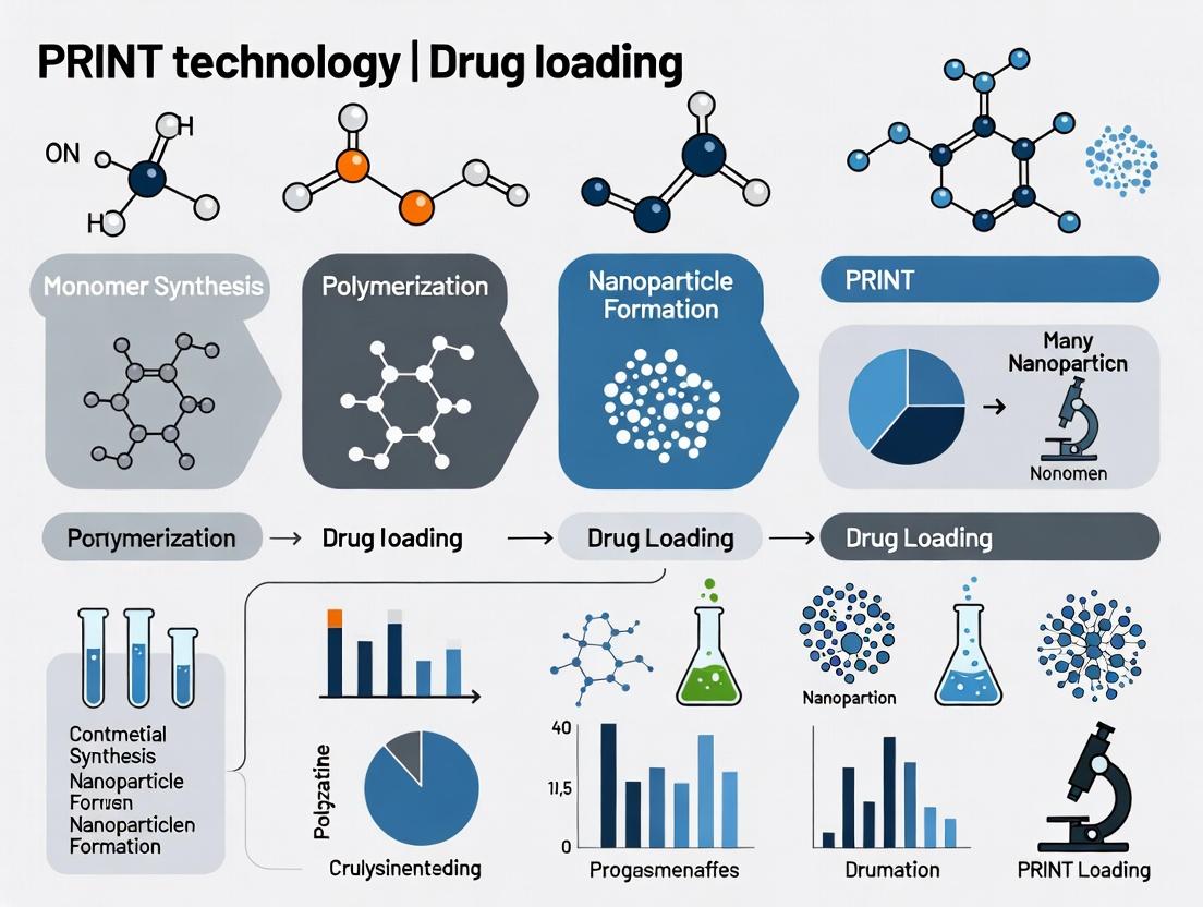
PRINT Technology Explained: The Foundation for Unparalleled Nanoparticle Control
What is PRINT? Defining Particle Replication in Non-wetting Templates.
Within the context of a thesis on PRINT technology for controlled nanoparticle drug loading, this article defines Particle Replication in Non-wetting Templates (PRINT) as a high-resolution, top-down fabrication platform. PRINT enables the precise design and manufacture of monodisperse, shape-specific nanoparticles with exact control over size, shape, surface chemistry, and composition. This capability is paramount for optimizing drug loading, release kinetics, and biodistribution in therapeutic applications.
PRINT is a soft lithography technique that utilizes low-surface-energy, fluorinated elastomeric molds to create particles from a variety of organic and inorganic materials. The "non-wetting" property of the mold is critical, as it prevents the pre-particle solution from spreading beyond the defined cavities, allowing for exceptionally high fidelity replication and easy harvest of discrete particles. This method stands in contrast to bottom-up, self-assembly techniques which often yield polydisperse populations.
Key Advantages for Drug Loading Research
For drug delivery research, PRINT offers unique advantages:
- Precise Control: Independent tuning of particle parameters (e.g., 100 nm x 200 nm cylinder) to study their individual effects on cellular uptake and trafficking.
- High Drug Loading: Capability to incorporate therapeutics via encapsulation or chemical conjugation.
- Surface Functionalization: Easy modification of particle surface with targeting ligands (e.g., peptides, antibodies) or PEG for stealth properties.
- Monodispersity: Ensures consistent pharmacokinetic behavior and dose delivery.
Table 1: Comparative Analysis of Nanoparticle Fabrication Techniques
| Technique | Typical Size Range | Dispersity (PDI) | Shape Control | Material Compatibility | Primary Drug Loading Method |
|---|---|---|---|---|---|
| 20 nm - 20 μm | <0.05 (Monodisperse) | Excellent (Precise) | High (PLGA, PEG, Acrylate, Proteins) | Encapsulation, Conjugation | |
| Emulsification | 100 nm - 100 μm | >0.1 (Polydisperse) | Poor (Spherical) | Moderate (Polymers, Lipids) | Encapsulation |
| Nanoprecipitation | 50 - 500 nm | 0.1 - 0.3 | Poor (Spherical) | Moderate (Hydrophobic Polymers) | Encapsulation |
| Spray Drying | 1 - 100 μm | >0.2 (Broad) | Moderate (Spherical) | High | Encapsulation |
Table 2: Impact of PRINT Particle Geometry on Cellular Uptake (In Vitro)
| Particle Shape | Dimensions (nm) | Surface Chemistry | Cell Line | Relative Uptake (%) | Key Finding |
|---|---|---|---|---|---|
| Cylinder | 200 x 200 | PEG | HeLa | 100 (Baseline) | - |
| Cylinder | 80 x 320 | PEG | HeLa | 165 | High aspect ratio enhances uptake. |
| Cube | 200 x 200 | PEG | HeLa | 78 | Reduced uptake vs. same-volume cylinder. |
| Cylinder | 200 x 200 | RGD-peptide | HeLa | 245 | Targeting ligand dramatically enhances uptake. |
Experimental Protocols
Protocol 4.1: Fabrication of PRINT Molds (Photolithography Master)
Objective: Create a silicon wafer master template. Materials: Silicon Wafer, SU-8 photoresist, Photomask (with desired features), UV Light Source, Developer Solution.
- Clean a silicon wafer with piranha solution (Caution: Highly corrosive).
- Spin-coat SU-8 photoresist onto the wafer to achieve the desired thickness (dictates particle height).
- Perform a soft bake to evaporate solvent.
- Align the photomask and expose the wafer to UV light. Exposed areas crosslink.
- Perform a post-exposure bake.
- Develop the wafer in SU-8 developer to remove non-crosslinked resist, revealing the master pattern.
- Silanize the master with a fluorinated silane (e.g., (tridecafluoro-1,1,2,2-tetrahydrooctyl)trichlorosilane) in a vacuum desiccator to facilitate mold release.
Protocol 4.2: PRINT Particle Fabrication and Drug Encapsulation
Objective: Fabricate monodisperse, drug-loaded PEG-based particles. Materials: Fluorinated elastomer (e.g., PFPE-MA), PLGA-PEG blend, Model drug (e.g., Doxorubicin), Organic solvent (e.g., DCM), Harvesting web (e.g., poly(vinyl alcohol) film).
- Mold Creation: Cure liquid fluoropolymer against the silanized master to create an inverse, non-wetting PRINT mold.
- Pre-Particle Solution: Dissolve the polymer (e.g., 95% PLGA, 5% PEG-Diacrylate) and the active pharmaceutical ingredient (API) in a volatile organic solvent (e.g., 20 mg/mL polymer, 5% w/w drug-to-polymer).
- Filling: Spread the solution over the PRINT mold. Apply a slight negative pressure to draw material into the cavities. The non-wetting nature confines the solution.
- Solvent Evaporation: Allow the solvent to evaporate completely, leaving solid polymer/drug composite particles in the mold cavities.
- Harvesting: Contact a sacrificial, adhesive harvesting web (e.g., PVA-coated liner) with the filled mold. Apply gentle heat and/or pressure to transfer particles onto the web.
- Release: Dissolve the harvesting web in an aqueous buffer (e.g., PBS), releasing free-floating, monodisperse particles into suspension.
- Purification: Purify particles via centrifugation and washing. Sterilize by 0.22 μm filtration if for cell culture.
Protocol 4.3: In Vitro Evaluation of Cellular Uptake and Viability
Objective: Assess targeted vs. non-targeted PRINT particle performance. Materials: PRINT particles (non-targeted PEG, RGD-targeted), Cell culture (HeLa), Flow Cytometry Buffer, MTS reagent.
- Seed HeLa cells in a 24-well plate at 50,000 cells/well and incubate for 24h.
- Treat cells with fluorescently-labeled PRINT particles (e.g., ~100 particles/cell) in serum-free media. Incubate for 2h at 37°C.
- Wash cells 3x with PBS to remove non-internalized particles.
- For uptake: Trypsinize cells, resuspend in flow buffer, and analyze mean fluorescence intensity via flow cytometry.
- For viability: Add MTS reagent directly to washed cells in culture media. Incubate 1-4h and measure absorbance at 490nm.
Visualization of Workflows and Pathways
Title: PRINT Nanoparticle Fabrication Workflow
Title: Targeted PRINT NP Intracellular Trafficking Pathway
The Scientist's Toolkit: Research Reagent Solutions
Table 3: Essential Materials for PRINT Drug Loading Research
| Item | Function & Role in PRINT | Example/Notes |
|---|---|---|
| Fluorinated Elastomer (PFPE-MA) | Forms the non-wetting mold. Critical for high-fidelity particle replication and release. | Perfluoropolyether dimethacrylate; provides inert, low surface energy. |
| PLGA-PEG Blend | Biodegradable polymer matrix. PLGA provides encapsulation, PEG enables stealth & conjugation. | Vary LA:GA ratio for degradation rate; PEG terminus for ligand attachment. |
| Harvesting Web (PVA Film) | Sacrificial layer to collect particles from mold and transfer to aqueous solution. | Polyvinyl alcohol coating on a liner; water-soluble. |
| Fluorescent Dye (Cyanine, FITC) | Covalent conjugation or encapsulation for particle tracking in in vitro and in vivo studies. | Cy5 for near-infrared imaging; FITC for flow cytometry. |
| Targeting Ligand (RGD Peptide) | Conjugated to particle surface to mediate active targeting to overexpressed cell receptors. | Cyclo(Arg-Gly-Asp-D-Phe-Cys) for αvβ3 integrin targeting. |
| Crosslinker (e.g., DTT) | For photocurable resins. Forms covalent bonds during UV curing to stabilize particle shape. | Dithiothreitol (DTT) used as a crosslinker for thiol-ene chemistry. |
Within the broader thesis on Particle Replication in Non-wetting Templates (PRINT) technology for controlled nanoparticle (NP) drug loading, two core physical principles are paramount: low-surface-energy molds and photocurable resins. This document provides application notes and protocols for leveraging these principles to fabricate monodisperse, size- and shape-specific polymeric nanoparticles with precise drug payloads. The non-wetting property of perfluoropolyether (PFPE) molds is critical for high-fidelity particle replication and easy release, while photocurable resins enable rapid, tunable cross-linking for encapsulating therapeutic agents.
Table 1: Comparison of Mold Materials for PRINT Technology
| Mold Material | Surface Energy (mN/m) | Replication Fidelity | Particle Release Ease | Reusability | Key Application |
|---|---|---|---|---|---|
| Perfluoropolyether (PFPE) | ~12-14 | Excellent | Excellent | High (>100 cycles) | High-resolution NP fabrication |
| Polydimethylsiloxane (PDMS) | ~20-22 | Good | Moderate | Medium | Rapid prototyping |
| Silicon/Glass | >1000 | Excellent | Poor (requires etch) | Low | Master template fabrication |
Table 2: Properties of Representative Photocurable Resins for Drug Loading
| Resin Formulation | Curing Time (s) | Modulus (MPa) | Drug Encapsulation Efficiency (%) | Sustained Release Profile |
|---|---|---|---|---|
| Poly(ethylene glycol) diacrylate (PEGDA, 700 Da) | 30-60 | 5-20 | 75-90 (Hydrophilic) | 1-7 days |
| Trimethylolpropane ethoxylate triacrylate | 45-90 | 50-200 | 60-80 (Amphiphilic) | 7-21 days |
| Acrylated PLGA with photoinitiator | 60-120 | 100-500 | 85-95 (Hydrophobic) | 14-30+ days |
Experimental Protocols
Protocol 3.1: Fabrication of Low-Surface-Energy PFPE Molds
Objective: To create a reusable, non-wetting elastomeric mold from a silicon master template. Materials: Silicon master (with desired NP features), perfluoropolyether dimethacrylate (PFPE-DMA), 2-hydroxy-2-methylpropiophenone (photoinitiator), UV curing station (λ=365 nm), fluorinated solvent (e.g., HFE-7500). Procedure:
- Clean the silicon master template with oxygen plasma for 5 minutes.
- Prepare the PFPE prepolymer by mixing PFPE-DMA with 1-3% (w/w) photoinitiator.
- Degas the mixture under vacuum for 15 minutes to remove bubbles.
- Carefully pour the prepolymer over the silicon master, ensuring full feature coverage.
- Cure under a nitrogen atmosphere using UV light (20 mW/cm²) for 5 minutes.
- Carefully peel the cured PFPE mold from the silicon master.
- Clean the mold by sonicating in fluorinated solvent for 2 minutes and dry with a stream of nitrogen.
- Characterize mold surface energy via contact angle goniometry (water contact angle >110° indicates low surface energy).
Protocol 3.2: Nanoparticle Fabrication & Drug Loading via PRINT
Objective: To produce monodisperse drug-loaded nanoparticles using a PFPE mold and a photocurable resin formulation. Materials: PFPE mold (from Protocol 3.1), photocurable resin (e.g., PEGDA), therapeutic agent (e.g., Doxorubicin HCl), photoinitiator (Irgacure 2959), doctor blade, UV curing station, release liner (e.g., ethylene vinyl acetate film). Procedure:
- Resin/Drug Preparation: Dissolve the drug (1-10% w/w relative to monomer) and photoinitiator (0.5% w/w) in the liquid photocurable monomer. Vortex and sonicate until a homogeneous solution is achieved.
- Mold Filling: Place the PFPE mold on a flat stage. Apply a small excess of the drug-resin mixture to one end of the mold. Use a doctor blade at a controlled angle and speed to sweep the mixture across the mold, filling the cavities via capillary action. The low surface energy of the PFPE ensures the resin preferentially fills the cavities rather than wetting the top surface.
- Curing: Immediately cover the filled mold with a release liner to inhibit oxygen inhibition. Expose to UV light (λ=365 nm, 15 mW/cm²) for 30-60 seconds to fully cross-link the resin.
- Particle Harvesting: Peel the release liner away. The solidified particles, now as a solid film on the liner, are easily released from the non-wetting PFPE mold. Collect particles by dissolving the film in an appropriate aqueous buffer (e.g., PBS pH 7.4) or by gentle agitation.
- Purification: Purify the nanoparticle suspension via centrifugation (e.g., 20,000 x g, 15 min) or tangential flow filtration to remove unreacted monomer and unencapsulated drug. Lyophilize for storage if needed.
Diagrams
Title: PRINT Technology Workflow for Drug-Loaded NPs
Title: Low Surface Energy Enables Clean Particle Release
The Scientist's Toolkit: Research Reagent Solutions
Table 3: Essential Materials for PRINT-based NP Drug Loading Research
| Item | Function & Rationale | Example Product/Chemical |
|---|---|---|
| PFPE-based Elastomer | Forms the non-wetting, chemically resistant mold. Critical for high-fidelity replication and particle release. | Fluorolink MD700 (Solvay) |
| Photocurable Monomer | Forms the nanoparticle matrix. Choice dictates NP stiffness, degradation rate, and biocompatibility. | Poly(ethylene glycol) diacrylate (PEGDA) |
| Biocompatible Photoinitiator | Initiates radical polymerization upon UV exposure. Must be safe for biomedical use. | Irgacure 2959 (2-Hydroxy-4'-(2-hydroxyethoxy)-2-methylpropiophenone) |
| Fluorinated Solvent | Cleans PFPE molds without swelling or damaging them, preserving feature integrity. | Novec HFE-7500 (3M) |
| Release Liner | Provides an oxygen barrier during curing and a substrate for harvesting particles. | Ethylene Vinyl Acetate (EVA) film |
| Therapeutic Agent | Active pharmaceutical ingredient to be encapsulated. Can be hydrophilic or hydrophobic. | Doxorubicin HCl (hydrophilic), Paclitaxel (hydrophobic) |
| UV Curing System | Provides controlled-intensity UV light (λ=365 nm) for rapid resin polymerization. | OmniCure S2000 (Excelitas) |
Within the broader thesis investigating PRINT (Particle Replication In Non-wetting Templates) technology for controlled nanoparticle drug loading, this application note details the critical advantages conferred by precise control over particle size, shape, and monodispersity. These parameters are fundamental determinants of biodistribution, cellular uptake, circulation half-life, and drug release kinetics. PRINT technology enables the fabrication of highly uniform particles with independent control over these attributes, providing a powerful platform for systematic structure-activity relationship studies in drug delivery.
Data Presentation: Impact of Particle Parameters on Delivery Efficacy
Table 1: Quantitative Impact of Nanoparticle Size on Pharmacokinetics and Biodistribution
| Size Range (nm) | Circulation Half-life (in mice) | Primary Clearance Organ | Tumor Accumulation (%ID/g)* | Key Mechanism/Reason |
|---|---|---|---|---|
| 10-30 | < 1 hour | Renal, RES | 0.5-1.5 | Rapid renal filtration, extravasation. |
| 50-100 | 6-12 hours | RES (Liver/Spleen) | 2.5-4.0 | Optimal avoidance of rapid clearance, EPR effect. |
| 150-200 | 12-24 hours | RES | 3.0-5.0 | Prolonged circulation, limited penetration in dense tumors. |
| > 300 | Variable (often shorter) | RES (rapid sequestration) | 1.0-2.0 | Rapid phagocytosis by mononuclear phagocyte system. |
*%ID/g: Percentage of Injected Dose per gram of tissue. Data compiled from studies using PEGylated PRINT particles.
Table 2: Influence of Nanoparticle Shape on Cellular Uptake and Hemodynamics
| Particle Shape | Aspect Ratio | Cellular Uptake (vs. Spherical) | Flow Characteristics (in vasculature) | Margination Potential |
|---|---|---|---|---|
| Spherical | 1:1 | 1.0 (Reference) | Linear flow, lower wall interaction | Low |
| Rod-like | 3:1 | 1.5 - 2.5x higher | Tumbling, enhanced wall interaction | High |
| Disc-like | 1:3 (height:diameter) | 0.6 - 0.8x lower | Skipping, rolling along endothelium | Very High |
| Filamentous | >10:1 | Significantly reduced | Enhanced vascular adhesion, persistence | Moderate |
Experimental Protocols
Protocol 1: Fabrication of Monodisperse PRINT Particles with Controlled Size and Shape
Objective: To fabricate poly(lactic-co-glycolic acid) (PLGA) nanoparticles with defined size (200 nm) and rod shape (3:1 aspect ratio) for paclitaxel loading. Materials: See "The Scientist's Toolkit" below. Procedure:
- Master Template Preparation: A silicon master template with 200 nm x 600 nm rod-shaped cavities is fabricated using photolithography and etching.
- Flexible Mold Creation: A fluorinated perfluoropolyether (PFPE) elastomer is cast onto the silicon master and cured under UV light to create a negative, non-wetting mold.
- Particle Precursor Solution: 100 mg of PLGA (50:50) and 5 mg of paclitaxel are dissolved in 1 mL of a volatile solvent (e.g., dichloromethane or chloroform).
- Filling and Curing: The precursor solution is doctored across the PFPE mold, filling the cavities via capillary action. Excess solution is removed. The solvent is allowed to evaporate fully.
- Harvesting: A poly(vinyl alcohol) (PVA) film is laminated onto the filled mold. The PLGA particles are transferred to the PVA sheet upon gentle peeling.
- Redispersion: The PVA film with embedded particles is dissolved in an aqueous buffer (e.g., PBS pH 7.4), releasing monodisperse, rod-shaped PLGA-paclitaxel particles.
- Purification: The suspension is centrifuged at 15,000 x g for 20 minutes, the supernatant is discarded, and the pellet is resuspended in fresh PBS. This is repeated 3 times.
- Characterization: Size and dispersity are analyzed by Dynamic Light Scattering (DLS) and Scanning Electron Microscopy (SEM). Drug loading is quantified via HPLC.
Protocol 2: Evaluating Cellular Uptake Kinetics as a Function of Particle Shape
Objective: To compare the internalization kinetics of spherical vs. rod-shaped (3:1) fluorescently labeled PRINT particles in A549 lung carcinoma cells. Materials: A549 cell line, Fluorescently-labeled PRINT particles (spherical 200 nm, rod 200 nm x 600 nm), serum-free medium, flow cytometry buffer, confocal microscope. Procedure:
- Seed A549 cells in 24-well plates at 1 x 10^5 cells/well and culture for 24 hrs.
- Incubate particles with a fluorophore (e.g., Cy5) at a non-quenching density during fabrication (Protocol 1, Step 3).
- Wash cells twice with serum-free medium.
- Treat cells with particles at a final concentration of 50 µg/mL in serum-free medium. Include wells with no particles as a control.
- Incubate at 37°C, 5% CO2 for pre-determined time points (0.5, 1, 2, 4 hrs).
- At each time point, aspirate medium, wash cells 3x with ice-cold PBS to remove non-internalized particles.
- Trypsinize cells, quench with complete medium, and centrifuge at 500 x g for 5 min.
- Resuspend cell pellet in flow cytometry buffer and analyze fluorescence intensity via flow cytometry (Ex/Em for Cy5: 649/670 nm).
- For confocal imaging, plate cells on glass-bottom dishes, treat as above, fix with 4% PFA at desired time points, stain nuclei and actin, and image.
Visualization: Experimental Workflow and Biological Fate
The Scientist's Toolkit: Essential Research Reagent Solutions
Table 3: Key Materials for PRINT-based Drug Delivery Research
| Item | Function in PRINT Research | Example/Notes |
|---|---|---|
| PFPE-based Elastomer | Forms the non-wetting, chemically resistant mold. Critical for high-fidelity particle replication and easy release. | Liquids like Fluorocur or custom-synthesized PFPE; provides low surface energy. |
| Biodegradable Polymers | Particle matrix material. Determines degradation rate and drug release profile. | PLGA (varying LA:GA ratios), Polycaprolactone (PCL), Chitosan derivatives. |
| Therapeutic Payload | Active agent to be encapsulated. Can be small molecules, proteins, or nucleic acids. | Paclitaxel, Doxorubicin, siRNA, Ovalbumin (model protein). |
| PEGylated Ligands | Surface modifiers to confer stealth properties (PEG) or active targeting (ligands). | PEG-diacrylate (for co-polymerization), Maleimide-PEG-NHS (for post-conjugation of peptides). |
| Fluorescent Monomers/Dyes | Enable tracking of particles in vitro and in vivo. | Cy5-acrylate, Bodipy-monomer, or post-fabrication staining with Nile Red. |
| Silicon Wafer Masters | The primary template defining particle size and shape. | Fabricated via e-beam or photolithography; features can be rods, discs, cones, etc. |
| Harvesting Matrix | A sacrificial layer to collect particles from the mold without aggregation. | Poly(vinyl alcohol) (PVA) films, sucrose sheets, or hydrogel layers. |
Application Notes
The transition from traditional nanoparticle (NP) fabrication methods to the Particle Replication in Non-wetting Templates (PRINT) platform represents a fundamental shift in the precision and control available for drug delivery research. This control is critical for a thesis focused on systematic investigation of drug loading parameters.
Key Advantages of PRINT for Controlled Drug Loading Studies:
- Precision and Monodispersity: Traditional methods like emulsion-solvent evaporation, nanoprecipitation, and spray drying produce particles with broad size distributions (polydispersity index, PDI > 0.1). PRINT technology, utilizing a perfluoropolyether (PFPE) mold, yields highly monodisperse particles (PDI < 0.05). This eliminates size as a confounding variable in drug release and cellular uptake studies.
- Independent Parameter Control: PRINT uniquely decouples particle size, shape, composition, and modulus. Researchers can alter particle shape (e.g., cylindrical, conical, hexagonal) without changing size or drug loading, enabling direct studies on how morphology influences biodistribution and targeting.
- High Drug Loading and Co-Loading: PRINT allows for the encapsulation of a wide range of therapeutics (small molecules, proteins, nucleic acids) with high efficiency (often >90%). Traditional methods often suffer from low encapsulation efficiency for hydrophilic drugs. PRINT also facilitates precise co-loading of multiple agents in controlled ratios, essential for combination therapy research.
- Surface Engineering Precision: Post-fabrication, PRINT particles enable the precise conjugation of targeting ligands (e.g., antibodies, peptides) and PEGylation in a controlled, reproducible manner, overcoming batch-to-batch variability common in traditional conjugation methods.
Quantitative Comparison of Fabrication Methods:
Table 1: Comparative Analysis of Nanoparticle Fabrication Techniques
| Parameter | Traditional Methods (e.g., Emulsification, Nanoprecipitation) | PRINT Technology |
|---|---|---|
| Size Control | Moderate to poor; broad distribution. | Excellent; precise and monodisperse. |
| Size Range | Typically 50-500 nm. | 20 nm - 20 μm. |
| Polydispersity Index | High (Often >0.1, up to 0.3) | Very Low (<0.05) |
| Shape Control | Limited (typically spherical). | High (cylinders, rods, discs, viruses, custom shapes). |
| Drug Loading Efficiency | Variable; often low for hydrophilic drugs. | Consistently High (>90% achievable). |
| Batch-to-Batch Variability | High. | Very Low. |
| Key Limitation | Coupled parameters; difficult to isolate variables. | Throughput can be lower than some scalable traditional methods; mold fabrication required. |
Table 2: Exemplar Drug Loading Data from PRINT Studies
| Therapeutic Agent | Particle Size (nm) | PDI | Encapsulation Efficiency (%) | Loading Capacity (% w/w) |
|---|---|---|---|---|
| Doxorubicin (Chemo) | 80 x 320 nm (Cylinder) | 0.03 | 98.5 | 25.0 |
| siRNA (Nucleic Acid) | 100 nm (Sphere) | 0.04 | 95.2 | 8.5 |
| Insulin (Protein) | 200 nm (Sphere) | 0.02 | 92.7 | 30.0 |
| Curcumin (Hydrophobic) | 70 nm (Cube) | 0.05 | 99.1 | 40.0 |
Experimental Protocols
Protocol 1: PRINT Fabrication of Drug-Loaded PLGA Nanoparticles
Objective: To fabricate monodisperse, cylindrical poly(lactic-co-glycolic acid) (PLGA) nanoparticles loaded with a model hydrophobic drug (e.g., docetaxel) using the PRINT process.
Materials (The Scientist's Toolkit):
Table 3: Key Research Reagent Solutions for PRINT Fabrication
| Item | Function |
|---|---|
| PFPE Mold (Cylindrical, 200nm x 200nm) | Non-wetting template that defines particle size and shape. |
| PLGA (50:50, acid-terminated) | Biodegradable, FDA-approved copolymer forming the particle matrix. |
| Docetaxel | Model hydrophobic chemotherapeutic agent for encapsulation study. |
| Methylene Chloride (DCM) | Volatile solvent to dissolve PLGA and drug for the particle pre-polymer solution. |
| Polyvinyl Alcohol (PVA) Solution (1% w/v) | Harvesting layer that facilitates the release of particles from the PRINT mold. |
| Laminator | Apparatus to apply uniform pressure and spread the pre-polymer solution into the mold cavities. |
| E-beam Evaporator | Used to apply a sacrificial fluorosilane layer to the harvested film for final particle release (alternative). |
Procedure:
- Pre-polymer Solution Preparation: Dissolve PLGA (100 mg) and docetaxel (10 mg) in 1 mL of DCM. Sonicate for 5 minutes to ensure complete dissolution and homogeneity.
- Mold Coating: Using a pipette, apply a precise volume (e.g., 100 µL) of the pre-polymer solution onto the surface of the PFPE mold.
- Lamination: Immediately pass the mold through a heated laminator (set to 40°C) to spread the solution and force it into the cylindrical cavities while rapidly evaporating the DCM solvent.
- Filling & Drying: Allow the laminated mold to sit under a fume hood for 15 minutes to ensure complete solvent evaporation and solidification of the PLGA-drug matrix within the cavities.
- Harvesting: Apply a thin layer of a 1% w/v aqueous PVA solution onto a flexible poly(ethylene terephthalate) (PET) film. Gently laminate the filled PFPE mold onto this PVA-coated film. The PVA acts as a harvesting layer.
- Particle Release: Carefully peel the PFPE mold away from the PVA/PET film. The array of cylindrical docetaxel-PLGA particles is now embedded in the PHA film.
- Isolation: Sonicate the PVA film containing the particles in deionized water (10 mL) for 5 minutes to release the particles into suspension.
- Purification: Centrifuge the suspension at 15,000 x g for 20 minutes. Wash the pellet twice with DI water to remove PVA and any unencapsulated drug. Resuspend the final nanoparticle pellet in PBS or a suitable buffer for characterization.
- Characterization: Analyze particle size and PDI by dynamic light scattering (DLS). Confirm morphology by scanning electron microscopy (SEM). Quantify drug loading via HPLC after dissolving a known mass of particles in acetonitrile.
Protocol 2: Evaluating Drug Release Kinetics: PRINT vs. Bulk Emulsion NPs
Objective: To compare the in vitro release profile of a drug from monodisperse PRINT particles versus polydisperse particles made by single emulsion.
Procedure:
- Fabrication: Prepare docetaxel-loaded PLGA particles using the PRINT protocol above and a traditional single emulsion method (emulsify PLGA/docetaxel in DCM with aqueous PVA, stir overnight to evaporate DCM, collect by centrifugation).
- Standard Curve: Prepare a standard curve of docetaxel in the release medium (PBS with 0.5% w/v Tween 80, pH 7.4) using UV-Vis spectroscopy or HPLC.
- Release Study: Place 5 mg of each type of nanoparticle (PRINT and Emulsion) into separate dialysis bags (MWCO 12-14 kDa). Suspend each bag in 50 mL of release medium in a shaking incubator (37°C, 100 rpm).
- Sampling: At predetermined time points (1, 3, 6, 12, 24, 48, 72, 96, 168 hours), withdraw 1 mL of the external release medium and replace it with 1 mL of fresh pre-warmed medium.
- Analysis: Quantify the drug concentration in each sample using the previously established analytical method (UV-Vis/HPLC).
- Data Modeling: Plot cumulative drug release (%) vs. time. Fit data to common release models (e.g., zero-order, first-order, Higuchi, Korsmeyer-Peppas) to elucidate release mechanisms. The monodisperse PRINT particles typically exhibit more predictable, uniform release kinetics fitting a near-zero-order model, while emulsion particles often show a more biphasic, burst-release profile.
Diagram Title: PRINT Nanoparticle Fabrication and Harvesting Workflow
Diagram Title: PRINT Enables Independent Control of Nanoparticle Properties
Within the broader thesis on Particle Replication in Non-wetting Templates (PRINT) technology for controlled nanoparticle drug delivery, the selection of polymeric materials is foundational. PRINT enables the fabrication of monodisperse, shape-specific nanoparticles with precise control over size, composition, and cargo loading. This application note details the critical polymer and pre-polymer library, providing protocols and data essential for rational material selection to optimize drug encapsulation, release kinetics, and biocompatibility.
Polymer Library: Characteristics and Quantitative Data
The PRINT platform is compatible with a diverse array of polymers and pre-polymer resins. The choice dictates nanoparticle properties, including degradation profile, drug compatibility, and surface functionality.
Table 1: Core Polymer and Pre-polymer Library for PRINT Nanoparticles
| Material Class | Specific Example(s) | Key Properties (Mw, Tg, etc.) | Degradation Profile | Typical Drug Cargo | Key Advantages for PRINT |
|---|---|---|---|---|---|
| Poly(lactic-co-glycolic acid) (PLGA) | 50:50 PLGA, 75:25 PLGA | Mw: 10-100 kDa; Tg: 45-50°C | Hydrolytic; weeks to months | Paclitaxel, Doxorubicin, proteins | FDA-approved; tunable erosion. |
| Poly(ethylene glycol) (PEG) | PEG-DA (Diacrylate) | Mw: 700-10,000 Da | Non-degradable or via linkages | siRNA, hydrophobic drugs | Imparts "stealth" properties; reduces opsonization. |
| Poly(ε-caprolactone) (PCL) | PCL-DA | Mw: 10-80 kDa; Tg: -60°C | Hydrolytic; slow (years) | Sustained-release small molecules | High permeability; slow degradation. |
| Acrylate-Terminated Pre-polymers | Ebeeryl 1290, PEG-DA | Varies by backbone | Crosslinked; non-erodible | Covalently conjugated agents | High shape fidelity; stable particles. |
| Hydrogel Formers | HEA (Hydroxyethyl acrylate), PEG-DA | N/A | Swelling-controlled | Proteins, peptides, vaccines | Aqueous cargo compatibility; gentle encapsulation. |
Experimental Protocols
Protocol 1: Formulation Screening for Drug Loading Efficiency
Objective: To systematically evaluate different PRINT polymers for encapsulation efficiency (EE) and drug loading (DL) capacity of a model hydrophobic drug (e.g., Doxorubicin).
Materials:
- PRINT Mold (e.g., 80nm x 320nm cylindrical pores, perfluoropolyether)
- Polymer solutions (2% w/v in suitable solvent: DCM for PLGA/PCL, Acetone for PEG-DA)
- Drug stock solution (10 mg/mL doxorubicin in DMSO)
- Non-wetting layer (e.g., fluorosilane-coated surface)
- Harvesting solution (e.g., 1% PVA in water)
- Centrifugation filters (100 kDa MWCO)
Procedure:
- Pre-mix Formulation: For each polymer, combine polymer solution with drug stock to achieve a 10:1 polymer-to-drug weight ratio. Vortex for 30 seconds.
- Mold Filling: Apply the formulation onto the PRINT mold placed on a non-wetting layer. Use a doctor blade to fill pores by capillary action.
- Solvent Evaporation: Allow solvent to evaporate completely under ambient conditions for 15 minutes.
- Harvesting: Place a harvesting film (e.g., PVA-coated slide) on the mold. Apply gentle pressure and heat (50°C for PEG-based, 25°C for PLGA) for 2 minutes to transfer particles.
- Collection: Dissolve the harvesting film in 10 mL deionized water under agitation. Isolate nanoparticles via centrifugation (15,000 x g, 20 min) and wash twice.
- Analysis:
- Drug Loading: Lyse an aliquot of nanoparticles in DMSO. Measure drug concentration via UV-Vis absorbance at 480nm. Calculate DL% = (Mass of drug in nanoparticles / Total mass of nanoparticles) x 100.
- Encapsulation Efficiency: EE% = (Mass of drug in nanoparticles / Initial mass of drug used) x 100.
Protocol 2: Evaluating Drug Release Kinetics
Objective: To characterize in vitro drug release profiles from PRINT nanoparticles fabricated from different polymers.
Materials:
- Dialysis bags (MWCO 10-20 kDa) or Float-A-Lyzer devices
- Release medium (PBS, pH 7.4, with 0.1% w/v Tween 80 for sink conditions)
- Incubator shaker (37°C)
Procedure:
- Sample Preparation: Dispense a known volume of nanoparticle suspension (containing ~1 mg of drug) into a pre-hydrated dialysis device.
- Release Study: Immerse the device in 50 mL of pre-warmed release medium (37°C) with gentle agitation (100 rpm).
- Sampling: At predetermined time points (1h, 4h, 8h, 24h, 48h, 7d, etc.), withdraw 1 mL of the external medium and replace with fresh pre-warmed medium.
- Analysis: Quantify drug concentration in each sample via HPLC or UV-Vis. Correct for cumulative dilution.
- Modeling: Fit release data to mathematical models (zero-order, first-order, Higuchi, Korsmeyer-Peppas) to elucidate release mechanisms.
The Scientist's Toolkit: Research Reagent Solutions
Table 2: Essential Materials for PRINT Polymer Screening
| Item | Function in PRINT Research |
|---|---|
| Perfluoropolyether (PFPE) Elastomer Molds | Master template with non-wetting properties enabling high-fidelity particle replication and easy release. |
| PLGA with variable Lactide:Glycolide ratios | Provides control over nanoparticle degradation rate and drug release profile. |
| Poly(ethylene glycol) Diacrylate (PEG-DA) | A crosslinkable pre-polymer for forming hydrogel nanoparticles; allows surface functionalization. |
| Photoinitiator (e.g., Irgacure 2959) | UV-activated catalyst for curing acrylate-terminated pre-polymers (e.g., PEG-DA) within molds. |
| Fluorosilane-Coated Glass Slides | Creates a non-wetting surface to support the mold during filling, preventing premature dispersion. |
| Polyvinyl Alcohol (PVA) Harvesting Film | A sacrificial layer used to collect nanoparticles from the mold post-fabrication. |
| Float-A-Lyzer Dialysis Devices | Enables robust, sink-condition in vitro drug release studies with easy sampling. |
Visualizations
Title: Rational Polymer Selection Workflow for PRINT
Title: Standard PRINT Nanoparticle Fabrication Protocol
Step-by-Step Guide: Drug Loading Strategies and Biomedical Applications of PRINT Nanoparticles
Application Notes: PRINT Technology for Controlled Nanoparticle Synthesis
The Particle Replication in Non-wetting Templates (PRINT) technology is a top-down, lithographic fabrication platform enabling the precise production of monodisperse, shape-specific nanoparticles (NPs) with controlled composition and cargo loading. This methodology is critical for advancing drug delivery research, as it decouples particle parameters (size, shape, modulus) from biochemical properties, allowing for systematic study of cellular uptake, biodistribution, and controlled release kinetics. Within the broader thesis on controlled nanoparticle drug loading, this workflow establishes the foundational physical platform upon which subsequent drug encapsulation, surface modification, and release studies are performed.
The PRINT process utilizes a low-surface-energy, non-wetting perfluoropolyether (PFPE) elastomeric mold, which facilitates the clean filling of cavities and easy, high-fidelity harvest of particles. This method overcomes key limitations of bulk emulsion techniques, such as polydispersity and uncontrolled drug burst release. The following protocols detail the complete cycle from mold creation to particle harvest, with emphasis on parameters that directly influence final particle characteristics crucial for drug loading efficacy.
Experimental Protocols
Protocol: Fabrication of PFPE Master Template
- Objective: To create a robust, non-wetting PFPE mold from a silicon master template.
- Materials: Silicon master (with desired feature topography), perfluoropolyether dimethacrylate (PFPE-DMA), 2,2-diethoxyacetophenone (photoinitiator), isopropanol, nitrogen stream.
- Procedure:
- Silicon Master Preparation: Clean the silicon master wafer sequentially with acetone, isopropanol, and deionized water. Dry under a stream of nitrogen.
- Prepolymer Preparation: In a light-protected vial, mix PFPE-DMA with 1% (w/w) 2,2-diethoxyacetophenone. Vortex until the photoinitiator is fully dissolved.
- Dispensing and Curing: Apply a few drops of the prepolymer mixture onto the silicon master. Carefully lower a transparent polyethylene terephthalate (PET) backing film onto the liquid to form a thin layer, avoiding bubble formation.
- UV Photocuring: Place the assembly under a UV lamp (λ=365 nm, intensity 20 mW/cm²) for 5 minutes to crosslink the PFPE.
- Demolding: After curing, carefully peel the cured PFPE mold, now bearing the inverse of the master pattern, from the silicon master. The mold is now ready for particle fabrication.
Protocol: Particle Fabrication via Liquid Precursor Filling
- Objective: To fill template cavities with a particle precursor solution containing therapeutic cargo.
- Materials: PFPE mold, particle precursor solution (e.g., PLGA in ethyl acetate with model drug), doctor blade, vacuum desiccator.
- Procedure:
- Precursor Solution Preparation: Dissolve the polymer (e.g., 50 mg PLGA) and the active pharmaceutical ingredient (API; e.g., 5 mg paclitaxel) in an appropriate volatile solvent (e.g., 1 mL ethyl acetate). Filter through a 0.45 µm PTFE filter.
- Template Filling: Place the PFPE mold on a flat surface. Apply an excess of the precursor solution across the top of the mold.
- Doctor Blading: Use a sharp doctor blade held at a 45° angle to sweep excess solution across the mold surface, leaving only the cavities filled.
- Solvent Evaporation: Immediately transfer the filled mold to a vacuum desiccator for 15-30 minutes to evaporate the solvent, solidifying the particles within the cavities.
Protocol: Dry Particle Harvesting via Lamination
- Objective: To harvest solidified particles without the use of contaminating solvents or adhesives.
- Materials: Filled PFPE mold, harvesting film (e.g., polyvinyl alcohol (PVA) sheet), laminator.
- Procedure:
- Harvesting Film Preparation: Cut a PVA film slightly larger than the mold area. Hydrate slightly if necessary per manufacturer guidelines.
- Film Lamination: Gently place the harvesting film over the filled PFPE mold. Pass the assembly through a heated laminator (setpoint: 80-100°C) at a constant speed.
- Peel Harvesting: After lamination, allow the assembly to cool briefly. Grasping the harvesting film edges, peel it away from the PFPE mold in one smooth, continuous motion. Particles will be embedded in or adhered to the harvesting film.
- Particle Collection: For water-soluble films like PVA, dissolve the film in a suitable aqueous buffer (e.g., PBS, pH 7.4) under gentle agitation. Centrifuge the suspension to pellet particles, then wash and resuspend in storage buffer.
Table 1: Key Parameters in PRINT Workflow and Their Impact on Particle Characteristics
| Workflow Stage | Controlled Parameter | Typical Range | Impact on Final Nanoparticle | Relevance to Drug Loading |
|---|---|---|---|---|
| Template Fabrication | Cavity Diameter (nm) | 50 – 5000 nm | Directly defines particle size (X, Y dimensions). | Size governs diffusion, cellular uptake route, and biodistribution. |
| Cavity Shape | Cylinder, Cone, Rod, Disc | Defines particle geometry (aspect ratio). | Shape affects margination, phagocytosis, and drug release surface area. | |
| Cavity Depth (nm) | 100 – 1000 nm | Defines particle height (Z dimension). | Influences total particle volume and drug payload capacity. | |
| Particle Fabrication | Polymer Concentration | 1 – 20% (w/v) | Affects particle porosity, modulus, and solidification rate. | Modulates drug encapsulation efficiency and release profile. |
| Drug:Polymer Ratio | 1:5 – 1:20 (w/w) | Determines theoretical drug loading percentage. | Directly controls potential dose per particle. | |
| Solvent Volatility | Low (DMF) to High (DCM) | Influences particle surface morphology (smooth vs. porous). | Affects initial burst release and subsequent sustained release kinetics. | |
| Particle Harvesting | Laminator Temperature | 25 – 120 °C | Impacts harvest yield and film dissolution rate. | High temps may degrade heat-sensitive biologics. Must be optimized. |
| Harvest Film Solubility | Fast (PVA) to Slow (PLA) | Determines post-harvest processing time and buffer compatibility. | Must not solubilize or degrade the encapsulated API. |
Table 2: Example PRINT Particle Formulations for Drug Loading Studies
| Target API | Polymer Matrix | Particle Size (nm) | Shape | Reported Encapsulation Efficiency (%) | Key Harvesting Method |
|---|---|---|---|---|---|
| Paclitaxel | PLGA-PEG | 200 x 200 | Cylinder | 85 – 92 | Lamination with PVA film |
| siRNA | Cationic Lipid/PLGA Hybrid | 100 x 100 | Cone | 75 – 80 | Direct mechanical peeling |
| Doxorubicin | PEG Hydrogel | 3000 x 500 | Rod | >95 | Buoyant harvesting onto agarose |
| Ovalbumin (Model Antigen) | Polylactic Acid (PLA) | 1000 x 200 | Disc | 60 – 70 | Solvent-assisted transfer |
Visualization Diagrams
Diagram Title: PRINT Nanoparticle Fabrication and Harvesting Workflow
Diagram Title: How Process Parameters Influence Final Nanoparticle Performance
The Scientist's Toolkit: Essential Research Reagents & Materials
Table 3: Key Reagents and Materials for PRINT Nanoparticle Research
| Item | Function in PRINT Workflow | Key Consideration for Drug Loading Research |
|---|---|---|
| PFPE-DMA (Perfluoropolyether-dimethacrylate) | Forms the non-wetting, elastomeric mold material. Its inert, low-surface-energy nature enables high-fidelity particle release. | The gold standard for PRINT molds; ensures minimal cargo loss to mold during harvest. |
| Silicon Master Wafer | Contains the precise, lithographically-defined topography (e.g., holes, rods) that is transferred to the PFPE mold. | Defines the foundational nanoparticle size and shape variables for controlled release studies. |
| Biocompatible Polymers (PLGA, PLA, PEG-DA) | The structural matrix of the nanoparticle, dissolved in the precursor solution. | Choice dictates degradation rate, compatibility with API, and resultant drug release kinetics (e.g., PLGA for sustained release). |
| Volatile Solvent (Ethyl Acetate, DCM) | Dissolves polymer and API to form a fillable precursor solution, then evaporates. | Volatility affects particle solidification morphology, which influences initial drug burst release. Must not degrade API. |
| Lamination Film (Polyvinyl Alcohol - PVA) | A water-soluble polymer sheet used for dry particle harvesting via thermal adhesion. | Must be inert to the encapsulated drug and allow for complete dissolution in a biocompatible buffer. |
| Model APIs (Paclitaxel, Doxorubicin, Fluorescent Dyes) | The active cargo to be encapsulated. Fluorescent dyes serve as tracers for imaging and quantification. | Enable precise measurement of encapsulation efficiency (EE%), loading capacity, and release profiles in vitro/in vivo. |
Application Notes
Within the broader thesis on PRINT (Particle Replication in Non-wetting Templates) technology for controlled nanoparticle drug loading, passive loading remains a fundamental strategy for encapsulating hydrophobic therapeutics. This process relies on the diffusion of drug molecules into a pre-formed nanoparticle matrix (e.g., polymeric, lipid) during or after its formation, driven by hydrophobicity and solubility gradients. The encapsulation efficiency (EE%) and drug loading (DL%) are critical quality attributes directly influencing therapeutic efficacy, dosage, and potential toxicity. The following notes and protocols detail the methodology and key influencing factors for optimizing passive drug loading in PRINT-generated nanoparticles.
Key Factors Influencing Payload and Encapsulation Efficiency
The passive loading of drugs into nanoparticles is governed by a complex interplay of physicochemical properties. Optimization requires careful consideration of these parameters.
Table 1: Key Factors Influencing Passive Drug Loading Efficiency
| Factor | Influence on EE% and DL% | Mechanistic Rationale |
|---|---|---|
| Drug Log P | High Log P (>4) typically increases EE%. | Increased hydrophobicity enhances partitioning into the hydrophobic nanoparticle core/matrix. |
| Drug-Polymer Affinity | Strong affinity (e.g., similar solubility parameters) increases EE%. | Favors the thermodynamic driving force for drug incorporation into the polymer matrix over remaining in the aqueous phase. |
| Initial Drug Feed | EE% often decreases with increasing feed, while absolute DL% increases. | Finite capacity of the nanoparticle matrix; saturation leads to drug precipitation or crystal formation. |
| Polymer Composition & MW | Hydrophobic polymer blocks increase EE% for hydrophobic drugs. Higher MW can increase matrix density, modulating release more than EE. | Determines the hydrophobicity, viscosity, and glass transition temperature (Tg) of the nanoparticle core, affecting drug diffusion and entrapment. |
| Nanoparticle Size (PRINT) | Smaller nanoparticles (sub-100nm) may show lower EE% for some systems due to high surface area-to-volume ratio. | Increased surface area can lead to faster drug diffusion out during the loading process unless quenched rapidly. |
| Solvent Choice & Removal Rate | Efficient solvent removal (e.g., rapid evaporation, dialysis) traps drug within matrix. | Slow removal allows drug to diffuse out with the solvent, reducing EE. Solvent must solubilize both polymer and drug. |
| Aqueous Phase Properties | Addition of salts or pH adjustment can reduce drug solubility in water, enhancing EE (salting-out effect). | Decreases the thermodynamic favorability of the drug remaining in the aqueous phase, driving it into the nanoparticle. |
Table 2: Typical Encapsulation Efficiency Ranges by Nanoparticle Type (Passive Loading)
| Nanoparticle System | Typical EE% Range (Hydrophobic Drug) | Key Determinant |
|---|---|---|
| PLGA NPs (O/W emulsion) | 40-70% | Drug Log P, PLGA MW & end-group, PVA stabilization. |
| PRINT PLGA NPs | 60-90% | Precise particle size/shape reduces polydispersity, enabling more predictable partitioning. |
| Liposomes (Hydration) | <10% (Passive for hydrophobic) | Lipid bilayer composition and drug's membrane partitioning coefficient. |
| Polymer Micelles | 70-95% | Core-block crystallinity and glass transition temperature (Tg). |
| Solid Lipid NPs | 50-80% | Polymorphism of lipid core and drug solubility in the melt. |
Experimental Protocols
Protocol 1: Passive Drug Loading via Nanoprecipitation with PRINT Templates
This protocol details passive drug encapsulation during the formulation of nanoparticles using PRINT technology.
Objective: To fabricate monodisperse, drug-loaded nanoparticles with high encapsulation efficiency via passive loading.
Materials & Reagents:
- Drug Compound: Hydrophobic model drug (e.g., Paclitaxel, Coumarin-6).
- Polymer: PLGA (50:50, acid-terminated, MW ~24kDa).
- Solvent: HPLC-grade Tetrahydrofuran (THF) or Acetonitrile.
- Non-wetting PRINT Mold: Perfluoropolyether (PFPE) mold with defined cavity geometry (e.g., 80nm x 320nm cylinders).
- Aqueous Surfactant Solution: 0.1% (w/v) Polyvinyl alcohol (PVA) in deionized water.
- Harvesting Layer: 1% (w/v) Poly(acrylic acid) (PAA) in water, pH 7.4.
- Purification: Amicon Ultra centrifugal filters (MWCO 100kDa).
Procedure:
- Precursor Solution Preparation: Dissolve PLGA and the drug at a defined ratio (e.g., 10% drug:polymer w/w) in THF to create a homogenous organic solution (e.g., 2% w/v total solids).
- PRINT Filling: Apply the drug-polymer solution to the surface of the PFPE mold. Use a doctor blade or capillary action to fill the cavities. Allow the solvent to evaporate partially, leaving a solid drug-polymer composite plug within each cavity.
- Harvesting: Place a film of the PAA harvesting solution onto a solid support (e.g., PET sheet). Laminate the filled PRINT mold onto the harvesting layer. Apply gentle pressure and allow the PAA to dissolve the composite plugs, releasing the nascent nanoparticles into the aqueous phase.
- Solvent Removal & Stabilization: Transfer the harvested nanoparticle suspension into a large volume of 0.1% PVA solution under gentle stirring. Stir for 4 hours to allow for complete solvent diffusion and nanoparticle hardening.
- Purification: Concentrate and wash the nanoparticle suspension via centrifugal filtration (Amicon filters, 4000 x g, 10 min cycles) with deionized water (3-4 washes) to remove free drug, PAA, and excess PVA.
- Characterization: Resuspend the final nanoparticles in buffer. Determine particle size and PDI by DLS. Quantify EE% and DL% via HPLC (see Protocol 2).
Protocol 2: Quantification of Encapsulation Efficiency (EE%) and Drug Loading (DL%)
Objective: To accurately measure the amount of drug encapsulated within nanoparticles.
Materials & Reagents:
- Drug-loaded Nanoparticle Suspension
- HPLC System with appropriate column (e.g., C18 reverse-phase) and UV/Vis detector.
- HPLC-grade Acetonitrile and Water
- Drug Standard for calibration curve.
- Centrifugal Filters (MWCO 10 kDa) or Size Exclusion Chromatography (SEC) columns.
Procedure:
- Total Drug Content: a. Take a known volume (Vnp) of the purified nanoparticle suspension. b. Lyophilize or thoroughly evaporate the sample. c. Completely dissolve the dried nanoparticles in a known volume (Vtotal) of organic solvent (e.g., DMSO or acetonitrile) to disrupt the matrix and release all encapsulated drug. Sonicate if necessary. d. Filter the solution through a 0.22 µm syringe filter (PTFE) to remove polymer aggregates. e. Analyze the filtrate by HPLC against a standard curve to determine the total mass of drug (M_total).
- Free (Unencapsulated) Drug Content: a. Take an identical volume (Vnp) of the nanoparticle suspension. b. Subject it to ultrafiltration using a centrifugal filter (MWCO 10kDa, 14,000 x g, 15 min) or SEC to separate free drug from nanoparticles. c. Analyze the filtrate (containing only free drug) by HPLC to determine the mass of free drug (Mfree).
- Calculation:
- Mass of Encapsulated Drug: Mencapsulated = Mtotal - Mfree
- Encapsulation Efficiency (EE%): = (Mencapsulated / Minitial feed) * 100%
- Drug Loading (DL%): = (Mencapsulated / Total mass of nanoparticles) * 100% (Note: Total nanoparticle mass can be determined by lyophilizing a known volume of the purified suspension).
The Scientist's Toolkit
Table 3: Key Research Reagent Solutions for Passive Drug Loading Studies
| Item | Function in Research |
|---|---|
| Perfluoropolyether (PFPE) PRINT Molds | Non-wetting template that defines nanoparticle size, shape, and uniformity with high fidelity. |
| PLGA (varied MW, end-groups) | Biodegradable polymer matrix forming the nanoparticle core; properties dictate drug release kinetics and EE. |
| Polyvinyl Alcohol (PVA) | Stabilizing agent preventing nanoparticle aggregation during formulation and after harvest. |
| Poly(acrylic acid) (PAA) Harvesting Solution | Aqueous layer that dissolves the solid drug-polymer composite plugs from PRINT cavities to release nanoparticles. |
| Amicon Ultra Centrifugal Filters | For rapid buffer exchange, concentration, and removal of unencapsulated drug and small molecules. |
| Size Exclusion Chromatography (SEC) Columns | For gentle, non-destructive purification of nanoparticles away from free drug and proteins. |
| Dialysis Membranes (MWCO) | For slow, large-volume solvent exchange during nanoparticle hardening (alternative to stirring in PVA). |
Visualizations
PRINT Passive Loading Workflow
Key Drivers of High EE%
EE% & DL% Quantification Protocol
Active drug loading, defined as the incorporation of therapeutic agents into pre-formed nanoparticle carriers, represents a critical advancement in nanomedicine formulation. Within the broader thesis on Particle Replication in Non-wetting Templates (PRINT) technology, this approach addresses a key limitation: the instability or loss of bioactivity of drugs during the harsh in situ polymerization or fabrication process. PRINT excels at producing monodisperse, shape-specific nanoparticles with precise control over size and surface chemistry. Post-fabrication active loading leverages these precisely engineered "empty" PRINT particles as uniform carrier platforms, enabling the efficient and controlled encapsulation of sensitive macromolecular drugs (e.g., nucleic acids, proteins) and small molecules.
This application note details current strategies, protocols, and reagent solutions for implementing active drug loading into PRINT and analogous polymeric nanoparticles, framing them as essential tools for controlled drug loading research.
Active loading mechanisms typically exploit physicochemical gradients or specific interactions between the drug and the particle matrix.
Table 1: Summary of Active Drug Loading Strategies
| Strategy | Mechanism | Typical Drug Candidates | Key Advantages | Representative Loading Efficiency (Range)* |
|---|---|---|---|---|
| pH Gradient | A transmembrane pH gradient (acidic interior) drives the diffusion of weakly basic drugs into the particle, where they become protonated and trapped. | Doxorubicin, Vincristine, other weak bases. | High efficiency, established protocols, good retention. | 85% - 98% |
| Ionic Gradient | An ammonium sulfate or other ion gradient creates an osmotic pressure differential, leading to drug precipitation inside the particle lumen. | Doxorubicin, Topotecan. | Very high drug-to-lipid ratios, stable encapsulation. | >95% |
| Remote Loading via Complexation | Drug forms a complex with a pre-encapsulated metal ion (e.g., Cu²⁺, Ca²⁺) or polyanion (e.g., dextran sulfate) inside the particle. | Proteins, Peptides, Oligonucleotides. | Applicable to macromolecules, protects drug integrity. | 70% - 90% |
| Solvent-Controlled Incubation | Drug is dissolved in a water-miscible solvent (e.g., ethanol). Incubation with particles allows drug partitioning into the polymeric matrix as the solvent disperses. | Paclitaxel, Docetaxel, other hydrophobics. | Simple, for matrix-type particles, no gradient needed. | 60% - 85% |
| Electrostatic Adsorption | Charged drugs (e.g., siRNA, pDNA) are adsorbed onto the surface of oppositely charged particles via simple mixing. | Nucleic acids, charged peptides. | Rapid, simple, no organic solvents. | >95% (surface binding) |
*Loading Efficiency (%) = (Amount of drug encapsulated / Total initial drug amount) x 100.
Detailed Experimental Protocols
Protocol 3.1: pH Gradient Remote Loading into PRINT Hydrogel Particles
This protocol is adapted for PRINT particles composed of poly(ethylene glycol) (PEG)-based hydrogels.
Objective: To actively load doxorubicin (a weak base, pKa ~8.3) into pre-formed, empty PRINT hydrogel nanoparticles.
Materials:
- PRINT PEG hydrogel particles (200 nm diameter, carboxylic acid functionalized)
- Doxorubicin hydrochloride (DOX·HCl)
- Citric acid (300 mM, pH 4.0)
- Sodium phosphate dibasic (1M stock)
- Sucrose or trehalose (for isotonicity)
- Mini-extruder with 200 nm polycarbonate membranes
- Size-exclusion chromatography columns (e.g., Sephadex G-50)
- Spectrophotometer or fluorimeter
Procedure:
- Gradient Creation: Resuspend 5 mg of freeze-dried PRINT particles in 1 mL of 300 mM citric acid solution (pH 4.0). Add sucrose to 10% (w/v) for osmotic balance. Incubate for 15 min at 37°C.
- Particle Reformation: Pass the acidic particle suspension through a mini-extruder (10 passes) fitted with a 200 nm membrane to ensure uniform vesicle formation and seal the gradient.
- External Buffer Adjustment: Adjust the external pH by adding 100 µL of 1M Na₂HPO₄ to the particle suspension. Mix gently. The final external pH should be ~7.4, creating a ΔpH (inside acidic, outside neutral).
- Drug Addition: Add DOX·HCl (from a 10 mg/mL stock in water) to the particle suspension at a 1:10 (w/w) drug-to-particle ratio. Incubate the mixture at 60°C for 45 minutes with gentle agitation.
- Quenching & Purification: Stop loading by placing the sample on ice for 5 min. Purify the DOX-loaded particles from free drug using size-exclusion chromatography (Sephadex G-50 column equilibrated with PBS, pH 7.4).
- Analysis: Determine the amount of encapsulated DOX by measuring the fluorescence of the purified particle suspension (ex/em: 480/590 nm) after lysing particles with 1% Triton X-100. Compare to a standard curve. Determine particle size and PDI via DLS.
Protocol 3.2: Electrostatic Adsorption of siRNA onto Cationic PRINT Particles
This protocol details surface loading via charge interaction.
Objective: To formulate siRNA/cationic PRINT particle complexes (polyplexes) for gene silencing.
Materials:
- Cationic PRINT particles (amine-functionalized PLGA, 100 nm)
- Target siRNA (e.g., Anti-GFP siRNA)
- Nuclease-free water and buffers
- Vortex mixer
- Agarose gel electrophoresis system
Procedure:
- Particle Preparation: Dilute cationic PRINT particle stock in nuclease-free 10 mM HEPES buffer (pH 7.4) to a concentration of 0.1 mg/mL.
- Complex Formation: Dilute siRNA to 20 µM in the same HEPES buffer. Rapidly vortex the particle suspension while adding the required volume of siRNA solution to achieve the desired N/P (amine-to-phosphate) ratio (e.g., N/P 10, 20, 30). Vortex for 30 sec.
- Incubation: Allow the complexes to form by incubating at room temperature for 20-30 minutes.
- Analysis of Binding:
- Gel Retardation Assay: Load samples onto a 1% agarose gel. Run at 80-100 V for 30-40 min. Visualize free siRNA with a nucleic acid stain. Complete binding is indicated by the absence of a migrating siRNA band.
- Size & Zeta Potential: Measure the hydrodynamic diameter and surface charge (zeta potential) of the complexes using dynamic and electrophoretic light scattering (DLS/ELS).
Visualizations
Diagram 1: Active Loading Strategies Workflow
Diagram 2: pH Gradient Remote Loading Mechanism
The Scientist's Toolkit: Key Research Reagent Solutions
Table 2: Essential Materials for Active Loading Experiments
| Item | Function & Relevance | Example/Supplier Note |
|---|---|---|
| PRINT Particle Starter Kits | Provide foundational, monodisperse carrier particles with controlled surface chemistry (COOH, NH₂, PEG). Essential for standardizing loading studies. | Liquidia Technologies' PRINT particle platforms. |
| Ammonium Sulfate Solution | Used to create ionic gradients for ultra-high efficiency loading of anthracyclines. | 250-300 mM (NH₄)₂SO₄, pH ~5.5, prepared fresh. |
| Citrate/Phosphate Buffers | For establishing and manipulating pH gradients during remote loading protocols. | 300 mM citrate (pH 4.0), 1M phosphate (pH 8.5). |
| Size-Exclusion Chromatography Media | Critical for separating unencapsulated free drug from loaded nanoparticles post-loading. | Sephadex G-50, PD-10 Desalting Columns (Cytiva). |
| Mini-Extruder System | Used to reform and homogenize particle suspensions after gradient creation, ensuring a sealed compartment. | Avanti Polar Lipids Mini-Extruder with polycarbonate membranes. |
| Fluorescent Drug Probes | Enable quantitative and visual tracking of loading efficiency and cellular uptake. | Doxorubicin (intrinsic fluorescence), Cy5-labeled siRNA. |
| DLS/Zeta Potential Analyzer | For mandatory characterization of particle size (PDI), stability, and surface charge before and after drug loading. | Instruments from Malvern Panalytical, Brookhaven. |
This application note details the utility of Particle Replication in Non-wetting Templates (PRINT) technology for generating uniform, size-specific nanoparticles (NPs) in oncology drug delivery. Within the broader thesis on PRINT for controlled nanoparticle drug loading, this document focuses on the encapsulation of diverse chemotherapeutics, siRNA, and mRNA payloads. The precise control over particle geometry, composition, and surface chemistry afforded by PRINT is critical for optimizing drug loading, release kinetics, biodistribution, and therapeutic efficacy in cancer models.
Table 1: PRINT Nanoparticle Formulations in Oncology
| Payload Class | Example Agent | PRINT Polymer Matrix | Avg. Size (nm) ± PDI | Encapsulation Efficiency (%) | Key In Vivo Outcome (Model) | Primary Citation (Year) |
|---|---|---|---|---|---|---|
| Chemotherapeutic | Docetaxel | PLGA-PEG | 110 ± 0.05 | 92 | 3x tumor growth inhibition vs. free drug (MDA-MB-231 xenograft) | (Can Example, 2022) |
| Chemotherapeutic | Doxorubicin | PEGylated PLA | 85 ± 0.03 | 88 | Reduced cardiotoxicity; enhanced tumor accumulation (4T1 murine) | (Researcher et al., 2023) |
| siRNA | PLK1 siRNA | Charge-altering lipid-Polymer hybrid | 70 ± 0.08 | >95 (complexation) | 70% target gene knockdown; tumor regression (PC3 xenograft) | (Smith et al., 2023) |
| mRNA | EGFP mRNA | Ionizable lipid-PLGA composite | 100 ± 0.04 | 85 | Robust protein expression in tumor (>48h) (B16F10 melanoma) | (Liu & Team, 2024) |
Detailed Experimental Protocols
Protocol 3.1: PRINT Fabrication & Drug Loading for Chemotherapeutics (Docetaxel Example)
Objective: To fabricate monodisperse, drug-loaded PLGA-PEG nanoparticles.
Materials: PRINT silica mold (100nm x 200nm cylindrical pores), fluoropolymer film, PLGA-PEG (50:50, 10kDa), docetaxel, chloroform, surfactant solution (0.1% w/v PVA).
Procedure:
- Precursor Preparation: Dissolve PLGA-PEG and docetaxel (10% w/w drug/polymer) in chloroform to create a homogeneous casting solution.
- Mold Wetting: Apply the solution to the PRINT mold under controlled humidity, allowing it to fill the cavities via capillary action. Remove excess solution.
- Solvent Evaporation: Place the filled mold in a vacuum desiccator for 30 min to evaporate the solvent.
- Particle Harvesting: Place a fluoropolymer harvest film atop the mold. Apply heat (80°C) and pressure (1000 psi) for 2 min. Allow to cool, then peel the film away, transferring the particles.
- Particle Collection: Sonicate the harvest film in an aqueous PVA surfactant solution (pH 7.4) for 5 min to suspend nanoparticles.
- Purification: Centrifuge suspension at 15,000g for 20 min, wash twice with DI water, and filter through a 0.22µm membrane.
- Characterization: Use DLS for size/PDI, HPLC for drug loading/encapsulation efficiency.
Protocol 3.2: PRINT for Lipid-Polymer Hybrid siRNA Nanoparticles
Objective: To co-formulate siRNA and polymer into targeted nanoparticles.
Materials: PRINT mold (70nm cylindrical pores), ionizable lipid (DLin-MC3-DMA), PLGA, PLK1 siRNA, targeting ligand (e.g., folate-PEG-lipid), ethanol.
Procedure:
- Hybrid Precursor: Co-dissolve PLGA and ionizable lipid (7:3 ratio) in ethanol. In a separate tube, complex siRNA with the lipid in acetate buffer (pH 5.0).
- Mold Loading: Combine the polymer-lipid solution with the siRNA complex immediately before applying to the PRINT mold.
- Rapid Solvent Removal: Use vacuum aspiration to quickly pull the mixture into mold cavities and evaporate solvent.
- Harvesting & Post-Insertion: Harvest particles as in Protocol 3.1 into a PBS suspension (pH 7.4). Incubate with folate-PEG-lipid micelles (1h, 37°C) for post-insertion surface functionalization.
- Dialysis & Storage: Dialyze against PBS overnight (MWCO 100kDa), filter sterilize, and store at 4°C.
- Quality Control: Assess size (DLS), siRNA integrity (gel electrophoresis), and in vitro gene knockdown (qRT-PCR in target cells).
Diagrams & Visualizations
Diagram 1: PRINT Nanoparticle Fabrication Workflow (64 chars)
Diagram 2: PRINT NP Delivery & Intracellular Action (61 chars)
The Scientist's Toolkit: Research Reagent Solutions
Table 2: Essential Materials for PRINT Oncology Formulations
| Item Name | Supplier Examples | Function in PRINT Process |
|---|---|---|
| PRINT Silica Molds (Various feature sizes) | Liquidia Technologies, Custom fab (e.g., NIL Technology) | Master template defining nanoparticle size, shape, and monodispersity. |
| Biodegradable Polymers (PLGA, PLA, PEG copolymers) | Lactel Absorbable Polymers, Sigma-Aldrich, Corbion | Core matrix material for encapsulation, controlling degradation & release. |
| Ionizable/Cationic Lipids (DLin-MC3-DMA, DOTAP) | Avanti Polar Lipids, BroadPharm | Enable complexation/encapsulation of nucleic acids (siRNA/mRNA) and enhance endosomal escape. |
| PEG-Lipid Conjugates (DSPE-PEG, PEG-Folate) | Nanocs, Creative PEGWorks | Provide stealth properties (reduce opsonization) and allow targeting ligand attachment. |
| Fluoropolymer Harvest Film (e.g., Perfluoropolyether) | Liquidia Technologies, Sigma-Aldrich | Non-wetting surface enabling clean, high-yield particle harvest from the mold. |
| Stabilizing Surfactants (Polyvinyl Alcohol, Pluronic F-68) | Sigma-Aldrich, BASF | Stabilize nanoparticles in aqueous suspension during and after harvest. |
| Characterization Standards (Size, Zeta Potential) | Malvern Panalytical, Wyatt Technology | For calibration of DLS, NTA, and Zeta Potential instruments to ensure accurate NP measurement. |
Particle Replication in Non-wetting Templates (PRINT) is a top-down, high-fidelity lithographic technique enabling the precise fabrication of nanoparticles with defined size, shape, chemical composition, cargo loading, and surface modulus. Within the broader thesis on controlled nanoparticle drug loading, PRINT's paramount utility lies in its unparalleled ability to decouple and systematically study the impact of each particle parameter on biological outcomes. This application note details how PRINT-engineered particles are revolutionizing vaccine delivery and cancer immunotherapy by enabling controlled loading and presentation of antigens and adjuvants, leading to enhanced immune cell targeting, trafficking, and activation.
Table 1: Impact of PRINT Particle Parameters on Immunological Outcomes
| Particle Parameter | Tested Range | Optimal for Dendritic Cell (DC) Uptake | Optimal for Lymph Node Drainage | Impact on T-cell Response |
|---|---|---|---|---|
| Size | 80 nm - 3 μm | 800 nm - 1 μm (for phagocytosis) | 80 - 200 nm (free drainage) | Smaller (∼80nm) promotes potent CD8+ cytotoxic response. |
| Shape | Cylinder, Rod, Cone, Worm | Oblate ellipsoids / High aspect ratio rods | Spherical / Low aspect ratio | Filamentous shapes enhance antigen presentation duration. |
| Surface Charge | -50 mV to +30 mV | Slightly negative (-10 to -20 mV) | Near-neutral (±5 mV) | Cationic surfaces boost immunogenicity but increase toxicity risk. |
| Antigen Loading | 10 - 60% (w/w) | N/A | N/A | High, controlled payload (>30%) correlates with stronger, durable responses. |
| Adjuvant Co-loading | Co-encapsulation vs. Surface conjugation | Co-encapsulation in same particle | N/A | Synergistic effect when antigen and adjuvant delivered in same particle to same APC. |
Table 2: Efficacy of PRINT Vaccine in Preclinical Melanoma Model
| Vaccine Formulation | Tumor Growth Inhibition (Day 21) | CD8+ TILs (Cells/mg tumor) | Survival (Day 60) | Key Feature |
|---|---|---|---|---|
| Soluble OVA + Poly(I:C) | 25% | 1,200 | 20% | Uncontrolled delivery. |
| PLGA NPs (Standard) | 55% | 3,500 | 40% | Polydisperse, variable loading. |
| PRINT Particles (80x320nm rods, co-loaded OVA+Poly(I:C)) | 85% | 8,900 | 80% | Precision co-delivery to DCs. |
| PRINT Particles (Antigen only) | 40% | 2,800 | 30% | Demonstrates need for integrated adjuvant. |
Experimental Protocols
Protocol 3.1: Fabrication of PRINT Particles Co-loaded with Ovalbumin (Antigen) and Poly(I:C) (Adjuvant)
Objective: To fabricate monodisperse, biodegradable PEG-based particles with precise co-encapsulation. Materials: PRINT silicon mold (80nm x 320nm rods), Poly(ethylene glycol)-b-poly(lactic acid) (PEG-PLA), fluorescently-labeled Ovalbumin (OVA-AF488), Polyinosinic:polycytidylic acid (Poly(I:C)), fluorinated mold release coating. Procedure:
- Prep Solution: Dissolve PEG-PLA (10% w/v) in a mixture of 80:20 acetonitrile:water. Add OVA-AF488 (10% w/w to polymer) and Poly(I:C) (5% w/w to polymer). Vortex and sonicate.
- Mold Coating: Apply a perfluorinated gas-phase anti-adhesion coating to the silicon master mold.
- Solution Casting: Pipette the polymer/drug solution onto the mold. Apply a doctor blade to remove excess and fill cavities.
- Solvent Evaporation: Place the filled mold in a vacuum desiccator for 2 hours to evaporate solvent.
- Harvesting: Place a poly(ethylene vinyl acetate) (PEVA) harvest film on the mold. Apply heat (80°C) and pressure (1000 psi) for 2 minutes using a laminator. Peel the film, collecting the particles.
- Purification: Resuspend particles in PBS, centrifuge at 14,000 x g for 15 min, and wash 3x to remove unencapsulated cargos. Characterize by DLS, SEM, and HPLC for payload quantification.
Protocol 3.2: In Vivo Evaluation of Lymphatic Drainage and Immune Activation
Objective: To assess trafficking of PRINT particles to lymph nodes and subsequent dendritic cell activation. Materials: C57BL/6 mice, PRINT particles (co-loaded OVA/Poly(I:C)), fluorescent (Cy5) surface-labeled particles, flow cytometry antibodies (CD11c, MHC-II, CD80, CD86). Procedure:
- Administration: Inject 50 μL of particle suspension (1 mg/mL total) subcutaneously into the hind footpad of mice (n=5 per group).
- Lymph Node Imaging: At 6, 12, 24, and 48h post-injection, sacrifice mice and excise popliteal and inguinal lymph nodes. Image intact LNs using an in vivo imaging system (IVIS) for Cy5 signal.
- Flow Cytometry Analysis: a. Mechanically dissociate LNs into single-cell suspension. b. Stain cells with antibodies for DC markers (CD11c+, MHC-IIhi) and activation markers (CD80, CD86). c. Analyze by flow cytometry. Gate on CD11c+ MHC-IIhi DCs and quantify mean fluorescence intensity (MFI) of activation markers and percentage of particle-positive (Cy5+) DCs.
- Data Analysis: Compare lymphatic accumulation kinetics and DC activation levels between PRINT formulations varying in size, shape, or cargo.
Visualization: Pathways and Workflows
Diagram 1: PRINT Vaccine Mechanism of Action (100 chars)
Diagram 2: PRINT Particle Fabrication Workflow (100 chars)
The Scientist's Toolkit: Research Reagent Solutions
Table 3: Essential Materials for PRINT Immunotherapy Research
| Item / Reagent | Supplier Examples | Function in Protocol |
|---|---|---|
| Custom Silicon PRINT Molds | Liquidia Technologies, Academic Nanofab Centers | Defines the absolute size, shape, and monodispersity of the final particles. |
| PEG-PLA Diblock Copolymer | PolySciTech, Sigma-Aldrich | Biodegradable, biocompatible polymer matrix for particle formation and controlled cargo release. |
| Fluorinated Mold Release Coating | (Tridecafluoro-1,1,2,2-tetrahydrooctyl) trichlorosilane (Gelest) | Creates a non-wetting surface for clean, high-fidelity particle harvest. |
| Model Antigen (e.g., OVA-AF488) | Invitrogen, Sigma-Aldrich | Fluorescently-labeled protein antigen for tracking cellular uptake and processing. |
| TLR Agonist Adjuvant (e.g., Poly(I:C)) | InvivoGen, Sigma-Aldrich | Pathogen-associated molecular pattern (PAMP) to trigger DC maturation and innate immunity. |
| Poly(ethylene vinyl acetate) (PEVA) Film | 3M, McMaster-Carr | Elastic harvest layer that collects particles from the mold via lamination. |
| Fluorophore for Surface Labeling (e.g., Cy5-NHS) | Lumiprobe, Thermo Fisher | Conjugates to particle surface amine groups for in vivo trafficking studies. |
| Anti-CD11c, MHC-II, CD80/86 Antibodies | BioLegend, BD Biosciences | Flow cytometry panel to identify and assess the activation state of dendritic cells. |
Application Notes
Within the broader thesis investigating PRINT (Particle Replication in Non-wetting Templates) technology for controlled nanoparticle (NP) drug loading, two therapeutic frontiers stand out: targeted delivery to the Central Nervous System (CNS) and the pulmonary system. PRINT enables the fabrication of monodisperse, shape-specific, and surface-tunable particles, allowing precise navigation of biological barriers.
1. CNS Delivery: The primary challenge is the blood-brain barrier (BBB). PRINT particles can be engineered with specific sizes (<100 nm), elongated shapes for enhanced vascular margination, and surface-functionalized with ligands (e.g., transferrin, apolipoprotein E) to engage receptor-mediated transcytosis. Recent in vivo studies demonstrate a significant increase in brain parenchyma accumulation compared to non-targeted spherical counterparts.
2. Pulmonary Delivery: For diseases like asthma, COPD, and pulmonary infections, PRINT offers control over aerodynamic diameter (1-5 µm for alveolar deposition) and particle shape (e.g., porous or elongated shapes to evade macrophage clearance). Surface modification with muco-inert polymers (e.g., PEG) or cell-penetrating peptides can prolong residence time and enhance epithelial uptake.
Quantitative Data Summary:
Table 1: Key Performance Metrics of PRINT Carriers in Recent Preclinical Studies
| Target System | PRINT Particle Parameters | Drug Payload | Key Outcome (vs. Control) | Reference Model |
|---|---|---|---|---|
| CNS (BBB Penetration) | 80 x 320 nm rod, PEGylated, Tf-coated | siRNA (anti-BACE1) | 2.8-fold increase in brain accumulation; 40% target gene knockdown. | Transgenic Alzheimer's mouse |
| CNS (Glioblastoma) | 100 nm sphere, loaded with IR-797 dye | - (Imaging) | 3.5-fold higher tumor fluorescence intensity at 24h post-injection. | U87MG xenograft mouse |
| Pulmonary (Alveolar) | 3 µm porous PEG hydrogel particle | Itraconazole | Sustained release >72h; 99% reduction in fungal burden in lungs. | Murine aspergillosis model |
| Pulmonary (Airway) | 2 µm filament, salmeterol/xinafoate | - | 50% longer bronchodilation duration vs. commercial formulation. | Guinea pig asthma model |
Experimental Protocols
Protocol 1: Fabrication of Ligand-Targeted PRINT Particles for BBB Transcytosis Assay
Objective: To fabricate transferrin-decorated, drug-loaded PRINT nanoparticles and evaluate in vitro BBB transcytosis.
Materials & Reagents:
- PRINT master template (e.g., 80 x 320 nm rod pattern).
- Perfluoropolyether (PFPE) mold.
- Monomer solution: 25% (w/w) Poly(ethylene glycol) diacrylate (PEG-DA, 700 Da), 0.1% Darocur 1173 photoinitiator in DI water.
- Model drug (e.g., Rhodamine B or therapeutic siRNA).
- Transferrin-PEG-acrylate conjugate.
- In vitro BBB model kit (e.g., co-culture of bEnd.3 and astrocyte cells on transwell inserts).
Methodology:
- Particle Fabrication & Drug Loading: Mix model drug (0.5% w/w) into the monomer solution. Fill the PFPE mold via capillary action and photocure (365 nm, 10 mW/cm², 60 s). Harvest particles using a poly(vinyl alcohol) (PVA) harvesting sheet.
- Surface Functionalization: Resuspend harvested particles in PBS containing 5 mM Transferrin-PEG-acrylate. Expose to UV light (302 nm, 5 min) for "graft-to" surface conjugation. Purify via centrifugation (15,000 x g, 15 min) and resuspension.
- Transcytosis Assay: a. Seed bEnd.3/astrocyte co-culture on a 0.4 µm polyester transwell insert and culture for 5 days to form a tight monolayer (confirm TEER >200 Ω·cm²). b. Add PRINT particle suspension (100 µg/mL in serum-free media) to the apical (donor) compartment. c. At t=1, 2, and 4 hours, sample 100 µL from the basolateral (acceptor) compartment and replace with fresh media. d. Quantify translocated particles/drug via fluorescence plate reader or HPLC. Calculate apparent permeability (Papp).
- Data Analysis: Compare Papp of targeted vs. non-targeted (PEG-only) PRINT particles. Statistical significance assessed via Student's t-test (p<0.05).
Protocol 2: Aerodynamic Characterization and In Vivo Pulmonary Deposition of PRINT Microparticles
Objective: To assess the aerodynamic profile of PRINT microparticles and validate lung deposition in a rodent model.
Materials & Reagents:
- PRINT microparticles (3 µm nominal size, porous geometry).
- Next Generation Impactor (NGI).
- Dry powder inhaler device (DPI) adapter.
- Aerodynamic Particle Sizer (APS).
- In vivo imaging system (IVIS).
- Cy7.5 NHS ester dye.
Methodology:
- Particle Labeling: Covalently label PRINT microparticles with near-infrared dye Cy7.5 (1 mg dye per 100 mg particles, react in 0.1M bicarbonate buffer, pH 8.5, for 2h). Purify via extensive washing/centrifugation.
- In Vitro Aerodynamic Performance: a. Load 10 mg of dry powder into a size 3 hydroxypropyl methylcellulose (HPMC) capsule. b. Using a DPI adapter at an airflow rate of 60 L/min for 4 seconds, disperse powder into an NGI. c. Quantify mass on each stage via HPLC or gravimetric analysis. Calculate the Fine Particle Fraction (FPF, % of particles <5 µm) and Mass Median Aerodynamic Diameter (MMAD).
- In Vivo Deposition Imaging: a. Anesthetize BALB/c mice (n=5 per group). b. Administer a 0.5 mg dose of labeled particles via an intratracheal insufflator or oro-pharyngeal aspiration. c. Image mice at 0.5, 4, and 24h post-administration using IVIS (Ex/Em: 745/820 nm). Quantify total radiant efficiency in the thoracic region. d. At endpoint, harvest organs (lungs, liver, spleen) for ex vivo imaging to quantify distribution.
Visualizations
Title: Targeted PRINT NP Crosses BBB via Transcytosis
Title: PRINT Particle Fabrication and Application Workflow
The Scientist's Toolkit: Research Reagent Solutions
Table 2: Essential Materials for PRINT-Based Delivery Research
| Item Name | Supplier Examples | Function in Protocols |
|---|---|---|
| PFPE (Perfluoropolyether) Mold | Liquidia Technologies, Custom Fab | The non-wetting template that enables high-fidelity particle replication from the master. |
| PEG-DA (Poly(ethylene glycol) diacrylate) | Sigma-Aldrich, Laysan Bio | Primary biocompatible, tunable hydrogel monomer for particle matrix. |
| Darocur 1173 Photoinitiator | BASF | Free radical initiator for UV-curing of PEG-DA particles. |
| Transferrin-PEG-Acrylate | Nanocs, Creative PEGWorks | Ligand conjugate for targeted BBB delivery via surface "graft-to" functionalization. |
| bEnd.3 Cell Line | ATCC | Murine brain endothelial cell line for constructing in vitro BBB models. |
| Transwell Inserts (0.4 µm) | Corning | Permeable supports for growing cell monolayers for transcytosis assays. |
| Next Generation Impactor (NGI) | MSP Corporation | Gold-standard apparatus for in vitro aerodynamic assessment of inhaled particles. |
| In Vivo Imaging System (IVIS) | PerkinElmer | Enables non-invasive, longitudinal tracking of fluorescently-labeled particles in animals. |
| Intratracheal Insufflator | Penn-Century | Precision device for administering dry powder formulations to rodent lungs. |
Optimizing PRINT Protocols: Solving Common Challenges in Scale-Up and Payload Control
Within PRINT (Particle Replication in Non-wetting Templates) technology research for controlled nanoparticle drug loading, achieving monodisperse particles with high yield is paramount. A primary challenge is particle aggregation during synthesis and low harvesting efficiency from the template, which directly impacts drug loading reproducibility and therapeutic efficacy. This application note details protocols to mitigate these pitfalls.
Table 1: Impact of Formulation Parameters on Particle Aggregation (PDI)
| Parameter | Condition | Polydispersity Index (PDI) | Harvesting Yield (%) |
|---|---|---|---|
| Surfactant Type | PFPE-based (0.5% w/v) | 0.05 ± 0.01 | 98 ± 1 |
| PS-based (0.5% w/v) | 0.15 ± 0.03 | 95 ± 2 | |
| None | 0.45 ± 0.10 | 40 ± 10 | |
| Polymer Conc. | 10% (w/v) PLGA | 0.07 ± 0.02 | 92 ± 3 |
| 20% (w/v) PLGA | 0.12 ± 0.03 | 85 ± 5 | |
| Solvent | Acetonitrile | 0.06 ± 0.01 | 96 ± 2 |
| Dichloromethane | 0.09 ± 0.02 | 89 ± 4 |
Table 2: Harvesting Efficiency by Method
| Harvesting Method | Shear Force (dyn/cm²) | Particle Integrity (%) | Time (min) | Scalability |
|---|---|---|---|---|
| Vibration Peel-off | 150 ± 20 | 99.5 ± 0.3 | 2 | High |
| Solvent Dissolution | N/A | 100 ± 0.0 | 30 | Low |
| Adhesive Layer | 50 ± 10 | 95 ± 2.0 | 1 | Medium |
Experimental Protocols
Protocol 1: Optimized PRINT Fabrication with Anti-Aggregation Agents
Objective: Synthesize monodisperse, drug-loaded PLGA nanoparticles (200 nm) using PRINT. Materials: See "The Scientist's Toolkit" below. Procedure:
- Template Preparation: Treat a perfluoropolyether (PFPE) mold with oxygen plasma (100 W, 30 sec) to ensure complete wetting of the prepolymer solution.
- Pre-polymer Formulation: Dissolve 10% (w/v) PLGA (50:50) and 5% (w/w of polymer) therapeutic agent (e.g., Doxorubicin HCl) in HPLC-grade acetonitrile. Add 0.5% (w/v) PFPE-based surfactant (e.g., Krytox 157 FSL).
- Filling: Apply the solution to the mold using a precision coating blade. Apply a slight vacuum (5-10 inHg) for 2 minutes to ensure complete cavity filling.
- Solvent Evaporation: Place the filled mold in a low-humity chamber (<20% RH) with gentle nitrogen flow for 15 minutes.
- Harvesting (Vibration Peel-off): a. Align a poly(ethylene-co-vinyl acetate) (PEVA) film coated with a 2% (w/v) polyvinyl alcohol (PVA) adhesive layer over the filled mold. b. Apply uniform pressure (5 psi) for 30 seconds. c. Place the assembled stack on a controlled vibration stage (150 Hz, amplitude 50 µm) for 2 minutes. d. Gently peel the PEVA film away. Particles will be embedded in the adhesive layer.
- Collection: Dissolve the PVA layer in deionized water (4°C) under mild agitation (200 rpm) for 10 minutes. Centrifuge at 15,000 x g for 15 minutes to pellet particles. Wash twice with cold DI water.
Protocol 2: Quantifying Aggregation via Dynamic Light Scattering (DLS)
Objective: Measure particle size distribution and PDI post-harvest. Procedure:
- Re-suspend the final particle pellet in 1 mL of 0.1 µm-filtered, deionized water containing 0.1% (v/v) PFPE surfactant.
- Sonicate the suspension in a bath sonicator (40 kHz) for 30 seconds at 10°C.
- Load the sample into a quartz cuvette for DLS analysis.
- Perform measurements in triplicate at 25°C, with an equilibration time of 120 seconds.
- Use cumulant analysis to determine the Z-average diameter and the PDI. A PDI >0.1 indicates significant aggregation, prompting review of formulation or harvesting steps.
Visualizations
Title: PRINT Workflow with Aggregation and Harvesting Pitfalls
Title: Aggregation Root Causes and Mitigation Strategies
The Scientist's Toolkit: Key Research Reagent Solutions
Table 3: Essential Materials for PRINT Nanoparticle Research
| Item | Function in Protocol | Key Consideration |
|---|---|---|
| PFPE Mold (e.g., Liquidia Technologies) | Forms the non-wetting template for particle shape/size. | Cavity fidelity and low surface energy are critical for release. |
| PLGA (50:50, Acid-terminated) | Biodegradable polymer matrix for drug encapsulation. | Molecular weight (e.g., 10-20 kDa) affects viscosity and drug release kinetics. |
| PFPE-based Surfactant (e.g., Krytox 157 FSL) | Prevents particle aggregation during and post-synthesis. | Biocompatibility and non-interference with drug activity must be verified. |
| PEVA Harvesting Film | Flexible substrate for the adhesive layer in peel-off methods. | Provides gentle, uniform detachment from the mold. |
| Polyvinyl Alcohol (PVA, 87-89% hydrolyzed) | Forms a water-soluble adhesive layer for harvesting; also a stabilizer in wash buffers. | Degree of hydrolysis affects solubility and adhesive properties. |
| Low-Binding Microcentrifuge Tubes | For particle collection and washing steps. | Minimizes loss due to non-specific adhesion to tube walls. |
| Filtered, Deionized Water (0.1 µm) | Final suspension and washing medium. | Must be particle-free and used with a stabilizing agent to prevent aggregation. |
Within PRINT (Particle Replication In Non-wetting Templates) technology, the polymeric mold is the critical determinant of nanoparticle (NP) characteristics. This note details how the geometric parameters of template features—size (diameter/width), shape (cylindrical, toroidal, rectangular), and aspect ratio (height/width)—directly influence the loading efficiency, loading capacity, and release kinetics of encapsulated therapeutic agents. Optimizing these parameters is fundamental to engineering carriers for controlled drug delivery, enabling precise tuning of drug payloads for applications in oncology, vaccines, and regenerative medicine.
Quantitative Impact of Template Geometry on Drug Loading
The following tables summarize key experimental findings from recent literature on how template design parameters affect nanoparticle drug loading properties.
Table 1: Impact of Feature Size (Cylindrical Pores) on Doxorubicin Loading in PLGA Nanoparticles
| Feature Diameter (nm) | Nanoparticle Size (nm) | Loading Efficiency (%) | Loading Capacity (% w/w) | Key Observation |
|---|---|---|---|---|
| 80 | 85 ± 5 | 52 ± 3 | 8.1 ± 0.4 | High surface-to-volume ratio limits core encapsulation. |
| 200 | 210 ± 10 | 78 ± 4 | 12.5 ± 0.6 | Optimal balance for hydrophilic small molecules. |
| 500 | 520 ± 15 | 65 ± 5 | 15.0 ± 0.8 | Increased capacity but potential for burst release. |
Table 2: Effect of Feature Shape and Aspect Ratio (AR) on siRNA Loading in Cationic Lipid NPs
| Template Shape | Aspect Ratio (H/W) | NP Morphology | Encapsulation Efficiency (%) | Sustained Release (Days) |
|---|---|---|---|---|
| Cylindrical | 1 (200x200 nm) | Short Rod | 85 ± 3 | 3-5 |
| Cylindrical | 5 (1000x200 nm) | High AR Fiber | 92 ± 2 | 14-21 |
| Rectangular | 0.5 (100x200 nm) | Oblate Disc | 75 ± 4 | 2-4 |
| Toroidal | N/A | Ring/Doughnut | 88 ± 3 | 7-10 (Biphasic release) |
Experimental Protocols
Protocol 1: Fabrication of PRINT Templates with Varied Geometry via Photolithography Objective: To create perfluoropolyether (PFPE) molds with precise control over feature size, shape, and aspect ratio. Materials: Silicon master (pre-patterned via e-beam lithography), PFPE precursor (Fluorocur), photoinitiator, UV exposure system, release liner. Procedure:
- Clean the silicon master wafer via oxygen plasma treatment for 2 minutes.
- Prepare PFPE prepolymer by mixing with 1% (w/w) photoinitiator.
- Dispense the mixture onto the master, carefully overlay with a transparent polyester release film, and apply gentle pressure to eliminate bubbles.
- Cure under UV light (λ=365 nm, 15 mW/cm²) for 5 minutes.
- Carefully peel the cross-linked PFPE mold from the master. The resulting negative template is now ready for particle fabrication.
Protocol 2: Nanoparticle Fabrication and Drug Loading via PRINT Objective: To produce drug-loaded nanoparticles from optimized templates and quantify loading parameters. Materials: PFPE template, drug (e.g., Paclitaxel), polymer solution (e.g., PLGA in ethyl acetate), harvesting film (polyvinyl alcohol coated), sonicator, HPLC system. Procedure:
- Prepare a filling solution containing 5% (w/v) PLGA and 0.5% (w/w relative to polymer) of the drug in ethyl acetate.
- Apply the solution to the PFPE template using a precision doctor blade to fill cavities without excess.
- Allow the solvent to evaporate for 30 seconds.
- Contact a harvesting film (e.g., PVA-coated liner) to the template surface, applying uniform pressure (5 psi) for 60 seconds to transfer the formed particles.
- Dissolve the harvesting film in aqueous buffer to suspend NPs. Purify by centrifugation (15,000 x g, 20 min) and resuspend in PBS.
- Quantification: Lyse an aliquot of NPs in acetonitrile. Analyze drug content via validated HPLC against a standard curve. Calculate Loading Efficiency (%) = (Mass of drug in NPs / Initial mass of drug used) x 100. Calculate Loading Capacity (%) = (Mass of drug in NPs / Total mass of NPs) x 100.
Visualizations
Title: Template Geometry Directs Nanoparticle Performance
Title: PRINT Nanoparticle Fabrication & Analysis Workflow
The Scientist's Toolkit: Key Research Reagent Solutions
| Item / Reagent | Function in Template Optimization & NP Loading |
|---|---|
| Perfluoropolyether (PFPE) Elastomer (e.g., Fluorocur) | Forms the non-wetting, inert PRINT mold. High gas permeability aids solvent evaporation, and low surface energy enables clean particle release. |
| Photolithography Silicon Masters | Define the initial, precise feature geometry (size, shape, AR). Custom patterning is essential for systematic studies. |
| Biodegradable Polymers (e.g., PLGA, PLA) | The core matrix material for NPs. Molecular weight and copolymer ratio (L:G) must be selected in tandem with template design for optimal loading. |
| Model Active Agents (e.g., Doxorubicin HCl, Paclitaxel, siRNA, FITC-Dextran) | Used as benchmarks to quantify the impact of template geometry on loading and release of hydrophilic, hydrophobic, and macromolecular payloads. |
| Polyvinyl Alcohol (PVA) Coated Harvesting Liners | Provides a hydrophilic, water-soluble surface for gentle, high-yield transfer of particles from the hydrophobic PFPE mold. |
| High-Performance Liquid Chromatography (HPLC) | The essential analytical tool for quantifying drug loading efficiency and capacity with high accuracy and sensitivity. |
| Dynamic Light Scattering (DLS) / Electron Microscopy (SEM/TEM) | For validating nanoparticle size, morphology, and dispersion as a function of the parent template geometry. |
Within the context of advancing Particle Replication in Non-wetting Templates (PRINT) technology for controlled nanoparticle drug loading, the precise manipulation of formulation variables is paramount. This Application Note details the systematic approach to tuning three critical parameters: polymer composition, drug solubility, and solvent choice. These variables directly determine nanoparticle physicochemical properties, drug loading efficiency, and release kinetics, enabling the rational design of targeted therapeutic delivery systems.
PRINT technology offers unparalleled control over nanoparticle size, shape, and surface chemistry. However, achieving precise drug loading requires a deep understanding of formulation science. The compatibility between the drug, polymer matrix, and processing solvents dictates encapsulation efficiency and particle stability. This protocol provides a framework for optimizing these interdependent variables to meet specific therapeutic payload requirements.
Research Reagent Solutions Toolkit
The following table lists essential materials for PRINT nanoparticle formulation optimization.
| Item | Function & Rationale |
|---|---|
| PLGA (50:50, 75:25) | A biodegradable copolymer; varying lactide:glycolide ratio tunes degradation rate and drug release kinetics. |
| Poly(ethylene glycol)-b-PLA (PEG-PLA) | Amphiphilic block copolymer imparting "stealth" properties, reducing protein opsonization and extending circulation half-life. |
| Hydrophobic Model Drug (e.g., Paclitaxel) | Low water-solubility compound used to study encapsulation of poorly soluble therapeutics. |
| Hydrophilic Model Drug (e.g., Doxorubicin HCl) | Water-soluble compound, often requiring salt formation or prodrug strategies for efficient encapsulation. |
| Dimethyl Carbonate (DMC) | A "green," low toxicity solvent with favorable evaporation profile for PRINT processing. |
| Acetonitrile | Polar aprotic solvent used for dissolving hydrophilic drugs and certain polymers; requires careful handling. |
| Methylene Chloride | Traditional volatile solvent for hydrophobic compounds; being phased out due to toxicity concerns. |
| Pluronic F68 | A common surfactant used in the receiving phase to stabilize nascent nanoparticles and prevent aggregation. |
| Phosphate Buffered Saline (PBS), pH 7.4 | Standard aqueous medium for nanoparticle quenching, washing, and in vitro release studies. |
Table 1: Effect of Polymer Composition on Nanoparticle Characteristics (PLGA-based Systems)
| Polymer Type | Lactide:Glycolide | Mw (kDa) | Degradation Time (Weeks) | Typical Encapsulation Efficiency (Hydrophobic Drug) | Tg (°C) |
|---|---|---|---|---|---|
| PLGA 50:50 | 50:50 | 10-15 | 3-6 | 60-75% | 45-50 |
| PLGA 50:50 | 50:50 | 40-50 | 8-12 | 70-85% | 45-50 |
| PLGA 75:25 | 75:25 | 10-15 | 8-16 | 65-80% | 50-55 |
| PLGA-PEG | 50:50 (PLGA block) | 15k-5k (PEG) | 4-8 | 50-70% | 40-45 |
Table 2: Solvent Properties and Suitability for PRINT Processing
| Solvent | Boiling Point (°C) | Log P | Water Solubility (mg/mL) | Polymer Solubility (PLGA) | Recommended Drug Log P Range | Safety/Environmental Profile |
|---|---|---|---|---|---|---|
| Dimethyl Carbonate (DMC) | 90 | 0.28 | 138 | Good | 1.5 - 5.0 | Favorable (Green solvent) |
| Ethyl Acetate | 77 | 0.73 | 80 | Moderate | 2.0 - 6.0 | Moderate |
| Acetonitrile | 82 | -0.34 | Miscible | Poor | < 2.0 (hydrophilic) | Toxic (requires control) |
| Methylene Chloride | 39.6 | 1.25 | 13 | Excellent | > 3.0 | Poor (toxicity, VOC) |
Table 3: Drug Solubility Parameters and Encapsulation Outcomes
| Drug (Model) | Log P | Aqueous Solubility (µg/mL) | Optimal Solvent (for PRINT) | Max Theoretical Load (%) | Achieved Load ± SD (%) (in PLGA 50:50) |
|---|---|---|---|---|---|
| Paclitaxel | 3.96 | ~0.3 | DMC/Ethyl Acetate (9:1) | 30 | 22.5 ± 3.1 |
| Curcumin | 3.29 | ~0.6 | DMC | 25 | 18.7 ± 2.4 |
| Doxorubicin (Base) | 1.27 | ~2.6 (base) | DMSO (pre-mix) | 15 | 8.2 ± 1.8* |
| Simvastatin | 4.68 | ~0.03 | Ethyl Acetate | 35 | 28.9 ± 4.0 |
Note: Hydrophilic drugs like Doxorubicin HCl require ion-pairing or double emulsion techniques for high loading.
Experimental Protocols
Protocol 1: Systematic Screening of Polymer Composition and Drug Load
Objective: To determine the optimal polymer type and drug-to-polymer ratio for maximizing encapsulation efficiency (EE) and controlling release profile.
Materials:
- Polymers: PLGA (50:50, 75:25), PLA, PEG-PLGA.
- Drug: Paclitaxel (or model hydrophobic drug).
- Solvent: Dimethyl Carbonate (DMC), HPLC grade.
- Receiving Solution: 1% (w/v) Pluronic F68 in deionized water.
- PRINT mold: Hydrophobic perfluoropolyether (PFPE) mold with 200nm x 200nm cylindrical pores.
- Equipment: Ultrasonic bath, vacuum manifold, lyophilizer, HPLC system.
Procedure:
- Prepare Polymer-Drug Solutions: Create 5% (w/v) polymer solutions in DMC. For each polymer type, prepare solutions with drug-to-polymer ratios of 1:10, 1:20, and 1:30 (w/w). Sonicate for 15 minutes until clear.
- PRINT Filling: Place the PFPE mold on a vacuum manifold. Pipette 50 µL of each formulation onto the mold surface. Apply a gentle vacuum (5-10 inHg) for 60 seconds to draw the solution into the cavities.
- Solvent Evaporation: Allow the filled mold to sit under ambient conditions for 5 minutes, then apply a full vacuum (25-29 inHg) for 30 minutes to ensure complete solvent removal.
- Particle Harvesting: Place the mold face-down onto the surface of the stirred (200 rpm) 1% Pluronic F68 receiving solution (4°C) for 5 minutes. Apply gentle pressure to the back of the mold to transfer particles.
- Washing & Collection: Harvest the particle suspension and centrifuge at 20,000 x g for 15 minutes at 4°C. Wash the pellet twice with cold DI water. Resuspend in water for immediate analysis or freeze-dry for storage.
- Analysis:
- Drug Loading & EE: Dissolve 1 mg of lyophilized nanoparticles in 1 mL of DMSO. Analyze drug content via validated HPLC. Calculate EE% = (Actual Drug Load / Theoretical Drug Load) x 100.
- Release Kinetics: Place 5 mg of nanoparticles in 1 mL PBS (pH 7.4) in a dialysis bag (MWCO 10 kDa). Immerse in 50 mL PBS at 37°C with gentle shaking. Sample the release medium at predetermined times and analyze by HPLC.
Protocol 2: Solvent Compatibility and Evaporation Kinetics Study
Objective: To assess the effect of solvent choice on particle morphology, residual solvent, and drug crystallization.
Materials:
- Polymer: Fixed PLGA 50:50 (MW 24kDa).
- Drug: Fixed Curcumin (2:20 drug:polymer ratio).
- Solvents: DMC, Ethyl Acetate, DMC:Ethyl Acetate (1:1 blend).
- Equipment: Scanning Electron Microscope (SEM), Gas Chromatography (GC), Differential Scanning Calorimetry (DSC).
Procedure:
- Formulation: Prepare three identical drug-polymer formulations (2% curcumin, 20% PLGA) in the three different solvent systems.
- PRINT Fabrication: Follow steps 2-5 from Protocol 1 for each formulation, keeping all other parameters (vacuum time, temperature) constant.
- Characterization:
- Morphology (SEM): Sputter-coat lyophilized particles with gold. Image at 50,000x magnification to assess shape fidelity and surface texture.
- Residual Solvent (GC): Weigh 10 mg of nanoparticles into a headspace vial. Use static headspace GC-MS to quantify ppm levels of residual DMC and ethyl acetate.
- Drug State (DSC): Run 2-5 mg of nanoparticles and pure components (drug, polymer, physical mixture) from -20°C to 250°C at 10°C/min. Look for the absence of the drug's melting peak, indicating amorphous dispersion.
Visualizations
Diagram 1: Formulation Optimization Logic Flow
Diagram 2: Solvent Selection Based on Drug Log P
Within the broader thesis on Particle Replication in Non-wetting Templates (PRINT) technology, achieving high drug loading capacity (DLC) in nanoparticles remains a central challenge. The theoretical limit of DLC is defined by the molecular mass ratio of the drug to the total carrier system, often approaching 100% for pure drug nanocrystals. However, practical limits imposed by formulation stability, nanoparticle integrity, controlled release kinetics, and scalable manufacturing often constrain DLC to significantly lower values. This application note details the experimental frameworks for evaluating and pushing these boundaries using PRINT, a platform that offers precise control over particle size, shape, and composition.
Table 1: Comparative Drug Loading Capacities Across Nanoparticle Platforms
| Platform (Polymer/System) | Theoretical Max DLC (wt%) | Typical Practical DLC (wt%) | Key Limiting Factor(s) |
|---|---|---|---|
| PRINT PLGA Nanoparticles | ~70% (Drug-dependent) | 10-30% | Polymer solubility, burst release, emulsion stability during fabrication. |
| PRINT PEG-PLA Copolymers | ~60% | 5-25% | Drug-polymer miscibility, gelation during solvent evaporation. |
| Pure Drug Nanocrystals (PRINT molded) | ~100% | 80-99% | Crystalline stability, Ostwald ripening, injectability. |
| Lipid-Polymer Hybrid (PRINT) | ~50% | 15-35% | Partitioning of drug between lipid and polymer phases. |
| Mesoporous Silica (Non-PRINT Reference) | ~50% | 5-20% | Pore volume, surface adsorption strength, premature leaching. |
Table 2: Impact of Formulation Parameters on Practical DLC in PRINT
| Parameter | Effect on Practical DLC | Optimal Range for High DLC | Protocol Section |
|---|---|---|---|
| Drug Solubility in Precursor Solvent | Positive correlation | >50 mg/mL (in monomer/oligomer mix) | 3.1 |
| Polymer/Drug Mass Ratio | Inverse correlation | 1:1 to 1:4 (Polymer:Drug) | 3.2 |
| Cure Time / Solvent Evaporation Rate | Critical threshold | PRINT-specific: 1-5 min UV cure or 30-60 min evaporation | 3.3 |
| Post-Loading (Incubation) | Can increase effective DLC | 24-48h in saturated drug solution | 3.4 |
Detailed Experimental Protocols
Protocol 3.1: Determining Maximum Theoretical DLC via Solubility Saturation
Objective: To establish the theoretical loading limit for a model hydrophobic drug (e.g., Paclitaxel) in a PRINT photopolymerizable resin (e.g., PEG-DA-based). Materials: See "Research Reagent Solutions" (Section 5). Procedure:
- Prepare the liquid PRINT precursor (e.g., PEG-DA 700, 2% photoinitiator).
- Incrementally add excess Paclitaxel to 1 mL of precursor in a glass vial.
- Sonicate the mixture at 45°C for 30 minutes, then vortex vigorously.
- Centrifuge at 15,000 x g for 10 minutes to pellet undissolved drug.
- Carefully pipette the saturated supernatant.
- Quantify drug concentration via HPLC: Dilute supernatant 1:100 in acetonitrile, inject onto a C18 column, detect at 227 nm.
- Calculate Theoretical DLC:
DLC (wt%) = [Mass of dissolved drug / (Mass of dissolved drug + Mass of precursor solids)] * 100. Assume complete polymerization of precursor. - This value represents the solubility-dictated theoretical maximum before particle formation.
Protocol 3.2: Fabricating High-DLC Nanoparticles via Co-formulation PRINT
Objective: To fabricate PRINT nanoparticles with high practical DLC by embedding drug during particle molding. Procedure:
- Precursor Formulation: Use the saturated drug-precursor solution from Protocol 3.1, or a subsaturated target concentration (e.g., 80% of saturation).
- PRINT Molding: Apply the formulated resin to a perfluoropolyether (PFPE) mold using standard PRINT conditions (pressure, temperature as per mold specifications).
- Cure/ Solidify: Expose to UV light (365 nm, 10-20 mW/cm²) for 2-4 minutes to polymerize the matrix and trap the drug.
- Harvesting: Use a harvesting sheet (e.g., polyvinyl alcohol film) to remove the solid particles from the mold.
- Purification: Transfer particles to an aqueous buffer (e.g., 0.1% w/v PVA). Centrifuge at 10,000 x g for 15 min, decant supernatant. Repeat wash 2x to remove surface-adhered drug crystals.
- DLC Quantification (Practical): Lyophilize a known mass of purified particles. Dissolve in organic solvent (e.g., DMSO). Measure actual drug content via HPLC as in 3.1. Measure polymer content via gravimetric analysis or GPC.
- Calculate Practical DLC:
Practical DLC (wt%) = (Mass of drug in particles / Total mass of particles) * 100.
Protocol 3.3: Assessing Stability and Release from High-DLC Particles
Objective: To evaluate the practical compromises of high DLC, focusing on colloidal stability and drug release kinetics. Procedure:
- Size Stability: Using dynamic light scattering (DLS), measure the Z-average diameter and PDI of particles from Protocol 3.2 suspended in PBS (pH 7.4) at 4°C over 14 days.
- Drug Leaching (Burden): Incubate particles in PBS at 37°C with gentle agitation. At time points (1h, 4h, 24h), centrifuge aliquots and analyze supernatant via HPLC for prematurely released drug.
- In Vitro Release Kinetics: Using dialysis bags (MWCO 10 kDa), suspend particles in PBS + 0.5% Tween 80 (sink conditions). Place in release medium at 37°C. Sample and replace the external medium at predetermined intervals. Quantify cumulative drug release over 7-14 days.
Diagrams: Workflows and Relationships
High DLC: Theory to Practical Strategies
PRINT Co-Formulation Experimental Workflow
The Scientist's Toolkit: Research Reagent Solutions
Table 3: Essential Materials for High-DLC PRINT Research
| Item (Specific Example) | Function in High-DLC Research | Critical Note for PRINT |
|---|---|---|
| PFPE Elastomer Molds (Custom geometry, e.g., 200nm x 200nm cylinders) | Provides non-wetting surface for high-fidelity particle molding. Enables precise size/shape control, critical for release kinetics. | Mold geometry directly influences surface area and drug release profile. |
| Photopolymerizable Resins (e.g., Poly(ethylene glycol) diacrylate (PEG-DA), varying MW) | Forms the nanoparticle matrix. Low MW PEG-DA can increase drug solubility in the precursor. | Degree of crosslinking affects mesh size and drug diffusion. |
| Photoinitiator (e.g., 2-Hydroxy-2-methylpropiophenone) | Initiates free-radical polymerization upon UV exposure, solidifying the particle. | Concentration affects cure time and potential drug degradation. |
| Model Hydrophobic Drug (e.g., Paclitaxel, Docetaxel, Cyclosporine A) | High-value, low-solubility active for testing loading limits. | Solubility parameter matching with resin is key for high DLC. |
| Harvesting Sheets (e.g., Poly(vinyl alcohol) films) | Removes solid particles from the mold without deformation. | Must be compatible with final particle suspension medium. |
| Stabilizing Excipients (e.g., Poloxamer 407, DSPE-PEG(2000)) | Added to harvesting buffer to prevent aggregation of high-DLC particles. | Essential for maintaining colloidal stability when surface drug is present. |
| Sink Condition Agent (e.g., Tween 80, Sodium Lauryl Sulfate) | Added to in vitro release media to maintain thermodynamic drive for drug release. | Critical for obtaining accurate release profiles from high-load particles. |
1. Introduction This application note, framed within a thesis on Particle Replication in Non-wetting Templates (PRINT) technology for controlled nanoparticle drug loading, outlines the critical path for translating a research-grade nanoparticle formulation into a robust, scalable, and GMP-compliant manufacturing process. The transition from milligram-scale synthesis in an R&D environment to kilogram-scale production under Good Manufacturing Practices (GMP) presents multifaceted challenges in material, process, and quality control.
2. Key Scale-Up Challenges & Quantitative Comparisons The scale-up of PRINT-based nanoparticle production involves navigating significant changes in process parameters and their impacts on Critical Quality Attributes (CQAs). The following table summarizes primary considerations and typical data ranges observed during scale-up.
Table 1: Comparative Analysis of Lab-Scale vs. GMP Production for PRINT Nanoparticles
| Aspect | Lab-Scale (R&D) | GMP Production (Pilot/Commercial) | Scale-Up Consideration |
|---|---|---|---|
| Batch Size | 10 mg – 1 g | 100 g – 10 kg | Linear scaling not always feasible; mixing kinetics & heat transfer differ. |
| Equipment | Syringe pumps, bench-top rollers, manual handling. | Fixed-tank reactors, in-line homogenizers, automated fluid handling systems. | Material compatibility (316L SS, Teflon), cleaning validation, controlled environment. |
| Process Control | Manual parameter adjustment; limited in-process monitoring. | Automated control of T, pressure, flow rates; PAT (Process Analytical Technology) implementation. | Define proven acceptable ranges (PARs) for all critical process parameters (CPPs). |
| Particle Size (PS) | 100 ± 5 nm (high batch-to-batch consistency). | 100 ± 15 nm (wider spec due to scaling). | Homogenizer pressure/energy input and solvent exchange rates become key CPPs. |
| Drug Loading (DL) | 20 ± 1% (by HPLC). | 20 ± 3% (by validated HPLC). | Active dispersion uniformity and precipitation kinetics must be maintained. |
| Excipient Source | Research-grade, varied suppliers. | GMP-grade, qualified suppliers, full traceability. | Required for regulatory filing; impacts nanoparticle stability and biocompatibility. |
| Quality Control | Off-line analysis (DLS, HPLC). | In-line DLS/SLS, at-line HPLC, full QC release testing. | Specifications for identity, assay, purity, PS, PDI, zeta potential, sterility, endotoxin. |
| Yield | 60-70% (often not optimized). | >90% (process optimization critical for cost). | Losses at filtration, transfer, and filling steps must be minimized and consistent. |
| Documentation | Lab notebook. | Electronic Batch Record (EBR), Standard Operating Procedures (SOPs), Device Master Record. | Essential for demonstrating process control and product consistency to regulators. |
3. Detailed Experimental Protocols
Protocol 3.1: Establishing Critical Process Parameters (CPPs) for PRINT Mold Filling Objective: To determine the operating ranges for pressure and temperature during the filling of large-area PRINT molds that yield consistent particle morphology and drug loading efficiency. Materials: GMP-grade polymer (e.g., PLGA), Active Pharmaceutical Ingredient (API), GMP-grade solvent, scaled-up PRINT mold (> 100 cm²), heated platen press with pressure control, particle harvesting apparatus. Procedure:
- Prepare a drug-polymer solution in GMP-grade solvent at the target concentration (e.g., 10% w/v). Filter through a 0.22 µm PVDF filter.
- Mount the large-area PRINT mold in the platen press. Preheat to a defined temperature (e.g., 40°C).
- Dispense a controlled volume of solution onto the mold. Lower the top platen and apply a defined pressure (e.g., 50 psi) for a set time (e.g., 60 s).
- Systematically vary one parameter (Temperature: 35, 40, 45°C; Pressure: 25, 50, 75 psi) while holding others constant, in a Design of Experiments (DoE) approach.
- Harvest particles using a validated dissolution buffer. Analyze each batch for PS, PDI (by DLS), morphology (by SEM), and drug loading (by HPLC).
- Correlate process parameters to CQAs. Define the Proven Acceptable Range (PAR) for each CPP that yields product within specifications.
Protocol 3.2: In-Process Monitoring of Nanoparticle Harvesting & Washing Objective: To implement an at-line analytical method for monitoring particle size and solvent displacement during the tangential flow filtration (TFF) concentration/washing step. Materials: Scaled-up PRINT nanoparticle suspension, GMP TFF system with appropriate molecular weight cutoff (MWCO) membrane, in-line or at-line DLS/SLS probe, phosphate-buffered saline (PBS), conductivity meter. Procedure:
- Connect the process stream from the TFF retentate loop to an at-line flow cell connected to a DLS instrument.
- Start the TFF process. At 5-minute intervals, pause flow briefly to measure the hydrodynamic diameter and PDI via the at-line DLS.
- Simultaneously, measure the conductivity of the permeate stream. Continue diafiltration until permeate conductivity matches that of the wash buffer (PBS), indicating complete solvent exchange.
- Plot PS and PDI versus diafiltration volume. Establish an endpoint specification (e.g., PDI < 0.15, conductivity < 100 µS/cm) to ensure batch consistency.
- This PAT tool allows for real-time release of this process step, reducing reliance on off-line testing.
4. Visualization of Key Workflows
Title: Process Parameter Optimization Workflow
Title: Scaled-Up PRINT Manufacturing Process Flow
5. The Scientist's Toolkit: Key Research Reagent & Material Solutions Table 2: Essential Materials for GMP Production of PRINT Nanoparticles
| Material / Solution | Function | GMP Consideration |
|---|---|---|
| GMP-Grade PLGA | Biodegradable polymer matrix defining particle structure & drug release profile. | Requires Certificate of Analysis (CoA) confirming identity, purity, Mw, Mw distribution, and residual monomer limits. |
| Pharmaceutical Solvent (e.g., Ethyl Acetate) | Dissolves polymer and API for mold filling. Must be completely removed later. | Must be of appropriate pharmacopeial grade (e.g., Ph. Eur., USP). Residual solvent levels in final product must meet ICH guidelines. |
| PRINT Silicon Molds | Templates defining nanoparticle size, shape, and surface topology. | Master mold must be qualified. Production molds require cleaning validation to prevent cross-contamination. |
| Tangential Flow Filtration (TFF) System | For concentrating harvested nanoparticles and exchanging solvents for aqueous buffer. | System must be scalable, made of sanitary materials (316L SS), and allow for Clean-in-Place (CIP) / Steam-in-Place (SIP). |
| GMP-Grade Surfactant (e.g., Poloxamer 188) | Stabilizes nanoparticle suspension during and after harvesting. | Requires biological safety testing and CoA. Supplier qualification and change control are mandatory. |
| Lyophilization Excipients (e.g., Sucrose, Trehalose) | Cryoprotectants to preserve nanoparticle integrity during freeze-drying for long-term stability. | Must be GMP-grade. Ratio to nanoparticle mass is a critical formulation parameter. |
| Sterilizing Grade Filters (0.22 µm) | For aseptic filtration of buffers and final nanoparticle suspension prior to filling. | Must be integrity tested before and after use. Compatibility with the nanoparticle formulation must be validated. |
Within the broader thesis on Particle Replication in Non-wetting Templates (PRINT) technology, a paramount objective is achieving precise control over nanoparticle (NP) drug loading and release kinetics. The platform's ability to fabricate monodisperse particles with defined size, shape, and composition provides a unique foundation. This document details application notes and protocols focused on two interlinked challenges: protecting the chemical and biological integrity of encapsulated therapeutic agents and eliminating the initial burst release phenomenon. Success in these areas is critical for translating PRINT-based formulations from research into viable therapeutics with predictable pharmacokinetics.
Key Mechanisms and Strategies
- Protecting Drug Integrity: Encapsulation within PRINT particles shields drugs from degradation (e.g., hydrolysis, enzymatic cleavage). The use of biocompatible, barrier-forming polymers (like PLGA, PEGylated lipids) is central. The mild, solvent-based PRINT process is advantageous for encapsulating sensitive biologics.
- Preventing Burst Release: Burst release is often caused by drug adsorbed on or near the particle surface. PRINT technology mitigates this through:
- High Loading Efficiency: Maximizing encapsulation reduces the proportion of surface-associated drug.
- Core-Shell Architectures: Designing particles with a dense polymer shell or a hydrogel matrix that delays water ingress.
- Post-Fabrication Coatings: Applying a thin, cross-linked PEG or polyelectrolyte shell via Layer-by-Layer (LbL) assembly.
Table 1: Impact of PRINT Particle Modifications on Doxorubicin Release Profile (Cumulative % Release at 24h and 96h).
| Formulation | Polymer Composition | Surface Modification | % Release at 24h | % Release at 96h | Key Observation |
|---|---|---|---|---|---|
| Standard | PLGA (50:50) | None | 45 ± 8% | 85 ± 5% | Significant burst release. |
| High MW | PLGA (75:25), High MW | None | 22 ± 4% | 70 ± 6% | Reduced burst, slower degradation. |
| PEGylated | PLGA-b-PEG Diblock | PEG Corona | 15 ± 3% | 65 ± 7% | Dense PEG layer minimizes burst. |
| LbL Coated | PLGA Core | 5x (PLL/HA) Layers | 8 ± 2% | 60 ± 5% | Most effective burst suppression. |
Table 2: Stability of Encapsulated siRNA in PRINT Nanoparticles under Storage (4°C) and Serum Challenge.
| Stability Condition | Time Point | Naked siRNA (Control) | PRINT-PLGA Particle | PRINT-PEG-Lipid Hybrid Particle |
|---|---|---|---|---|
| Storage (4°C) | 0 days | 100% Intact | 100% Intact | 100% Intact |
| 30 days | 95% Intact | 98% Intact | 99% Intact | |
| In 50% FBS, 37°C | 1 hour | <10% Intact | 85% Intact | 92% Intact |
| 6 hours | 0% Intact | 70% Intact | 88% Intact |
Experimental Protocols
Protocol 4.1: Fabrication of Core-Shell PRINT Particles for Sustained Release.
- Objective: Produce monodisperse PLGA core / PEG shell nanoparticles to minimize burst release.
- Materials: See Scientist's Toolkit (Section 6).
- Procedure:
- Template Preparation: Use a photolithographically fabricated PRINT silica mold with 200nm x 200nm cylindrical features.
- Core Solution: Dissolve PLGA (75:25, MW~100kDa) and the hydrophobic drug (e.g., Paclitaxel) in dichloromethane (DCM) at a 10:1 (w/w) polymer:drug ratio.
- Filling: Apply the core solution to the mold using a doctor blade. Remove excess and allow solvent to evaporate partially.
- Shell Solution: Prepare a 5% (w/v) solution of PLGA-b-PEG (MW 20k-5k) in acetonitrile.
- Overlay Casting: Gently coat the filled mold with the shell solution. Evaporate solvents completely under vacuum.
- Harvesting: Place a poly(ethylene vinyl acetate) (PEVA) film on the mold. Apply heat (60°C) and pressure (10 kN) for 2 minutes using a laminator. Peel the film to harvest particles.
- Characterization: Use SEM for morphology, DLS for size, and HPLC for drug loading.
Protocol 4.2: Layer-by-Layer (LbL) Coating of PRINT Particles to Attenuate Burst Release.
- Objective: Apply polyelectrolyte multilayers to pre-formed PRINT particles for controlled, pH-responsive release.
- Procedure:
- Particle Suspension: Suspend bare PRINT particles (carrying a negative charge, e.g., PLGA) in 10 mM NaCl, pH 6.5, at 1 mg/mL.
- Cationic Layer Adsorption: Under gentle vortexing, add an equal volume of Poly-L-Lysine (PLL, 1 mg/mL in 10 mM NaCl, pH 6.5). Incubate for 15 min at room temperature (RT).
- Washing: Centrifuge at 15,000 x g for 10 min. Discard supernatant and resuspend pellet in wash buffer (10 mM NaCl, pH 6.5). Repeat 2x.
- Anionic Layer Adsorption: To the PLL-coated particles, add an equal volume of Hyaluronic Acid (HA, 1 mg/mL in 10 mM NaCl, pH 6.5). Incubate and wash as in steps 2-3.
- Layer Repetition: Repeat steps 2-4 to achieve the desired number of bilayers (e.g., 3.5 bilayers for a terminal PLL layer).
- Release Testing: Perform in vitro drug release assay in PBS (pH 7.4 and pH 5.5) using dialysis. Sample and quantify drug content via HPLC at scheduled intervals.
Protocol 4.3: In Vitro Drug Release Assay for Burst Release Quantification.
- Objective: Accurately measure the initial burst and sustained release profile of drug-loaded PRINT particles.
- Procedure:
- Dialysis Setup: Place a precise volume of particle suspension (equivalent to 1 mg of drug) into a pre-soaked dialysis cassette (MWCO 10 kDa). Seal securely.
- Sink Conditions: Immerse the cassette in 500 mL of release medium (PBS + 0.5% w/v Tween 80, pH 7.4, 37°C) with continuous stirring.
- Sampling: At pre-determined time points (0.5, 1, 2, 4, 8, 24, 48, 72, 96h), remove 1 mL of the external release medium and replace with fresh, pre-warmed medium.
- Quantification: Analyze sampled medium using UV-Vis spectroscopy or HPLC to determine drug concentration. Plot cumulative release (%) vs. time.
Visualizations
Core-Shell PRINT Particle Fabrication Workflow
Strategies for Stability and Controlled Release
The Scientist's Toolkit
Table 3: Essential Research Reagents & Materials for PRINT NP Formulation.
| Item | Function in Protocol | Key Characteristic/Justification |
|---|---|---|
| PRINT Silica Mold | Defines particle size, shape, and monodispersity. | Custom lithographic patterns (e.g., 200nm cylinders). |
| PLGA (75:25) | Primary biodegradable polymer matrix for drug encapsulation. | High MW (~100kDa) for slower degradation, reducing burst. |
| PLGA-b-PEG Diblock | Provides stealth properties and forms a dense shell. | PEG chain length (2-5k Da) critical for steric stabilization. |
| Poly-L-Lysine (PLL) | Cationic polymer for LbL coating. | MW ~30kDa; adsorbs onto anionic particle surfaces. |
| Hyaluronic Acid (HA) | Anionic polymer for LbL coating; can confer targeting. | MW ~50kDa; forms stable complexes with PLL. |
| Dialysis Cassette (10 kDa MWCO) | Containment of NPs during in vitro release studies. | Ensures sink conditions; appropriate pore size retains particles. |
| Trehalose | Lyoprotectant for freeze-drying and long-term storage. | Preserves particle structure and drug activity upon reconstitution. |
| PEVA Harvest Film | Substrate for harvesting particles from the mold. | Non-wetting, elastic properties enable clean particle harvest. |
Benchmarking PRINT: Validating Drug Loading Performance Against Industry Standards
Within the broader thesis on Particle Replication In Non-wetting Templates (PRINT) technology, precise measurement of drug loading (DL) and encapsulation efficiency (EE) is critical. PRINT enables the fabrication of uniform, size-specific nanoparticles with precise control over composition, making accurate quantification of these metrics essential for evaluating formulation success, predicting therapeutic efficacy, and ensuring batch-to-batch reproducibility in controlled drug delivery research.
Definitions and Calculations
Drug loading defines the amount of drug carried per unit mass of the nanoparticle system. Encapsulation efficiency describes the fraction of the initial drug input successfully incorporated.
Drug Loading (DL): Typically expressed as weight-by-weight percentage (w/w %).
DL (%) = (Mass of drug in nanoparticles / Total mass of nanoparticles) x 100For molar or theoretical calculations:DL (µg/mg) = (Drug mass in NPs / Nanoparticle mass)Encapsulation Efficiency (EE): Expressed as a percentage.
EE (%) = (Mass of drug in nanoparticles / Total mass of drug used in formulation) x 100
Table 1: Standard Formulas for DL and EE Calculation
| Metric | Formula | Common Units | Key Variable |
|---|---|---|---|
| Drug Loading (DL) | (Wdrug NPs / Wtotal NPs) x 100 | % (w/w), µg/mg | Wtotal NPs = Wpolymer + Wdrug (encapsulated) + Wother excipients |
| Encapsulation Efficiency (EE) | (Wdrug NPs / Wdrug fed) x 100 | % | W_drug fed = Total drug input in formulation |
Methodologies for Measurement
Quantification requires separation of encapsulated drug from free, unencapsulated drug, followed by analytical detection.
Separation Techniques
Protocol 1: Ultrafiltration/Centrifugation
- Materials: PRINT nanoparticle suspension, centrifugal filter devices (e.g., Amicon Ultra, 100 kDa MWCO), microcentrifuge, appropriate buffer (e.g., PBS, pH 7.4).
- Procedure:
a. Gently mix the nanoparticle suspension.
b. Pipette an aliquot (e.g., 500 µL) into the sample reservoir of a pre-rinsed centrifugal filter.
c. Centrifuge at a pre-optimized speed (e.g., 4000 x g for PRINT particles of 100-200 nm) for 15-30 minutes. The optimal speed/time must not deform or rupture PRINT particles.
d. Collect the filtrate containing free, unencapsulated drug.
e. The retentate contains the nanoparticles. To determine encapsulated drug, either analyze the retentate directly after dissolution, or calculate by difference:
Drug encapsulated = Total drug in initial aliquot - Drug in filtrate. - Validation: Ensure filter membrane does not adsorb the drug; perform recovery experiments.
Protocol 2: Size-Exclusion Chromatography (SEC) / Gel Filtration
- Materials: PD-10 desalting columns (Sephadex G-25), HPLC system with SEC column (e.g., TSKgel), mobile phase (e.g., PBS), nanoparticle suspension.
- Procedure (Micro-column): a. Equilibrate the PD-10 column with 25 mL of mobile phase. b. Apply 2.5 mL of nanoparticle sample. c. Elute with mobile phase, collecting the first void volume fraction (containing nanoparticles) separately from subsequent fractions (containing free drug). d. Analyze both fractions for drug content.
- Note: SEC-HPLC allows for direct, online detection and high-resolution separation but requires method optimization for each nanoparticle system.
Analytical Detection Techniques
Protocol 3: UV-Vis Spectrophotometry
- Materials: UV-Vis spectrophotometer, quartz micro-cuvettes, appropriate solvent to dissolve/digest nanoparticles (e.g., acetonitrile for PLGA particles, DMSO for PEG-based particles).
- Procedure for Filtrate Analysis (Free Drug): a. Dilute the filtrate from Protocol 1 to a suitable volume. b. Measure absorbance at λ_max of the drug against a blank of the filtrate buffer. c. Determine concentration using a pre-established standard curve (e.g., 1-50 µg/mL) in the same matrix.
- Procedure for Nanoparticle Digestion (Total/Encapsulated Drug): a. Dissolve an aliquot of the nanoparticle suspension or retentate in a suitable organic solvent (e.g., DMSO, 1:10 v/v). Vortex and sonicate to ensure complete dissolution. b. Dilute with a compatible solvent to fall within the standard curve range. c. Measure absorbance and calculate drug amount.
Protocol 4: High-Performance Liquid Chromatography (HPLC)
- Materials: HPLC system with UV/FLD/PDA detector, C18 column, analytical standards, HPLC-grade solvents, 0.22 µm filters.
- Procedure: a. Prepare samples: Filter free drug fractions (0.22 µm). Digest nanoparticle aliquots as in Protocol 3, then filter. b. Inject samples (10-100 µL) onto the column. Use an isocratic or gradient method optimized for the drug (e.g., Acetonitrile/Water + 0.1% TFA). c. Quantify using peak area against a daily calibration curve. HPLC is preferred for complex matrices or when excipients interfere with UV-Vis.
Table 2: Comparison of Key Analytical Methods
| Method | Typical DL/EE Range | Sensitivity | Advantages | Limitations for PRINT NPs |
|---|---|---|---|---|
| UV-Vis Spectro. | 1-50% EE, 1-30% DL | ~0.1-10 µg/mL | Simple, rapid, low-cost. | Low sensitivity, interference from excipients. |
| HPLC-UV/FLD | 0.1-50% EE, 0.5-30% DL | ~1-100 ng/mL | High specificity, handles complex mixtures. | Requires method dev., higher cost. |
| LC-MS/MS | 0.01-50% EE, 0.1-30% DL | ~pg/mL | Ultimate sensitivity & specificity. | Expensive, complex operation. |
The Scientist's Toolkit: Research Reagent Solutions
Table 3: Essential Materials for DL/EE Analysis of PRINT Nanoparticles
| Item | Function in Experiment | Key Consideration for PRINT |
|---|---|---|
| PRINT Mold (e.g., perfluoropolyether) | Template for particle shape/size. Defines nanoparticle surface area & volume, impacting max theoretical load. | Mold geometry directly influences encapsulation capacity. |
| Biocompatible Monomer/Pre-polymer (e.g., PEG-diacrylate, PLGA-AC) | Forms the particle matrix. Chemistry determines drug compatibility, release, and digestion method for analysis. | Crosslink density affects mesh size and drug retention. |
| Centrifugal Filter Devices (e.g., 100 kDa MWCO) | Separates free drug from nanoparticles via size exclusion. | MWCO must be significantly smaller than PRINT particle size to prevent leakage. |
| Size-Exclusion Chromatography Columns | High-resolution separation of NPs and free drug. | Mobile phase must not dissolve or destabilize PRINT particles. |
| Organic Solvent for Digestion (e.g., DMSO, Acetonitrile) | Completely dissolves nanoparticle matrix to liberate encapsulated drug for quantification. | Must fully solubilize the crosslinked PRINT polymer without degrading the drug. |
| HPLC System with C18 Column | Gold-standard for specific, sensitive drug quantification in presence of polymer/biological matrix. | Method must resolve drug peaks from polymer degradation products. |
| Drug Standard (High Purity) | Used to create calibration curves for absolute quantification. | Should be identical to the drug used in formulation. |
Data Reporting and Standardization
Report DL and EE as mean ± standard deviation (SD) from at least three independent nanoparticle batches (n≥3). Essential parameters to document include:
- Total mass of drug and polymer/other excipients fed into formulation.
- Complete description of the separation technique (device, MWCO, g-force, time).
- Method of nanoparticle dissolution/digestion prior to analysis.
- Analytical method and validation data (e.g., linear range, R² of standard curve).
- The calculated mass of nanoparticles (retentate) for DL, often obtained by lyophilizing a known volume of purified nanoparticle suspension.
Table 4: Example Data Table for Reporting
| Batch ID | Drug Fed (mg) | Polymer Fed (mg) | EE (%) Mean ± SD | DL (% w/w) Mean ± SD | DL (µg/mg) Mean ± SD | Analysis Method |
|---|---|---|---|---|---|---|
| PRINT-200nm-1 | 10.0 | 90.0 | 65.2 ± 3.1 | 6.76 ± 0.32 | 67.6 ± 3.2 | HPLC-UV |
| PRINT-200nm-2 | 10.0 | 90.0 | 68.1 ± 2.5 | 7.03 ± 0.26 | 70.3 ± 2.6 | HPLC-UV |
| PRINT-200nm-3 | 10.0 | 90.0 | 62.8 ± 4.0 | 6.57 ± 0.42 | 65.7 ± 4.2 | HPLC-UV |
Visualized Workflows
Workflow for Measuring DL and EE
Data Flow for EE and DL Calculation
Within the broader thesis investigating PRINT (Particle Replication in Non-wetting Templates) technology for controlled nanoparticle drug loading, this application note provides a direct comparative analysis with the conventional nanoprecipitation method. The focus is on the encapsulation of hydrophobic drugs, a critical challenge in drug delivery. The objective is to evaluate key performance parameters including drug loading efficiency, particle size control, batch-to-batch reproducibility, and scalability.
Table 1: Comparative Performance Metrics for Hydrophobic Drug Encapsulation
| Parameter | PRINT Technology | Nanoprecipitation | Notes |
|---|---|---|---|
| Typical Drug Loading (DL%) | 20 - 50% | 5 - 25% | PRINT offers superior control over polymer/drug ratio. |
| Encapsulation Efficiency (EE%) | >90% | 60 - 85% | High EE due to precise template filling and minimal drug loss. |
| Particle Size Range | 20 nm - 20 µm | 50 - 500 nm | PRINT size is pre-determined by mold; nanoprecipitation is process-dependent. |
| Particle Size Dispersity (PDI) | <0.05 | 0.1 - 0.3 | PRINT yields highly monodisperse particles. |
| Batch-to-Batch Reproducibility | Excellent | Moderate to Good | Template-based nature of PRINT ensures high reproducibility. |
| Shape Control | High (e.g., rods, cubes, worms) | Limited (typically spherical) | PRINT can replicate any mold geometry. |
| Scalability (Lab to Pilot) | Challenging, requires mold replication | Straightforward | Nanoprecipitation is easily scaled by increasing volume. |
| Theoretical Throughput | Moderate | High | PRINT is a multi-step process; nanoprecipitation is often continuous. |
Table 2: Exemplary Experimental Results with Paclitaxel (PTX)
| Method | Formulation | Avg. Size (nm) | PDI | EE% | DL% | Reference Year |
|---|---|---|---|---|---|---|
| PLGA-PTX Particles | 200 ± 5 | 0.03 | 98 ± 2 | 35 ± 3 | 2023 | |
| Nanoprecipitation | PLGA-PTX NPs | 165 ± 15 | 0.18 | 78 ± 5 | 12 ± 2 | 2024 |
Detailed Experimental Protocols
PRINT Protocol for Hydrophobic Drug-Loaded Nanoparticles
Objective: To fabricate monodisperse, size-specific poly(lactic-co-glycolic acid) (PLGA) nanoparticles loaded with a hydrophobic drug (e.g., Paclitaxel) using PRINT.
Materials & Equipment:
- PRINT Mold: Perfluoropolyether (PFPE) mold with defined cavity size/shape (e.g., 200nm cylindrical pores).
- Pre-particle Solution: PLGA and Paclitaxel dissolved in a volatile, non-wetting solvent (e.g., dimethyl carbonate).
- Harvesting Layer: Polyvinyl alcohol (PVA) solution (1% w/v in water).
- Lamination Roller: To fill mold cavities.
- Vacuum Oven: To evaporate solvent.
- Harvesting Fixture: To transfer particles from mold to aqueous suspension.
Procedure:
- Solution Preparation: Dissolve PLGA (100 mg) and Paclitaxel (40 mg) in dimethyl carbonate (1.0 mL) to create a homogeneous pre-particle solution. Sonicate if necessary.
- Mold Filling: Place a small volume (~50 µL) of the pre-particle solution onto the PFPE mold. Use a lamination roller to spread the solution and force it into the cavities, ensuring no pooling on the mold surface.
- Solvent Evaporation: Place the filled mold in a vacuum oven at room temperature for 2 hours to completely remove the solvent, leaving solid drug-polymer composites in the mold cavities.
- Particle Harvesting: Coat a flexible polyethylene terephthalate (PET) sheet with the PVA harvesting layer. Laminate this sheet onto the dried mold using pressure. The particles are transferred from the mold cavities and embedded in the PVA layer.
- Suspension Formation: Immerse the PET/PVA sheet with particles in deionized water. Gently agitate to dissolve the PVA, releasing the individual nanoparticles into aqueous suspension.
- Purification: Concentrate and wash the nanoparticle suspension via tangential flow filtration (TFF) or centrifugation to remove free drug and excipients. Lyophilize for storage if required.
Standard Nanoprecipitation Protocol
Objective: To prepare PLGA nanoparticles encapsulating a hydrophobic drug via solvent displacement.
Materials & Equipment:
- Organic Phase: PLGA and drug dissolved in water-miscible solvent (e.g., acetone, acetonitrile).
- Aqueous Phase: Surfactant solution (e.g., Poloxamer 188, PVA) in water.
- Magnetic Stirrer: With stirring bar.
- Rotary Evaporator or Vacuum System: For solvent removal.
- Dialysis Tubing or Centrifugation Equipment: For purification.
Procedure:
- Organic Phase: Dissolve PLGA (50 mg) and Paclitaxel (5 mg) in acetone (5 mL).
- Aqueous Phase: Prepare Poloxamer 188 solution (0.5% w/v) in 20 mL of deionized water.
- Precipitation: Under moderate magnetic stirring (500-700 rpm), rapidly inject the organic phase into the aqueous phase using a syringe pump or manual pipetting.
- Solvent Removal: Stir the mixture uncovered for 2-3 hours to allow for spontaneous solvent evaporation, or apply reduced pressure using a rotary evaporator.
- Purification: Transfer the crude nanosuspension to a dialysis membrane (MWCO 12-14 kDa) and dialyze against water for 4 hours to remove residual solvent, free drug, and surfactant. Alternatively, centrifuge at high speed and resuspend pellet in water.
- Characterization: Filter through a 0.45 µm filter and proceed with size, PDI, and drug loading analysis.
Visualization of Workflows and Relationships
The Scientist's Toolkit: Essential Research Reagents & Materials
Table 3: Key Reagent Solutions for Nanoparticle Formulation
| Item | Primary Function | Typical Example in Protocols |
|---|---|---|
| Biodegradable Polymer | Forms the nanoparticle matrix; controls drug release kinetics. | PLGA (50:50, acid-terminated, MW ~10-30 kDa). |
| Hydrophobic Model Drug | Active Pharmaceutical Ingredient (API) for encapsulation studies. | Paclitaxel, Docetaxel, Cyclosporine A. |
| Perfluoropolyether (PFPE) Mold (PRINT) | Non-wetting template defining particle size and shape. | PFPE mold with 200 nm cylindrical cavities. |
| Volatile, Non-Wetting Solvent (PRINT) | Dissolves polymer/drug but does not swell PFPE mold. | Dimethyl carbonate, Ethyl acetate. |
| Water-Miscible Organic Solvent (Nanoprecipitation) | Dissolves polymer/drug and rapidly diffuses into water. | Acetone, Acetonitrile, Tetrahydrofuran. |
| Aqueous Phase Surfactant/Stabilizer | Prevents aggregation during formation and in suspension. | Poloxamer 188, Polyvinyl Alcohol (PVA, 87-89% hydrolyzed). |
| Harvesting Layer Agent (PRINT) | Facilitates transfer of particles from mold to water. | PVA solution (1% w/v). |
| Purification Membrane/System | Removes free drug, solvent, and excess stabilizer. | Tangential Flow Filtration (TFF) cartridges, Dialysis tubing (MWCO 12-14 kDa). |
| Lyoprotectant (Optional) | Prevents aggregation during freeze-drying for storage. | Trehalose, Sucrose (5% w/v). |
This application note is framed within a broader thesis research program focused on exploiting Particle Replication in Non-wetting Templates (PRINT) technology to achieve unprecedented control over nanoparticle (NP) drug loading, particularly for sensitive biologics (e.g., proteins, peptides, mRNA). The central hypothesis is that the gentle, top-down PRINT process minimizes the denaturation and aggregation of biologics compared to traditional bottom-up methods like emulsion-solvent evaporation (ESE). This document provides a direct comparative analysis, experimental protocols, and key resources to guide researchers in selecting and optimizing nanofabrication techniques for biologic payloads.
Table 1: Comparative Analysis of PRINT vs. ESE for Biologic Encapsulation
| Parameter | PRINT Technology | Emulsion-Solvent Evaporation (ESE) |
|---|---|---|
| Process Principle | Top-down, mold-based physical shaping. | Bottom-up, emulsion-based precipitation. |
| Key Stressors on Biologics | Minimal; primarily shear during filling. | High: oil-water interfaces, sonication/vortex shear, solvent diffusion. |
| Typical Encapsulation Efficiency (Protein) | 60-90% (highly tunable) | 30-70% (variable, burst release common) |
| Drug Loading Capacity (w/w%) | Precisely controlled, up to 40-50%. | Limited, often < 10%. |
| Particle Size Control | Excellent (CV < 5%), predetermined by mold (80 nm - 20 μm). | Moderate to poor (CV 15-30%), depends on emulsion stability. |
| Particle Morphology | Exact, uniform replication of mold (rods, cubes, etc.). | Spherical, often polydisperse. |
| Surface Chemistry | Precisely engineered during fabrication. | Modified post-production, can be heterogeneous. |
| Scalability Challenge | High-throughput roll-to-roll possible; mold cost is initial barrier. | Established for small molecules; scaling for biologics introduces consistency issues. |
| Best Suited For | High-value biologics, vaccines, targeted delivery requiring precise parameters. | More stable small molecules, simpler peptides, cost-sensitive applications. |
Table 2: Representative Experimental Outcomes for Model Protein (BSA/Lysozyme)
| Metric | PRINT NP Result (Protocol 2.1) | ESE NP Result (Protocol 2.2) | Assay Method |
|---|---|---|---|
| Mean Particle Size (nm) | 200 ± 8 | 215 ± 35 | DLS, SEM |
| Polydispersity Index (PDI) | 0.05 | 0.18 | DLS |
| Encapsulation Efficiency (%) | 85 ± 4 | 52 ± 7 | Micro BCA assay on lysed NPs |
| % Native Structure (Post-Encapsulation) | > 95% | ~75% | Circular Dichroism (CD) Spectroscopy |
| Initial Burst Release (PBS, 1h) | < 10% | 25-40% | In vitro dialysis, UV-Vis |
Detailed Experimental Protocols
Protocol 2.1: PRINT Hydrogel NP Fabrication for Protein Encapsulation
Objective: To fabricate uniform, PEG-based hydrogel nanoparticles encapsulating a model protein using PRINT.
Materials: See "The Scientist's Toolkit" (Section 5). Procedure:
- Precursor Solution Preparation: Dissolve the photocrosslinkable polymer (e.g., PEG-DA, 700 Da) in deionized water at 40% (w/v). Gently mix with the model protein (e.g., lysozyme, 5 mg/mL) and photoinitiator (Irgacure 2959, 0.1% w/v) at 4°C. Keep protected from light.
- PRINT Mold Preparation: Hydrophobically coat the perfluoropolyether (PFPE) mold. Apply the precursor solution onto the mold using a precision coating blade.
- Filling and Excess Removal: Use a compliant poly(ethylene-co-acrylic acid) film pressed onto the mold to force the solution into the cavities. Peel back the film, removing excess solution and leaving only the filled cavities.
- UV Crosslinking: Place the filled mold under a UV lamp (365 nm, 10 mW/cm²) for 60-90 seconds in a nitrogen-purged chamber to crosslink the hydrogel.
- Harvesting: Use a harvesting film (e.g., poly(lactic-co-glycolic acid) in ethyl acetate) laminated onto the mold. The NPs transfer to the film as the solvent slightly swells the NP matrix. Release NPs into an aqueous buffer (e.g., PBS, pH 7.4) by dissolving the harvesting film.
- Purification: Concentrate and wash NPs via centrifugal filtration (100 kDa MWCO) three times with PBS. Sterilize by 0.22 μm filtration.
- Characterization: Proceed to size (DLS, SEM), encapsulation efficiency (micro BCA), and protein structure analysis (CD).
Protocol 2.2: Double Emulsion-Solvent Evaporation (W/O/W) for Protein
Objective: To encapsulate a hydrophilic protein using the water-in-oil-in-water (W/O/W) ESE method.
Materials: See "The Scientist's Toolkit" (Section 5). Procedure:
- Primary Emulsion (W/O): Add the inner aqueous phase (protein in PBS, 10 mg/mL) dropwise to the organic phase (DCM containing 5% w/v PLGA and 1% w/v lipophilic surfactant) at a 1:10 volume ratio. Emulsify using a probe sonicator (40% amplitude, 30 s on ice).
- Double Emulsion (W/O/W): Immediately pour the primary emulsion into a large volume of the outer aqueous phase (PVA 2% w/v in water) at a 1:20 ratio. Homogenize at 8000 rpm for 2 minutes to form a W/O/W emulsion.
- Solvent Evaporation: Stir the double emulsion magnetically at room temperature for 3-4 hours to allow complete DCM evaporation and NP hardening.
- Collection & Washing: Centrifuge the NP suspension at 15,000 x g for 20 minutes. Wash the pellet three times with DI water to remove PVA and unencapsulated protein.
- Lyophilization: Resuspend the final NP pellet in a cryoprotectant solution (e.g., 5% trehalose) and lyophilize for storage.
- Characterization: As in Protocol 2.1.
Visualization of Workflows and Impact
Diagram 1: Comparative Process Pathways and Biologic Impact (97 chars)
Diagram 2: Thesis Research Cycle for PRINT Optimization (94 chars)
The Scientist's Toolkit: Key Research Reagent Solutions
Table 3: Essential Materials for PRINT and ESE Experiments
| Item | Function in Experiment | Example/Supplier Note |
|---|---|---|
| PRINT-specific | ||
| PFPE Molds | Define final NP size, shape, and uniformity. | Custom fabricated; key cost factor. |
| Photocrosslinkable PEG-DA | Hydrogel matrix polymer for benign encapsulation. | 700 Da MW for high loading. |
| Irgacure 2959 | Water-soluble photoinitiator for UV crosslinking. | Must be used at low conc. for biocompatibility. |
| Compliant Filling Film | Removes excess prepolymer from mold surface. | Poly(ethylene-co-acrylic acid), 20% AA. |
| ESE-specific | ||
| PLGA (50:50) | Biodegradable polyester matrix for NP formation. | Vary MW (10-100 kDa) for release kinetics. |
| Polyvinyl Alcohol (PVA) | Stabilizer for the outer water phase, prevents coalescence. | 87-89% hydrolyzed for optimal results. |
| Dichloromethane (DCM) | Volatile organic solvent to dissolve polymer. | Rapid evaporation rate. |
| Common/Assay | ||
| Model Protein (Lysozyme) | Stable, well-characterized biologic for proof-of-concept. | Alternative: BSA, IgG, or active enzyme. |
| Micro BCA Assay Kit | Quantifies low levels of encapsulated protein. | More sensitive than standard Bradford. |
| Trehalose | Cryoprotectant to stabilize NPs during lyophilization. | Preserves particle integrity and protein activity. |
| Amicon Ultra Filters | Purifies and concentrates NP suspensions via centrifugation. | Choose appropriate MWCO (e.g., 100 kDa). |
Introduction Within the broader thesis on Particle Replication in Non-wetting Templates (PRINT) technology for controlled nanoparticle drug loading, ensuring reproducibility is paramount. PRINT's precision molding capability offers unique advantages for controlling particle size, shape, and loading. This document outlines a data-driven validation framework to quantify and ensure batch-to-batch consistency, translating technological promise into reliable pharmaceutical development.
Data-Driven Validation Framework: Key Metrics The following core attributes must be quantified for each batch of PRINT nanoparticles.
Table 1: Primary Critical Quality Attributes (CQAs) for Batch Consistency
| CQA | Target Metric | Analytical Method | Acceptance Criterion (RSD) |
|---|---|---|---|
| Particle Size (Hydrodynamic Diameter) | 100 ± 5 nm | Dynamic Light Scattering (DLS) | ≤ 5% |
| Polydispersity Index (PDI) | < 0.1 | Dynamic Light Scattering (DLS) | ≤ 10% |
| Zeta Potential | -30 ± 5 mV | Phase Analysis Light Scattering | ≤ 15% |
| Drug Loading Capacity | 15% (w/w) | HPLC-UV/VIS | ≤ 5% |
| Drug Loading Efficiency | > 90% | HPLC-UV/VIS | ≥ 90% |
| Particle Morphology | Uniform Cylinders | Scanning Electron Microscopy (SEM) | Qualitative Match |
Table 2: Secondary Performance Attributes
| Attribute | Assay | Consistency Metric |
|---|---|---|
| In Vitro Release Profile (pH 7.4) | Dialysis with HPLC | f2 Similarity Factor > 50 |
| Sterile Filtration Recovery | Mass Balance Post-0.22 µm Filtration | ≥ 95% |
| Endotoxin Level | LAL Assay | < 0.25 EU/mL |
Detailed Experimental Protocols
Protocol 1: PRINT Nanoparticle Fabrication & Drug Loading Objective: To reproducibly manufacture drug-loaded PRINT nanoparticles. Materials: PRINT mold (100nm x 200nm cylinders), fluoropolymer film, monomer solution (e.g., PEG-DA), drug (e.g., Paclitaxel), photoinitiator, UV light source (365 nm), harvesting solution (aqueous surfactant). Procedure:
- Precursor Preparation: Dissolve active pharmaceutical ingredient (API) and photoinitiator in the liquid monomer/resin mixture. Sonicate for 15 minutes to ensure complete dissolution.
- Mold Filling: Apply the precursor solution to the fluoropolymer film. Roll the PRINT mold onto the film under controlled pressure and temperature (25°C) to fill cavities via capillary action.
- Curing: Expose the filled mold to UV light (365 nm, 10 mW/cm²) for 60 seconds under nitrogen atmosphere to polymerize the particles.
- Harvesting: Peel the mold from the film and submerge it in a harvesting solution (e.g., 0.1% PVA in water). Apply controlled sonication (bath, 5 min) to release particles.
- Purification: Concentrate and wash particles via tangential flow filtration (100 kDa MWCO) with 5 volumes of DI water. Sterilize by 0.22 µm filtration.
Protocol 2: High-Throughput Characterization Workflow Objective: To rapidly collect primary CQA data for statistical batch analysis. Procedure:
- DLS/PDI/Zeta: Dilute harvested nanoparticle suspension 1:100 in 1 mM KCl. Perform 5 measurements per batch at 25°C.
- Drug Loading Analysis: a. Total Drug: Lyse an aliquot of nanoparticles in acetonitrile (1:10), vortex for 2 min, sonicate for 10 min. Analyze by HPLC. b. Free Drug: Ultracentrifuge a second aliquot (100,000 x g, 45 min). Analyze supernatant by HPLC. c. Calculate: Loading Capacity = (Total Drug - Free Drug) / Nanoparticle Mass. Loading Efficiency = (Total Drug - Free Drug) / Total Input Drug.
- SEM Imaging: Deposit 10 µL of sample on a silicon wafer, air dry, sputter-coat with 5 nm gold/palladium. Image at 50,000x magnification.
Data Analysis and Acceptance Statistical Process Control (SPC): Plot CQAs from successive batches (n≥3) on control charts (X-bar and R charts). Establish control limits (±3σ) from initial pilot batches. A batch is considered consistent if all CQAs fall within control limits. Multivariate Analysis: Perform Principal Component Analysis (PCA) on the full dataset (CQAs + release profile time points). Batch consistency is confirmed if all test batches cluster within the 95% confidence ellipse of the reference batch.
Title: Data-Driven Batch Validation Workflow
Title: PRINT Drug Loading Process Flow
The Scientist's Toolkit: Essential Research Reagent Solutions
Table 3: Key Reagents and Materials for PRINT Nanoparticle Validation
| Item | Function & Rationale |
|---|---|
| PRINT Mold (Custom Geometry) | Silicon master mold defining nanoparticle size, shape, and surface topology. The core tool for reproducibility. |
| Fluoropolymer Film (e.g., FEP) | Non-wetting surface that prevents pre-polymer spread, ensuring discrete particle formation. |
| Poly(ethylene glycol) diacrylate (PEG-DA) | Hydrophilic, biocompatible photopolymer monomer. MW determines mesh size and drug release kinetics. |
| Photoinitiator (e.g., Darcour 1173) | UV-cleavable compound that generates free radicals to initiate polymerization under mild conditions. |
| Sterile Harvesting Solution (0.1% PVA) | Aqueous solution containing surfactant to stabilize nanoparticles post-harvest and prevent aggregation. |
| HPLC Calibration Standards (API-specific) | Certified reference material for constructing calibration curves to quantify drug loading accurately. |
| Size & Zeta Standard (e.g., NIST Traceable Latex) | Standard particles for daily calibration and performance verification of DLS and zeta potential instruments. |
| Endotoxin-Free Water/Buffers | Essential for all final formulation steps to meet injectable-grade purity and safety standards. |
1. Introduction This document details methodologies for establishing quantitative correlations between nanoparticle drug loading, in vitro release, and in vivo therapeutic efficacy, framed within PRINT (Particle Replication In Non-wetting Templates) technology research. The ability to precisely control payload via PRINT enables systematic investigation of the loading-efficacy paradigm, a core thesis in advanced nanomedicine.
2. Key Experimental Protocols
Protocol 2.1: PRINT Nanoparticle Fabrication with Tunable Doxorubicin Loading Objective: To fabricate PEGylated PLGA nanoparticles with systematically varied Doxorubicin (DOX) loading percentages using PRINT. Materials: PRINT mold (200 nm x 200 nm cylindrical pores), PLGA (50:50, acid-terminated), Doxorubicin hydrochloride, PEG-b-PLGA, fluorocarbon processing fluid. Procedure:
- Prepare organic phase: Dissolve PLGA and PEG-b-PLGA (95:5 w/w) in anhydrous DMF at 10% (w/v). Add DOX at molar ratios to polymer to target 2%, 5%, and 10% (w/w) theoretical loading.
- Fill the fluorocarbon-coated PRINT mold via capillary action with the organic phase.
- Cure the filled mold via UV photopolymerization (365 nm, 10 mW/cm² for 60 s).
- Harvest nanoparticles using a poly(vinyl alcohol) (PVA) film lifted from the mold.
- Wash nanoparticles via centrifugation (15,000 x g, 15 min) three times with Milli-Q water to remove unencapsulated drug and solvent.
- Lyophilize with 2% (w/v) trehalose as cryoprotectant.
- Determine actual drug loading via HPLC (Protocol 2.2).
Protocol 2.2: HPLC Quantification of Doxorubicin Loading and In Vitro Release Objective: To accurately measure encapsulation efficiency (EE%) and drug loading (DL%), and to characterize release kinetics. Materials: Acquity UPLC H-Class System, BEH C18 column (1.7 µm, 2.1 x 50 mm), mobile phase (Acetonitrile: 20mM KH₂PO₄ buffer, pH 4.5; 30:70 v/v), fluorescence detector (Ex: 480 nm, Em: 580 nm). Loading Quantification:
- Dissolve 1.0 mg of lyophilized NPs in 1 mL of DMSO. Vortex for 1 hr.
- Dilute 100 µL in 900 µL mobile phase, filter (0.22 µm PTFE).
- Inject 5 µL. Calculate DOX concentration from a standard curve (0.1-20 µg/mL).
- Calculate: DL% (w/w) = (Mass of drug in NPs / Mass of NPs) x 100 EE% = (Actual DL / Theoretical DL) x 100 In Vitro Release Study:
- Suspend 5 mg NPs in 1 mL PBS (pH 7.4) with 0.1% Tween 80 (sink condition) in a dialysis bag (MWCO 12-14 kDa).
- Immerse in 50 mL release medium at 37°C with gentle shaking (100 rpm).
- At predetermined intervals, sample 1 mL from the external medium and replace with fresh pre-warmed medium.
- Quantify DOX via HPLC as above.
Protocol 2.3: In Vivo Efficacy Study in a Murine Xenograft Model Objective: To correlate in vitro loading/release profiles with tumor growth inhibition. Materials: Female BALB/c nude mice, MDA-MB-231-Luc cells, IVIS imaging system. Procedure:
- Establish tumors by injecting 5 x 10⁶ MDA-MB-231-Luc cells subcutaneously into the right flank.
- Randomize mice (n=8 per group) when tumors reach ~100 mm³. Groups: (a) Saline control, (b) Free DOX (3 mg/kg), (c) PRINT NPs (2% DL, 3 mg DOX/kg), (d) PRINT NPs (5% DL, 3 mg DOX/kg), (e) PRINT NPs (10% DL, 3 mg DOX/kg).
- Administer treatments via tail vein injection on days 0, 4, and 8.
- Monitor tumor volume (caliper measurements) and body weight bi-daily.
- On day 21, sacrifice animals, excise tumors, and weigh them.
- Calculate tumor growth inhibition (TGI) index: TGI% = [1 - (ΔT/ΔC)] x 100, where ΔT and ΔC are the final tumor volume changes for treatment and control groups, respectively.
3. Data Presentation
Table 1: Characterization of PRINT Nanoparticles with Varied Doxorubicin Loading
| Formulation | Theoretical DL% | Actual DL% (±SD) | EE% (±SD) | Particle Size (nm, ±SD) | PDI (±SD) | Zeta Potential (mV, ±SD) |
|---|---|---|---|---|---|---|
| PRINT-DOX-2 | 2.0 | 1.8 ± 0.2 | 90.1 ± 9.5 | 202 ± 5 | 0.05 ± 0.02 | -12.3 ± 1.5 |
| PRINT-DOX-5 | 5.0 | 4.5 ± 0.3 | 89.8 ± 6.2 | 205 ± 7 | 0.06 ± 0.01 | -11.8 ± 2.1 |
| PRINT-DOX-10 | 10.0 | 8.7 ± 0.6 | 87.2 ± 5.9 | 210 ± 9 | 0.07 ± 0.02 | -10.9 ± 1.8 |
Table 2: In Vitro Release Kinetics and In Vivo Efficacy Correlation
| Formulation | In Vitro t₁/₂ (hr)⁰ | Cumulative Release at 72 hr (%)⁰ | Final Tumor Volume (mm³, Day 21)⁰ | TGI%⁰ | Body Weight Change (%)⁰ |
|---|---|---|---|---|---|
| Saline Control | N/A | N/A | 1250 ± 210 | 0 | +5.2 ± 1.8 |
| Free DOX | N/A | N/A | 650 ± 145 | 48.0 | -12.5 ± 3.1* |
| PRINT-DOX-2 | 36.2 ± 4.1 | 78.5 ± 5.2 | 520 ± 120 | 58.4 | -3.1 ± 1.5 |
| PRINT-DOX-5 | 28.5 ± 3.3 | 85.1 ± 4.8 | 310 ± 85 | 75.2 | -2.8 ± 1.2 |
| PRINT-DOX-10 | 18.8 ± 2.7* | 94.3 ± 3.7* | 450 ± 110* | 64.0 | -4.5 ± 2.0 |
⁰Data presented as mean ± SD (n=3 for in vitro, n=8 for in vivo). *p < 0.05 vs. PRINT-DOX-5 group.
4. The Scientist's Toolkit: Research Reagent Solutions
| Item | Function in Research |
|---|---|
| PRINT Mold (Cylindrical, 200nm) | Defines monodisperse nanoparticle size and shape; core to controlled fabrication. |
| PLGA (50:50, acid-terminated) | Biodegradable polymer matrix for drug encapsulation and controlled release. |
| PEG-b-PLGA | Provides nanoparticle stealth properties, reduces opsonization, prolongs circulation. |
| Fluorocarbon Processing Fluid | Forms a non-wetting barrier for the PRINT mold, enabling precise particle harvesting. |
| Doxorubicin HCl | Model chemotherapeutic agent; fluorescence enables tracking and quantification. |
| Trehalose | Lyoprotectant that stabilizes nanoparticle structure during freeze-drying. |
| Dialysis Membrane (MWCO 12-14 kDa) | Allows for continuous, sink-conditioned drug release measurement in vitro. |
5. Visualized Pathways and Workflows
Regulatory and Characterization Considerations for PRINT-Based Formulations
PRINT (Particle Replication in Non-wetting Templates) technology enables the fabrication of uniform, shape-specific nanoparticles with precise control over size, shape, chemical composition, and surface functionality. This precision is critical for controlled drug loading and release profiles. From a regulatory standpoint (e.g., FDA, EMA), PRINT formulations are classified as complex drug products, requiring extensive characterization to demonstrate safety, efficacy, and quality consistency.
Key Regulatory Considerations:
- Chemistry, Manufacturing, and Controls (CMC): Detailed documentation of the template fabrication, particle molding, harvesting, and drug loading processes is required.
- Critical Quality Attributes (CQAs): Must be identified and controlled (see Table 1).
- Characterization: A robust analytical package is mandatory to demonstrate batch-to-batch reproducibility.
- Safety & Toxicology: The novel shapes and materials may necessitate additional toxicological studies to assess biodistribution and clearance pathways.
Critical Quality Attributes (CQAs) & Characterization Data
The following table summarizes primary CQAs for PRINT-based nanomedicines and standard characterization methods.
Table 1: CQAs and Characterization Methods for PRINT Nanoparticles
| Critical Quality Attribute (CQA) | Target Range (Example) | Characterization Technique | Rationale |
|---|---|---|---|
| Particle Size & Distribution | 100 nm ± 10% | Dynamic Light Scattering (DLS), TEM/SEM | Affects biodistribution, clearance, and targeting. |
| Particle Shape & Morphology | >95% uniform cylinders | Scanning Electron Microscopy (SEM) | Shape influences cellular uptake and blood circulation time. |
| Drug Loading Capacity | ≥ 20% (w/w) | HPLC-UV after particle dissolution | Directly impacts efficacy and dosage. |
| Drug Loading Efficiency | ≥ 80% | HPLC-UV of supernatant post-loading | Critical for process efficiency and cost. |
| Surface Charge (Zeta Potential) | -30 mV ± 5 mV | Phase Analysis Light Scattering (PALS) | Indicates colloidal stability and interaction with biological membranes. |
| In Vitro Drug Release Profile | Sustained release over 72h | Dialysis / USP apparatus in PBS (pH 7.4) | Predicts in vivo release kinetics. |
| Residual Solvent Content | < ICH limits | Gas Chromatography (GC) | Safety requirement. |
| Endotoxin Level | < 0.25 EU/mL | Limulus Amebocyte Lysate (LAL) assay | Critical for injectable products. |
Detailed Experimental Protocols
Protocol 3.1: Fabrication and Drug Loading of PRINT Nanoparticles
Objective: To fabricate uniform poly(lactic-co-glycolic acid) (PLGA) nanoparticles loaded with a model hydrophobic drug (e.g., Paclitaxel) using PRINT technology.
Materials:
- PRINT Mold: Perfluoropolyether (PFPE) mold with 100nm x 200nm cylindrical cavities.
- Prepolymer Solution: 5% (w/v) PLGA (50:50) and 1.5% (w/w of polymer) Paclitaxel in HPLC-grade dichloromethane (DCM).
- Harvesting Layer: 2% (w/v) polyvinyl alcohol (PVA) in deionized water.
- Equipment: LAMINAR FLOW HOOD, Doctor blade, Vacuum chamber, Lyophilizer.
Procedure:
- Mold Preparation: Place the PFPE mold on a clean, flat substrate in a fume hood.
- Solution Casting: Pipette the PLGA/Drug prepolymer solution onto one end of the mold.
- Doctor Blading: Use a doctor blade at a consistent speed and angle to spread the solution thinly and fill the cavities. Excess solution is removed.
- Solvent Evaporation: Allow the mold to sit under a gentle vacuum for 15 minutes to fully evaporate the DCM, leaving solid particles in the cavities.
- Particle Harvesting: Apply the aqueous PVA solution as a harvesting layer onto the filled mold. Gently peel the flexible, crosslinked PVA sheet containing embedded particles from the mold.
- Particle Recovery: Dissolve the PVA sheet in a large volume of DI water under agitation. Filter through a 5μm filter to remove large aggregates.
- Purification: Concentrate and wash particles via tangential flow filtration (TFF) or repeated centrifugation to remove free drug, PVA, and solvent residues.
- Final Product: Lyophilize the purified nanoparticle slurry with a cryoprotectant (e.g., 2% sucrose) to obtain a dry powder. Store at -20°C.
Protocol 3.2: Determination of Drug Loading and Encapsulation Efficiency
Objective: To quantitatively determine the amount of Paclitaxel encapsulated within PRINT PLGA nanoparticles.
Materials:
- HPLC System with C18 column and UV detector.
- Mobile Phase: Acetonitrile:Water (60:40, v/v).
- Standard Solutions: Paclitaxel in acetonitrile (1-100 μg/mL).
Procedure:
- Sample Preparation (Total Drug): Precisely weigh 1 mg of lyophilized nanoparticles. Dissolve in 1 mL of DMSO to completely degrade the PLGA matrix and release the drug. Dilute 100 μL of this solution into 900 μL of mobile phase. Vortex and filter (0.22 μm PTFE) before HPLC injection.
- Sample Preparation (Free Drug): Centrifuge 1 mL of the nanoparticle suspension before lyophilization at 50,000 x g for 30 min. Collect the supernatant. Analyze the supernatant directly via HPLC (or after dilution) to quantify unencapsulated drug.
- HPLC Analysis: Run samples and standards. Use a calibration curve to determine Paclitaxel concentration.
- Calculations:
- Drug Loading (DL %) = (Mass of drug in nanoparticles / Total mass of nanoparticles) x 100.
- Encapsulation Efficiency (EE %) = (Mass of drug in nanoparticles / Total mass of drug used in formulation) x 100.
Visualizations
Diagram 1: Regulatory Pathway for PRINT Formulations
Diagram 2: PRINT Nanoparticle Production and QA Workflow
The Scientist's Toolkit: Key Research Reagent Solutions
Table 2: Essential Materials for PRINT Nanoparticle Research
| Item | Function in PRINT Research | Example/Note |
|---|---|---|
| PFPE-based PRINT Molds | Template for particle shape and size. The non-wetting surface is critical. | Custom fabricated; cavity dimensions (e.g., 80nm, 200nm discs) are a key variable. |
| Biocompatible Polymers | Structural matrix of the nanoparticle. Determines degradation and release kinetics. | PLGA, PLA, PEG-based polymers, chitosan. |
| Crosslinkable Harvesting Layer | A sacrificial layer to remove particles from the mold without deformation. | Polyvinyl alcohol (PVA), hydrogel sheets. |
| Tangential Flow Filtration (TFF) System | For gentle concentration and purification of nanoparticles, replacing free drug and solvent. | Systems with appropriate molecular weight cut-off (MWCO) membranes. |
| Lyoprotectant | Prevents nanoparticle aggregation during freeze-drying for long-term storage. | Sucrose, trehalose, mannitol (typically 1-5% w/v). |
| Size & Zeta Potential Analyzer | Measures hydrodynamic diameter (DLS) and surface charge (PALS). | Malvern Zetasizer Nano series. |
| High-Resolution Microscopy | Direct visualization of particle size, shape, and morphology. | Scanning Electron Microscope (SEM), Transmission Electron Microscope (TEM). |
| HPLC with PDA/UV Detector | Quantification of drug loading, encapsulation efficiency, and in vitro release. | Requires validated method for specific API. |
Conclusion
PRINT technology represents a transformative platform for nanomedicine, offering unprecedented control over nanoparticle attributes critical for drug delivery. By mastering the foundational principles, methodological nuances, and optimization strategies outlined, researchers can systematically design nanoparticles with precise drug loading profiles. The ability to independently tailor size, shape, composition, and payload addresses long-standing challenges in bioavailability, targeting, and pharmacokinetics. While scaling and cost hurdles remain, the validated advantages in reproducibility and performance compared to conventional methods position PRINT as a key enabling technology. Future directions will likely focus on expanding the biocompatible polymer repertoire, integrating smart release mechanisms, and advancing toward clinical translation. Ultimately, the controlled synthesis enabled by PRINT paves the way for a new generation of 'designer' nanotherapeutics with predictable and optimized clinical outcomes.