Targeting the Unreachable: Advanced CRISPR-Cas9 Delivery Strategies for Neurodegenerative Disease Therapy
This article provides a comprehensive analysis of CRISPR-Cas9 delivery systems specifically engineered for treating neurodegenerative diseases (NDDs) like Alzheimer's, Parkinson's, and Huntington's.
Targeting the Unreachable: Advanced CRISPR-Cas9 Delivery Strategies for Neurodegenerative Disease Therapy
Abstract
This article provides a comprehensive analysis of CRISPR-Cas9 delivery systems specifically engineered for treating neurodegenerative diseases (NDDs) like Alzheimer's, Parkinson's, and Huntington's. Targeting researchers and drug development professionals, it explores the fundamental challenge of crossing the blood-brain barrier (BBB), details cutting-edge viral and non-viral delivery platforms (AAVs, LNPs, exosomes), and examines critical optimization parameters for efficiency and safety. The content further compares the efficacy and translational potential of different strategies, evaluates preclinical validation models, and discusses the regulatory and clinical pathway forward, synthesizing the current state and future trajectory of gene editing in neurology.
The Blood-Brain Barrier Challenge: Why Delivering CRISPR to the Brain is a Monumental Hurdle
Within the broader thesis on CRISPR-Cas9 delivery for neurodegenerative disease research, the precise definition of genetic targets is a critical prerequisite. Success in gene editing-based therapeutic and mechanistic studies hinges on accurately identifying and validating the specific genes, variants, and pathogenic mechanisms involved in Alzheimer's disease (AD), Parkinson's disease (PD), and Huntington's disease (HD). This document provides detailed application notes and protocols for defining these targets, supported by current data and methodologies.
The following tables summarize key validated and emerging genetic targets, based on recent genome-wide association studies (GWAS), multi-omics analyses, and functional studies.
Table 1: Primary Monogenic & Polygenic Targets in Neurodegenerative Diseases
| Disease | High-Effect Risk Gene(s) (Monogenic) | Key Variant(s) | Associated Risk (OR*/Penetrance) | Key Polygenic Risk Factors (from recent GWAS) |
|---|---|---|---|---|
| Alzheimer's (AD) | APP, PSEN1, PSEN2 | Numerous missense (e.g., APP Swedish KM670/671NL) | Near-complete penetrance for autosomal dominant forms | >80 loci identified. Top hits: APOE-ε4 (OR: 3-15), BIN1, CLU, ABCA7, TREM2 (R47H OR: ~2-3) |
| Parkinson's (PD) | SNCA, LRRK2, GBA, PRKN, PINK1 | LRRK2 G2019S, GBA N370S/L444P, SNCA multiplications | LRRK2 G2019S: ~1-2% general PD, 13-30% familial; GBA: OR ~5-10 | >90 risk loci identified. Top hits: SNCA, MAPT, GCH1, STK39 |
| Huntington's (HD) | HTT | CAG repeat expansion (>36 repeats) | 100% penetrance for >40 repeats. Inverse correlation between repeat length and age of onset. | Monogenic disorder. Modifier genes include: MSH3, FAN1, MLH1 |
*OR: Odds Ratio
Table 2: Emerging Targets from Functional Genomics & Transcriptomics (Last 2 Years)
| Disease | Emerging Target Gene | Evidence Type (CRISPR Screen, scRNA-seq, etc.) | Proposed Pathogenic Role |
|---|---|---|---|
| AD | USP25 | CRISPRi knockdown in microglia models | Modulates microglial inflammatory response and Aβ clearance. |
| AD | MEF2C | scRNA-seq of tauopathy models | Neuronal resilience factor; its network is disrupted in AD. |
| PD | SHISA9 | CRISPR-Cas9 knockout in dopaminergic neurons | Regulates neuronal excitability and vulnerability to mitochondrial stress. |
| PD | KANK1 | iPSC-derived neuron CRISPR screen | Involved in lysosomal function and LRRK2 pathway. |
| HD | PMS1 | Genetic modifier studies & CRISPR validation | DNA mismatch repair gene; influences somatic CAG instability. |
Experimental Protocols for Target Validation
Protocol 3.1: Functional Validation of GWAS Hits in iPSC-Derived Neurons using CRISPR-Cas9
Objective: To establish causality of a GWAS-identified risk variant (e.g., a non-coding variant near BIN1) in AD pathogenesis. Materials: See "Scientist's Toolkit" Section 5. Workflow:
- Identification & Design: Use epigenomic data (ATAC-seq, H3K27ac ChIP) from disease-relevant cell types to map the variant to a putative enhancer. Design two CRISPR-Cas9 strategies:
- Strategy A (Deletion): Design sgRNAs to excise the entire putative enhancer region (1-3 kb).
- Strategy B (Base Editing): For precise single-nucleotide conversion, design a base editor (e.g., ABE8e) sgRNA to install the protective allele.
- Delivery: Electroporate ribonucleoprotein (RNP) complexes of HiFi Cas9 protein and synthetic sgRNAs into control human iPSCs.
- Cloning & Genotyping: Single-cell clone and expand edited iPSCs. Validate edits by PCR (for deletions) or Sanger sequencing (for base edits).
- Differentiation: Differentiate isogenic edited and unedited iPSC lines into cortical neurons using a standardized 60-day protocol.
- Phenotypic Assays:
- qRT-PCR & Western Blot: Assess BIN1 expression and protein levels.
- RNA-seq: Perform transcriptomic profiling to identify downstream pathway alterations.
- Functional Assay: Subject neurons to Aβ oligomer insult and measure tau phosphorylation (p-tau S202/T205) via ELISA.
- Analysis: Compare phenotypes between risk allele, protective allele, and enhancer-deleted lines to establish variant function.
Protocol 3.2:In VivoCRISPR Screen for Genetic Modifiers of HTT Aggregation
Objective: To identify genes that, when knocked down, modulate mutant HTT (mHTT) aggregation in a mouse model. Materials: AAV9-CRISPRi (dCas9-KRAB-MeCP2) pooled sgRNA library (~3 guides/gene, targeting 500 chromatin-related genes), Q175 knock-in HD model mice. Workflow:
- Library Packaging: Package the sgRNA library into AAV9 with the CRISPRi effector. Titrate virus.
- Stereotaxic Injection: At 1 month of age, bilaterally inject AAV9-CRISPRi library (~1e11 vg) into the striatum of Q175 mice (n=10) and wild-type controls (n=5).
- Phenotyping & Tissue Harvest: At 6 months, conduct motor behavioral tests (rotarod). Perfuse and harvest striatal tissue.
- Sorting & Sequencing: Dissociate striatal tissue. Using FACS, sort neurons (NeuN+) into two bins based on mHTT aggregation load (low vs. high, using an aggregate-specific antibody like MW1). Extract genomic DNA from sorted pools.
- sgRNA Amplification & NGS: PCR amplify integrated sgRNA sequences from each pool and subject to next-generation sequencing (NGS).
- Bioinformatic Analysis: Use MAGeCK or similar algorithms to identify sgRNAs enriched in the "low aggregation" pool (protective hits) versus "high aggregation" pool (enhancer hits).
Visualizations
Title: Genetic Target ID & Validation Workflow
Title: HD Pathways & Genetic Modifiers
The Scientist's Toolkit: Research Reagent Solutions
| Item | Function & Application | Example Product/ID |
|---|---|---|
| HiFi Cas9 Protein | High-fidelity Cas9 variant for clean editing with reduced off-target effects, essential for iPSC work. | IDT Alt-R HiFi S.p. Cas9 Nuclease V3 |
| Base Editor AAV Kit | All-in-one AAV system for in vivo or in vitro precise single-nucleotide editing. | BE4max-AAV from Addgene (#140003) + sgRNA cloning vector. |
| CRISPRi AAV Pooled Library | For in vivo knockdown screens; includes dCas9-KRAB and focused sgRNA library in AAV format. | Custom library (e.g., CRISPRi v2, human chromatin-modifying genes) packaged in AAV9. |
| iPSC-to-Neuron Differentiation Kit | Reproducible generation of disease-relevant neuronal subtypes (cortical, dopaminergic). | Thermo Fisher Gibco STEMdiff Alzheimer's Disease Kit. |
| AAV Serotype Selection Kit | For testing optimal AAV capsids for delivery to specific brain regions and cell types. | Takara AAVance AAV Serotype Screening Kit. |
| Mutant Protein Aggregation Sensor | Cell line expressing a fluorescently tagged disease protein (e.g., HTT-exon1) for aggregation quantification. | HD iPSC-derived neurons with mHTT-Q72 reporter. |
| sgRNA Cloning & Validation Kit | Streamlined workflow for synthesizing, cloning, and sequencing verifying sgRNA expression cassettes. | Synthego Synthetic sgRNA EZ Kit + U6 amplification primers. |
Within the context of developing CRISPR-Cas9-based therapies for neurodegenerative diseases (NDs) like Alzheimer's and Parkinson's, the blood-brain barrier (BBB) represents the most significant delivery challenge. It is a highly selective, dynamic interface that protects the central nervous system (CNS) from toxins and pathogens while maintaining homeostasis. Effective delivery of gene-editing machinery across this "fortress" requires a deep understanding of its anatomy and function. This note details the structure, key pathways, and experimental protocols for BBB research relevant to neurotherapeutic delivery.
Structural and Functional Composition of the Neurovascular Unit (NVU)
The BBB is not merely a barrier of endothelial cells but a functional unit composed of multiple cell types—the Neurovascular Unit (NVU).
Table 1: Cellular Components of the Neurovascular Unit (NVU)
| Component | Primary Function in BBB Integrity | Relevance to CRISPR Delivery |
|---|---|---|
| Brain Microvascular Endothelial Cells (BMECs) | Form tight junction (TJ) complexes (claudin-5, occludin, ZO-1); express efflux transporters (P-gp, BCRP). | Primary barrier to particle/cargo entry; target for transient modulation or receptor-mediated transcytosis (RMT). |
| Pericytes | Embedded in basement membrane; regulate capillary diameter, TJ formation, and endothelial signaling. | Modulation of pericyte coverage can affect permeability; potential cellular targets for therapy. |
| Astrocytes (End-feet) | Ensheath ~99% of BBB vasculature; release factors that induce and maintain TJ properties. | Source of trophic signals; astrocyte dysfunction in NDs may compromise BBB. |
| Microglia | Resident immune cells of CNS; surveil and respond to injury/infection. | Can become reactive in NDs, contributing to neuroinflammation and BBB disruption. |
| Neurons | Provide metabolic demands; influence blood flow and BBB properties via neurotransmitters. | Ultimate target cell population for neurodegenerative disease gene editing. |
| Basement Membrane | Extracellular matrix (collagen IV, laminin) providing structural support and cell anchoring. | Physical and biochemical hurdle; composition changes with age and disease. |
Table 2: Key Quantitative Characteristics of the Healthy Human BBB
| Parameter | Approximate Value / Characteristic | Method of Determination |
|---|---|---|
| Surface Area | ~20 m² | Morphometric analysis |
| Endothelial Transendothelial Electrical Resistance (TEER) | 1500-2000 Ω·cm² (in vivo) | Measurement of ionic permeability |
| Paracellular Pore Size | <1 nm | Tracer studies (e.g., sucrose) |
| Number of Tight Junction Strands | 4-6 | Electron microscopy |
| Capillary Density | ~650 km/kg brain tissue | Histological quantification |
Key Pathways Governing BBB Integrity and Transport
Diagram 1: BBB Transport & Junction Pathways
Research Reagent Solutions Toolkit
Table 3: Essential Reagents and Tools for BBB/CRISPR Delivery Research
| Category | Item | Function / Application |
|---|---|---|
| In Vitro BBB Models | Primary human BMECs, Immortalized cell lines (hCMEC/D3, bEnd.3), iPSC-derived BMECs | Establish physiologically relevant barriers for permeability and transport studies. |
| TEER Measurement | Epithelial Voltohmmeter (EVOM) with STX2 chopstick electrodes | Quantify real-time barrier integrity in Transwell models. |
| Paracellular Tracers | Sodium Fluorescein (376 Da), FITC-Dextrans (4-70 kDa), 14C-Sucrose | Assess passive paracellular permeability. |
| Transcytosis Ligands | Transferrin, Angiopep-2, Anti-Transferrin Receptor Antibody (OX26) | Study and exploit Receptor-Mediated Transcytosis (RMT) for cargo delivery. |
| Efflux Transporter Substrates/Inhibitors | Rhodamine 123 (P-gp substrate), Ko143 (BCRP inhibitor) | Evaluate active efflux mechanisms. |
| CRISPR-Cas9 Delivery Vehicles | Lipid Nanoparticles (LNPs), Adeno-Associated Virus (AAV) serotypes (e.g., AAV9, AAV-PHP.eB), Polymeric nanoparticles | Physicochemical optimization for CNS tropism and BBB penetration. |
| Tight Junction Modulators | AT-1002 (zonula occludens toxin derivative), Mannitol (hyperosmolar) | Research tools for transiently modulating TJ opening (reversible). |
| Immunofluorescence Markers | Anti-Claudin-5, Anti-Occludin, Anti-ZO-1, Anti-GFAP (astrocytes), Anti-PDGFR-β (pericytes) | Visualize NVU components and TJ complexes. |
Experimental Protocols
Protocol 1: Establishing and Validating anIn VitroBBB Model Using iPSC-Derived BMECs
Objective: To generate a high-TEER, physiologically relevant human BBB model for screening CRISPR delivery vectors. Materials: iPSC-derived BMEC differentiation kit, Collagen IV & Fibronectin, Transwell inserts (0.4 μm pore, 12-well), TEER meter, Tracer molecules. Procedure:
- Coating: Dilute Collagen IV (400 μg/mL) and Fibronectin (100 μg/mL) in PBS. Coat Transwell inserts (apical side) and plate bottom (basolateral side). Incubate at 37°C for ≥2 hours. Aspirate before seeding.
- Cell Seeding: Thaw and seed iPSC-derived BMECs at a density of 1.0 x 10^6 cells/cm² onto the coated apical chamber in complete medium. Add medium to the basolateral chamber.
- Culture & Induction: 24 hours post-seeding, switch to induction medium containing retinoic acid (RA) to enhance TJ formation. Culture for 48-72 hours, changing medium daily.
- TEER Measurement: Sterilize STX2 electrodes in 70% ethanol and equilibrate in medium. Place the longer electrode in the basolateral chamber and the shorter in the apical chamber, ensuring no contact with the membrane. Record resistance (Ω). Subtract the value of a cell-free coated insert (background). Multiply by the membrane area (e.g., 1.12 cm² for a 12-well insert) to obtain Ω·cm².
- Validation - Permeability Assay: Prepare tracer (e.g., 10 μM sodium fluorescein) in assay buffer (HBSS with 10 mM HEPES). Add to the apical chamber. Sample 100 μL from the basolateral chamber at T=0, 30, 60, 90, 120 minutes, replacing with fresh buffer. Quantify tracer concentration via fluorescence plate reader. Calculate Apparent Permeability (Papp) in cm/s: Papp = (dQ/dt) / (A * C0), where dQ/dt is the flux rate, A is membrane area, and C0 is the initial apical concentration. Acceptance Criteria: A validated model should achieve TEER >1000 Ω·cm² and Papp for sodium fluorescein < 3.0 x 10^-6 cm/s.
Diagram 2: iPSC-BMEC Model Workflow
Protocol 2: Evaluating CRISPR-Cas9 Nanoparticle Transcytosis Across anIn VitroBBB Model
Objective: To quantify the transport and efficacy of CRISPR-Cas9 lipid nanoparticles (LNPs) targeting a neuronal gene. Materials: Fluorescently labeled or qPCR-able CRISPR-LNPs (e.g., sgRNA against APP, Cas9 mRNA), validated in vitro BBB model (from Protocol 1), qPCR reagents for target genomic DNA, Western blot supplies for protein knockdown. Procedure:
- Dosing: Apply CRISPR-LNPs (e.g., at 50 μg/mL mRNA dose) to the apical (blood) compartment in serum-free assay buffer. Include control LNPs (non-targeting scramble).
- Transcytosis Sampling: Incubate at 37°C. At designated timepoints (1, 2, 4, 8, 24h), collect the entire basolateral volume. Replace with fresh buffer. Measure cargo in basolateral samples via:
- For fluorescent LNPs: Fluorescence intensity.
- For nucleic acids: RT-qPCR for Cas9 mRNA or ddPCR for sgRNA.
- Post-Transcytosis Cellular Analysis: At experiment endpoint (e.g., 48-72h), lyse cells on the Transwell membrane. Analyze:
- Genomic editing: Extract genomic DNA. Perform T7 Endonuclease I assay or NGS on PCR-amplified target locus.
- Protein knockdown: Perform Western blot on target protein (e.g., APP) from cell lysates.
- Barrier Integrity Monitoring: Measure TEER before and after the experiment. A >20% drop indicates significant barrier disruption by the formulation. Calculations: Determine Percent Transported (% of applied dose) and Permeability Coefficient (Papp) for the LNP cargo. Correlate with downstream editing efficiency.
Strategies for CRISPR-Cas9 Delivery Across the BBB
The BBB's selectivity necessitates sophisticated delivery strategies for CRISPR-Cas9 systems.
Table 4: Delivery Strategies for BBB Penetration
| Strategy | Mechanism | Example (Current Research) | Pros & Cons for Neurodegenerative Therapy |
|---|---|---|---|
| Receptor-Mediated Transcytosis (RMT) | Exploitation of endogenous receptor pathways (e.g., TfR, LDLR, InsR) on BMECs. | AAV-PHP.eB (engineered capsid), Anti-TfR antibody-Cas9 fusion proteins. | Pro: High selectivity, potential for widespread CNS distribution. Con: Limited cargo capacity for viral vectors, receptor saturation. |
| Adsorptive-Mediated Transcytosis (AMT) | Relies on electrostatic interaction between cationic delivery vectors and anionic BMEC surface. | Cationic lipid or polymer-based Cas9 RNP nanoparticles. | Pro: Simple formulation, applicable to various cargos. Con: Lower selectivity, potential cytotoxicity. |
| Transient Barrier Modulation | Reversible, temporal disruption of TJs to increase paracellular permeability. | Co-administration with hyperosmolar mannitol or TJ modulator peptides. | Pro: Allows larger cargo passage. Con: Risk of neurotoxicity, uncontrolled influx of plasma components. |
| Cell-Based "Trojan Horse" | Use of carrier cells (e.g., monocytes, mesenchymal stem cells) that naturally traffic across the BBB. | Engineered monocytes carrying Cas9 RNPs. | Pro: Biological targeting, sustained release potential. Con: Complex manufacturing, control of editing in carrier cells. |
| Direct CNS Administration | Bypassing the BBB via intrathecal or intracerebroventricular injection. | Intrathecal delivery of AAV9-CRISPR in SOD1-ALS models. | Pro: Guaranteed CNS delivery, high local concentration. Con: Invasive, limited global brain distribution. |
The intricate structure and function of the BBB present a formidable but not insurmountable challenge for delivering CRISPR-Cas9 therapeutics for neurodegenerative diseases. A systematic approach combining advanced in vitro modeling, a deep understanding of transport pathways, and innovative vector engineering is essential. The protocols and frameworks outlined here provide a foundation for researchers to rigorously test and develop the next generation of brain-penetrant gene-editing therapies.
In neurodegenerative diseases (NDs), the blood-brain barrier (BBB) undergoes specific pathophysiological alterations. Chronic neuroinflammation, oxidative stress, and the loss of pericytes lead to dysregulation of tight junction proteins (e.g., Claudin-5, Occludin, ZO-1) and upregulation of specific transport pathways. This "diseased" BBB presents a transient, non-uniform window of enhanced permeability that can be strategically exploited for the delivery of large therapeutic cargoes like CRISPR-Cas9 systems. The key is to target the endogenous molecular footprints of the disease state rather than inducing broad, non-specific disruption.
Table 1: Disease-Specific Alterations in BBB Permeability
| Neurodegenerative Disease | Primary Pathophysiological Drivers | Key BBB Alterations | Estimated Pore Size/Enhancement |
|---|---|---|---|
| Alzheimer's Disease (AD) | Aβ deposition, neuroinflammation, pericyte degeneration. | Downregulation of tight junctions, upregulation of RAGE, LRP1 dysfunction. | Paracellular permeability increase ~2-3 fold; pore radius >20 nm. |
| Parkinson's Disease (PD) | α-synuclein pathology, neuroinflammation. | Dysregulated P-glycoprotein efflux, inflammatory cytokine-induced leakage. | Enhanced transcytosis; ~1.5-2.5 fold increase in small molecule flux. |
| Amyotrophic Lateral Sclerosis (ALS) | Neuroinflammation, oxidative stress, VEGF upregulation. | VEGF-mediated increased vascular permeability, tight junction redistribution. | Significant enhancement in passive diffusion; model-dependent variable fold-change. |
The Scientist's Toolkit: Research Reagent Solutions
Table 2: Essential Reagents for Studying Diseased BBB Permeability & Delivery
| Reagent / Material | Supplier Examples | Function in Experimentation |
|---|---|---|
| Primary Human Brain Microvascular Endothelial Cells (HBMECs) | ScienCell, Cell Systems | Gold-standard for constructing in vitro human BBB models. |
| Transwell Permeable Supports (3.0 µm pore) | Corning | Physical scaffold for co-culture BBB models to measure trans-endothelial electrical resistance (TEER) and permeability. |
| Recombinant Human TNF-α & IL-1β | PeproTech, R&D Systems | To induce a pro-inflammatory state mimicking neuroinflammation, downregulating tight junctions. |
| Anti-Claudin-5 / Occludin / ZO-1 Antibodies | Invitrogen, Abcam | Immunostaining or Western Blot to quantify tight junction integrity. |
| Fluorescent Tracers (NaF, 10 kDa & 70 kDa Dextrans) | Thermo Fisher | To measure size-dependent paracellular permeability. |
| In Vivo MRI Contrast Agent (Gadolinium-based) | Generic | For non-invasive imaging and quantification of BBB leakage in animal models. |
| CRISPR-Cas9 RNP Complex (with fluorescent tag) | Synthego, IDT | Model therapeutic cargo to assess delivery efficiency across diseased BBB models. |
Experimental Protocols
Protocol 3.1: Inducing a Disease-Relevant BBB PhenotypeIn Vitro
Objective: To create an in vitro BBB model with permeability characteristics mimicking Alzheimer's disease. Materials: HBMECs, Astrocyte-conditioned media, Transwell inserts (24-well, 3.0 µm), TEER meter, cytokine mix (TNF-α 10 ng/mL + IL-1β 5 ng/mL), fluorescent dextrans. Procedure:
- Seed HBMECs on collagen-coated Transwell inserts at 50,000 cells/cm². Culture with astrocyte-conditioned media in the basolateral chamber for 5-7 days.
- Monitor BBB integrity by measuring TEER daily. Use an insert without cells as background.
- Once TEER stabilizes >150 Ω·cm² (indicating intact barrier), add the cytokine mix to the apical chamber.
- Incubate for 48 hours. Measure TEER at 24h and 48h.
- Permeability Assay: At 48h, add fluorescently tagged 10 kDa dextran (1 mg/mL) to the apical chamber. Sample 100 µL from the basolateral chamber at 60, 120, and 180 minutes.
- Quantify fluorescence (Ex/Em: 490/520 nm). Calculate Apparent Permeability (Papp) using the formula: Papp = (dQ/dt) / (A * C₀), where dQ/dt is the flux rate, A is the membrane area, and C₀ is the initial apical concentration.
- Fix cells for immunocytochemistry against Claudin-5 to visualize tight junction disruption.
Protocol 3.2: Assessing CRISPR-Cas9 RNP Delivery Across the Diseased BBB Model
Objective: To quantify the transport efficiency of CRISPR-Cas9 ribonucleoprotein (RNP) complexes. Materials: SpyCas9 protein, synthetic sgRNA (targeting a safe-harbor locus like AAVS1), fluorescent dye (e.g., Cy5) for labeling, diseased in vitro BBB model (from Protocol 3.1). Procedure:
- Complex Formation: Label SpyCas9 with Cy5 using a commercial protein labeling kit. Complex labeled Cas9 with sgRNA at a 1:2 molar ratio in serum-free medium. Incubate at 25°C for 15 min to form RNPs.
- Delivery: Apply the RNP complex solution (100 nM final Cas9 concentration) to the apical chamber of the cytokine-treated (diseased) and untreated (control) BBB models.
- Transport Phase: Incubate for 6 hours at 37°C. Collect media from the basolateral chamber.
- Quantification: Measure Cy5 fluorescence in the basolateral media. Compare flux to the free dye control. Normalize fluorescence to the total protein applied.
- Validation: Recover cells from the basolateral chamber (representing "brain side") and analyze by flow cytometry for Cy5+ events to quantify endothelial transport and potential cellular uptake.
Visualization: Pathways and Workflows
Title: Exploiting Inflammatory BBB Breakdown for Delivery
Title: In Vitro BBB Permeability & Delivery Assay Workflow
Within CRISPR-Cas9-based research for neurodegenerative diseases (NDs) like Alzheimer's, Parkinson's, and ALS, effective delivery to the central nervous system (CNS) remains a paramount challenge. The blood-brain barrier (BBB) severely restricts systemic access, necessitating evaluation of both direct intracranial injections and systemic routes with engineered vehicles. This application note compares intracerebral (IC), intrathecal (IT), and intravenous (IV) delivery methodologies, providing protocols and analysis for preclinical research.
Quantitative Comparison of Delivery Routes
Table 1: Comparative Analysis of CNS Delivery Routes for CRISPR-Cas9 Vectors
| Parameter | Intracerebral (IC) | Intrathecal (IT) | Intravenous (IV) |
|---|---|---|---|
| Invasive? | Highly invasive (stereotactic surgery) | Minimally invasive (lumbar puncture) | Non-invasive |
| BBB Bypass? | Direct bypass | Partial bypass via CSF | Requires active crossing/engineering |
| Therapeutic Distribution | Highly localized (<2mm from injection site) | Widespread along neuraxis, cortical surface | Potentially global but highly variable |
| Typical Vector Formats | AAV (e.g., AAV9, AAVrh.10), LV, nanoparticles | AAV (e.g., AAV9, AAVhu68), ASOs, LNPs | Engineered AAVs (AAV-BR1), Trojan horse LV, BBB-penetrant LNPs |
| Onset of Action | Rapid (days) | Moderate (days-weeks) | Slowest (weeks) |
| Key Advantages | High local concentration, precise targeting | Broader CSF distribution, less invasive than IC | Systemic reach, potential to treat peripheral manifestations |
| Major Limitations | Limited diffusion, surgical trauma, multi-target burden | Limited parenchymal penetration, CSF clearance | Low CNS biodistribution (<1% typical), off-target exposure, immune response |
| Primary Use Case in NDs | Focal pathology (e.g., substantia nigra in PD) | Diseases with widespread CSF-accessible targets (e.g., SMA, tauopathy) | Diseases requiring whole-CNS + peripheral correction (e.g., Huntington's) |
Table 2: Representative Biodistribution Data (% of Injected Dose per Gram Tissue)
| Tissue | IC (AAV9-Cas9) | IT (AAVhu68-Cas9) | IV (AAV-PHP.eB-Cas9) |
|---|---|---|---|
| Injected Brain Region | 10,000 - 50,000* | 100 - 500 | 10 - 50 |
| Contralateral Brain | 10 - 100 | 50 - 200 | 5 - 30 |
| Spinal Cord | < 10 | 200 - 1000 | 5 - 25 |
| Liver | < 1 | 5 - 20 | 5000 - 20000 |
| Spleen | < 0.5 | 2 - 10 | 1000 - 5000 |
| *Note: * Values for IC are ng of vector genome per gram tissue; other routes are % of total dose/g. Data are illustrative composites from recent literature. |
Experimental Protocols
Protocol 1: Stereotactic Intracerebral Injection of CRISPR-Cas9 AAV in Mice
Objective: To deliver AAV-CRISPR vectors directly to a specific brain region (e.g., striatum for Huntington's disease models). Materials: Adult mouse, stereotactic frame, anesthesia, heating pad, hair clippers, betadine/ethanol, sterile instruments, Hamilton syringe (10 µL), pulled glass capillary needle, AAV vector (≥1e12 vg/mL), brain atlas, drill, bone wax, sutures. Procedure:
- Anesthetize mouse and secure in stereotactic frame with ear bars.
- Shave scalp, sterilize with betadine/ethanol.
- Make a midline sagittal incision to expose the skull.
- Identify Bregma and use it as a reference point. Calculate anteroposterior (AP), mediolateral (ML), and dorsoventral (DV) coordinates for your target.
- Drill a small burr hole at the calculated (AP, ML) coordinate.
- Load AAV vector into Hamilton syringe. Lower needle slowly to the target DV coordinate.
- Infuse vector at a rate of 100 nL/min (typical volume: 1-2 µL).
- Wait 5-10 minutes post-infusion before slowly retracting the needle.
- Close the incision with sutures or wound clips.
- Monitor animal until fully recovered.
Protocol 2: Intrathecal (Lumbar) Injection of CRISPR Lipid Nanoparticles (LNPs) in Rats
Objective: To deliver CRISPR-Cas9 mRNA/sgRNA encapsulated in LNPs via the cerebrospinal fluid (CSF). Materials: Rat, anesthesia, hair clippers, heating pad, sterile saline, LNP formulation (0.5-1 mg/kg mRNA), 30G insulin syringe, flexible tubing adapter, povidone-iodine. Procedure:
- Anesthetize rat and place in prone position on heating pad.
- Shave and disinfect the lumbar area (L5-L6 region).
- Palpate the iliac crests to identify the L5-L6 intervertebral space.
- Hold the rat firmly. Insert the 30G needle at a ~30° angle pointing cranially into the intervertebral space.
- A sudden "tail flick" or clear CSF reflux confirms entry into the intrathecal space.
- Slowly inject the LNP formulation (recommended volume: 20-50 µL for rats).
- Withdraw the needle and apply light pressure.
- Place animal in a clean cage and monitor for neurological deficits.
Protocol 3: Intravenous Delivery of BBB-Penetrant AAV-CRISPR in Non-Human Primates (NHPs)
Objective: Systemic administration of engineered AAV capsids (e.g., AAV-PHP.B variants) for global CNS editing. Materials: NHP, appropriate anesthesia/restraint, sterile alcohol wipes, engineered AAV vector (1e13 - 1e14 vg/kg), saline, infusion pump, catheter, vital signs monitor. Procedure:
- Establish venous access (saphenous or femoral vein) and secure a catheter.
- Dilute the AAV vector dose in sterile saline to a suitable infusion volume (e.g., 10 mL/kg).
- Connect the vector solution to an infusion pump via the catheter.
- Initiate slow intravenous infusion (e.g., over 30-60 minutes) while continuously monitoring vital signs.
- Flush the catheter with saline post-infusion.
- Recover animal and conduct regular blood draws for vector biodistribution and immunogenicity studies over subsequent weeks.
Visualizations
Title: Decision Flow: CRISPR-Cas9 CNS Delivery Routes
Title: IV and IT Delivery Pathways to the CNS
The Scientist's Toolkit: Key Research Reagent Solutions
Table 3: Essential Materials for CNS CRISPR-Cas9 Delivery Studies
| Item | Function & Rationale | Example Vendors/Catalog |
|---|---|---|
| Sterotaxic Frame with Digital Atlas | Precise 3D targeting of brain nuclei for IC injections. | RWD, Kopf, Neurostar |
| Recombinant AAV Serotypes | Viral vectors with varying tropism (AAV9: broad CNS; AAV-PHP.eB: enhanced BBB crossing in mice). | Addgene, Vigene, Vector Biolabs |
| CRISPR-Cas9 LNP Kits | Pre-formulated lipids for encapsulating mRNA/sgRNA; enables rapid IT/IV screening. | Precision NanoSystems, Aldevron |
| In Vivo Imaging System (IVIS) | Tracks biodistribution of fluorescently tagged vectors or edited cells post-delivery. | PerkinElmer, LI-COR |
| CSF Collection Kits (Rodent/NHP) | For sampling CSF post-IT delivery to assess vector concentration and biomarkers. | Micruvette, Sarstedt |
| Next-Gen Sequencing Kits | Assess on-target editing efficiency and off-target profiles in extracted CNS tissue. | Illumina, IDT, Twist Bioscience |
| Anti-AAV Neutralizing Antibody Assay | Measures host immune response critical for interpreting IV/IT efficacy. | ELISA kits from Progen, AAVanced |
| BBB Permeability Assay Kit (in vitro) | Pre-screen engineered vectors/LNPs for BBB crossing potential before in vivo studies. | Cellial, Pharma, Millipore |
| Neurohistology Antibodies | Validate target protein knockdown/modification (e.g., anti-pTau, anti-alpha-synuclein). | Abcam, Cell Signaling, BioLegend |
Within the overarching thesis focused on CRISPR-Cas9 delivery for neurodegenerative disease research, the selection of a safe and efficient gene delivery vehicle is paramount. Adeno-associated viruses (AAVs) have emerged as the leading in vivo transduction vector for neuronal targets due to their low immunogenicity, sustained expression, and the availability of diverse serotypes with unique tropisms. This primer details the application of AAVs for neuronal transduction, providing essential protocols and considerations for their use in delivering CRISPR-Cas9 components to the central and peripheral nervous systems.
Key AAV Properties and Serotype Selection for Neuronal Targets
The efficacy of AAV-mediated neuronal transduction is heavily dependent on the selection of the appropriate serotype, route of administration, and promoter. The following table summarizes critical quantitative data for commonly used neuronal tropic AAV serotypes.
Table 1: Neuronal Tropism and Properties of Select AAV Serotypes
| Serotype | Primary Receptor | Key Neuronal Targets | Typical Titer for In Vivo Use (vg/mL) | Onset of Expression | Peak Expression | Relative Immunogenicity |
|---|---|---|---|---|---|---|
| AAV1 | N-linked sialic acid | Broad CNS & PNS, astrocytes | 1x10^12 - 1x10^13 | 7-10 days | 2-4 weeks | Low |
| AAV2 | HSPG | Broad CNS (widespread) | 1x10^12 - 1x10^13 | 10-14 days | 3-6 weeks | Moderate |
| AAV5 | PDGFR / Sialic acid | Cortical neurons, photoreceptors | 1x10^12 - 1x10^13 | 10-14 days | 3-6 weeks | Low |
| AAV6 | HSPG / Sialic acid | Motor neurons (intramuscular) | 1x10^12 - 1x10^13 | 5-7 days | 2-3 weeks | Low-Moderate |
| AAV8 | LamR | Broad CNS, efficient in hippocampus | 1x10^12 - 1x10^13 | 5-7 days | 2-4 weeks | Low |
| AAV9 | LamR / Galactose | Broad CNS & PNS, crosses BBB | 1x10^12 - 1x10^13 | 5-7 days | 2-4 weeks | Low |
| AAV-DJ | Hybrid (multiple) | Broad CNS & PNS (enhanced) | 1x10^12 - 1x10^13 | 7-10 days | 2-4 weeks | Moderate |
| AAV-PHP.eB / .B | Modified capsid (LY6A) | Enhanced CNS tropism (mice) | 1x10^12 - 1x10^13 | 7-10 days | 2-4 weeks | Low |
| AAV-retro | Modified capsid | Efficient retrograde transport (PNS→CNS) | 1x10^12 - 1x10^13 | 10-14 days | 4-6 weeks | Low |
Experimental Protocols
Protocol 1: Intracerebroventricular (ICV) or Intraparenchymal Stereotaxic Injection of AAV in Neonatal Mice
This protocol is optimized for widespread CNS transduction, particularly useful for developmental disease models.
Materials: Purified AAV preparation (titer ≥ 1x10^13 vg/mL), neonatal mice (P0-P2), stereotaxic injector with a finely pulled glass capillary needle (tip diameter ~50-80 µm), ice pack, anesthesia equipment (isoflurane or hypothermia on ice), sutures, analgesic (e.g., buprenorphine), sterile PBS.
Procedure:
- Anesthetize: Place P0-P2 pup on ice for 3-5 minutes until unresponsive to toe pinch.
- Position: Secure the pup in a modified stereotaxic frame using molded clay. Clean the scalp with 70% ethanol.
- Injection: Using the stereotaxic apparatus, identify the bregma. For ICV injection, target coordinates: -0.5 mm AP, ±1.0 mm ML from bregma, -1.5 mm DV. For intraparenchymal (e.g., striatum), adjust accordingly.
- Load: Back-fill the glass capillary with mineral oil, then load the AAV solution (~2-3 µL).
- Inject: Lower the needle to the target depth. Infuse 1-2 µL of virus at a rate of 100 nL/min using a microinjection pump.
- Withdraw: Wait 2 minutes post-injection to prevent backflow, then slowly withdraw the needle.
- Recovery: Suture the scalp if necessary. Warm the pup on a heating pad until fully active and return to the dam. Administer subcutaneous analgesic.
- Analysis: Allow 2-4 weeks for robust transgene expression before analysis.
Protocol 2: Systemic (Intravenous) Delivery of AAV9 for Global CNS Transduction
This protocol leverages AAV9's ability to cross the blood-brain barrier (BBB) in neonates and, to a lesser extent, in adults, enabling whole-body transduction.
Materials: Purified AAV9 preparation (high titer, ≥ 5x10^13 vg/mL), adult or neonatal mice, heating lamp, 31G insulin syringes, sterile saline, restraint device (for adults).
Procedure:
- Preparation: Dilute AAV stock in sterile, cold PBS to the desired dose (e.g., 1x10^11 - 5x10^11 vg/g body weight for neonates; 1x10^12 - 5x10^12 vg total for adults). Keep on ice.
- Neonate (P0-P2): Anesthetize briefly on ice. Inject intravenously into the superficial temporal vein or facial vein using a 31G needle. Volume should not exceed 50 µL. Allow recovery on a heating pad.
- Adult: Warm the mouse under a heating lamp to dilate the tail vein. Restrain the mouse and inject the calculated volume (up to 200 µL) slowly into the lateral tail vein. Monitor for distress.
- Post-procedure: Return animal to its cage. Monitor for any acute adverse reactions.
- Analysis: Expression in the CNS typically peaks 3-4 weeks post-injection. Perfusion and tissue harvest should be performed at the desired time point.
Protocol 3: Titration of AAV Genomic Titer via Quantitative PCR (qPCR)
Accurate titer determination is critical for dose consistency.
Materials: AAV sample, DNase I, Proteinase K, SYBR Green or TaqMan qPCR master mix, primers/probe targeting a conserved region (e.g., polyA signal, ITR), plasmid standard of known concentration, thermal cycler.
Procedure:
- Digest: Incubate 5 µL of AAV sample with 2 U of DNase I for 30 min at 37°C to remove unpackaged DNA.
- Inactivate: Heat-inactivate DNase I at 75°C for 10 min.
- Release Genome: Add Proteinase K (final 0.5 mg/mL) and SDS (final 0.1%) and incubate at 56°C for 1 hour, then 95°C for 10 min to release and denature viral genomes.
- Prepare Standard: Serially dilute the plasmid standard (linearized) from 10^7 to 10^1 copies/µL.
- qPCR Setup: Perform qPCR reactions in triplicate for standards and digested AAV samples (diluted 1:10-1:1000). Use appropriate cycling conditions.
- Analyze: Generate a standard curve from the plasmid dilutions. Use the curve to determine the genomic copy number (GC) in the AAV sample. Calculate titer: Titer (vg/mL) = (GC x Dilution Factor x Total Elution Volume) / Volume of AAV lysed.
The Scientist's Toolkit: Research Reagent Solutions
Table 2: Essential Reagents for AAV Neuronal Transduction Experiments
| Item | Function & Rationale |
|---|---|
| AAV Helper-Free Expression System | Triple transfection kit (Rep/Cap plasmid, Helper plasmid, ITR-containing transgene plasmid) for producing high-titer, replication-incompetent AAV without wild-type adenovirus contamination. |
| Capsid-Specific Antibodies | For immunostaining or ELISA to confirm serotype identity, track transduction patterns, or quantify viral particle integrity. |
| Neuron-Specific Promoter Plasmids | e.g., Synapsin (Syn1), CaMKIIα, hThy1, or MeCP2 promoters to drive strong, neuron-specific transgene expression, minimizing off-target glial expression. |
| CRISPR-Cas9 AAV Compatible Constructs | Dual or single-vector systems (e.g., SaCas9 fits in a single AAV with gRNA; SpCas9 often requires dual AAVs for donor templates or split-inteins) designed for AAV's ~4.7 kb packaging limit. |
| Iodixanol Gradient Medium | For high-purity AAV purification via ultracentrifugation, resulting in higher specific infectivity and lower cellular toxicity compared to CsCl gradients. |
| Heparin Affinity Chromatography Columns | For purifying serotypes with heparan sulfate proteoglycan affinity (e.g., AAV2, AAV3, AAV6), providing a rapid, scalable method. |
| Endotoxin Removal Resin | Critical for in vivo applications; reduces inflammatory responses that can confound neurodegenerative disease models and lower transduction efficiency. |
| In Vivo Grade Sterile PBS | For final vector formulation and dilution; must be endotoxin-free and at physiological pH for injections. |
| Stereotaxic Injection System | With digital coordinate readouts and microinjection pumps (e.g., Nanoject) for precise, reproducible delivery of AAV into specific brain regions of rodent models. |
| Titer-Specific qPCR Assay Kits | Pre-validated primer/probe sets targeting ITRs for accurate, serotype-agnostic quantification of viral genome titer, essential for dosing. |
Visualizations
AAV Neuronal Transduction Workflow
CRISPR AAV Delivery System Components
AAV Neuron Entry Pathway
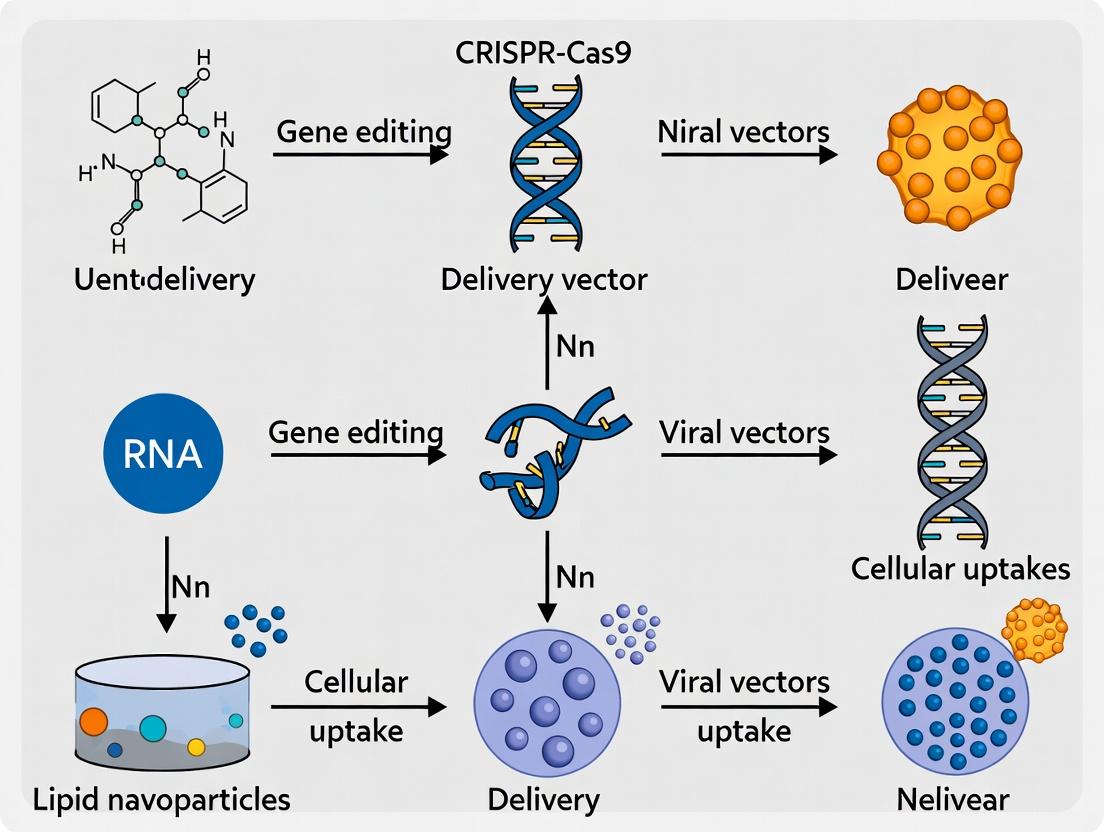
Engineering the Cargo: Viral and Non-Viral CRISPR-Cas9 Delivery Platforms in Action
Within the framework of a thesis focused on CRISPR-Cas9 delivery for neurodegenerative disease research, the selection of an appropriate Adeno-Associated Virus (AAV) serotype is a critical first step. The efficacy and specificity of gene editing are predicated on the precise delivery of CRISPR components to the relevant pathological cell types (e.g., neurons, astrocytes, microglia) in affected brain regions. AAVs offer a versatile platform, but their natural and engineered capsids exhibit distinct tropisms, governed by differential interactions with cell surface receptors, ability to cross barriers, and intracellular trafficking. This application note provides a comparative analysis of key AAV serotypes and detailed protocols for their evaluation in preclinical brain targeting.
Quantitative Serotype Comparison Table
The following table summarizes the relative tropism and key characteristics of commonly used AAV serotypes for central nervous system (CNS) applications, based on recent in vivo studies in rodents and non-human primates (NHPs).
Table 1: Comparative Tropism and Properties of AAV Serotypes for CNS Delivery
| Serotype | Primary CNS Cell Tropism (Relative Efficiency) | Strongly Targeted Brain Regions | Common Administration Routes | Notes & Key Receptors |
|---|---|---|---|---|
| AAV9 | Neurons (High), Astrocytes (Mod), Microglia (Low) | Cortex, Striatum, Spinal Cord, Cerebellum | Intravenous (IV), Intracerebroventricular (ICV), Intraparenchymal | Crosses BBB efficiently; Galactose, LamR, NRP1 |
| AAVrh.10 | Neurons (Very High), Astrocytes (Mod) | Cortex, Striatum, Substantia Nigra | IV, ICV | Similar to AAV9 with enhanced neuronal tropism in some regions. |
| AAV-PHP.eB | Neurons (High) | Widespread Cortex, Striatum, Thalamus | Systemic (IV) | Engineered variant; enhanced BBB crossing in C57BL/6J mice via Ly6a. |
| AAV-PHP.S | Peripheral & Sensory Neurons (High), CNS (Low) | Dorsal Root Ganglia | Systemic (IV) | Engineered for PNS targeting; limited CNS penetration. |
| AAV1 | Neurons (High), Astrocytes (Low) | Local spread around injection site | Intraparenchymal | Excellent for local transduction; N-linked sialic acid. |
| AAV2 | Neurons (Mod-High) | Local spread around injection site | Intraparenchymal | Classic serotype; heparan sulfate proteoglycan (HSPG). |
| AAV5 | Neurons (High), Astrocytes (Mod), Photoreceptors | Cortex, Local spread | Intraparenchymal, ICV | PDGFR, Sialic acid (2,3-linked). |
| AAV-DJ | Neurons (Mod), Astrocytes (Mod-High) | Widespread from injection site | Intraparenchymal | Chimeric capsid; broad tropism; HSPG, Lactose. |
| AAV6 | Astrocytes (High in some studies) | Local spread | Intraparenchymal | Utilizes HSPG and sialic acid; tropism can be variable. |
Experimental Protocols
Protocol 1: Rapid In Vivo Screening of AAV Serotype Tropism via Intracranial Injection
Objective: To compare the cellular tropism of different AAV serotypes for a specific brain region of interest (e.g., striatum for Huntington's disease models).
Materials:
- Purified AAV vectors (serotypes 1, 2, 5, 6, 9, DJ, etc.) encoding a reporter gene (e.g., EGFP, mCherry) under a ubiquitous promoter (CAG or CMV).
- Stereotaxic apparatus.
- Hamilton syringe (10 µL) with a 33-gauge needle.
- Adult C57BL/6 mice (8-12 weeks old).
- Anesthesia (Isoflurane).
- Perfusion pump and fixatives.
Procedure:
- Vector Preparation: Dilute all AAV vectors to an identical genomic titer (e.g., 1x10^12 vg/mL) in sterile PBS.
- Stereotaxic Surgery: Anesthetize mouse and secure in stereotaxic frame. Identify bregma and calculate coordinates for unilateral striatal injection (e.g., AP: +0.5 mm, ML: -2.0 mm, DV: -3.5 mm).
- Injection: Make a burr hole. Load 2 µL of AAV vector into the Hamilton syringe. Lower the needle to the target DV coordinate at a slow, steady rate. Infuse the virus at a rate of 0.2 µL/min. After infusion, wait 5 minutes before slowly retracting the needle.
- Post-op & Incubation: Suture the wound and allow animals to recover. Provide analgesia. Allow 3-4 weeks for robust transgene expression.
- Tissue Collection & Analysis: Perfuse mice transcardially with PBS followed by 4% PFA. Extract brains, post-fix, and section (40-50 µm) using a vibratome.
- Immunohistochemistry: Stain free-floating sections with antibodies against: Reporter (e.g., anti-GFP), Neuronal marker (NeuN), Astrocyte marker (GFAP), Microglia marker (Iba1). Use appropriate fluorescent secondary antibodies.
- Imaging & Quantification: Acquire high-resolution confocal images of the injection site. Use image analysis software (e.g., ImageJ, Imaris) to calculate the percentage of reporter-positive cells that co-localize with each cell type marker (n=3-5 animals per serotype).
Protocol 2: Assessing Systemic CNS Delivery via Tail Vein Injection
Objective: To evaluate the efficiency and regional distribution of BBB-crossing AAV serotypes (e.g., AAV9, AAV-PHP.eB) for whole-brain transduction.
Materials:
- AAV9-CAG-EGFP and AAV-PHP.eB-CAG-EGFP (1x10^11 vg/g body weight in 100 µL PBS).
- Mouse restrainer.
- 29-gauge insulin syringe.
- Heat lamp.
Procedure:
- Vector Administration: Warm mouse tail under a heat lamp for 1-2 minutes to dilate veins. Restrain mouse and inject the AAV preparation slowly into a lateral tail vein.
- Incubation: Allow 4-5 weeks for maximal CNS expression.
- Tissue Processing: Perfuse and fix animals as in Protocol 1. Section the brain to obtain coronal slices covering olfactory bulb to cerebellum.
- Whole-Brain Imaging: For gross assessment, image whole-brain slices under a fluorescent stereo microscope.
- Quantitative Analysis: Perform IHC as in Protocol 1. Quantify the number of transduced cells (EGFP+) per mm^2 in predefined regions (cortex, striatum, hippocampus, cerebellum, thalamus). Also, analyze the cellular tropism profile in each region via co-staining.
Visualizations
Title: AAV Serotype Selection Workflow for CRISPR Delivery
Title: AAV Cellular Uptake and Trafficking Pathway
The Scientist's Toolkit: Key Research Reagent Solutions
Table 2: Essential Materials for AAV Serotyping Experiments
| Item | Function & Rationale |
|---|---|
| High-Titer, Purified AAV Preps (Serotypes 1,2,5,6,9,rh.10,DJ,PHP.eB) | Core reagents. Must be CsCl or HPLC-purified to ensure purity and accurate titering for comparative studies. |
| Ubiquitous Promoter Plasmids (pAAV-CAG, pAAV-CBh) | To drive expression of reporter genes (EGFP, tdTomato) or CRISPR machinery independent of cell-type-specific promoters during tropism screening. |
| Stereotaxic Injection System | For precise, repeatable intracerebral delivery of AAV into defined brain coordinates in rodents. |
| Cell-Type-Specific Antibodies (Anti-NeuN, GFAP, Iba1, Olig2) | For immunohistochemical identification of neurons, astrocytes, microglia, and oligodendrocytes to quantify AAV tropism. |
| Confocal Microscope | Essential for high-resolution, multi-channel imaging to analyze co-localization of AAV reporter signal with cell markers. |
| qPCR Kit for AAV Genome Quantification (TaqMan probes for ITR) | To measure vector genome copies in different brain regions post-systemic injection, assessing biodistribution. |
| Ly6a (SCARA4) Genotyping Assay | Critical when using engineered capsids like PHP.eB, as their efficiency is mouse strain-dependent (high in C57BL/6J Ly6a+, low in others). |
The therapeutic application of CRISPR-Cas9 in neurodegenerative disease research is fundamentally constrained by the limited packaging capacity of adeno-associated virus (AAV) vectors, the most common delivery vehicle. Standard Streptococcus pyogenes Cas9 (SpCas9) exceeds the AAV cargo limit of ~4.7 kb. This application note details two primary strategies—split-Cas9 systems and miniaturized editors—to overcome this barrier, framed within the context of developing gene therapies for conditions like Huntington's, ALS, and Alzheimer's disease.
Table 1: Comparison of CRISPR-Cas Systems for AAV Packaging
| System | Total Size (kb) | AAV Packaging Requirement | Editing Efficiency (In Vivo CNS) | Key Advantage | Primary Limitation |
|---|---|---|---|---|---|
| Full-length SpCas9 | ~4.2 kb (Cas9) + gRNA (~0.5 kb) | Dual-AAV (Cas9 + gRNA/Donor) | ~5-15% (reported in mouse brain) | High activity, well-characterized | Requires cumbersome dual-vector co-delivery |
| saCas9 | ~3.3 kb (Cas9) + gRNA | Single AAV (with short promoter) | ~10-25% | Fits in single AAV with regulatory elements | Larger PAM (NNGRRT), more off-targets than SpCas9 |
| Cas12f (Cas14) | ~1.5-2.0 kb (Cas protein) + gRNA | Single AAV with ample space | ~1-10% (reporter systems) | Ultra-compact, single AAV delivery | Very low efficiency in mammalian cells |
| Split-SpCas9 (Intein) | ~Split halves (~2.1 kb each) | Dual-AAV (N-half + C-half/gRNA) | ~2-20% (highly variable) | Uses full SpCas9, better fidelity | Reconstitution efficiency limits potency |
| Prime Editor (PE) | PE2: ~6.3 kb | Dual or Triple AAV | ~5-30% in CNS (depends on design) | Precise editing, no DSBs | Severe packaging challenge, complex system |
Table 2: Recent In Vivo CNS Delivery Efficiency Data (2023-2024)
| Delivery System | Target/Model | Reported Editing Efficiency | Vector Dose (vg/kg) | Key Metric |
|---|---|---|---|---|
| Dual-AAV SaCas9 | HTT in Q175 mouse | 12.7% allele reduction (striatum) | 2.5e13 (total) | Reduction of mutant HTT protein |
| Dual-AAV Split-SpCas9 | mHTT in zQ175 mouse | Up to 45% indels (bulk tissue) | 5e12 per vector | High variability between neurons |
| Single AAV Anc80L65-saCas9 | BACE1 in APP/PS1 mouse | 35% indels (hippocampus) | 1e13 | Reduced amyloid plaques |
| Dual AAV PE (PEmax) | MECP2 in mouse brain | ~15% precise correction | 2.4e13 (total) | Proof-of-concept for precise editing |
Detailed Experimental Protocols
Protocol 3.1: Production of Dual-AAV for Intein-Mediated Split-SpCas9
Objective: To produce AAV vectors for reconstituting full-length SpCas9 via intein splicing in vivo. Materials:
- Plasmids: pAAV-Nhalf-Cas9 (N-terminal half with split intein), pAAV-Chalf-Cas9 (C-terminal half with split intein + gRNA expression cassette).
- HEK293T cells, PEI transfection reagent, AAV rep/cap plasmids (serotype 9 or PHP.eB for CNS), pHelper plasmid.
- Iodixanol gradient solutions, Amicon Ultra-15 centrifugal filters.
Procedure:
- Cell Seeding: Seed fifteen 15-cm dishes with HEK293T cells at 70% confluency.
- Transfection Mix: For each dish, prepare:
- 10 µg of either pAAV-Nhalf or pAAV-Chalf plasmid.
- 10 µg of AAV Rep/Cap plasmid (serotype defined).
- 20 µg of pHelper plasmid.
- 120 µL of PEI (1 mg/mL) in 2 mL Opti-MEM. Incubate 15 min, add dropwise to cells.
- Harvest: At 72 hr post-transfection, scrape cells, pellet by centrifugation. Resuspend in lysis buffer (150 mM NaCl, 50 mM Tris-HCl, pH 8.5), freeze-thaw 3x.
- Purification: Clarify lysate by centrifugation. Layer supernatant on iodixanol step gradient (15%, 25%, 40%, 60%). Ultracentrifuge at 350,000 x g for 1.5 hr.
- Collection & Concentration: Extract the 40% iodixanol fraction. Concentrate using a 100K MWCO Amicon filter. Buffer exchange to PBS + 0.001% Pluronic F-68.
- Titering: Quantify genomic titer (vg/mL) via ddPCR using primers against the WPRE or polyA signal.
Protocol 3.2: In Vivo Delivery and Analysis in a Neurodegenerative Mouse Model
Objective: To assess gene editing in the mouse CNS using split-Cas9 AAVs. Materials:
- Adult C57BL/6 mice or relevant disease model (e.g., zQ175 for Huntington's).
- Stereotaxic injection apparatus, Hamilton syringe.
- Dual-AAV preparations (N-half & C-half/gRNA), titer-matched (~1e13 vg/mL each).
- Perfusion pump, 4% PFA, cryostat, antibodies for IHC.
Procedure:
- Stereotaxic Injection: Anesthetize mouse, secure in stereotaxic frame. Identify coordinates for target region (e.g., Striatum: AP +0.5 mm, ML ±2.0 mm, DV -3.0 mm from Bregma).
- Virus Mix: Combine N-half and C-half AAVs in a 1:1 ratio (total volume 2-3 µL). Load into injection syringe.
- Infusion: Lower needle to target depth, infuse at 0.2 µL/min. Wait 5 min post-injection before slow needle withdrawal.
- Tissue Harvest: At 4-8 weeks post-injection, perfuse transcardially with PBS followed by 4% PFA. Extract brain, post-fix overnight, cryoprotect in 30% sucrose.
- Analysis:
- Sectioning: Cut 40 µm coronal sections on a cryostat.
- Immunohistochemistry: Stain for Cas9 (to confirm expression) and neuronal markers (NeuN). Use fluorescence in situ hybridization (FISH) for indel detection at the target locus if antibodies are unavailable.
- Deep Sequencing: Microdissect injected region, extract genomic DNA. Amplify target locus by PCR and perform next-generation sequencing (NGS) to quantify indel percentage.
Visualizations
Diagram Title: Split-Cas9 Intein Splicing Workflow
Diagram Title: Strategies to Overcome AAV Size Limits
The Scientist's Toolkit: Research Reagent Solutions
| Reagent / Material | Vendor Examples (2024) | Function in Experiment |
|---|---|---|
| AAVpro Helper Free System | Takara Bio | All-in-one plasmid system for high-titer AAV9 production in HEK293T cells. |
| pAAV-saCas9-U6-gRNA (PHP.eB) | Addgene (#135998) | Ready-to-pack single AAV plasmid expressing saCas9 and gRNA for CNS targeting. |
| Intein-Split SpCas9 Plasmids (N/C halves) | Addgene (#134963, #134964) | Essential for producing dual-AAV split-Cas9 systems. |
| AAV Serotype PHP.eB Cap Plasmid | Vigene Biosciences | Provides enhanced CNS tropism in rodents for more efficient neuronal transduction. |
| AAVance Concentration Kits | MilliporeSigma | Rapid concentration and purification of AAV from cell lysates. |
| ddPCR AAV Genome Titer Kit | Bio-Rad | Accurate, absolute quantification of AAV vector genome titer without standards. |
| NEBNext Ultra II FS DNA Library Prep Kit | NEB | Prepares amplicon libraries from edited genomic DNA for NGS-based indel analysis. |
| Cas9 (7A9-3A3) Mouse mAb | Cell Signaling Tech (#14697) | Antibody for detecting Cas9 protein expression in transfected/infected cells and tissue via IHC/IF. |
| Neurotrac Stereotaxic Dye | Precision Instruments | Visual confirmation of injection site accuracy during surgery. |
Within the thesis framework of developing CRISPR-Cas9 delivery systems for neurodegenerative disease research, Lipid Nanoparticles (LNPs) have emerged as a leading non-viral platform. Their modularity allows for rational design to overcome two paramount barriers: (1) crossing the blood-brain barrier (BBB) and (2) achieving cell-specific targeting within the central nervous system (CNS), such as neurons, astrocytes, or microglia. This document outlines key formulation strategies, quantitative benchmarks, and detailed protocols for evaluating LNP performance in CNS applications.
Key Application Notes:
- Ionizable Cationic Lipid is Central: The ionizable lipid determines endosomal escape efficiency, the critical rate-limiting step for functional intracellular delivery of CRISPR ribonucleoprotein (RNP) or mRNA.
- PEGylation is a Double-Edged Sword: While polyethylene glycol (PEG)-lipids stabilize LNPs and prevent rapid clearance, their presence can hinder cellular uptake and endosomal escape. Employing cleavable PEG-lipids (e.g., via enzyme-sensitive linkages) is a strategic priority.
- Targeting Requires a Multi-Faceted Approach: Passive targeting via size control (~80-120 nm) and charge modulation (slightly negative to neutral zeta potential) promotes BBB penetration. Active targeting requires the conjugation of ligands (e.g., peptides, antibody fragments) to the LNP surface, often via PEG-lipid terminals.
- Payload Matters: Encapsulating CRISPR-Cas9 mRNA plus sgRNA is currently more efficient than direct RNP encapsulation, but RNP delivery offers faster action and reduced immunogenicity risks.
Table 1: Benchmarking LNP Formulations for CNS Delivery
| Formulation Parameter | Standard System (Liver-Tropic) | Optimized CNS-Penetrant System | Functional Impact |
|---|---|---|---|
| Average Size (nm) | 70-90 nm | 80-120 nm | Favors enhanced permeability and retention (EPR) at the BBB. |
| Polydispersity Index | < 0.15 | < 0.2 | Maintains batch homogeneity and consistent biodistribution. |
| Zeta Potential | Slightly negative (~ -5 mV) | Near-neutral to slightly negative (-3 to +2 mV) | Reduces non-specific clearance, improves BBB interaction. |
| Ionizable Lipid pKa | ~6.4-6.6 | ~6.6-6.9 | Optimal endosomal disruption within milder endosomal pH of neural cells. |
| PEG-Lipid % (mol) | 1.5 - 2.5% | 0.5 - 1.5% (Cleavable) | Minimizes "PEG dilemma," improves cellular uptake post-BBB crossing. |
| Encapsulation Efficiency (CRISPR mRNA) | > 90% | > 85% | High payload protection is critical for in vivo efficacy. |
| In Vivo CNS Tropism (vs. Liver) | Liver: > 95% | Liver: ~70-80%; Brain: 3-5% fold increase | Modest absolute brain accumulation (~1-2% ID/g) is often therapeutically relevant. |
Table 2: Targeting Ligands for CNS Cell Types
| Target Cell | Ligand Type | Example Sequence/Target | Conjugation Method | Key Benefit |
|---|---|---|---|---|
| BBB Endothelium (Transcytosis) | Peptide | Angiopep-2 (TFFYGGSRGKRNNFKTEEY) | Maleimide-thiol to PEG terminus | Binds LRP1 receptor for receptor-mediated transcytosis. |
| Neurons | Peptide | Tet1 (HLNILSTLWKYR) | Maleimide-thiol to PEG terminus | Binds ganglioside GT1b, internalized via retrograde transport. |
| Astrocytes | Antibody Fragment | Anti-GFAP scFv | Strain-promoted azide-alkyne cycloaddition (SPAAC) | High specificity for glial fibrillary acidic protein. |
| Microglia | Peptide | MG1 (CSSRTMHHC) | Maleimide-thiol to PEG terminus | Selectively binds to microglial surface markers. |
Experimental Protocols
Protocol 1: Microfluidic Formulation of CRISPR-LNPs Objective: Reproducibly produce stable, size-controlled LNPs encapsulating CRISPR-Cas9 mRNA. Materials: See Scientist's Toolkit. Procedure:
- Prepare the Aqueous Phase: Dilute CRISPR-Cas9 mRNA (and sgRNA if separate) in 50 mM citrate buffer, pH 4.0, to a final concentration of 0.1 mg/mL.
- Prepare the Lipid Phase: Combine ionizable cationic lipid, DSPC, cholesterol, and PEG-lipid (with or without functionalized terminal) in ethanol at a molar ratio of 50:10:38.5:1.5. Use a total lipid concentration of 10 mM.
- Formulation: Using a staggered herringbone or microfluidic mixer (e.g., NanoAssemblr), set the Aqueous:Organic flow rate ratio to 3:1. Pump both phases at a total flow rate of 12 mL/min to ensure rapid mixing.
- Dialyze: Immediately collect the effluent in a dialysis cassette (MWCO 20 kDa). Dialyze against 1X PBS (pH 7.4) for 4 hours at 4°C, with one buffer change after 2 hours.
- Concentrate & Filter: Concentrate LNPs using centrifugal filters (100 kDa MWCO). Sterile-filter through a 0.22 µm PES membrane.
- Characterize: Measure hydrodynamic diameter and PDI by DLS. Determine encapsulation efficiency using a Ribogreen assay.
Protocol 2: In Vitro BBB Transwell Model Assay Objective: Assess LNP ability to cross a simulated BBB. Materials: hCMEC/D3 cell line, Transwell inserts (3.0 µm pore), TEER measurement system. Procedure:
- Culture BBB Monolayer: Seed hCMEC/D3 cells on collagen-coated Transwell inserts at 50,000 cells/cm². Culture for 5-7 days until TEER values stabilize > 40 Ω·cm².
- Test LNP Transport: Add LNPs (50 µg lipid/mL) to the apical (luminal) chamber. Incubate at 37°C.
- Sample & Quantify: At T=1, 2, 4, and 8 hours, collect 100 µL from the basolateral (abluminal) chamber. Replenish with fresh medium.
- Analyze: Quantify payload (mRNA) in basolateral samples via qRT-PCR. Calculate apparent permeability (Papp) using the formula:
Papp = (dQ/dt) / (A * C0), where dQ/dt is the transport rate, A is the membrane area, and C0 is the initial apical concentration. - Validate Integrity: Monitor TEER before and after experiment. Use lucifer yellow (1 mM) as a paracellular leakage control.
Diagrams
Title: LNP Journey for CRISPR CNS Delivery
Title: Core Experimental Workflow for CNS LNPs
The Scientist's Toolkit
Table 3: Essential Research Reagent Solutions
| Item | Function & Rationale | Example/Supplier |
|---|---|---|
| Ionizable Cationic Lipid | Core structural lipid; protonates in endosome to enable membrane disruption and payload release. Critical for endosomal escape. | DLin-MC3-DMA (MC3), SM-102, ALC-0315 (Acuitas). |
| Helper Lipids (DSPC, Cholesterol) | DSPC enhances structural integrity and fusogenicity. Cholesterol stabilizes LNP bilayer and promotes cellular uptake. | 1,2-distearoyl-sn-glycero-3-phosphocholine (DSPC). |
| PEG-Lipid (Functionalizable) | Provides steric stabilization, controls size, and offers a conjugation point for targeting ligands. Cleavable versions (e.g., disulfide) are preferred. | DMG-PEG2000, DSG-PEG2000 (maleimide-terminated). |
| Microfluidic Mixer | Enables reproducible, scalable, and rapid mixing for forming monodisperse, stable LNPs. | NanoAssemblr (Precision NanoSystems), microfluidic chips. |
| Dialysis Cassette | Removes ethanol and exchanges buffer post-formulation without damaging fragile LNPs. | Slide-A-Lyzer (MWCO 20 kDa, Thermo Fisher). |
| Ribogreen Assay Kit | Quantifies encapsulated vs. free nucleic acid payload to determine encapsulation efficiency (%) . | Quant-iT RiboGreen RNA Assay (Thermo Fisher). |
| hCMEC/D3 Cell Line | A well-characterized human immortalized brain endothelial cell line for modeling the BBB in vitro. | Merck Millipore. |
| TEER Measurement System | Measures Trans-Endothelial Electrical Resistance to validate the integrity of the BBB monolayer. | EVOM2 Voltohmmeter (World Precision Instruments). |
| Strain-Promoted Conjugation Kit | Enables efficient, copper-free click chemistry for conjugating sensitive ligands (e.g., antibodies) to LNPs. | DBCO-PEG-Lipid and Azide-modified ligand. |
Within the broader thesis on CRISPR-Cas9 delivery for neurodegenerative diseases, exosomes and extracellular vesicles (EVs) present a transformative, bio-inspired platform. These natural lipid nanovesicles facilitate intercellular communication and can be engineered to cross the blood-brain barrier (BBB), delivering therapeutic CRISPR cargo to neuronal targets. This document provides detailed application notes and protocols for harnessing EVs in this context.
EV Biogenesis, Engineering, and Cargo Loading: Core Mechanisms
Exosomes (30-150 nm) originate from the endosomal pathway, specifically the inward budding of multivesicular bodies (MVBs). Their native composition of tetraspanins (CD63, CD81), lipids, and adhesion molecules confers inherent stability, low immunogenicity, and tropism. For therapeutic use, they must be engineered to package and deliver CRISPR-Cas9 components (Cas9 mRNA/sgRNA or ribonucleoprotein (RNP)).
Diagram 1: EV Biogenesis & Engineering for CRISPR
Table 1: Primary Methods for CRISPR-Cas9 Loading into EVs
| Loading Method | Mechanism | Typical Loading Efficiency | Advantages | Disadvantages |
|---|---|---|---|---|
| Transfection of Donor Cells | Transfect parent cells with plasmid DNA/mRNA encoding Cas9 and sgRNA; EVs bud with cargo. | 5-20% (protein) | Simple, maintains natural EV biogenesis. | Low efficiency, heterogeneous cargo, off-target cell modulation. |
| Electroporation | Electric pulses create pores in isolated EVs to allow Cas9 RNP entry. | 10-25% (RNP) | Direct RNP loading, good for pre-formed complexes. | Can cause EV aggregation, cargo aggregation. |
| Sonication | Ultrasound disrupts EV membrane, allowing cargo diffusion, followed by membrane repair. | 15-30% (RNP) | Higher efficiency than electroporation for some cargoes. | Harsh, may alter EV surface characteristics. |
| Chemical Transfection | Use of commercial transfection reagents with isolated EVs. | 10-20% (mRNA) | Commercially available, simple protocol. | Potential reagent toxicity, need for purification. |
| Active Packaging via Fusion Proteins | Engineer Cas9 with EV-sorting domains (e.g., CD63-GFP, C1C2 domain of lactadherin). | 20-40% (protein) | High specificity, controlled loading. | Requires genetic engineering of donor cells. |
Protocol: Production and Electroporation of EVs for Cas9 RNP Delivery
A. EV Isolation from HEK-293T or MSC Culture (Ultracentrifugation Protocol)
Objective: Isolate EVs from conditioned medium for subsequent engineering. Materials: See "Research Reagent Solutions" below. Procedure:
- Cell Culture: Culture HEK-293T or mesenchymal stem cells (MSCs) in EV-depleted FBS medium to 80% confluency.
- Conditioned Medium Collection: Harvest culture medium after 48 hours. Centrifuge at 300 × g for 10 min (pellet cells), then 2,000 × g for 20 min (dead cells), then 10,000 × g for 30 min (4°C) to remove large debris.
- Ultracentrifugation: Filter supernatant through a 0.22 µm PES filter. Ultracentrifuge at 100,000 × g, 4°C for 70 min. Discard supernatant.
- EV Wash & Resuspension: Gently resuspend pellet in large volume of sterile PBS. Perform a second ultracentrifugation at 100,000 × g, 4°C for 70 min. Resuspend final EV pellet in 100-200 µL sterile PBS. Aliquot and store at -80°C.
- Characterization: Perform Nanoparticle Tracking Analysis (NTA) for size/concentration, Western Blot for markers (CD63, TSG101, Alix), and TEM for morphology.
B. Electroporation of Cas9 RNP into Isolated EVs
Objective: Actively load pre-assembled Cas9 protein and sgRNA complex into isolated EVs. Procedure:
- RNP Complex Assembly: Assemble Cas9 protein (e.g., 10 µg) and target sgRNA (molar ratio 1:3) in duplex buffer. Incubate at 25°C for 10 min.
- Electroporation Setup: Mix 50 µg EVs (in PBS) with assembled RNP complex (total volume ≤ 100 µL). Transfer to a 2 mm electroporation cuvette.
- Electroporation: Apply a single pulse (500 V, 5 ms) using a square-wave electroporator. Immediately add 500 µL of pre-warmed complete medium and incubate at 37°C for 30 min for membrane recovery.
- Purification: To remove unloaded RNP, layer the mixture on a qEV original size-exclusion column (Izon). Collect EV-rich fractions (typically 7-9). Concentrate if necessary using a 100 kDa MWCO centrifugal filter.
- Quality Control: Verify loading via Western Blot for Cas9, measure particle concentration (NTA), and test functional delivery in a reporter cell line.
Diagram 2: EV Electroporation & Purification Workflow
Application Notes: Targeting EVs to Neurons for Neurodegenerative Disease Models
Table 2: Strategies for Neuron-Specific EV Targeting
| Targeting Motif | Conjugation Method | Ligand/Peptide | Intended Receptor on Neuron | Thesis Application Rationale |
|---|---|---|---|---|
| RVG Peptide | Genetic fusion to EV membrane protein (Lamp2b) or chemical conjugation. | Rabies Virus Glycoprotein (29 aa) | Nicotinic Acetylcholine Receptor | Well-established for BBB crossing and neuronal targeting. |
| Neuron-Specific Aptamer | Click chemistry to EV surface lipids or to engineered tetraspanin-fused SNAP-tag. | RNA/DNA aptamer (e.g., against NCAM) | Neuron-specific surface markers (NCAM) | High specificity, reduces off-target glial delivery. |
| Lamp2b Fusion | Genetic engineering of donor cells to express Lamp2b fused to targeting peptide. | Various (e.g., RVG, BDNF) | Corresponding receptor | Utilizes endogenous EV biogenesis for surface display. |
| MSC Innate Tropism | None (use naïve MSC-EVs). | Native surface proteins (e.g., integrins) | Endothelial/Neuronal adhesion molecules | Leverages natural MSC-EV homing to sites of injury (e.g., neuroinflammation). |
Key Consideration: Post-loading and engineering, always re-characterize EV size, concentration, and surface markers to ensure engineering steps did not compromise EV integrity.
The Scientist's Toolkit: Research Reagent Solutions
Table 3: Essential Materials for EV-CRISPR Research
| Item | Function/Description | Example Product/Cat. No. |
|---|---|---|
| EV-Depleted FBS | Fetal bovine serum processed to remove bovine EVs, essential for clean EV production. | Gibco Exosome-Depleted FBS (A2720801) |
| Ultracentrifuge | Instrument for differential centrifugation to isolate EVs via high g-force. | Optima XPN-100 (Beckman Coulter) |
| Size Exclusion Columns | For gentle purification of EVs from free protein/RNP after loading. | qEVoriginal 70nm (Izon Science) |
| Nanoparticle Tracker | Measures EV size distribution and concentration via light scattering. | NanoSight NS300 (Malvern) |
| Cas9 Nuclease | Recombinant S. pyogenes Cas9 protein for RNP complex assembly. | TrueCut Cas9 Protein v2 (A36498) |
| Electroporator | Square-wave electroporator for efficient RNP loading into EVs. | Gene Pulser Xcell (Bio-Rad) |
| Tetraspanin Antibodies | For Western Blot validation of EV markers (CD63, CD81, TSG101). | Anti-CD63 (TS63), Abcam |
| Lamp2b-RVG Plasmid | Donor construct for engineering EVs with neuronal targeting peptide. | pLJM1-EGFP-Lamp2b-RVG (Addgene #122254) |
| 0.22 µm PES Filter | Sterile filtration of conditioned medium prior to UC. | Stericup (Millipore) |
| 100kDa MWCO Filter | Concentrates EV suspensions post-SEC. | Amicon Ultra-4 Centrifugal Filter (UFC810024) |
Within the thesis on CRISPR-Cas9 delivery for neurodegenerative diseases, a principal challenge is bypassing the blood-brain barrier (BBB). Focused ultrasound (FUS) coupled with systemically injected microbubbles (MBs) offers a non-invasive, transient, and spatially targeted method for BBB disruption (BBBD), enabling the entry of viral vectors (e.g., AAV) or lipid nanoparticles carrying CRISPR-Cas9 payloads. This application note details the current protocols and quantitative insights for integrating this technology into preclinical research.
Table 1: Typical FUS Parameters for Preclinical BBB Disruption
| Parameter | Typical Range | Common Setting (Example) | Notes |
|---|---|---|---|
| Frequency | 0.25 - 1.5 MHz | 0.5 MHz | Lower frequencies increase focal volume and BBB effect. |
| Peak Negative Pressure (PNP) | 0.3 - 0.8 MPa | 0.45 MPa | Pressure calibrated in situ; critical for safety/efficacy. |
| Pulse Length | 5 - 20 ms | 10 ms | |
| Pulse Repetition Frequency (PRF) | 1 - 10 Hz | 2 Hz | |
| Sonication Duration | 60 - 120 s per target | 90 s | |
| Microbubble Dose | 1x10^7 - 1x10^8 bubbles/kg | 2x10^7 bubbles/kg | Definity or similar; bolus injection. |
Table 2: Outcomes of FUS-MB Mediated Delivery for Neurodegenerative Research
| Measured Outcome | Typical Result Range | Key Determining Factors | Assay/Detection Method |
|---|---|---|---|
| BBBD Duration (Transience) | 4 - 12 hours | PNP, MB dose, age/animal model. | Contrast-enhanced MRI (T1w). |
| Vector (AAV) Delivery Efficiency | 10-100x increase in target region vs. contralateral. | Serotype, time of injection relative to FUS. | Bioluminescence, qPCR, IHC. |
| Cas9 Editing Efficiency In Vivo | Varies (e.g., 5-40% allele modification) | Vector, guide RNA design, promoter, time point. | NGS, T7E1 assay, in situ hybridization. |
| Neuron-Specific Transduction | >70% of delivered cells (with AAV-PHP.eB/CamKIIa) | Vector serotype and promoter selection. | Immunohistochemistry co-localization. |
Experimental Protocols
Protocol 1: Preclinical Setup and FUS-MB Mediated BBBD in Mice
Objective: To transiently disrupt the BBB in a targeted brain region (e.g., hippocampus for Alzheimer's research) to enable vector entry.
Materials:
- Anesthetized mouse (e.g., C57BL/6) on stereotaxic frame with warming pad.
- MRI-guided or optically registered FUS system (e.g., RK-100, FUS Instruments).
- Ultrasound coupling gel/degassed water column.
- Definity or custom lipid-shelled microbubbles.
- MRI contrast agent (e.g., Gd-DTPA).
- Small animal MRI system (optional but recommended for targeting/confirmation).
Procedure:
- Anesthesia & Preparation: Anesthetize mouse (isoflurane/O2). Secure head in stereotaxic apparatus. Depilate scalp. Apply coupling medium.
- Targeting: Use pre-acquired MRI scans to register FUS transducer coordinates to the target brain region (e.g., unilateral hippocampus). Alternatively, use anatomical landmarks with a pre-calibrated system.
- Microbubble Administration: Dilute commercial microbubbles in saline to desired concentration. Inject via tail vein as a bolus (e.g., 100 µL) immediately prior to sonication.
- Sonication: Initiate FUS sonication using parameters from Table 1. Monitor for stable vital signs.
- BBBD Confirmation (Optional): 5 minutes post-FUS, inject MRI contrast agent. Acquire T1-weighted MRI scans to confirm localized BBBD as hyperintense signal.
- Vector Administration: Immediately or within 30-120 minutes post-BBBD, intravenously inject the CRISPR-Cas9 delivery vector (e.g., 1x10^11 vg AAV9).
Protocol 2: Assessment of CRISPR-Cas9 Delivery and Editing Post-FUS
Objective: To quantify vector biodistribution and gene editing efficiency following FUS-MB facilitated delivery.
Materials:
- Tissue homogenizer.
- RNA/DNA extraction kits.
- qPCR system and TaqMan probes for vector genome quantification.
- Next-generation sequencing (NGS) platform or T7 Endonuclease I assay kit.
- Antibodies for immunohistochemistry (Neuronal nuclei, Cas9 protein, target protein).
Procedure:
- Tissue Collection: At appropriate endpoint (e.g., 2-4 weeks for expression, 8+ weeks for stable editing), perfuse animal transcardially with PBS. Dissect brain into targeted and contralateral regions, and other organs (liver, spleen) for biodistribution.
- Vector Biodistribution: Homogenize tissues. Extract genomic DNA. Perform absolute quantification of vector genomes per diploid genome (vg/dg) using qPCR with serotype-specific primers/probes against the vector genome and a reference host gene.
- Editing Efficiency Analysis:
- NGS: Design amplicons spanning the target genomic locus. PCR amplify from extracted DNA, prepare NGS library, and sequence. Analyze reads for indel percentages.
- T7E1 Assay: PCR amplify target region from mixed alleles. Heat-denature and re-anneal PCR products. Digest with T7 Endonuclease I, which cleaves heteroduplex DNA formed by WT and edited strands. Analyze fragment sizes on gel; calculate approximate editing efficiency.
- Immunohistochemical Validation: Fix brain in 4% PFA, section (40 µm). Stain for Cas9 protein and neuronal marker (NeuN). Image with confocal microscopy to confirm neuronal co-localization.
The Scientist's Toolkit: Research Reagent Solutions
Table 3: Essential Materials for FUS-Mediated CRISPR Delivery
| Item | Function | Example Product/Note |
|---|---|---|
| FUS System with Targeting | Precisely delivers acoustic energy to disrupt BBB in a focal volume. | RK-100 (FUS Instruments); Image-guided systems. |
| Clinical Grade Microbubbles | Oscillate under FUS, mechanically stressing capillary walls to open TJs. | Definity (Lantheus); SonoVue. |
| AAV Vector (CRISPR-Cas9) | Delivers genetic cargo; serotype choice affects tropism and immune response. | AAV9, AAV-PHP.eB, AAV-retro for specific trafficking. |
| MRI Contrast Agent (Gd-based) | Validates location and degree of BBBD via leakage into brain parenchyma. | Gadovist; Magnevist. |
| In Vivo Imaging System | Tracks vector biodistribution and transgene expression longitudinally. | IVIS Spectrum (if luciferase reporter); MRI. |
| TaqMan qPCR Assay | Quantifies vector genome copies in tissue with high specificity and sensitivity. | Custom assays for ITR or Cas9 sequence. |
| Next-Generation Sequencer | Gold-standard for quantifying precise genome editing efficiency and profiles. | Illumina MiSeq; for deep amplicon sequencing. |
| Cas9-Specific Antibody | Detects Cas9 protein presence and cellular localization in situ. | Clone 7A9-3A3 (Cell Signaling); anti-SpCas9. |
Visualizations
Title: Workflow for FUS CRISPR Delivery
Title: FUS Microbubble Mechanism for BBB Opening
Title: Key Steps for Experimental Validation
This Application Note details strategies for CRISPR-Cas9-mediated genome editing within the context of developing cell-based therapies for neurodegenerative diseases (NDs) such as Parkinson's, Huntington's, and ALS. Two principal paradigms exist: In Vivo editing, where genetic corrections are made directly within a patient's body, and Ex Vivo editing, where patient-derived cells are genetically modified outside the body and then transplanted back. The choice of strategy hinges on disease pathophysiology, target cell type, delivery challenges, and safety profile.
Ex Vivo Advantages: Precise control over editing efficiency, thorough pre-transplant characterization, and the ability to select and expand correctly edited clones. It is particularly suited for editing autologous hematopoietic stem cells (HSCs), mesenchymal stem cells (MSCs), or induced pluripotent stem cell (iPSC)-derived neuronal progenitors. In Vivo Advantages: Avoids complex cell manufacturing, enables targeting of hard-to-culture or non-dividing cells (e.g., mature neurons, glia), and is potentially applicable to a broader patient population without individualized cell products.
For NDs, ex vivo strategies often focus on engineering supportive cells (e.g., astrocytes expressing neurotrophic factors) or correcting patient-derived iPSCs. In vivo strategies aim to directly edit neurons in situ to correct mutations or modulate disease-associated genes.
Quantitative Data Comparison
Table 1: Comparative Analysis of In Vivo vs. Ex Vivo Editing Strategies for Neurodegenerative Disease Applications
| Parameter | In Vivo Strategy | Ex Vivo Strategy |
|---|---|---|
| Primary Target Cells | Neurons, astrocytes, microglia in situ | Patient-derived iPSCs, HSCs, MSCs, fibroblasts in culture |
| Typical Delivery Vector | AAV, lipid nanoparticles (LNPs) | Electroporation, nucleofection, viral transduction (LV, RV) |
| Average Editing Efficiency | Variable (1-60%), highly tissue/delivery dependent | High (often >70% for in vitro assays) |
| Off-Target Risk Assessment | Challenging; requires deep sequencing of target tissue | More straightforward; performed on clonal populations pre-transplant |
| Immune Response Concern | High (to Cas9, AAV, edited cells) | Moderate (to edited cells post-transplant) |
| Manufacturing Complexity | Lower (vector production) | Higher (cell culture, editing, QC, expansion) |
| Time to Therapeutic Effect | Potentially faster | Slower (includes cell manufacturing time) |
| Key Challenge | Safe & efficient delivery to CNS; immunogenicity | Maintaining cell viability, identity, and function post-editing |
| Thesis Context Example | AAV9-Cas9/gRNA injection to silence mutant HTT in striatum | Transplant of CRISPR-corrected, patient iPSC-derived dopaminergic neurons for PD |
Table 2: Example Editing Outcomes for ND-Relevant Genes (Recent Data)
| Target Gene (Disease) | Strategy | Cell/Model Type | Efficiency (% INDEL or Correction) | Key Readout |
|---|---|---|---|---|
| HTT (Huntington's) | In Vivo (AAV-CRISPR) | Mouse striatum | ~40% INDEL reduction in mutant HTT protein | Behavioral improvement, reduced mHTT aggregates |
| SNCA (Parkinson's) | Ex Vivo (CRISPRi) | Patient-derived iPSCs | >90% repression of α-synuclein | Reduced pathological α-synuclein in differentiated neurons |
| SOD1 (ALS) | In Vivo (LNP-mRNA) | SOD1G93A mouse spinal cord | ~50% reduction in mutant SOD1 mRNA | Delayed disease onset, extended survival |
| APP (Alzheimer's) | Ex Vivo (Base Editing) | Human iPSC-derived neurons | ~35% precise point correction | Decreased pathogenic Aβ42/40 ratio |
Detailed Experimental Protocols
Protocol 3.1: Ex Vivo Editing of Patient Fibroblasts for iPSC Generation and Neuronal Differentiation
Objective: Create an isogenic, gene-corrected iPSC line from a patient with a monogenic ND.
- Skin Biopsy & Fibroblast Culture: Culture patient dermal fibroblasts in DMEM + 10% FBS.
- CRISPR RNP Complex Formation: For a 10 µL nucleofection reaction, combine 5 µg of purified S.p. Cas9 protein with 3 µg of synthetic sgRNA (targeting the mutation site). Incubate 10 min at RT.
- Electroporation & HDR: Resuspend 1x10^5 fibroblasts in 100 µL P3 Primary Cell Solution (Lonza). Add RNP complex and 2 µg of ssODN HDR template containing the correction and a silent restriction site. Electroporate using program DS-150 on a 4D-Nucleofector.
- Recovery & Single-Cell Cloning: Plate cells in fibroblast medium. After 48 hrs, trypsinize and seed at ~1 cell/well in 96-well plates. Expand clones for 2-3 weeks.
- Genotype Screening: Perform PCR on genomic DNA from each clone. Use restriction fragment length polymorphism (RFLP) assay (if silent site introduced) and Sanger sequencing to identify corrected isogenic clones.
- Reprogramming: Using a non-integrating Sendai virus vector kit (e.g., CytoTune-iPS 2.0), reprogram corrected fibroblasts to iPSCs. Characterize pluripotency markers (Nanog, OCT4) and karyotype.
- Directed Differentiation: Differentiate corrected and uncorrected iPSCs into relevant neuronal lineage (e.g., midbrain dopaminergic neurons using dual SMAD inhibition). Assess functional rescue (electrophysiology, disease-relevant assays).
Protocol 3.2: In Vivo Editing in a Mouse Model of Neurodegeneration via AAV Delivery
Objective: Perform in vivo knockdown of a dominant disease allele in the adult mouse CNS.
- Vector Design & Production: Clone a SaCas9 or compact Cas9 variant (for AAV packaging) and a disease allele-specific gRNA into an AAV vector genome under separate U6 and GFAP/CamKIIa promoters (for glial/neuronal specificity). Package into AAV9 or AAV-PHP.eB capsids via triple transfection in HEK293 cells and purify via iodixanol gradient.
- Animal Stereotactic Surgery: Anesthetize adult disease model mice (e.g., SOD1G93A). Secure in stereotactic frame. Perform craniotomy. Inject 2 µL of purified AAV-CRISPR (titer ≥ 1x10^13 vg/mL) bilaterally into the target region (e.g., striatum: AP +1.0 mm, ML ±2.0 mm, DV -3.5 mm from bregma) at 0.4 µL/min.
- Post-Op & Expression Wait Period: Allow 4-6 weeks for robust Cas9/gRNA expression and editing.
- Tissue Harvest & Analysis: Perfuse mice transcardially with PBS. Dissect out injected brain regions. For genomic analysis: extract DNA, perform T7 Endonuclease I (T7EI) or ICE analysis to quantify INDEL frequency. For molecular analysis: perform Western blot or immunofluorescence to assess reduction in mutant protein levels and evaluate neurodegenerative markers.
Visualizations
Title: Ex Vivo vs In Vivo CRISPR Workflow for NDs
Title: Overcoming CNS Delivery Barriers for In Vivo Editing
The Scientist's Toolkit: Research Reagent Solutions
Table 3: Essential Reagents for Editing Patient-Derived Cells for Transplantation
| Reagent / Solution | Vendor Examples | Function in Protocol |
|---|---|---|
| S.p. Cas9 Nuclease, HiFi | IDT, Thermo Fisher | High-fidelity nuclease for precise cutting; reduces off-target effects in ex vivo editing. |
| Chemically Modified sgRNA | Synthego, IDT | Enhanced stability and reduced immunogenicity; crucial for both RNP complex formation and AAV packaging. |
| ssODN HDR Template | IDT, Genewiz | Single-stranded DNA donor template for precise knock-in or point mutation correction during ex vivo editing. |
| 4D-Nucleofector X Unit & P3 Kit | Lonza | System for efficient delivery of CRISPR RNP into hard-to-transfect primary patient cells (fibroblasts, stem cells). |
| mTeSR Plus / Essential 8 | STEMCELL Technologies | Defined, feeder-free medium for maintaining genomic stability of human iPSCs post-editing. |
| AAVpro Purification Kit | Takara Bio | All-in-one system for purification and concentration of high-titer AAV vectors for in vivo studies. |
| T7 Endonuclease I | NEB | Enzyme for mismatch cleavage assay; rapid genotyping to estimate editing efficiency in bulk populations. |
| AAV9 or AAV-PHP.eB Capsids | Addgene, Vigene | Serotypes with enhanced tropism for CNS cells for in vivo delivery of CRISPR components. |
| Neurobasal/B-27 Supplement | Thermo Fisher | Base medium for the differentiation and long-term maintenance of edited neurons from iPSCs. |
| Rho-associated kinase (ROCK) inhibitor Y-27632 | Tocris | Improves survival of single dissociated cells (e.g., after nucleofection or during cloning). |
Balancing Efficiency and Safety: Optimizing Delivery for Specificity and Minimizing Off-Targets
Therapeutic delivery of CRISPR-Cas9 components to the central nervous system (CNS) represents a promising frontier for treating neurodegenerative diseases like Alzheimer's, Parkinson's, and ALS. The primary challenge lies in the blood-brain barrier (BBB), a highly selective semipermeable border that prevents most circulating agents from entering the brain. Simultaneously, systemically administered delivery vectors, such as lipid nanoparticles (LNPs) or viral vectors, are rapidly sequestered by the liver and spleen, significantly reducing the available dose for brain targeting. This application note details strategies and protocols to optimize dosage and biodistribution for CNS-targeted CRISPR-Cas9 delivery, aiming to maximize brain uptake while minimizing hepatic sequestration.
The following table summarizes current strategies, their mechanisms, and quantitative outcomes based on recent literature.
Table 1: Strategies for Optimizing Brain Uptake and Reducing Liver Sequestration
| Strategy | Mechanism of Action | Typical Vector | Quantitative Impact on Brain Uptake (vs. baseline) | Quantitative Impact on Liver Reduction (vs. baseline) | Key References (Recent) |
|---|---|---|---|---|---|
| Surface Functionalization (Ligand Targeting) | Engages receptor-mediated transcytosis (RMT) at BBB (e.g., via Transferrin, LDL, Insulin receptors). | LNP, AAV, Polymer | Increase of 2-5x in brain parenchyma; up to 50x increase in endothelial cells. | Variable; depends on ligand. Some can increase off-target accumulation. | Kumar et al., 2023; Zhou et al., 2022 |
| Charge Modulation (Neutral/Slightly Negative) | Reduces non-specific electrostatic interactions with anionic heparan sulfate proteoglycans on liver sinusoids. | LNP | ~3x increase in brain delivery. | Can reduce liver uptake by 40-60%. | Dobrowolski et al., 2022; Cheng et al., 2021 |
| PEGylation Density & PEG Shield Shedding | Initial PEG corona reduces opsonization and liver uptake. Acid-cleavable or targeting ligand-revealing PEG enhances BBB crossing. | LNP | Up to 10x increase with cleavable PEG + targeting. | Initial long PEG reduces liver uptake by ~70%; cleavable design avoids dose-limiting pharmacokinetics. | Rosenblum et al., 2020; Terstappen et al., 2021 |
| Dose Optimization (Pre-dosing/Decoy) | Saturation of peripheral uptake mechanisms (e.g., with empty carriers or competing ligands) to saturate liver macrophages. | LNP, AAV | 2-3x increase in brain fluorescence or gene expression. | Can reduce liver sequestration by 30-50%. | Suzuki et al., 2022; Weiss et al., 2023 |
| Administration Route (Intracerebroventricular/Intrathecal) | Direct bypass of BBB and systemic circulation. | AAV, LNPs | High local concentration; minimal systemic distribution. | Liver exposure reduced by >90%. | Meyer et al., 2023; ClinicalTrials.gov |
| Vector Engineering (Capsid Selection/Engineering) | Use of natural/engineered AAV capsids with tropism for brain endothelial cells or neurons (e.g., AAV-PHP.eB, AAV.CAP-B10). | AAV | Engineered capsids can increase CNS transduction by 100-1000x in mice vs. AAV9. | Specific variants show reduced hepatotropism. | Goertsen et al., 2022; Tabebordbar et al., 2023 |
Experimental Protocols
Protocol 1: Formulation and Characterization of Brain-Targeted CRISPR-LNPs
Objective: Prepare and characterize lipid nanoparticles (LNPs) encapsulating Cas9 mRNA and sgRNA, functionalized with a BBB-targeting ligand.
Materials:
- Ionizable lipid (e.g., DLin-MC3-DMA, SM-102), DSPC, Cholesterol, PEG-lipid (DMG-PEG2000), and Maleimide-PEG2000-DSPE.
- Targeting ligand (e.g., Transferrin peptide derivative, TfR scFv) with a free thiol group.
- Cas9 mRNA and target sgRNA.
- Microfluidic mixer (e.g., NanoAssemblr).
- PBS (pH 7.4), Zeta Potential Analyzer, DLS/Nanoparticle Tracker.
- Agarose gel electrophoresis equipment.
Method:
- Lipid Mix Preparation: Dissolve ionizable lipid, DSPC, cholesterol, and PEG-lipid in ethanol at molar ratios (e.g., 50:10:38.5:1.5). For targeted LNPs, include 0.5 mol% Maleimide-PEG2000-DSPE in place of an equivalent amount of DMG-PEG2000.
- Aqueous Phase Preparation: Dilute Cas9 mRNA and sgRNA in citrate buffer (pH 4.0) at a total N/P ratio of ~6.
- Formulation: Use a microfluidic mixer to combine the aqueous and ethanol phases at a 3:1 flow rate ratio (aqueous:ethanol). The total flow rate should be ≥12 mL/min.
- Buffer Exchange & Purification: Dialyze or use tangential flow filtration against PBS (pH 7.4) for 4 hours at 4°C to remove ethanol and achieve neutral pH.
- Ligand Conjugation (Post-Insertion): Incubate Maleimide-containing LNPs with thiolated targeting ligand (10:1 molar ratio ligand:maleimide) for 2 hours at room temperature. Purify via size-exclusion chromatography.
- Characterization: Measure particle size (PDI) and zeta potential via DLS. Confirm RNA encapsulation efficiency (>90%) using a Ribogreen assay. Verify ligand conjugation via gel shift assay or ELISA.
Protocol 2: Quantitative Biodistribution Study in Mice
Objective: Quantify the organ-specific distribution of a CRISPR delivery vector after systemic administration.
Materials:
- Formulated CRISPR-LNPs or AAV (with fluorescent dye-labeled cargo or luciferase reporter).
- Wild-type or disease-model mice (e.g., C57BL/6).
- In vivo Imaging System (IVIS) for luminescence/fluorescence.
- qPCR machine and reagents, tissue homogenizer.
- Organ collection set: Saline, 4% PFA, RNA/DNA isolation kits.
Method:
- Dosing: Administer test article via tail vein injection at a standardized dose (e.g., 1 mg/kg mRNA or 1e13 vg/kg AAV). Include a non-targeted control group.
- In Vivo Imaging: At predetermined time points (e.g., 1, 4, 24, 72h), anesthetize mice and image using IVIS (for luciferase/fluorescence signal).
- Tissue Collection: At terminal time points, perfuse mice transcardially with 20 mL cold PBS. Harvest brain, liver, spleen, lungs, and kidneys.
- Quantitative Analysis (qPCR for Vector Genome/Gene Editing): a. DNA Analysis: Isolate genomic DNA from tissue sections. Perform qPCR using primers specific for the Cas9 transgene or a unique vector sequence. Express results as vector genomes (vg) per µg of total DNA. b. RNA/Editing Analysis: Isolate RNA, synthesize cDNA, and perform qPCR/droplet digital PCR (ddPCR) to quantify Cas9 mRNA expression or measure indel frequency at the target locus using mismatch-cleavage assays (T7E1/Surveyor) or next-generation sequencing.
- Data Normalization: Express biodistribution data as "% of injected dose per gram of tissue" or as a relative ratio (Brain/Liver) to assess targeting efficiency.
Protocol 3: Assessing Functional Gene Editing in the Brain
Objective: Measure on-target CRISPR-Cas9 editing efficacy in the CNS following optimized delivery.
Materials:
- Tissue from Protocol 2.
- Frozen tissue homogenizer.
- Genomic DNA extraction kit.
- T7 Endonuclease I (T7E1) or Surveyor Nuclease.
- NGS library prep kit for amplicon sequencing.
- Agarose gel electrophoresis system.
Method:
- DNA Extraction: Homogenize brain regions (cortex, striatum, hippocampus) and extract high-quality genomic DNA.
- Target Locus Amplification: Perform PCR to amplify a ~500-800bp region surrounding the CRISPR target site.
- Heteroduplex Formation: Denature and reanneal the PCR products to form heteroduplexes in mismatched DNA from edited alleles.
- Nuclease Digestion: Digest the heteroduplexes with T7E1 or Surveyor nuclease, which cleaves at mismatch sites.
- Analysis: Run digested products on an agarose gel. Calculate the indel percentage using band intensity (formula: % indel = 100 × [1 - sqrt(1 - (b+c)/(a+b+c))], where a is the undigested band, b and c are cleavage products).
- Confirmation by NGS: For precise quantification, subject PCR amplicons to next-generation sequencing. Analyze reads for insertions and deletions around the cut site using tools like CRISPResso2. Report % editing frequency per brain region.
Diagrams
Diagram Title: Strategy to Maximize Brain Uptake and Reduce Liver Sequestration
The Scientist's Toolkit: Research Reagent Solutions
Table 2: Essential Reagents for Brain-Targeted CRISPR-Cas9 Delivery Studies
| Item | Function & Rationale | Example Product/Catalog |
|---|---|---|
| Ionizable Cationic Lipids | Critical for LNP self-assembly and endosomal escape of nucleic acids. Structure influences efficacy and tropism. | SM-102 (MedChemExpress, HY-108566), DLin-MC3-DMA (Avanti, 880151) |
| Functional PEG-Lipids | Provides steric stabilization, reduces liver uptake. Maleimide-terminated versions allow ligand conjugation. | DMG-PEG2000 (Avanti, 880151), DSPE-PEG(2000)-Maleimide (Nanocs, PG1-SM-2k) |
| BBB Targeting Ligands | Enables receptor-mediated transcytosis across the BBB. Crucial for brain-specific delivery. | Transferrin (human, Sigma T0665), T7 Peptide (GenScript), CDX (RetroNectin) |
| AAV Capsid Variants | Engineered capsids with enhanced CNS tropism and reduced hepatotropism for viral delivery. | AAV-PHP.eB (Addgene #103005), AAV.CAP-B10 (Vigene) |
| In Vivo Luciferin | Substrate for bioluminescence imaging (IVIS) to track biodistribution of luciferase-reporting vectors. | D-Luciferin potassium salt (PerkinElmer, 122799) |
| Ribogreen/Quant-iT Assay | Fluorescent nucleic acid stain for accurately quantifying encapsulation efficiency of RNA in LNPs. | Quant-iT RiboGreen RNA Assay Kit (Invitrogen, R11490) |
| T7 Endonuclease I | Enzyme for detecting CRISPR-induced indels via mismatch cleavage; standard for initial editing assessment. | T7 Endonuclease I (NEB, M0302S) |
| Next-Gen Sequencing Kit | For deep, quantitative analysis of on-target and off-target editing frequencies from tissue amplicons. | Illumina DNA Prep Kit (Illumina, 20018705) |
The promise of CRISPR-Cas9 for correcting genetic mutations underlying neurodegenerative diseases (e.g., Huntington's, ALS, Alzheimer's) is immense. However, effective in vivo delivery, primarily via Adeno-Associated Virus (AAV) vectors, faces two major immunogenic barriers: pre-existing humoral and cellular immunity to AAV capsids, and adaptive immunity to the bacterial-derived Cas9 nuclease. These immune responses can severely attenuvectord transgene expression, eliminate edited cells, and pose significant safety risks. This Application Note details protocols and strategies to quantify and navigate these hurdles within preclinical research frameworks.
Assessing Pre-Existing Immunity to AAV
Quantitative Data on AAV Seroprevalence
Pre-existing neutralizing antibodies (NAbs) against common AAV serotypes significantly limit patient eligibility and transduction efficiency.
Table 1: Global Seroprevalence of Anti-AAV Neutralizing Antibodies (NAb Titers ≥1:5)
| AAV Serotype | General Population Prevalence (%) | Regional Variation (High) | Key References (2020-2024) |
|---|---|---|---|
| AAV1 | 30-40% | - | Elmore et al., 2023 |
| AAV2 | 50-70% | Up to 80% in Asia | Li et al., 2024; Boutin et al., 2024 Update |
| AAV5 | 30-40% | Lower in EU (<35%) | nona et al., 2022 |
| AAV8 | 35-50% | Higher in Africa (>55%) | Ferreira et al., 2024 |
| AAV9 | 40-60% | - | George et al., 2023 |
| AAVrh.10 | 20-35% | - | Clinical trial data aggregate |
Protocol:In VitroNeutralization Assay for AAV NAbs
Objective: Determine the titer of neutralizing antibodies in subject serum/plasma against a specific AAV serotype.
Materials (Research Reagent Solutions):
- Subject Serum/Plasma: Heat-inactivated at 56°C for 30 min.
- AAV Vector: AAV-GFP reporter vector (serotype of interest), titer ≥1e12 vg/mL.
- Cell Line: HEK293T or HeLa cells (highly permissive for most AAV serotypes).
- Assay Medium: DMEM + 2% FBS.
- Control: AAV-GFP + naive serum (negative control); AAV-GFP + known positive serum (positive control).
- Detection: Flow cytometer for GFP+ cell quantification.
Procedure:
- Serum Dilution: Prepare 2-fold serial dilutions of test serum (e.g., 1:5 to 1:1280) in assay medium in a 96-well plate.
- Virus-Serum Incubation: Mix an equal volume of AAV-GFP (MOI ~10,000 vg/cell) with each serum dilution. Incubate at 37°C for 1 hour.
- Cell Infection: Seed cells at 70% confluence 24h prior. Aspirate growth medium and add 100µL of virus-serum mixture to cell monolayers (in triplicate). Incubate at 37°C for 72h.
- Analysis: Harvest cells, resuspend in PBS, and analyze by flow cytometry. The NAb titer is defined as the serum dilution that reduces GFP+ cells by 50% (IC50) compared to the naive serum control.
Navigating Cas9 Immunity
Quantitative Data on Anti-Cas9 Immune Responses
Immunogenicity data for commonly used Streptococcus pyogenes Cas9 (SpCas9).
Table 2: Prevalence of Pre-Existing Adaptive Immunity to Bacterial Cas9 Proteins
| Cas9 Variant | % Donors with Pre-Existing Antibodies | % Donors with Reactive T-Cells | Mitigation Strategy Success (Preclinical) |
|---|---|---|---|
| Wild-Type SpCas9 | >78% | 46-89% | Low |
| S. aureus Cas9 (SaCas9) | >65% | ~40% | Moderate |
| Engineered "Stealth" Cas9* | <10% (by design) | <15% (by design) | High (in animal models) |
| Data aggregated from Charlesworth et al. (2019), Wagner et al. (2021), and Simhadri et al. (2024). *Stealth variants involve epitope deletion or humanization. |
Protocol: IFN-γ ELISpot for Cas9-Specific T-Cell Response
Objective: Detect and quantify Cas9-reactive T-cells in peripheral blood mononuclear cells (PBMCs).
Materials (Research Reagent Solutions):
- PBMCs: Isolated from donor blood via Ficoll density gradient.
- Peptides: Overlapping 15-mer peptide pools spanning the entire SpCas9 protein.
- ELISpot Plate: Pre-coated with anti-human IFN-γ antibody.
- Stimulation Controls: Phytohemagglutinin (PHA, positive control), DMSO (negative control).
- Detection System: Biotinylated detection antibody, Streptavidin-ALP, and BCIP/NBT substrate.
Procedure:
- Plate Preparation: Block ELISpot plate with serum-free medium for 1 hour at 37°C.
- Cell Stimulation: Seed 2.5e5 PBMCs/well. Add Cas9 peptide pools (1µg/mL per peptide). Include positive (PHA) and negative (DMSO) control wells. Perform in triplicate.
- Incubation: Incubate plate for 40-48 hours at 37°C, 5% CO₂ in a humidified incubator.
- Spot Development: Follow manufacturer's protocol for IFN-γ detection (wash, add detection Ab, then Streptavidin-ALP, followed by substrate).
- Quantification: Enumerate spots using an automated ELISpot reader. Results are expressed as Spot Forming Cells (SFC) per 10^6 PBMCs after subtracting background from negative control.
Integrated Mitigation Strategies for Preclinical Studies
Table 3: Strategy Comparison for Overcoming Pre-Existing Immunity
| Strategy | Target (AAV/Cas9) | Mechanism | Pros | Cons | Current Stage |
|---|---|---|---|---|---|
| Serotype Switching | AAV | Use rare/non-human serotype (e.g., AAVrh74, AAV-LK03) | Simple, rapid | Limited portfolio, may have tropism issues | Standard practice |
| Empty Capsid Decoy | AAV | Co-administer empty capsids to adsorb NAbs | Can reduce neutralization | High capsid load, potential increased immunogenicity | Late preclinical |
| Plasmapheresis/Immunodepletion | AAV | Physically remove NAbs pre-dosing | Potentially effective for high-titer patients | Transient, invasive, costly | Clinical trials |
| Cas9 Humanization | Cas9 | Replace immunogenic epitopes with human sequences | Reduces de novo immune recognition | May impact activity or structure | Early research |
| Epitope Masking | Cas9 | Fuse Cas9 with IgG Fc domain to block phagocytosis | Shields protein, extends half-life | Large fusion, delivery challenges | Proof-of-concept |
| Transient Immunosuppression | Both | Short-course steroids, mTOR inhibitors | Broad suppression, well-known drugs | Non-specific, side effects | Common in trials |
The Scientist's Toolkit: Essential Research Reagents
| Item | Function/Application | Example Vendor/Product |
|---|---|---|
| AAV Neutralization Assay Kit | Standardized, luciferase-based kit for consistent NAb titer measurement. | Promega (Rapidject), BioVision |
| Pre-Packaged AAV Serotype Panel | Allows rapid in vitro screening of multiple serotypes for lowest NAb interference. | Vector Biolabs, Vigene Biosciences |
| Cas9 (Sp & Sa) Peptide Libraries | Overlapping peptide pools for comprehensive T-cell ELISpot or intracellular cytokine staining. | JPT Peptide Technologies |
| Human PBMC Isolation Kit | Rapid isolation of viable PBMCs from whole blood for immune assays. | STEMCELL Technologies (Lymphoprep), Miltenyi Biotec |
| High-Sensitivity IFN-γ ELISpot Kit | For detecting low-frequency, Cas9-reactive T-cell responses. | Mabtech, R&D Systems |
| Engineered "Immune-Silent" Cas9 Expression Plasmid | For testing humanized/deimmunized Cas9 variants in vitro. | Addgene (various), GenScript |
| Adjuvant-free In Vivo Grade AAV | For animal studies minimizing vector-induced inflammation. | PackGene Biotech, Virovek |
Visualizing Workflows and Strategies
Title: Preclinical Immunogenicity Screening & Mitigation Workflow
Title: Immunity Barriers & Mitigation Paths for CNS CRISPR Therapy
Application Notes: Repair Pathways and Editing Strategies
Genome editing in post-mitotic neurons presents a unique challenge due to their predominantly quiescent cell state. The canonical homology-directed repair (HDR) pathway is largely inactive, shifting the balance toward non-homologous end joining (NHEJ). This application note compares these pathways and details novel strategies to overcome this bottleneck for neurodegenerative disease research.
Quantitative Comparison of NHEJ vs. HDR Efficiency in Neurons
The following table summarizes key quantitative findings from recent studies in primary neurons and in vivo models.
Table 1: Editing Outcome Frequencies in Post-Mitotic Neurons
| Editing Approach | System (Delivery) | Target Gene | Efficiency Range | Primary Outcome | Key Limitation | Citation (Year) |
|---|---|---|---|---|---|---|
| CRISPR-Cas9 (NHEJ) | Primary Mouse Neurons (AAV) | Dnmt1 | 15-30% | Indel formations, gene knockout | Predominant small deletions; uncontrolled outcomes. | Liu et al. (2023) |
| CRISPR-Cas9 (HDR) | iPSC-Derived Neurons (RNP) | SNCA | 0.5-2% | Precise point correction | Extremely low due to cell cycle absence. | Lin et al. (2022) |
| Base Editor (C->T) | Rat Brain in vivo (AAV) | Bdnf | ~35-50% | Base substitution | Cytosine context and bystander editing. | Lee et al. (2024) |
| Prime Editor | Human iPSC-Neurons (LV) | PSEN1 | ~5-15% | Small insertions/deletions | Efficiency varies with pegRNA design and PE version. | Chen et al. (2023) |
| Dual AAV-Cas9 (NHEJ) | Mouse Cortex in vivo (AAV) | Mapt | Up to 70% (indels) | Exon deletion for knockout | High efficiency but stochastic indels. | Wang et al. (2023) |
Table 2: Novel Editor Performance Metrics
| Editor Type | Key Advantage for Neurons | Typical Edit Precision | Risk of DSBs | Primary Application in Neuro Research |
|---|---|---|---|---|
| Base Editors (BE4max) | High efficiency without DSBs; works in non-dividing cells. | Single base change (C->T, A->G). | Very Low | Modeling or correcting point mutations (e.g., APP, TREM2). |
| Prime Editors (PE2/PE5) | Versatile; can introduce all transition/transversion mutations, small ins/dels. | High precision at target site. | None | Correcting pathogenic SNVs and small variants (e.g., LRRK2, PINK1). |
| CRISPR-Cas9* / HDR Enhancers | Potentially higher HDR rates. | Precise, templated edits. | High (with Cas9*) | Used with cell-cycle modulators in dividing progenitors prior to differentiation. |
| NHEJ-Mediated Templated Insertion | Uses active NHEJ for small tag insertion. | Moderate precision. | High | Knock-in of short epitope tags or loxP sites in neuronal genomes. |
Detailed Experimental Protocols
Protocol 1: AAV-Mediated Base Editing in Primary Cortical Neurons
Objective: To achieve efficient C-to-T base conversion in a gene of interest (e.g., Bdnf) in cultured post-mitotic mouse cortical neurons.
Key Reagent Solutions:
- AAV-PHP.eB Vectors: Serotype for efficient neuronal transduction in vitro and in vivo. One AAV carries ABE8e or BE4max editor, the other carries the sgRNA.
- Primary Cortical Neurons: Isolated from E16-E18 mouse embryos.
- Neurobasal+/B-27 Medium: For long-term neuronal maintenance.
- High-Fidelity Polymerase & Sequencing Primers: For amplicon sequencing validation.
- T7 Endonuclease I or ICE Analysis Tool: For initial efficiency assessment if indels are produced.
Procedure:
- Neuron Culture: Plate primary cortical neurons on poly-D-lysine-coated plates in Neurobasal Plus medium supplemented with B-27, GlutaMAX, and penicillin/streptomycin. Culture for 7-10 days in vitro (DIV) to ensure full maturation and post-mitotic state.
- AAV Preparation: Combine AAVs encoding the base editor (e.g., AAV-ABE8e) and the target-specific sgRNA (AAV-U6-sgRNA). Use a molar ratio of 1:2 (editor:sgRNA). Perform a viral titer check via qPCR.
- Transduction: At DIV 7, add the AAV mixture to neurons at a final total genomic copy (gc) of 1x10^5 gc/cell in fresh medium. Include a control with GFP-only AAV.
- Incubation: Allow editing to proceed for a minimum of 14 days, with half-medium changes every 4 days.
- Genomic DNA Extraction: At DIV 21, harvest cells. Extract gDNA using a silica-membrane column kit.
- Analysis: Amplify the target locus by PCR. Submit products for Sanger sequencing, followed by analysis with the BEAT or EditR tool, or perform next-generation amplicon sequencing for quantitative assessment of base conversion frequency and purity.
Protocol 2:In VivoEvaluation of NHEJ vs. Prime Editing in Mouse Brain
Objective: To compare indel formation (NHEJ) versus precise correction (Prime Editing) efficiencies following stereotactic injection.
Key Reagent Solutions:
- Dual AAV Systems: For Cas9/sgRNA (NHEJ) or Prime Editor/pegRNA-ngRNA (PE).
- Stereotactic Frame: For precise intracerebral injection.
- Hamilton Syringe & Glass Microneedle: For viral delivery.
- PFA Perfusion & Cryostat: For tissue fixation and sectioning.
- PCR Kit & NGS Library Prep Kit: For deep sequencing from tissue.
Procedure:
- Viral Production: Package SpCas9 + Mapt-sgRNA into two separate AAV9 vectors (dual system) for NHEJ. Package PE2 + Mapt-specific pegRNA + ngRNA into two AAV-PHP.eB vectors.
- Stereotactic Surgery: Anesthetize adult mice and secure in the frame. Identify coordinates for the hippocampus or cortex. Inject 1-2 µL of purified virus (titer ~1x10^13 gc/mL) per site at a slow, constant rate (100 nL/min). Retract needle after 5 minutes.
- Recovery & Expression: Allow 4-6 weeks for robust expression and editing in vivo.
- Tissue Harvest: Perfuse transcardially with PBS followed by 4% PFA. Extract brain, post-fix, and cryoprotect in 30% sucrose. Section tissue (40 µm thick) on a cryostat.
- Genomic Analysis: Microdissect the injection site from fresh-frozen tissue. Extract gDNA. Perform two-step PCR to add NGS barcodes to the target amplicon. Sequence on a MiSeq. Analyze NHEJ outcomes (indel spectra) with CRISPResso2 and prime editing outcomes with PE-Analyzer.
Pathway and Workflow Diagrams
Title: DSB Repair Pathway Choice in Post-Mitotic Neurons
Title: Strategic Approaches to Enhance Neuronal Genome Editing
The Scientist's Toolkit: Key Research Reagents & Materials
| Item Name | Supplier Examples | Function in Neuronal Editing | Critical Application Note |
|---|---|---|---|
| AAV Serotypes (PHP.eB, 9, DJ) | Addgene, Vigene, in-house production | High-efficiency neuronal transduction; essential for in vivo and difficult in vitro models. | PHP.eB shows superior tropism for mouse CNS; AAV9 is standard for broad CNS delivery. |
| SpCas9 Plasmids (NLS-optimized) | Addgene (#138147) | Provides the core endonuclease. NLS optimization critical for nuclear import in neurons. | Use dual-AAV "split-intein" systems for packaging with large editors (e.g., PE2). |
| ABE8e & BE4max Plasmids | Addgene (#138489, #138497) | Enable efficient A-to-G or C-to-T conversion without inducing DSBs. | ABE8e offers faster kinetics; BE4max has wider editing windows. Use appropriate sgRNA design tools. |
| Prime Editor 2 (PE2) System | Addgene (#132775) | Enables all 12 base-to-base conversions, insertions, deletions without DSBs. | Efficiency heavily depends on pegRNA design (extension length, RT template). Use pegRNA design tools. |
| Neurobasal Plus / B-27 Plus | Thermo Fisher | Optimized medium for long-term survival and health of primary neurons. | "Plus" formulations increase neuronal viability and editing window. Essential for post-transduction health. |
| Recombinant Adeno-Associated Virus (rAAV) Purification Kit | Takara, Omega Bio-tek | Purifies and concentrates AAV vectors from producer cell lysates. | High-purity, endotoxin-free preps are vital for in vivo studies to minimize immune response. |
| In Vivo JetPEI | Polyplus-transfection | Synthetic polymer for non-viral plasmid DNA delivery. | Can be used for rapid screening of editors in mouse brain before committing to AAV production. |
| CRISPResso2 / BEAT / PE-Analyzer | Open Source (GitHub) | Bioinformatics tools for quantifying NHEJ indels, base editing, and prime editing outcomes from sequencing data. | Must use appropriate tool for editor type. Essential for accurate efficiency and product purity analysis. |
Within the broader scope of a thesis focused on CRISPR-Cas9 delivery for neurodegenerative disease (NDD) research, precise spatiotemporal control of genome editing is paramount. Uncontrolled, constitutive expression of Cas9 increases off-target effects and immune responses, which is particularly critical in long-lived, post-mitotic neurons. This application note details the strategic use of inducible promoter systems and Self-Inactivating (SIN) vectors to achieve tight temporal regulation of CRISPR components, enabling precise interrogation of gene function and therapeutic modeling in NDDs like Alzheimer's, Parkinson's, and Huntington's disease.
Core Principles: Promoters & Self-Inactivation
Promoter Selection: The choice of promoter dictates the timing, cell specificity, and magnitude of Cas9/sgRNA expression.
- Constitutive Promoters (e.g., CMV, EF1α): Provide strong, continuous expression. Useful for stable cell line generation but offer no temporal control.
- Inducible Promoters: Enable expression only upon administration of a specific inducer, allowing precise timing of editing events.
- Chemically-Induced: Tetracycline/doxycycline (Tet-On/Off), rapamycin (dimerizer systems).
- Physically-Induced: Heat-shock, light-activated promoters.
Self-Inactivating (SIN) Systems: These are engineered viral vectors (primarily Lentiviral and AAV) where the enhancer/promoter elements in the Long Terminal Repeat (LTR) are deleted. Upon integration/reverse transcription, this deletion is transferred to the 5' LTR, rendering the integrated provirus transcriptionally inactive for viral genes. This enhances safety by preventing reactivation and allows the insertion of a specific, regulated internal promoter to drive transgene (Cas9) expression without interference.
Quantitative Comparison of Key Inducible Systems
Table 1: Comparison of Major Temporal Regulation Systems for CRISPR-Cas9 Delivery
| System | Inducer/Trigger | Mechanism | Key Metrics (Typical Performance) | Advantages for NDD Research | Limitations |
|---|---|---|---|---|---|
| Tet-On 3G | Doxycycline (Dox) | Dox binds rtTA, activating TRE promoter. | Induction Fold-Change: >1,000xActivation Kinetics: ~24h post-DoxLeakiness: Very low basal expression | High dynamic range; reversible; well-validated in neuronal cultures & in vivo. | Requires continuous Dox administration for sustained activity; potential for Dox side effects in vivo. |
| Cumate Switch | Cumate | Cumate binds CymR, derepressing CuO promoter. | Induction Fold-Change: ~500xActivation Kinetics: ~12h post-cumateLeakiness: Low basal expression | Low basal activity; cumate is inert in mammalian systems. | Less widespread adoption; inducer cost. |
| Dimerizer Systems (e.g., iCas9) | Rapamycin/AP21967 | Inducer dimerizes FRB & FKBP domains, activating Cas9. | Induction Fold-Change: ~50-100xActivation Kinetics: ~1-2h post-rapamycin | Rapid on/off kinetics; reversible. | Moderate fold-change; potential for endogenous mTOR pathway interference (rapamycin). |
| AAV-SIN Systems | N/A (Architecture) | SIN design prevents constitutive viral transcription. | Titer Reduction vs Wild-Type: ~0.5-1 logSpecificity: >90% expression from internal promoter | Enhances specificity of internal inducible promoter; improves safety profile for in vivo use. | Requires careful design and packaging; lower titers possible. |
Detailed Protocols
Protocol 4.1: Developing a Doxycycline-Inducible, SIN Lentiviral CRISPR-Cas9 System for Neuronal Cells
Objective: To generate a self-inactivating lentivirus for doxycycline-regulated Cas9 expression in primary rodent cortical neurons.
Research Reagent Solutions & Materials:
- Plasmid Backbone: pLVX-Tet3G-Puro (Tet-On 3G transactivator), pLVX-TRE3G-SFFV-Cas9-2A-Blast (Inducible Cas9).
- Packaging Plasmids: psPAX2 (Gag/Pol), pMD2.G (VSV-G).
- Cells: HEK293T (packaging), E18 rat primary cortical neurons.
- Reagents: Polyethylenimine (PEI), Hexadimethrine bromide (Polybrene), Doxycycline hyclate, Neuronal maintenance media (Neurobasal + B27).
- Critical Controls: pLVX-TRE3G-SFFV-dGFP (Inducible GFP for optimization).
Method:
- Vector Co-transfection for Virus Production:
- Seed HEK293T cells in 10-cm dishes to reach 70-80% confluency in 24h.
- Prepare DNA mix for one dish: 10 µg transfer plasmid (pLVX-TRE3G-Cas9), 7.5 µg psPAX2, 2.5 µg pMD2.G in 500 µL serum-free DMEM.
- Prepare PEI mix: 40 µL PEI (1 mg/mL) in 500 µL serum-free DMEM. Incubate 5 min.
- Combine DNA and PEI mixes, vortex, incubate 20 min at RT. Add dropwise to cells.
- Replace media with 8 mL fresh DMEM + 10% FBS after 6-8h.
- Virus Harvest & Concentration:
- Collect supernatant at 48h and 72h post-transfection. Pool and filter through a 0.45 µm PES filter.
- Concentrate virus via ultracentrifugation (25,000 rpm, 2h, 4°C) or using PEG-it virus precipitation solution. Resuspend pellet in 200 µL PBS + 1% BSA. Aliquot and store at -80°C.
- Neuronal Transduction & Induction:
- Plate primary cortical neurons in 24-well plates at 500,000 cells/well.
- At DIV 3, add concentrated virus (MOI ~5-10) and Polybrene (4 µg/mL) to culture media.
- At DIV 7, add doxycycline (1 µg/mL final concentration) to induce Cas9 expression. Include -Dox controls.
- Harvest cells for analysis (genomic DNA cleavage assay, Western blot for Cas9) at 24h, 48h, and 72h post-induction.
Protocol 4.2: Validating Temporal Control and Editing Efficiency
Objective: To quantify on-target editing and leakiness of the inducible SIN system.
Method:
- Western Blot for Cas9 Kinetics:
- Lyse neurons at specified time points post-Dox addition in RIPA buffer.
- Run 30 µg total protein on 4-12% Bis-Tris gel, transfer to PVDF membrane.
- Probe with anti-Cas9 monoclonal antibody (1:2000) and anti-β-III-Tubulin (loading control). Use HRP-conjugated secondary antibodies and chemiluminescent detection.
- T7 Endonuclease I (T7EI) Assay for Editing Efficiency:
- Harvest genomic DNA 7 days post-induction.
- PCR-amplify a ~500-800bp region flanking the target site from the Snca (α-synuclein) gene.
- Hybridize PCR products: Denature at 95°C, slowly reanneal to form heteroduplexes.
- Digest with T7EI enzyme for 30 min at 37°C. Analyze fragments on a 2% agarose gel. Calculate indel percentage.
- RNA-seq for Off-Target Analysis:
- Perform RNA-seq on poly-A selected RNA from induced (+Dox) and uninduced (-Dox) neurons.
- Align reads to the reference genome and quantify differential gene expression.
- Key Analysis: Compare -Dox sample to untransduced control to assess basal leakiness. Compare +Dox to -Dox to identify any off-target transcriptional effects of Cas9 expression/editing.
Visualizations
Diagram Title: Workflow for SIN Lentiviral CRISPR-Cas9 System with Doxycycline Induction
Diagram Title: Leakiness and Off-Target Analysis Decision Flow
The Scientist's Toolkit
Table 2: Essential Research Reagents for Temporal CRISPR Control in Neuronal Models
| Item | Function & Relevance | Example Product/Catalog |
|---|---|---|
| pLVX-Tet3G Inducible Expression Systems | Provides the optimized Tet-On 3G transactivator and responsive TRE3G promoter plasmids for building inducible vectors. | Takara Bio #631350 |
| 3rd Generation SIN Lentiviral Packaging Mix | Pre-mixed plasmids (pMDLg/pRRE, pRSV-Rev, pMD2.G) for producing safer, self-inactivating lentivirus with high titer. | Addgene #12263, #12259, #12260 |
| AAVpro Helper Free System (Serotype 9) | For generating SIN AAV vectors; AAV9 exhibits strong tropism for central nervous system cells in vivo. | Takara Bio #6230 |
| Doxycycline Hyclate | The inducer molecule for Tet-On systems. Water-soluble formulation is ideal for in vitro and in vivo administration. | Sigma-Aldrich #D9891 |
| CloneAmp HiFi PCR Premix | High-fidelity PCR for amplifying genomic target sites for T7EI or next-generation sequencing (NGS) validation assays. | Takara Bio #639298 |
| T7 Endonuclease I | Enzyme for detecting Cas9-induced indel mutations via mismatch cleavage in heteroduplex DNA. | NEB #M0302S |
| NEBNext Ultra II DNA Library Prep Kit | For preparation of sequencing libraries to assess on-target efficiency and genome-wide off-targets via NGS. | NEB #E7645S |
| Neurobasal-A Medium + B-27 Supplement | Serum-free medium optimized for long-term survival and health of primary neuronal cultures. | Gibco #10888022 + #17504044 |
| Polyethylenimine (PEI), Linear, 25kDa | High-efficiency, low-cost transfection reagent for plasmid delivery into HEK293T packaging cells. | Polysciences #23966-1 |
The therapeutic application of CRISPR-Cas9 for neurodegenerative diseases (e.g., Alzheimer's, Huntington's, ALS) requires unparalleled precision. Off-target editing in post-mitotic neurons could have irreversible, deleterious consequences. This Application Note details the integration of high-fidelity Cas9 variants and refined gRNA design rules to achieve the specificity demanded for in vitro and in vivo models of neurodegeneration. The protocols are framed within a broader thesis aiming to develop safe and effective CRISPR-based gene correction strategies for monogenic forms of dementia.
High-Fidelity Cas9 Variants: Quantitative Comparison
Recent protein engineering efforts have yielded Cas9 variants with dramatically reduced off-target activity while retaining robust on-target editing. The table below summarizes key engineered variants and their performance characteristics.
Table 1: Comparison of High-Fidelity Streptococcus pyogenes Cas9 (SpCas9) Variants
| Variant Name | Key Mutations | Reported Reduction in Off-Target Activity (vs. WT SpCas9) | On-Target Efficiency (Relative to WT) | Primary Mechanism | Primary Citation |
|---|---|---|---|---|---|
| SpCas9-HF1 | N497A, R661A, Q695A, Q926A | >85% across tested sites | Comparable to WT for many targets | Weakened non-specific DNA contacts | Kleinstiver et al., Nature, 2016 |
| eSpCas9(1.1) | K848A, K1003A, R1060A | >90% across tested sites | Comparable to WT for many targets | Alters positive charge to reduce non-target strand binding | Slaymaker et al., Science, 2016 |
| HypaCas9 | N692A, M694A, Q695A, H698A | ~70-80% reduction | Often higher than HF1 or eSpCas9 | Stabilizes fidelity-enhancing conformational state | Chen et al., Nature, 2017 |
| evoCas9 | 32 mutations from directed evolution | >100-fold reduction in mean off-targets | Highly variable; can be lower | Broad remodeling of DNA interaction interface | Casini et al., Nature Biotech, 2018 |
| SuperFi-Cas9 | R221K, N394K, K848A, K1003A | >500-fold reduction for "risky" off-targets | Comparable to WT | Slows down and verifies binding at mismatched sites | Bravo et al., Science, 2022 |
Improved gRNA Design Rules & Algorithms
Optimal gRNA design is critical for minimizing off-target risk. The rules have evolved beyond simple seed region analysis.
Table 2: Modern gRNA Design Parameters for Enhanced Specificity
| Parameter | Recommendation | Rationale |
|---|---|---|
| Seed Region (PAM-proximal 8-12 nt) | Minimize genomic duplication; avoid runs of 4+ identical bases. | This region is most sensitive to mismatches; unique sequences reduce off-target potential. |
| Off-Target Prediction | Use algorithms (CFD, MIT) to score and rank all potential off-target sites. | In silico prediction is the first critical filter for gRNA selection. |
| gRNA Length | Consider truncated gRNAs (17-18 nt) for ultra-high fidelity applications. | Shorter gRNAs are more sensitive to mismatches, increasing specificity but potentially lowering on-target efficiency. |
| Chemical Modifications | Incorporate 2'-O-methyl-3'-phosphorothioate (MS) at terminal bases. | Enhances nuclease stability and can slightly reduce off-target effects in some contexts. |
| Poly-T Sequences | Avoid 4 or more consecutive T's. | Can cause premature termination for U6 polymerase III promoters. |
Protocol: Validating Specificity for Neuronal Cell Gene Editing
Application Note AN-ND-01: Comprehensive On- and Off-Target Analysis in iPSC-Derived Neurons
Objective: To evaluate the specificity of a high-fidelity Cas9 variant paired with an optimized gRNA for editing a disease-relevant allele (e.g., HTT) in induced pluripotent stem cell (iPSC)-derived cortical neurons.
Part A: Nucleofection and Editing
- Design gRNAs: Using the gene of interest, design 3-5 gRNAs with the highest on-target and lowest off-target scores using CHOPCHOP, Benchling, or CRISPick.
- Clone Expression Constructs: Clone each gRNA into a U6-expression plasmid. Use a separate plasmid expressing a high-fidelity Cas9 variant (e.g., HypaCas9) tagged with a nuclear localization signal (NLS) and a puromycin resistance gene driven by a constitutive promoter.
- Differentiate iPSCs: Differentiate control iPSCs into cortical neuron progenitors using established dual-SMAD inhibition protocols (14-21 days).
- Nucleofection: At day 10 of differentiation, harvest 1x10^6 progenitors. Co-nucleofect (e.g., Lonza P3 Primary Cell Kit) with 2 µg of Cas9 plasmid and 1 µg of gRNA plasmid.
- Selection & Maturation: 48 hours post-nucleofection, add puromycin (1 µg/mL) for 48 hours to select transfected cells. Allow cells to mature into neurons for an additional 14 days.
Part B: On-Target Efficiency Assessment (T7 Endonuclease I Assay)
- Genomic DNA Extraction: Harvest genomic DNA from edited and unedited control neurons using a silica-column based kit.
- PCR Amplification: Design primers flanking the on-target site (amplicon 400-600 bp). Perform PCR.
- Heteroduplex Formation: Denature and reanneal PCR products in a thermal cycler (95°C for 10 min, ramp to 85°C at -2°C/s, then to 25°C at -0.1°C/s).
- Digestion: Treat reannealed DNA with T7EI enzyme (NEB) for 1 hour at 37°C.
- Analysis: Run products on a 2% agarose gel. Cleavage bands indicate indels. Calculate editing efficiency: % indel = 100 * [1 - sqrt(1 - (b+c)/(a+b+c))], where a is undigested band intensity, b and c are cleavage product intensities.
Part C: Genome-Wide Off-Target Screening (GUIDE-seq)
- dsODN Transfection: During nucleofection (Step A.4), include 100 pmol of blunt-ended, phosphorothioate-protected dsODN tag.
- Genomic DNA Extraction & Processing: Extract genomic DNA 72 hours post-nucleofection. Shear DNA, prepare sequencing libraries with specific primers to capture dsODN-integrated sites.
- Sequencing & Analysis: Perform high-throughput sequencing (Illumina). Use the GUIDE-seq analysis pipeline (PMID: 26630009) to identify and rank off-target sites.
- Validation: For top 5-10 predicted off-target sites, perform targeted deep sequencing (see Part D) to quantify off-target editing frequency.
Part D: Targeted Deep Sequencing for Off-Target Validation
- Amplicon Design: Design primers for the on-target site and each potential off-target site identified by GUIDE-seq or in silico prediction.
- PCR & Barcoding: Perform two-step PCR to add Illumina adapter sequences and sample-specific barcodes.
- Sequencing: Pool amplicons and sequence on a MiSeq (2x250 bp).
- Data Analysis: Align reads to reference genomes. Use CRISPResso2 or similar tool to quantify insertion/deletion frequencies at each locus. Define significant off-target activity as indel frequency >0.1% with statistical significance over control.
Title: Workflow for Validating CRISPR Specificity in Neurons
The Scientist's Toolkit: Research Reagent Solutions
Table 3: Essential Reagents for High-Fidelity CRISPR-Cas9 Experiments
| Reagent / Material | Function / Purpose | Example Supplier / Cat. No. (Representative) |
|---|---|---|
| High-Fidelity Cas9 Expression Plasmid | Source of the engineered nuclease with reduced off-target activity. | Addgene (#114167 for HypaCas9, #71814 for eSpCas9(1.1)) |
| gRNA Cloning Vector (U6 promoter) | Backbone for expressing the designed guide RNA. | Addgene (#41824 - pSpCas9(BB)-2A-Puro) |
| Chemically Competent Cells | For plasmid amplification and cloning. | NEB 5-alpha (C2987) |
| iPSC Line (Control) | Disease-relevant, genetically characterized cell source for differentiation. | WiCell, Coriell Institute |
| Neuronal Differentiation Kit | For reproducible generation of cortical neurons from iPSCs. | STEMdiff Cortical Neuron Kit (Stemcell Tech, #08600) |
| Nucleofector Kit for Stem/Neural Cells | High-efficiency delivery of CRISPR plasmids to hard-to-transfect neurons. | Lonza P3 Primary Cell Kit (V4XP-3024) |
| Puromycin Dihydrochloride | Selection antibiotic for cells expressing the Cas9 plasmid. | Thermo Fisher (A1113803) |
| GUIDE-seq dsODN | Double-stranded oligodeoxynucleotide tag for genome-wide off-target capture. | IDT (Alt-R CRISPR-Cas9 GUIDE-seq Oligo) |
| T7 Endonuclease I | Enzyme for detecting indel mutations via mismatch cleavage. | NEB (M0302) |
| Next-Generation Sequencing Kit | For deep sequencing of on- and off-target sites. | Illumina MiSeq Reagent Kit v3 (MS-102-3001) |
| CRISPR Analysis Software | For designing gRNAs and analyzing sequencing data. | Benchling, CRISPResso2, Cas-OFFinder |
Title: Mechanistic Pathway of High-Fidelity Cas9 Action
Protocol: RapidIn VitroSpecificity Screening (RISS)
Application Note AN-ND-02: Pre-Validation in Immortalized Cell Lines
Objective: To rapidly benchmark and select the best gRNA/Cas9 variant pair before committing to lengthy neuronal differentiation.
Procedure:
- Cell Seeding: Seed HEK293T cells (easily transfected) in a 96-well plate.
- Multiplex Transfection: Co-transfect each well with a constant amount of HiFi-Cas9 plasmid and one of the candidate gRNA plasmids (from Section 4, Part A.1) using a lipid-based transfection reagent.
- Harvest: 72 hours post-transfection, harvest genomic DNA directly in the plate using a quick lysis buffer (e.g., 50mM NaOH, 5 minutes at 95°C, then neutralize with Tris-HCl).
- Two-Step PCR:
- Step 1: Use the lysate as template for a multiplex PCR that simultaneously amplifies the on-target locus and 3-5 top predicted off-target loci in a single reaction per gRNA.
- Step 2: Use the Step 1 product as template for a second, indexing PCR to add Illumina sequencing adapters and barcodes.
- Pooled Deep Sequencing & Analysis: Pool all barcoded amplicons and run on a MiSeq. Analyze data with CRISPResso2 to generate a specificity ratio (On-Target % Indel / Mean Off-Target % Indel) for each gRNA/variant. Proceed with the top candidate to the neuronal protocol.
Addressing Scalability and Manufacturing Challenges for Clinical-Grade Delivery Systems
Within the research framework for CRISPR-Cas9-based therapies targeting neurodegenerative diseases (e.g., Alzheimer's, Parkinson's, ALS), the translation from promising in vitro results to clinical efficacy is critically dependent on the development of robust, scalable, and clinically viable delivery systems. This document details application notes and protocols focused on overcoming key scalability and Good Manufacturing Practice (GMP) challenges associated with viral vector and lipid nanoparticle (LNP) production for central nervous system (CNS) delivery.
Key Scalability Challenges & Quantitative Analysis
The transition from research-scale to clinical-scale manufacturing presents distinct hurdles for both viral and non-viral delivery platforms.
Table 1: Scalability Challenges for Major Delivery Platforms
| Challenge Category | Adeno-Associated Virus (AAV) | Lipid Nanoparticles (LNPs) |
|---|---|---|
| Raw Material Sourcing | GMP-grade plasmids, helper viruses, cell lines; serum-free media. | GMP-grade ionizable lipids, PEG-lipids, cholesterol, phospholipids. |
| Process Scalability | Adherent cell (HEK293) scale-up vs. suspension transition; harvest efficiency. | Microfluidic mixer throughput & consistency; buffer exchange at >100L scale. |
| Purification & Yield | Affinity & ion-exchange chromatography; empty/full capsid separation (often <30% full). | Tangential flow filtration (TFF); encapsulation efficiency (~70-90% for mRNA). |
| Quality Control (QC) | Potency (transduction units), empty/full ratio, sterility, adventitious agents. | Size (PDI <0.2), encapsulation efficiency, endotoxin, sterility. |
| Storage & Stability | Thermal instability; requires -80°C storage; cryopreservation formulations. | 2-8°C storage target; lyophilization challenges for RNA stability. |
| Cost of Goods (COGs) | Extremely high (~$10⁵ - $10⁶ per GMP batch for early phase). | Lower, but significant for custom ionizable lipids & GMP RNA. |
Table 2: Comparative Metrics for Scalable Production Methods (Current Benchmarks)
| Production Method | Typical Scale (Research) | Target Clinical Scale | Estimated Yield (AAV) / Output (LNP) | Key Scalability Limitation |
|---|---|---|---|---|
| AAV - HEK293 Adherent | 10-layer CellSTACKs | Limited (Nested factories) | ~10¹⁴ vp/batch | Surface area dependency; labor-intensive. |
| AAV - HEK293 Suspension | 1L bioreactor | 200L - 500L bioreactor | ~10¹⁶ vp/batch | Cell density, infection kinetics, aggregation. |
| AAV - Baculovirus/Sf9 | 1L shaker flask | 50L - 200L bioreactor | ~10¹⁵ vp/L | RNAi machinery activation; post-translational modifications differ from mammalian. |
| LNP - Bench Microfluidics | 1 mL/min total flow | Not scalable directly | ~1 mL formulation | Throughput and heat dissipation. |
| LNP - In-Line Mixing | 10 mL/min | 100+ mL/min (continuous) | 10s-100s of liters/day | Turbulent flow control; consistent mixing efficiency. |
Application Notes & Detailed Protocols
Protocol: Scalable AAV9 Production Using Suspension HEK293 Cells for CNS Targeting
Application Note: AAV9 is a leading serotype for crossing the blood-brain barrier. This protocol outlines a scalable, transient transfection process in suspension cells.
I. Materials & Pre-Production
- Cell Line: Suspension-adapted HEK293F cells.
- Media: Chemically defined, serum-free medium (e.g., FreeStyle 293 Expression Medium).
- GMP-Grade Plasmids: AAV Rep/Cap (serotype 9), adenoviral helper, and ITR-flanked CRISPR-Cas9 transgene plasmids.
- Transfection Reagent: Linear 40kDa PEI.
- Bioreactor: Single-use bioreactor (SUB) with pH, DO, and temperature control.
II. Production Process
- Cell Expansion: Expand cells in shake flasks to obtain sufficient inoculum for the production bioreactor. Maintain viability >95%.
- Bioreactor Seeding: Seed SUB at a density of 0.5-1.0 x 10⁶ cells/mL in production medium.
- Transfection: At cell density of 2.0-3.0 x 10⁶ cells/mL: a. Dilute the three plasmid DNAs (1:1:1 molar ratio, total DNA 1 mg/L) in fresh medium. b. Separately dilute PEI (3:1 PEI:DNA ratio) in medium. c. Combine DNA and PEI solutions, incubate 15-20 min, then add directly to the bioreactor.
- Harvest: 72 hours post-transfection, cool culture to 4°C. Separate cells from supernatant via continuous centrifugation. Retain both fractions for recovery (cell-associated AAV).
- Lysis & Clarification: Resuspend cell pellet in lysis buffer (e.g., Triton X-100, Benzonase). Incubate, then clarify using depth filtration (0.2 µm).
III. Purification (Affinity Chromatography)
- Equilibrate AVB Sepharose column with PBS-Mg.
- Load clarified lysate onto the column.
- Wash with 10 column volumes (CV) of PBS-Mg + 500 mM NaCl.
- Elute with 50 mM glycine, pH 2.5, and immediately neutralize eluate with 1 M Tris, pH 8.5.
- Concentrate and buffer exchange into final formulation buffer (e.g., PBS + 0.001% Pluronic F68) using tangential flow filtration (100 kDa MWCO).
- Perform 0.2 µm sterile filtration.
Protocol: Scale-Up of CRISPR-LNP Formulation via Piston-Driven In-Line Mixing
Application Note: This protocol details the clinical-scale production of ionizable lipid-based LNPs encapsulating Cas9 mRNA and sgRNA for neuronal cell transfection.
I. Materials
- Lipids: GMP-grade ionizable lipid (e.g., DLin-MC3-DMA), DSPC, cholesterol, PEG-lipid (DMG-PEG2000).
- Aqueous Phase: Cas9 mRNA & sgRNA in citrate buffer (pH 4.0).
- Organic Phase: Lipids dissolved in ethanol.
- Equipment: Dual-syringe or multi-jet in-line mixer; tangential flow filtration (TFF) system.
II. Formulation Process
- Solution Preparation: a. Prepare lipid stock in ethanol to a total lipid concentration of 10-50 mM. b. Prepare nucleic acid solution in 25 mM citrate buffer (pH 4.0) at a concentration of 0.1-0.2 mg/mL.
- In-Line Mixing: a. Set aqueous:organic flow rate ratio to 3:1 (vol/vol). Total flow rate is scaled from development (e.g., 12 mL/min) to production (e.g., 120 mL/min). b. Simultaneously pump the aqueous and organic phases through a confined tee or multi-jet mixer. c. Collect the crude LNP suspension in a vessel containing 5x volume of PBS (pH 7.4) under gentle stirring.
- Buffer Exchange & Concentration: a. Use a TFF system with a 100-200 kDa MWCO membrane. b. Diafilter against 10-20 volumes of PBS, pH 7.4, to remove ethanol, exchange buffer, and concentrate the final LNP product.
- Sterile Filtration: Pass the concentrated LNP dispersion through a 0.22 µm sterile filter.
III. QC Sampling: Immediately sample for analysis of particle size (DLS), PDI, RNA encapsulation efficiency (Ribogreen assay), and endotoxin.
The Scientist's Toolkit: Key Research Reagent Solutions
Table 3: Essential Materials for Scalable CRISPR Delivery System Development
| Reagent/Material | Function & Importance for Scalability |
|---|---|
| Suspension-Adapted HEK293 Cells | Enables scale-up in stirred-tank bioreactors, removing dependency on adherent culture surfaces. |
| GMP-Grade Plasmid Kits | Ensures high-purity, endotoxin-free DNA for transfection, critical for reproducibility and regulatory compliance. |
| Linear PEI (40 kDa) | Cost-effective, scalable transfection reagent for large-volume AAV production in suspension culture. |
| Chemically Defined Media | Serum-free, animal component-free media supports consistent growth and reduces lot variability and contamination risk. |
| Ionizable Lipid (e.g., SM-102, DLin-MC3-DMA) | Key LNP component for endosomal escape; GMP-grade is essential for clinical translation. |
| Microfluidic or In-Line Mixers | Enables reproducible, high-throughput LNP formation with controlled size and low PDI. |
| Tangential Flow Filtration (TFF) System | Scalable method for concentrating and buffer-exchanging both AAV and LNP products. |
| AVB or POROS Capture Chromatography | Scalable, high-capacity affinity resin for purification of full AAV capsids from empty ones. |
Visualization of Processes & Relationships
Diagram 1: Path to Clinical-Grade CRISPR Delivery System
Diagram 2: Scalable AAV Manufacturing Workflow
Diagram 3: Clinical-Scale LNP Production via In-Line Mixing
Bench to Bedside: Validating Efficacy in Preclinical Models and Comparing Translational Potential
Within the overarching thesis on CRISPR-Cas9 delivery for neurodegenerative disease (NDD) research, establishing robust in vivo proof-of-concept (POC) is a critical translational hurdle. This necessitates animal models that faithfully recapitulate key pathological hallmarks, from protein aggregation and neuronal loss to behavioral deficits. While transgenic rodents offer genetic and logistical tractability, large animal models provide superior neuroanatomical, physiological, and pharmacokinetic fidelity to humans. These Application Notes detail the selection criteria, experimental protocols, and analytical frameworks for utilizing these complementary models in preclinical POC studies for CRISPR-based NDD therapies.
Model Selection & Comparative Analysis
Key Selection Criteria for NDD Research
Selection hinges on the specific NDD (e.g., Alzheimer's (AD), Parkinson's (PD), Huntington's (HD), ALS) and the POC question (efficacy, biodistribution, safety).
Quantitative Model Comparison
Table 1: Comparative Analysis of Animal Models for NDD CRISPR POC Studies
| Feature | Transgenic Mice (e.g., APP/PS1, TauP301S) | Transgenic Rats (e.g., BACHD, TgF344-AD) | Large Animals (Non-Human Primate, Swine) |
|---|---|---|---|
| Genetic Fidelity | High (multiple human transgenes possible) | High (larger brain enables more complex genetics) | Moderate (transgenesis challenging; often wild-type or AAV-mediated gene delivery) |
| Neuroanatomical Fidelity | Low (lissencephalic, small size) | Moderate (gyrencephalic, larger sub-regions) | Very High (size, connectivity, gyrencephaly similar to human) |
| Pathological Timeline | Rapid onset/ progression (3-12 months) | Moderate progression (9-18 months) | Slow, age-dependent (years) |
| CSF Volume & Sampling | ~35 µL, terminal sampling only | ~150 µL, serial sampling possible | >5 mL, routine serial sampling |
| CRISPR Delivery Challenge | Easier (whole-brain penetrance possible) | Moderate (larger volume requires optimization) | High (precise targeting required, large volume) |
| Behavioral Cognitive Testing | Established but limited repertoire (e.g., Morris water maze) | Richer repertoire (e.g., complex touchscreens) | Highly analogous to human cognitive testing |
| Typical N (per group) | 10-15 | 8-12 | 2-4 |
| Study Duration | 3-6 months | 6-12 months | 12-24 months+ |
| Primary POC Utility | Target validation, mechanism, initial efficacy/safety | Expanded phenotyping, pharmacokinetics/pharmacodynamics | Biodistribution, dosing, route of administration, translational safety |
Detailed Experimental Protocols
Protocol 2.1: Stereotactic Intracerebroventricular (ICV) Delivery of CRISPR-Cas9 Components in Neonatal Mice
Application: Widespread CNS targeting for early-onset pathogenic models (e.g., SOD1-G93A for ALS). Materials: P0-P2 transgenic mouse pups, stereotaxic injector (e.g., Nanoject III), pulled glass capillary needles, CRISPR ribonucleoprotein (RNP: Cas9 protein + sgRNA) or AAV vectors, ice, anesthesia (isoflurane), surgical tools. Procedure:
- Anesthetize pup on ice for 2-3 minutes until immobile. Place on cooling stage.
- Load ~2-3 µL of CRISPR formulation (e.g., AAV9-sgRNA/Cas9, ~1e13 vg/mL) into needle.
- Visually landmark bregma. Insert needle 2mm lateral and 1mm rostral to lambda at a 45° angle.
- Inject 2 µL total volume at a rate of 100 nL/sec.
- Retract needle slowly, wait 60 sec. Place pup on warm plate for recovery before returning to dam.
- Monitor longitudinally for phenotype modulation (e.g., motor function, survival).
Protocol 2.2: Convection-Enhanced Delivery (CED) into the Striatum of Transgenic Rats
Application: Precise targeting of deep brain structures relevant to HD or PD. Materials: Adult transgenic rat (e.g., BACHD), stereotaxic frame, osmotic pump or syringe pump, stepped cannula, MRI contrast agent (e.g., Gd-DTPA) for co-infusion, MRI scanner. Procedure:
- Anesthetize rat and secure in stereotaxic frame. Perform aseptic craniotomy.
- Calculate striatal coordinates (e.g., AP: +0.5 mm, ML: ±2.8 mm, DV: -5.0 mm from bregma).
- Load CRISPR formulation (e.g., AAV5-CRISPRi) mixed with 1 mM Gd-DTPA into syringe connected to infusion cannula via tubing.
- Lower cannula to target DV. Initiate infusion via pump at 0.2 µL/min for total volume of 10 µL.
- Post-infusion, leave cannula in place for 10 minutes before slow retraction.
- Perform in vivo MRI immediately to verify distribution volume of the co-infused agent.
- Perfuse and analyze tissue at endpoint (e.g., 4-8 weeks) for editing efficiency (NGS) and htt aggregate load (immunohistochemistry).
Protocol 2.3: MRI-Guided Intraparenchymal Delivery in Non-Human Primate (NHP)
Application: Translational POC for targeted gene editing in a gyrencephalic brain. Materials: Adult cynomolgus macaque, clinical MRI scanner, neuro-navigation system, implantable guide tube, recessed needle, programmable infusion pump (e.g., MRI-compatible), CRISPR AAV vector. Procedure:
- Acquire pre-operative T1/T2-weighted MRI scans. Plan trajectory to target (e.g., prefrontal cortex) avoiding vasculature and sulci.
- Under general anesthesia, mount head in stereotaxic frame integrated with neuro-navigation. Drill burr hole and implant guide tube to pre-calculated depth.
- Position animal in MRI. Insert recessed infusion needle through guide tube, connected to pump via long tubing.
- Begin low-rate infusion (0.5-1 µL/min) of AAV-CRISPR vector. Acquire intermittent MRI scans to monitor potential backflow.
- After total dose delivery (e.g., 100 µL), wait 15-30 minutes before needle retraction.
- Monitor animal post-op with serial MRI, CSF sampling, and behavioral assessments over 3-6 months before terminal histological analysis.
The Scientist's Toolkit: Key Research Reagent Solutions
Table 2: Essential Reagents for CRISPR-Cas9 POC in Animal Models
| Reagent / Material | Function in NDD POC Studies | Example Product / Note |
|---|---|---|
| AAV Serotypes (9, PHP.eB, Rh10, 5) | CNS-tropic delivery vectors for CRISPR components. Serotype determines cellular tropism and spread. | AAV9-CB7-Cas9 (broad CNS), AAV-PHP.eB-CAG-sgRNA (enhanced murine tropism) |
| CRISPR-Cas9 Ribonucleoprotein (RNP) | Transient editing activity, reduces off-target and immunogenicity risks compared to viral DNA delivery. | Alt-R S.p. Cas9 Nuclease V3 complexed with modified sgRNA |
| sgRNA Design & Synthesis Tools | Design of guides targeting NDD-related genes (e.g., APP, SNCA, HTT, C9orf72). Chemical modifications enhance stability. | IDT Alt-R CRISPR-Cas9 sgRNA, Synthego SYNTHEsg |
| In Vivo JetCas9 | A polymeric nanoparticle for non-viral, systemic delivery of CRISPR-Cas9 plasmids to the brain. | Polyplus-transfection product; used for passive targeting. |
| Tissue Clearing Reagents | Enable 3D imaging of editing efficiency and pathology in intact rodent brains. | ScaleS, CUBIC reagents for clearing; validated for plaque/aggregate imaging. |
| Next-Gen Sequencing Kits | Quantify on-target editing efficiency and detect off-target events in genomic DNA from brain regions. | Illumina MiSeq for amplicon sequencing; GUIDE-seq kits for off-target profiling. |
| Pathology Antibodies | Detect and quantify NDD hallmarks (phospho-Tau, Aβ plaques, α-synuclein, mutant huntingtin aggregates). | Phospho-Tau (Ser202, Thr205) AT8, Anti-Aβ (6E10), Anti-Huntingtin (EM48) |
| CSF Collection Kits | Serial sampling to monitor biomarkers (neurofilament light chain, Tau) in response to therapy in large models. | Specific kits for rat cisternal or NHP lumbar puncture. |
Visualization: Workflows and Pathways
Diagram 1: CRISPR-Cas9 POC Workflow in Animal Models
Title: POC Workflow and Model Selection Logic
Diagram 2: CRISPR Mechanism & Therapeutic Target Pathway in AD
Title: CRISPR Targets in Alzheimer's Disease Pathway
Within the broader thesis on developing CRISPR-Cas9 therapies for neurodegenerative diseases (NDs) such as Huntington's disease (HD), Amyotrophic Lateral Sclerosis (ALS), and Alzheimer's disease (AD), accurately measuring therapeutic efficacy is paramount. This document outlines critical application notes and protocols for assessing three fundamental, interdependent metrics: genomic editing rates, target protein reduction, and phenotypic rescue in relevant cellular and animal models.
Table 1: Representative CRISPR-Cas9 Efficacy Metrics in Neurodegenerative Disease Models
| Disease Model | Target Gene | Delivery Method | Editing Rate (%) | Protein Reduction (%) | Phenotypic Rescue Metric | Reference (Year) |
|---|---|---|---|---|---|---|
| HD (Q175 mice) | HTT (mutant allele) | AAV9-saCas9 | 10-40% (striatum) | ~50-60% (mHTT) | Improved motor coordination on rotarod | (2023) |
| ALS (SOD1G93A mice) | SOD1 | AAV9-Cas9 + gRNA | ~50% (spinal cord) | ~60% (SOD1 protein) | Delayed onset, extended survival | (2024) |
| AD (APP/PS1 mice) | APP | Lipid Nanoparticle (mRNA/gRNA) | ~70% (in vitro neurons) | ~60% (Aβ plaques) | Improved cognitive performance in maze tests | (2023) |
| Prion Disease (Cells) | PRNP | Electroporation (RNP) | >90% (N2a cells) | >95% (PrPSc) | Abolished prion replication | (2024) |
| FTD/ALS (C9orf72 iPSC-Neurons) | C9orf72 (repeat expansion) | Lentiviral dCas9-KRAB | N/A (epigenetic) | ~70% (repeat RNA foci) | Reduced dipeptide protein aggregates | (2023) |
Experimental Protocols
Protocol 1: Quantifying Editing Rates via Next-Generation Sequencing (NGS)
Objective: To precisely determine the frequency of indels or specific edits at the target genomic locus. Materials: Purified genomic DNA, PCR primers flanking target site, high-fidelity PCR mix, NGS library prep kit, bioinformatics pipeline (e.g., CRISPResso2). Procedure:
- Extract gDNA: Isolate high-quality genomic DNA from treated and control cells/tissue using a silica-column based method.
- Amplify Target Locus: Perform PCR (98°C for 30s; 35 cycles of 98°C/10s, 60°C/30s, 72°C/30s; final extension 72°C/2min) using locus-specific primers with overhangs compatible with your NGS library prep.
- Prepare NGS Library: Clean PCR amplicons and use a streamlined kit (e.g., Illumina Nextera XT) to attach indices and sequencing adapters. Pool libraries equimolarly.
- Sequence & Analyze: Run on a MiSeq or similar platform for high-depth (>10,000x) amplicon sequencing. Analyze fastq files with CRISPResso2 to calculate percentage of reads with indels and allele-specific modifications.
Protocol 2: Measuring Target Protein Reduction by Immunoassay
Objective: To quantify the knockdown of the target protein (e.g., mutant huntingtin, SOD1, tau) following editing. Materials: Cell or tissue lysate, target-specific antibody pair (for ELISA) or antibody (for Western), sandwich ELISA kit components or Western blot reagents, plate reader/chemiluminescence imager. Procedure (for Meso Scale Discovery (MSD) ELISA):
- Prepare Lysates: Lyse cells/tissue in RIPA buffer with protease inhibitors. Centrifuge and quantify total protein.
- Plate Assay: Coat MSD plate with capture antibody overnight. Block with assay buffer.
- Incubate: Add normalized lysates and protein standard to wells. Incubate with shaking for 2h.
- Detect: Add detection antibody conjugated with MSD SULFO-TAG for 1h, followed by MSD GOLD Read Buffer. Measure electrochemiluminescence signal on an MSD imager.
- Analyze: Normalize target protein levels to total protein or a housekeeping protein. Express treated samples as a percentage of untreated control.
Protocol 3: Assessing Phenotypic Rescue in a Neuronal Survival Assay
Objective: To evaluate functional recovery in vulnerable cell types, such as motor neurons or cortical neurons. Materials: Patient-derived iPSC motor neurons, 96-well plates, CellTiter-Glo Luminescent Viability Reagent, plate-reading luminometer. Procedure:
- Differentiate & Edit: Differentiate iPSCs into motor neurons (e.g., expressing TDP-43 mutation). Deliver CRISPR-Cas9 components at Day 7 of differentiation.
- Induce Stress & Culture: At Day 30, introduce a relevant stressor (e.g., glutamate, oxidative stress inducer) to a subset of wells. Culture for an additional 72 hours.
- Measure Viability: Equilibrate plates to room temperature. Add CellTiter-Glo Reagent in an equal volume to culture medium. Shake for 2min, incubate for 10min, then record luminescence.
- Calculate Rescue: Survival is defined as luminescence in stressed wells relative to non-stressed controls. Phenotypic rescue is the significant improvement in survival of edited neurons compared to non-edited neurons under stress.
Mandatory Visualization
Title: Three Pillars of CRISPR Efficacy Assessment
Title: From Gene Edit to Phenotypic Rescue Pathway
The Scientist's Toolkit: Essential Research Reagents
Table 2: Key Reagent Solutions for CRISPR Efficacy Analysis
| Reagent/Material | Function in Efficacy Assessment | Example Product/Note |
|---|---|---|
| High-Fidelity PCR Master Mix | Amplifies target locus for NGS with minimal error, critical for accurate editing quantification. | Thermo Fisher Platinum SuperFi II, NEB Q5. |
| CRISPResso2 Software | Open-source bioinformatics pipeline for precise quantification of genome editing outcomes from NGS data. | Runs locally or via web portal. |
| Meso Scale Discovery (MSD) Assay Kits | Ultrasensitive, multiplex immunoassays for quantifying target protein reduction (e.g., total/phospho-tau). | Superior dynamic range for CNS biomarkers. |
| iPSC Differentiation Kit | Generates disease-relevant neuronal cell types (motor neurons, cortical neurons) for phenotypic assays. | STEMdiff Motor Neuron Kit. |
| CellTiter-Glo 3D | Luminescent assay for measuring 3D cell viability (e.g., in organoids or spheroids), indicating phenotypic rescue. | Optimized for complex cellular models. |
| Recombinant AAV Serotype 9 | Efficient in vivo delivery vector for Cas9/gRNA to the central nervous system in rodent models. | High CNS tropism. |
| Lipid Nanoparticles (LNPs) | For delivery of Cas9 mRNA and gRNA in vivo or to hard-to-transfect primary neurons. | Custom formulations available. |
| Validated Target-Specific Antibodies | Essential for protein detection via Western, ELISA, or immunofluorescence. Critical for protein reduction metric. | Anti-huntingtin (mEM48), Anti-SOD1, Anti-Tau. |
Application Notes
This analysis provides a comparative evaluation of three leading delivery platforms for CRISPR-Cas9 systems in the context of neurodegenerative disease (ND) research. Efficient delivery to the central nervous system (CNS) remains a critical barrier. The notes below synthesize current data on Adeno-Associated Viruses (AAVs), Lipid Nanoparticles (LNPs), and Exosomes, with a focus on their applicability for in vitro and in vivo ND models.
AAVs: AAVs are the most established viral vectors for CNS gene therapy, with several serotypes (e.g., AAV9, AAV-PHP.eB) demonstrating tropism for neurons and the ability to cross the blood-brain barrier (BBB) to some extent. Their primary advantage is long-term, stable transgene expression from episomal DNA, which is crucial for chronic ND models. However, immunogenicity concerns, pre-existing neutralizing antibodies, and a limited cargo capacity (~4.7 kb) that struggles with bulky CRISPR-Cas9 constructs are significant drawbacks.
LNPs: LNPs have been revolutionized by their success in mRNA COVID-19 vaccines. For CRISPR delivery, they can encapsulate mRNA encoding Cas9 and a guide RNA (gRNA). They offer a large payload capacity, rapid production, low immunogenicity, and no risk of genomic integration. Recent formulations are being engineered for CNS targeting via surface functionalization. A key limitation is their transient expression profile, which may require repeated administration for sustained editing in ND models, and potential toxicity at high doses.
Exosomes: Exosomes are natural extracellular vesicles that mediate intercellular communication. They inherently possess low immunogenicity, can cross the BBB, and can be engineered to display targeting ligands. Their biocompatibility is high. However, large-scale production and precise loading of large CRISPR-Cas9 ribonucleoproteins (RNPs) or genetic material remain technically challenging. Payload efficiency is generally lower than AAVs or LNPs, but they offer a uniquely natural delivery mechanism.
Comparative Data Tables
Table 1: Core Platform Characteristics for CRISPR-Cas9 CNS Delivery
| Feature | AAVs | LNPs | Exosomes |
|---|---|---|---|
| Payload Type | DNA (ssAAV, scAAV) | mRNA, gRNA; RNP (adv.) | mRNA, gRNA, RNP, DNA |
| Max Payload Capacity | ~4.7 kb | Virtually unlimited (modular) | ~200 nm particle size limit; cargo-dependent |
| Immunogenicity | Moderate-High (capsid/transgene) | Low-Moderate (lipid reactogenicity) | Very Low (native vesicles) |
| Genomic Integration Risk | Very Low (episomal) | None | None |
| Production Scalability | Moderate (cell culture-based) | High (chemical synthesis) | Low-Moderate (cell culture-derived) |
| BBB Penetration (Native) | Serotype-dependent (AAV9, PHP series good) | Poor (requires targeting) | Good (natural tropism) |
| Expression Kinetics Onset | Slow (weeks) | Rapid (hours to days) | Moderate (days) |
| Expression Duration | Long-term (months-years) | Transient (days-weeks) | Short to Moderate (days) |
| Typical CRISPR Format | All-in-one vector, split systems | Cas9 mRNA + sgRNA | Cas9 RNP, Cas9 mRNA + sgRNA |
Table 2: Quantitative Performance Metrics in Preclinical ND Models
| Metric | AAVs | LNPs | Exosomes |
|---|---|---|---|
| In Vivo Neuronal Transduction Efficiency (%) | 60-90% (CNS-wide, serotype-dep.) | 15-50% (with targeted formulations) | 10-40% (engineered) |
| Editing Efficiency In Vivo (% indels) | 5-40% (stable expression) | 20-80% (transient peak) | 5-30% |
| Durability of Editing (Months) | 6-24+ | <1 | 1-3 (estimated) |
| Neutralizing Antibody Prevalence in Humans (%) | 30-70% (serotype-dep.) | ~1-5% (PEG-specific) | Data limited; expected low |
| Acute Inflammatory Response | Moderate | Low-Moderate (dose-dep.) | Minimal |
Detailed Experimental Protocols
Protocol 1: Evaluating AAV9-CRISPR Efficacy in a Murine Tauopathy Model Objective: To assess long-term knockdown of mutant human tau expression in the hippocampus. Materials: AAV9 vector encoding SaCas9 and tau-targeting gRNA (all-in-one), stereotaxic injection apparatus, adult tau transgenic mice, PCR reagents, IHC antibodies for tau and neuronal markers. Procedure:
- Vector Preparation: Purify AAV9 via iodixanol gradient ultracentrifugation; titer via qPCR.
- Stereotaxic Injection: Anesthetize mouse; inject 2 µL of AAV9 (1x10^13 vg/mL) bilaterally into the dorsal hippocampus (coordinates from Bregma: AP -2.0 mm, ML ±1.5 mm, DV -1.8 mm) at 0.2 µL/min.
- Post-op & Monitoring: Monitor for 4 weeks.
- Tissue Harvest & Analysis: At 3 months post-injection, perfuse-fix brain. Analyze one hemisphere by IHC for tau load and neuronal health. Isolate genomic DNA from the other for targeted deep sequencing of the tau locus to quantify indel percentage.
Protocol 2: Formulation and Testing of Brain-Targeted LNPs for Cas9 mRNA Delivery Objective: To formulate and validate LNPs encapsulating Cas9 mRNA and a Huntington's disease (HTT) allele-specific gRNA. Materials: Ionizable lipid (e.g., DLin-MC3-DMA), DSPC, cholesterol, PEG-lipid, Cas9 mRNA, microfluidic mixer, maleimide-functionalized targeting ligand (e.g., transferrin), size/zeta potential analyzer. Procedure:
- LNP Formulation: Prepare lipid mixture in ethanol and aqueous mRNA solution. Use a microfluidic device to mix at a 3:1 aqueous:ethanol ratio (total flow rate 12 mL/min) to form LNPs. Dialyze against PBS.
- Surface Functionalization: Incubate LNPs with thiolated targeting ligand (e.g., transferrin) via maleimide-thiol chemistry for 1h at RT. Purify via size-exclusion chromatography.
- Characterization: Measure particle size (~80-100 nm), PDI (<0.2), encapsulation efficiency (>90%).
- In Vivo Testing: Administer via tail-vein injection (0.5 mg mRNA/kg) to HD model mice. Harvest brain regions 48h post-injection. Quantify Cas9 protein by Western blot and HTT gene editing by next-generation sequencing of the target region.
Protocol 3: Isolation and Loading of Neuronal-Targeting Exosomes with Cas9 RNP Objective: To generate exosomes loaded with Cas9 RNP for allele-specific editing in patient-derived iPSC neurons. Materials: HEK293T cells stably expressing CD47/Lamp2b fusion, serum-free media, differential ultracentrifugation equipment, Cas9 protein, sgRNA, cholesterol-modified gRNA, transfection reagent. Procedure:
- Exosome Production & Harvest: Culture HEK293T-CD47/Lamp2b cells in exosome-depleted media for 48h. Collect conditioned media.
- Exosome Isolation: Clear media via centrifugation (2000g, 30min). Filter (0.22 µm). Ultracentrifuge at 100,000g for 70 min at 4°C. Wash pellet in PBS and repeat ultracentrifugation. Resuspend in PBS.
- RNP Loading via Electroporation: Mix purified exosomes with pre-complexed Cas9 protein and sgRNA (with cholesterol tag) in electroporation buffer. Electroporate at 350V, 150µF. Incubate 30 min at 37°C for recovery.
- Validation & Application: Characterize exosomes by NTA and Western blot (CD81, TSG101). Treat iPSC-derived neurons containing a familial AD mutation. After 7 days, extract DNA for sequencing analysis of editing efficiency and assess amyloid precursor protein (APP) processing via ELISA.
Diagrams (Graphviz DOT Scripts)
Title: AAV-CRISPR in vivo workflow for ND models
Title: Key safety and practical considerations by platform
Title: Mechanisms of BBB crossing for delivery platforms
The Scientist's Toolkit: Essential Reagent Solutions
| Item | Function & Application in CRISPR-ND Delivery |
|---|---|
| AAV Serotype Kits (e.g., AAV9, PHP.eB) | Pre-packaged capsids for pseudotyping; essential for optimizing CNS tropism and evasion of pre-existing immunity in animal models. |
| Ionizable Cationic Lipids (e.g., DLin-MC3-DMA, SM-102) | Core component of LNPs; enables efficient encapsulation and endosomal escape of mRNA/RNP payloads. |
| PEG-Lipids (e.g., DMG-PEG2000) | Provides LNP surface stealth properties, modulating pharmacokinetics and allowing for subsequent functionalization. |
| Targeting Ligands (e.g., Transferrin, RVG peptide) | Conjugated to LNP or exosome surface to enhance receptor-mediated uptake by brain endothelial cells or neurons. |
| Exosome Isolation Kits (Polymer-based) | Enable rapid, standardized isolation of exosomes from cell culture media, though ultracentrifugation remains the gold standard. |
| Cholesterol-tagged sgRNA | Chemical modification that promotes spontaneous insertion into the lipid bilayer of exosomes or LNPs, facilitating RNP loading. |
| CRISPR-Cas9 Ribonucleoprotein (RNP) Complex | Pre-assembled Cas9 protein and sgRNA; the most rapid-acting and potentially safer editing modality, suitable for exosome/LNP delivery. |
| Next-Generation Sequencing (NGS) Panel for Off-Target Analysis | Critical for assessing editing specificity; panels focused on known neuronal gene loci and predicted off-target sites are essential. |
| Stereotaxic Injector with Digital Console | Provides precision for direct intracranial delivery of AAVs or LNPs/exosomes into specific brain regions of rodent ND models. |
| iPSC Neuronal Differentiation Kits | Generate relevant human neuronal subtypes (cortical, dopaminergic) from patient-derived cells for in vitro screening of delivery platforms. |
Application Notes
The successful application of CRISPR-Cas9 in neurodegenerative disease (ND) research hinges on establishing causal links between specific gene edits, quantifiable molecular changes, and clinically relevant functional or cognitive outcomes. This requires a multi-modal biomarker strategy. The core hypothesis is that precision genome editing will correct or modulate pathogenic pathways, leading to measurable changes in fluid and imaging biomarkers that subsequently correlate with improvements in behavioral and cognitive readouts in preclinical models.
Table 1: Biomarker Classes for Correlating CRISPR Outcomes in ND Research
| Biomarker Class | Specific Examples (Quantitative Readouts) | Correlation Target |
|---|---|---|
| Molecular (CSF/Plasma) | Aβ42/40 ratio (pg/mL), pTau-181 (pg/mL), Neurofilament Light Chain (NfL) (pg/mL), α-synuclein (ng/mL), mutant HTT lowering (% reduction). | Target engagement, disease pathology modulation. |
| Functional Neuroimaging | Resting-state fMRI (functional connectivity strength), FDG-PET (glucose metabolism in specific regions), TSPO-PET (microglial activation, Standardized Uptake Value [SUV]). | Network-level functional recovery, neuroinflammation status. |
| Histopathological | Plaque load (#/mm²), Tangle density (#/mm²), Lewy body count, synaptic density (synaptophysin IHC intensity). | Direct anatomical correction. |
| Electrophysiological | LTP magnitude (% baseline), oscillatory power in gamma band (μV²/Hz), auditory evoked potential latency (ms). | Neuronal circuit function. |
| Cognitive/Behavioral | Morris Water Maze (escape latency in sec), Novel Object Recognition (Discrimination Index), Rotarod (latency to fall in sec), Gait analysis (stride length cm). | Integrated functional outcome. |
Critical to this approach is longitudinal sampling. A significant change in a molecular biomarker (e.g., 40% reduction in CSF NfL) at 4 weeks post-CRISPR intervention should precede or coincide with observable improvements in functional connectivity (e.g., 25% increase in hippocampal-cortical fMRI correlation) and ultimately manifest as a 30% improvement in cognitive task performance at 12 weeks.
Protocols
Protocol 1: Longitudinal Multi-Omic Biomarker Profiling in a CRISPR-Treated Rodent Model of Alzheimer's Disease
Objective: To quantify CRISPR-mediated editing effects on the amyloid pathway and downstream neuroinflammatory signals, correlating these with cognitive performance.
Materials & Reagents:
- APP/PS1 transgenic mice (or a model receiving intracranial AAV-CRISPR-Cas9/sgRNA targeting Bace1).
- CSF collection kit (e.g., cisternal puncture setup).
- Multiplex immunoassay panels (e.g., Luminex or MSD for Aβ peptides, cytokines).
- RNA-Seq library prep kit (e.g., Illumina Stranded mRNA Prep).
- Morris Water Maze apparatus and tracking software.
Procedure:
- Baseline Characterization (Week 0): Subject mice to baseline CSF draw (≤10 μL), run behavioral battery (MWM acquisition phase), and sacrifice a cohort for baseline histology.
- CRISPR Intervention (Week 2): Perform stereotactic intracranial injection of AAV9-CRISPR-Cas9 and AAV9-sgRNA targeting Bace1 into the hippocampus and cortex.
- Longitudinal Sampling (Weeks 6, 12, 18): a. CSF Collection: At each timepoint, collect CSF via cisternal puncture under anesthesia. Process for multiplex analysis. b. Behavioral Testing: Conduct MWM probe trials and novel object recognition tests.
- Endpoint Analysis (Week 24): Perfuse mice. Hemibrain for RNA-Seq (snRNA-Seq preferred) and hemibrain for IHC (Aβ, Iba1, GFAP).
- Data Correlation: Perform Pearson correlation analysis between: Bace1 editing efficiency (% from NGS), CSF Aβ42 levels, microglial activation gene signature score from RNA-Seq, and MWM probe trial quadrant time.
Protocol 2: Correlating Striatal Gene Editing with Motor Function via Imaging and Digital Biomarkers in a Huntington's Disease Model
Objective: To link HTT allele-specific knockdown with metabolic imaging and automated motor phenotyping.
Materials & Reagents:
- zQ175 knock-in HD model mice.
- AAVrh10-CRISPR-Cas9/sgRNA targeting mutant HTT SNP.
- In vivo imaging system for bioluminescence/fluorescence (if using reporter).
- Small animal PET/CT with [18F]FDG.
- Digital gait analysis system (e.g., TreadScan or DigiGait).
Procedure:
- Surgical Delivery: Inject AAV vectors stereotactically into the striatum of 2-month-old zQ175 mice.
- Weekly Digital Gait Analysis: Starting 4 weeks post-injection, record high-speed video of mice walking on a transparent treadmill. Software extracts >50 parameters (stride length, swing/stance phase variability, hindlimb base of support).
- FDG-PET Imaging (Week 12): Fast mice, inject [18F]FDG, acquire PET/CT images under anesthesia. Quantify standardized uptake value ratio (SUVR) in striatum normalized to cerebellum.
- Terminal Analysis (Week 16): a. Molecular Analysis: Process striatal tissue for deep sequencing of the HTT locus to determine editing efficiency and for Western blot of mutant HTT protein levels. b. Histology: Immunostaining for DARPP-32 (striatal neuron health) and EM48 (HTT aggregates).
- Correlation: Generate a multivariate model correlating: 1) % mutant HTT editing, 2) striatal FDG-PET SUVR, 3) coefficient of variation in stride length, and 4) aggregate count per neuron.
The Scientist's Toolkit: Key Research Reagent Solutions
| Item | Function in Biomarker Development for CRISPR-ND Research |
|---|---|
| AAV Serotype (e.g., AAV9, AAVrh.10, PHP.eB) | Enables efficient in vivo delivery of CRISPR machinery to the CNS with specific tropism for neurons/glia. Critical for achieving therapeutic editing levels. |
| Next-Gen Sequencing Assay (Indel Detection by Decomposition) | Quantifies CRISPR editing efficiency and specificity at the target locus from bulk or single-cell genomic DNA. Essential for dose-response correlation. |
| Single-Nucleus RNA-Seq (snRNA-Seq) Kits | Profiles transcriptional changes resulting from gene editing in specific CNS cell types (neurons, microglia, astrocytes), identifying pathway-level biomarker signatures. |
| Ultra-Sensitive Immunoassay Plates (e.g., MSD S-PLEX) | Measures low-abundance biomarkers (e.g., specific Aβ forms, phosphorylated tau) in small-volume CSF samples from rodent models with high sensitivity. |
| Small Animal Stereotactic Frame with Digital Alignment | Ensures precise, reproducible intracranial delivery of CRISPR vectors to target brain regions for consistent biomarker generation across cohorts. |
| Automated Behavioral Phenotyping Platforms | Provides high-throughput, objective digital biomarkers (gait dynamics, nest-building complexity) minimizing observer bias for robust correlation with molecular data. |
Visualizations
Title: Biomarker Correlation Workflow for CRISPR Studies
Title: Causal Pathway from Gene Editing to Functional Outcome
Within the broader thesis on CRISPR-Cas9 delivery for neurodegenerative disease (NDD) research, this article provides a structured review of the current clinical trial landscape. It details active and planned trials, presents key experimental protocols for related preclinical research, and offers a toolkit for scientists working in this translational field. The focus is on genetic interventions targeting the root causes of NDDs such as Huntington's disease (HD), Alzheimer's disease (AD), Amyotrophic Lateral Sclerosis (ALS), and Parkinson's disease (PD).
Table 1: Active and Planned CRISPR Clinical Trials for NDDs (as of 2024)
| Trial Identifier/Name | Target Disease | Target Gene/Pathway | Delivery System | Intervention Type | Phase | Status | Sponsor/Institution |
|---|---|---|---|---|---|---|---|
| NCT05032196 | Early-stage Huntington's Disease | HTT (mutant allele) | AAV (intrastriatal injection) | CRISPR-Cas9 gene editing (excision) | Phase 1/2 | Active, not recruiting | University of California, San Diego / Locanabio |
| NCT04601051 | Alzheimer's Disease (APOE4-related) | APOE4 | Lipid Nanoparticles (systemic) | CRISPR base editing | Phase 1 | Recruiting | Beam Therapeutics |
| NCT05358717 | Amyotrophic Lateral Sclerosis (C9orf72) | C9orf72 (GGGGCC repeat expansion) | AAV (intrathecal injection) | CRISPR-Cas9 disruption | Phase 1 | Planned/Submitted | Vertex Pharmaceuticals |
| (Pre-IND) | Parkinson's Disease (GBA-related) | GBA1 | AAV (intracranial) | CRISPR activation (CRISPRa) | Preclinical | Planned | Sangamo Therapeutics |
| NCT05211739 | Transthyretin Amyloidosis (Polyneuropathy) | TTR | Lipid Nanoparticles (systemic) | CRISPR-Cas9 knockout (in vivo) | Phase 1 | Active (Related NDD model) | Intellia Therapeutics |
Application Notes & Protocols
Protocol 1: In Vitro Validation of CRISPR Guides in Patient-Derived Neurons
Objective: To validate gRNA efficiency and specificity for a target NDD gene (e.g., HTT, APP, SNCA) in a physiologically relevant cell model. Workflow:
- Cell Culture: Maintain human iPSCs carrying the NDD-associated mutation. Differentiate into cortical neurons using a standardized neural induction protocol (e.g., dual-SMAD inhibition).
- gRNA Design & Cloning: Design 3-5 gRNAs targeting the mutant allele or a safe harbor locus for gene insertion. Clone into an all-in-one AAV-CRISPR-Cas9 (SaCas9 or smaller variant) plasmid with a fluorescent reporter.
- Transfection: Transfect neurons at day 30 of differentiation using a neuron-specific transfection reagent.
- Analysis (72 hrs post-transfection):
- Efficiency: Isolate genomic DNA. Perform T7 Endonuclease I assay or next-generation sequencing (NGS) of the target locus to calculate indel percentage.
- Specificity: Perform NGS-based GUIDE-seq or CIRCLE-seq to assess off-target editing.
- Phenotypic Readout: Immunocytochemistry for disease-relevant proteins (e.g., mutant HTT aggregates, phosphorylated tau). Measure neuronal viability via MTT assay.
In vitro CRISPR Validation in NDD Neurons
Protocol 2: Intrastriatal Delivery and Assessment in a Rodent NDD Model
Objective: To assess the efficacy and safety of a CRISPR therapeutic delivered via stereotactic injection in a rodent model (e.g., HD knock-in, tauopathy). Workflow:
- Vector Production: Package the validated CRISPR construct (from Protocol 1) into an AAV serotype (e.g., AAV9, AAVrh.10) or prepare lipid nanoparticles (LNPs) containing Cas9 mRNA/gRNA.
- Surgery: Anesthetize adult transgenic mice. Perform stereotactic surgery to bilaterally inject the vector (1-2 µL per site) into the striatum/hippocampus.
- Longitudinal Monitoring (4, 8, 12 weeks):
- Behavior: Rotarod, open field, Morris water maze (context-dependent).
- In vivo Imaging: MRI for anatomy, optional PET for metabolic activity.
- Terminal Analysis (12 weeks):
- Molecular: Harvest brain. Section. Perform NGS on micro-dissected tissue for editing validation. Measure mutant protein reduction via Western blot.
- Histopathology: Immunohistochemistry for disease markers, glial activation (GFAP, Iba1), and neuronal health (NeuN). Assess vector biodistribution via in situ hybridization for Cas9 mRNA.
- Safety: Full histopathology of off-target organs (liver, spleen) if using systemic LNP delivery. Serum cytokine analysis.
In vivo CRISPR Delivery & Assessment Workflow
The Scientist's Toolkit: Key Research Reagent Solutions
| Reagent/Material | Function in NDD CRISPR Research | Example Vendor/Product |
|---|---|---|
| Patient-derived iPSC Lines | Provide a genetically relevant human neuronal background for in vitro gRNA validation and toxicity studies. | Cedars-Sinai iPSC Core, Jackson Laboratory. |
| Neural Differentiation Kits | Standardize the generation of cortical, dopaminergic, or motor neurons from iPSCs for consistent assays. | Thermo Fisher STEMdiff, Gibco PSC Neural Induction Medium. |
| AAV Serotype Kits (e.g., AAV9, AAV-PHP.eB) | Enable efficient transduction of neurons in vitro and in vivo; critical for delivery optimization. | Addgene (pre-packaged AAV), Vigene Biosciences. |
| LNP Formulation Reagents | For screening ionizable lipids and preparing Cas9 mRNA/gRNA LNPs for systemic or CNS delivery studies. | Precision NanoSystems NxGen Microfluidic Mixer. |
| CRISPR Validation Kits | Streamline analysis of editing efficiency (T7E1) and specificity (off-target detection) in complex genomic DNA. | IDT Alt-R Genome Editing Detection kit, Takara GUIDE-seq kit. |
| Neuropathology Antibodies | Essential for assessing phenotypic rescue (e.g., reduction in pTau, mutant HTT aggregates) and safety (gliosis). | BioLegend, Abcam, Cell Signaling Technology. |
| In vivo Stereotactic Instruments | Ensure precise, reproducible delivery of CRISPR agents to specific brain regions in rodent models. | Kopf Instruments, World Precision Instruments. |
| NGS for Targeted Deep Sequencing | Gold-standard for quantifying on-target editing and identifying off-target events in bulk tissue or single cells. | Illumina MiSeq, IDT xGen amplicon panels. |
Key Signaling Pathways in CRISPR-Based NDD Interventions
CRISPR Intervention in NDD Pathogenic Cascades
Regulatory and Safety Considerations for First-in-Human CNS Gene Editing Trials
This application note details the specific regulatory, ethical, and safety frameworks governing the initiation of first-in-human (FIH) clinical trials for CNS-targeted gene editing therapies, specifically using CRISPR-Cas9 for neurodegenerative diseases. It is contextualized within a broader thesis on optimizing delivery vectors and editing strategies for conditions like Alzheimer's disease (AD), Parkinson's disease (PD), and Amyotrophic Lateral Sclerosis (ALS). The transition from preclinical research to clinical application requires meticulous planning to address unique CNS challenges, including blood-brain barrier (BBB) penetration, off-target editing in post-mitotic neurons, and immune responses to editing components.
The following tables consolidate critical quantitative data and benchmarks from recent guidance and trials.
Table 1: Preclinical Data Requirements for FDA/EMA IND/CTA Submission
| Requirement Category | Specific Metrics & Endpoints | Typical Benchmark / Threshold |
|---|---|---|
| Proof of Concept | Target engagement in vivo; Reduction of pathogenic protein (e.g., Aβ, tau, α-synuclein). | >50% reduction in target protein in relevant CNS region. |
| Biodistribution | Vector genome (vg) copies per diploid genome in target vs. off-target tissues (liver, gonads). | CNS: Therapeutic levels (e.g., >10 vg/dg). Off-target: <1% of liver vg/dg relative to CNS. |
| Off-Target Analysis | Number of predicted and validated off-target sites via CIRCLE-seq or GUIDE-seq. | <5 validated off-target sites with INDEL frequency <0.1% in relevant cell types. |
| Immunogenicity | Anti-Cas9 antibody titers pre- and post-dosing; Cytokine release profiling. | No anaphylaxis; manageable cytokine levels (e.g., IL-6, IFN-γ) within 2x baseline. |
| Tumorigenicity Risk | Integration frequency (for viral vectors); Proliferation assays in stem cell populations. | Vector integration <0.1% of cells; no clonal expansion in 6-month follow-up. |
| Toxicology (GLP) | NOAEL (No Observable Adverse Effect Level) in two species (e.g., NHP & rodent). | Defined dose (vg/kg or total vg) with >10-fold safety margin from proposed human dose. |
Table 2: Proposed Clinical Monitoring Parameters for FIH CNS Gene Editing Trial
| Monitoring Phase | Primary Safety Endpoints | Frequency & Methodology |
|---|---|---|
| Pre-Dosing | Neutralizing antibodies to Cas9 and viral vector (if used); Baseline MRI/CSF biomarkers. | Screening, Day -28. ELISA, neutralizing assay. |
| Acute (0-48hrs) | Cytokine storm, fever, CNS inflammation (headache, nausea). | Q6hrs for 48hrs. Serum cytokines, clinical exam. |
| Short-Term (1-12 Wks) | MRI for inflammation/edema; CSF for neurofilament light chain (NfL) - axonal injury marker. | Weeks 1, 4, 12. Volumetric MRI, ultrasensitive NfL assay. |
| Long-Term (1-5 Yrs) | Oncogenic surveillance; Neurological function; Off-target editing in accessible cells (PBMCs). | Every 6 months for 2y, then annually. Whole-genome sequencing of PBMC clones, clinical rating scales. |
Detailed Experimental Protocols
Protocol 1: Comprehensive Off-Target Analysis via CIRCLE-seq Objective: To identify and quantify genome-wide off-target sites for a given sgRNA in human iPSC-derived neurons. Materials: Isolated genomic DNA from edited cells, CIRCLE-seq kit (e.g., Integrated DNA Technologies), NGS platform. Procedure:
- Genomic DNA Isolation & Shearing: Extract high-molecular-weight gDNA. Shear to ~300 bp using a focused-ultrasonicator.
- Circularization: Treat sheared gDNA with end-repair, A-tailing, and ligation using a splinter oligo to form single-stranded DNA circles. Purify.
- In Vitro Cleavage: Incubate circularized DNA with recombinant Cas9:sgRNA ribonucleoprotein (RNP) complex (100 nM) for 16h at 37°C.
- Adapter Ligation & Amplification: Linearize cleaved circles, ligate NGS adapters, and PCR-amplify resulting fragments.
- NGS & Bioinformatic Analysis: Sequence on Illumina platform. Map reads to reference genome (hg38). Identify peaks of read ends, which indicate cleavage sites. Validate top 10-20 candidate sites by amplicon sequencing in original cell type.
Protocol 2: Immunogenicity Assessment in Non-Human Primates (NHPs) Objective: To measure humoral and cellular immune responses to Cas9 and AAV capsid following intracerebroventricular (ICV) delivery. Materials: Cynomolgus macaques, AAV9-CRISPR-Cas9 construct, ELISA kits for anti-Cas9/AAV9, IFN-γ ELISpot kit. Procedure:
- Pre-bleed: Collect serum and PBMCs prior to dosing for baseline measurements.
- Dosing: Administer test article via ICV infusion under MRI guidance.
- Serial Sampling: Collect serum and PBMCs at Days 7, 14, 28, 56, and 90 post-dose.
- Humoral Response (ELISA): Coat plates with recombinant Cas9 or AAV9 empty capsids. Incubate with serially diluted serum. Detect with anti-monkey IgG-HRP. Report titers as EC50.
- Cellular Response (ELISpot): Isolate PBMCs. Stimulate with Cas9 or capsid peptide libraries. Perform IFN-γ ELISpot. Count spot-forming units (SFU) per million cells. A significant response is >50 SFU/10^6 cells and 2x background.
Protocol 3: GLP Toxicology Study in Naïve Rats & NHPs Objective: To establish the NOAEL and define target organ toxicity. Materials: Sprague-Dawley rats, Cynomolgus macaques, test and control articles, clinical pathology analyzers, histopathology equipment. Procedure:
- Study Design: 3 dose groups (low, mid, high) + vehicle control. N=10/sex/group for rats; N=4/sex/group for NHPs. High dose ≥10x proposed human dose (on a vg/kg basis).
- Route of Administration: Mimic clinical route (e.g., intrathecal or ICV).
- In-Life Observations: Daily clinical observations, detailed neurological exams weekly. Body weight, food consumption.
- Terminal Endpoints: Full necropsy at scheduled sacrifices (e.g., Day 30, 90). Collect and weigh all major organs. Preserve brain, spinal cord, liver, spleen, gonads in formalin for H&E staining.
- Clinical Pathology: Hematology, serum chemistry, coagulation at baseline and termination. Include CNS-specific markers in CSF (NfL, GFAP).
Diagrams: Workflows and Considerations
Title: FIH Regulatory Pathway from Preclinical to Post-Trial
Title: Post-Dosing Safety Monitoring & Feedback Loop
The Scientist's Toolkit: Research Reagent Solutions
| Item | Function & Application in CNS FIH Prep |
|---|---|
| iPSC-Derived Neurons (Disease-specific) | Provide a genetically relevant human cell model for on/off-target assessment, potency assays, and mechanistic studies. |
| Recombinant SpCas9 Protein (Research Grade) | For in vitro cleavage assays (CIRCLE-seq) and generating standard curves for immunogenicity assays. |
| AAV9 or AAVrh.10 Serotype Capsid ELISA Kit | Quantifies anti-capsid antibody titers in patient serum pre- and post-dosing to assess immune status. |
| Anti-Cas9 Monoclonal Antibody | Essential positive control for developing and validating patient anti-Cas9 antibody detection assays (ELISA, Western). |
| Neurofilament Light Chain (NfL) Simoa Assay Kit | Ultrasensitive digital ELISA for quantifying axonal injury biomarker NfL in CSF, a critical safety pharmacodynamic marker. |
| CIRCLE-seq Kit | All-in-one solution for unbiased, genome-wide off-target profiling; includes adapters, enzymes, and control sgRNA. |
| Guide RNA (sgRNA) Synthesis Kit (IVT) | For rapid, cost-effective synthesis of research sgRNAs for specificity screening and optimization. |
| Cas9 Mouse Monoclonal (7A9-3A3) | Useful for immunohistochemistry to detect Cas9 protein distribution in preclinical tissue sections. |
| Human IFN-γ ELISpotPRO Kit | Validated for NHP and human PBMCs to measure T-cell responses to Cas9 or viral capsid antigens. |
| Liquid Chromatography-Mass Spectrometry (LC-MS) | For precise quantification of CRISPR components and metabolites in bioanalytical assays as part of pharmacokinetic (PK) studies. |
Conclusion
The journey of delivering CRISPR-Cas9 to the brain for neurodegenerative diseases is rapidly evolving from a conceptual challenge to a tangible therapeutic frontier. Success hinges on a multi-faceted strategy: ingeniously engineered delivery vehicles that breach or bypass the BBB, platforms optimized for neuronal specificity and safety, and robust preclinical validation in relevant models. While AAVs lead current efforts, LNPs and exosomes offer promising alternatives with advantages in packaging, immunogenicity, and manufacturing. The critical path forward requires parallel advancements in vector design, patient stratification, and sensitive biomarker development to monitor efficacy in clinical trials. As these delivery technologies mature, they will not only enable precise genetic corrections but also pave the way for synergistic approaches, such as regulating neuroinflammation or enhancing neuroprotection, fundamentally expanding the toolkit for combating previously untreatable neurological disorders.