T-Cell Dependent vs. T-Cell Independent Immunogenicity: A Foundational Guide for Drug Development and Safety Assessment
This article provides a comprehensive overview of T-cell dependent (TD) and T-cell independent (TI) immunogenicity, tailored for researchers, scientists, and drug development professionals.
T-Cell Dependent vs. T-Cell Independent Immunogenicity: A Foundational Guide for Drug Development and Safety Assessment
Abstract
This article provides a comprehensive overview of T-cell dependent (TD) and T-cell independent (TI) immunogenicity, tailored for researchers, scientists, and drug development professionals. It begins with the fundamental immunology of TD and TI antigen recognition, B-cell activation, and resultant immune responses, including memory formation. It then explores state-of-the-art methodologies for assessing both pathways, their application in preclinical and clinical immunogenicity risk assessment, and strategies for mitigating unwanted immunogenicity. The article concludes with comparative analyses, validation frameworks, and discusses future implications for advancing the safety and efficacy of biologics, vaccines, and novel therapeutic modalities.
Understanding the Fundamentals: How TD and TI Antigens Drive Distinct Immune Responses
This whitepaper serves as an in-depth technical guide to the fundamental distinction between T-cell dependent (TD) and T-cell independent (TI) antigens, framed within a broader thesis on the principles of immunogenicity research. A precise understanding of this dichotomy is critical for researchers, scientists, and drug development professionals designing novel vaccines, biologics, and immunotherapies. The nature of the antigen dictates the quality, magnitude, and memory of the humoral immune response, directly influencing therapeutic efficacy and durability.
Core Definitions and Classification
Antigens are classified based on their ability to elicit antibody responses with or without CD4+ T helper (Th) cell cooperation.
T-Cell Dependent (TD) Antigens
These are typically proteins or complex macromolecules that require presentation by antigen-presenting cells (APCs) to naïve CD4+ T cells. The subsequent cognate T-B cell interaction provides critical signals for B cell activation, leading to germinal center (GC) formation, affinity maturation, class switch recombination (CSR), and generation of long-lived plasma cells and memory B cells.
T-Cell Independent (TI) Antigens
TI antigens can stimulate B cells directly, without substantive CD4+ T cell help. They are subdivided into two categories:
- TI-1 Antigens: Possess intrinsic mitogenic properties (e.g., LPS) that can polyclonally activate B cells, particularly at high concentrations.
- TI-2 Antigens: Comprise highly repetitive structures (e.g., bacterial capsular polysaccharides, polymeric proteins) that extensively cross-link the B cell receptor (BCR), delivering strong activation signals.
Quantitative Comparison of Key Characteristics
Table 1: Comparative Analysis of TD and TI Antigen Properties
| Characteristic | T-Cell Dependent (TD) Antigens | T-Cell Independent Type 1 (TI-1) | T-Cell Independent Type 2 (TI-2) |
|---|---|---|---|
| Chemical Nature | Proteins, glycoproteins, hapten-carrier complexes | Bacterial lipopolysaccharide (LPS), other mitogens | Highly repetitive epitopes: polysaccharides, viral capsids |
| Key Responding B Cell | Follicular (FO) B cells (B-2) | Marginal Zone (MZ) and FO B cells | Primarily Marginal Zone (MZ) and B-1 B cells |
| Affinity Maturation | Extensive, via Germinal Centers | Minimal to absent | Very limited |
| Immunoglobulin Class Switch | Robust (to IgG, IgA, IgE) | Yes, but limited diversity | Primarily to IgG3 (mouse) / IgG2 (human) |
| Memory B Cell Generation | Strong and long-lived | Weak or absent | Poor |
| Response in Immunodeficiency | Absent in T-cell deficiency | Largely intact | Absent in infants, XLP patients |
| Example | Tetanus toxoid, SARS-CoV-2 Spike protein | E. coli LPS, Brucella abortus | Pneumococcal polysaccharide, Ficoll |
Signaling Pathways and Cellular Interactions
Diagram 1: Signaling Pathways for TD and TI Antigens.
Key Experimental Protocols
Protocol: Assessing TD vs. TI Humoral ResponsesIn Vivo
Objective: To classify an unknown antigen and characterize the elicited antibody response. Methodology:
- Animal Models: Use wild-type (C57BL/6) and T-cell deficient (e.g., Nu/Nu nude or Tcrd-/-) mice (n=5-10/group).
- Immunization:
- Experimental Groups: (1) PBS control, (2) TD antigen control (e.g., OVA, 100 µg), (3) TI-1 control (LPS, 10 µg), (4) TI-2 control (NP-Ficoll, 20 µg), (5) Test antigen.
- Formulation: Administer antigen in appropriate adjuvant (e.g., Alum for TD) or PBS (for TI antigens) via intraperitoneal (i.p.) or subcutaneous (s.c.) route.
- Sample Collection: Collect serum pre-immunization (day 0) and post-immunization (e.g., day 7, 14, 28, 56).
- Readouts:
- Antigen-Specific ELISA: Measure total antigen-specific IgM, IgG, and subclasses (IgG1, IgG2b/c, IgG3).
- ELISPOT: Quantify antibody-secreting cells (ASCs) in spleen and bone marrow.
- Flow Cytometry: Analyze germinal center B cells (B220+GL7+Fas+) and plasma cells (B220lowCD138+) in spleen.
- Interpretation: A response absent in T-cell deficient mice indicates TD nature. A robust early IgM/IgG3 response in deficient mice suggests TI-2. Polyclonal activation suggests TI-1.
Protocol:In VitroB Cell Activation Assay
Objective: To dissect direct B cell activation requirements. Methodology:
- B Cell Isolation: Isolate naïve splenic B cells from mouse spleen using magnetic-activated cell sorting (MACS) for CD43- or CD19+ cells (>95% purity).
- Culture: Plate 2x10^5 B cells/well in 96-well U-bottom plates with RPMI-1640 + 10% FBS.
- Stimulation Conditions:
- Negative Control: Media only.
- Positive Controls: Anti-IgM F(ab')2 (10 µg/mL, TI-2 mimic), LPS (10 µg/mL, TI-1), CD40L + IL-4 (TD mimic).
- Test Conditions: Titrated concentrations of test antigen +/- anti-CD40 (clone HM40-3) and cytokines (IL-4, IL-5).
- Incubation: Culture for 72h at 37°C, 5% CO2.
- Readouts:
- Proliferation: [3H]-thymidine incorporation or CFSE dilution via flow cytometry.
- Activation Markers: Surface CD69, CD86, MHC-II via flow cytometry at 24-48h.
- Class Switch: Induction of AID (AICDA) mRNA by qRT-PCR at 48h.
The Scientist's Toolkit: Key Research Reagents
Table 2: Essential Reagents for TD/TI Immunogenicity Research
| Reagent / Material | Category | Function / Application | Example Product/Catalog |
|---|---|---|---|
| NP-Ovalbumin & NP-Ficoll | Model Antigens | Gold-standard TD (NP-OVA) and TI-2 (NP-Ficoll) antigens for controlled immunization studies. | Biosearch Technologies; N-5051, N-5052 |
| Ultra-pure LPS (E. coli) | TI-1 Antigen | A well-characterized TI-1 antigen and TLR4 agonist for control stimulations. | InvivoGen; tlrl-3pelps |
| Recombinant Mouse CD40L | T-cell Help Mimic | Used in vitro with cytokines to provide Signal 2 for TD B cell activation studies. | BioLegend; 767002 |
| Anti-Mouse CD40 (HM40-3), Agonistic | T-cell Help Mimic | Agonistic antibody used in vivo or in vitro to provide CD40 signaling. | BioLegend; 102802 |
| IL-4 & IL-5 Cytokines | Cytokines | Critical cytokines for B cell growth, survival, and class switching to IgG1/IgE. | PeproTech; 214-14, 215-15 |
| Magnetic Cell Separation Kits (Mouse) | Cell Isolation | For isolation of pure naive B cells (e.g., CD43-), T cells, or other subsets. | Miltenyi Biotec; 130-049-801 |
| ELISA Kits (Ig Isotypes) | Assay | For quantifying antigen-specific or total antibody isotypes in serum/culture supernatant. | SouthernBiotech; various |
| Flow Antibodies: B220, GL7, Fas, CD138 | Cell Analysis | Key for identifying Germinal Center B cells (B220+GL7+Fas+) and plasma cells (B220lowCD138+). | BioLegend; 103212, 144604, 152608, 142502 |
Critical Considerations and Therapeutic Implications
The TD/TI framework directly informs vaccine design. Traditional polysaccharide vaccines (TI-2) are less effective in infants. Conjugate vaccines, which link polysaccharide to a protein carrier (converting TI to TD), have revolutionized protection against Haemophilus influenzae type b and pneumococcus. In biologic drug development, understanding this dichotomy is essential for predicting immunogenicity risk: protein therapeutics risk inducing TD, unwanted immune responses, while repetitive structures in gene therapies or enzyme replacements may pose a TI-2 risk. Modern adjuvant strategies often aim to deliberately engage TD pathways for durable, high-affinity immunity.
Cellular and Molecular Mechanisms of TD Antigen Recognition and B-Cell Activation
This whitepaper details the core cellular and molecular mechanisms underlying T-cell dependent (TD) antigen recognition and the subsequent activation of B lymphocytes. Within the broader thesis of immunogenicity research, TD responses represent the sophisticated arm of adaptive immunity, characterized by high-affinity antibody production, immunoglobulin class switching, and the generation of long-lived memory B cells and plasma cells. This process requires a carefully orchestrated, multi-step interaction between antigen-presenting cells (APCs), CD4+ T helper (Th) cells, and B cells, culminating in the formation of germinal centers (GCs). Understanding these mechanisms is fundamental for rational vaccine design, the development of biologics, and therapeutic modulation of immune responses in autoimmunity and cancer.
Core Molecular Mechanisms
Antigen Acquisition, Processing, and Presentation by B Cells
A B cell’s antigen receptor (BCR) is a membrane-bound immunoglobulin (mIg) non-covalently associated with the Igα/Igβ (CD79a/CD79b) heterodimer. Upon engagement with a specific native protein antigen (epitope), the BCR-antigen complex is internalized via receptor-mediated endocytosis. The antigen is trafficked to late endosomes, degraded into peptides (typically 12-20 amino acids), and loaded onto Major Histocompatibility Complex class II (MHC-II) molecules. This peptide-MHC-II complex is then transported to the B cell surface for presentation to CD4+ T cells.
Key Quantitative Data: Antigen Uptake and Presentation
| Parameter | Typical Value/Range | Experimental Context | Reference (Example) |
|---|---|---|---|
| BCR affinity (KD) for TD antigen | nM to pM range | Measured by Surface Plasmon Resonance (SPR) | (Tolar et al., Nat Rev Imm, 2017) |
| Time to peptide-MHC-II display post-BCR engagement | 30 - 120 minutes | In vitro using B cell lines with labeled antigen | (Aluvihare et al., Immunity, 1997) |
| Number of specific pMHC-II complexes per B cell | 10^2 - 10^4 | Using peptide-MHC-II specific antibodies or tetramers | (Dad et al., J Immunol, 2015) |
The Immunological Synapse and T Cell Help
Antigen-specific recognition occurs at a structured interface called the immunological synapse (IS) between the B and T cell. The T cell receptor (TCR) engages the peptide-MHC-II complex, while accessory molecules provide critical co-stimulatory signals.
Primary Signals:
- Signal 1: TCR-pMHC-II interaction.
- Signal 2: CD40L (CD154) on the activated T cell binding to CD40 on the B cell. This is the paramount co-stimulatory signal for TD activation.
- Cytokine Signal (Signal 3): Polarized secretion of cytokines (e.g., IL-4, IL-21, IFN-γ) from the T cell dictates B cell differentiation fate (e.g., antibody class switching to IgE/IgG1, or IgG2a).
Key Quantitative Data: Synaptic Interactions
| Interaction Pair | Bond Lifetime (Off-rate, koff) | Role in Synapse Stability | Reference (Example) |
|---|---|---|---|
| TCR - pMHC-II | ~1 - 10 s⁻¹ | Determines T cell activation threshold | (Yokosuka et al., Immunity, 2005) |
| CD40L - CD40 | High avidity, sustained | Essential for GC formation and survival | (Elgueta et al., Immunol Rev, 2009) |
| ICAM-1 - LFA-1 | Variable, integrin activation-dependent | Adhesion, synaptic structure | (Dustin et al., Ann Rev Imm, 2004) |
Intracellular Signaling Cascades
Concurrent signaling from the BCR and CD40 initiates synergistic pathways that drive B cell proliferation, survival, and differentiation.
BCR Signaling: Engaged BCRs cluster and activate Src-family kinases (e.g., Lyn), which phosphorylate the Immunoreceptor Tyrosine-based Activation Motifs (ITAMs) on Igα/Igβ. This recruits and activates Syk, triggering the PLC-γ, PI3K, and MAPK (ERK, JNK, p38) pathways.
CD40 Signaling: Trimerized CD40 recruits TNF Receptor-Associated Factors (TRAFs), particularly TRAF2, TRAF3, TRAF5, and TRAF6. This leads to activation of the canonical and non-canonical NF-κB pathways (p50/RelA and p52/RelB complexes), as well as the MAPK and PI3K pathways.
Diagram Title: Integrated BCR and CD40 Signaling Pathways in B Cell Activation
Germinal Center Reaction and Terminal Differentiation
Activated B cells proliferate intensely to form germinal centers (GCs), which are specialized microanatomical sites within lymphoid follicles. Here, B cells undergo:
- Somatic Hypermutation (SHM): Introduction of point mutations in the variable regions of immunoglobulin genes.
- Affinity Maturation: A selection process where B cells with higher-affinity BCRs for the antigen receive survival signals from T follicular helper (Tfh) cells, outcompeting lower-affinity clones.
- Class Switch Recombination (CSR): DNA recombination event that changes the antibody isotype (from IgM/IgD to IgG, IgA, or IgE) while preserving antigen specificity.
Selected high-affinity B cells then differentiate into long-lived memory B cells or antibody-secreting plasma cells.
Key Quantitative Data: Germinal Center Dynamics
| Process/Parameter | Value/Range | Measurement Method | Reference (Example) |
|---|---|---|---|
| GC B cell division rate | Every 6-12 hours | In vivo using fluorescent labeling (CFSE) | (Victora et al., Cell, 2012) |
| SHM rate | ~10⁻³ per base pair per generation | Sequencing of Ig V-regions from single GC B cells | (Wei et al., Science, 2020) |
| Plasma cell antibody secretion rate | Up to 10⁴ molecules/cell/second | In vitro ELISPOT & secretion assays | (Radbruch et al., Nat Rev Imm, 2006) |
Experimental Protocols
Protocol: In Vitro TD B Cell Activation Assay
Purpose: To study primary B cell activation, proliferation, and differentiation upon receiving T cell help.
Materials:
- Purified naïve B cells from mouse spleen or human peripheral blood (e.g., using CD43- or CD19+ magnetic bead separation).
- Antigen-specific CD4+ T cells (e.g., from TCR transgenic mice) or polyclonal T cells activated with anti-CD3/CD28.
- TD antigen: Soluble protein (e.g., Ovalbumin, KLH) at defined concentrations.
- Culture medium: RPMI-1640 + 10% FBS + L-glutamine + β-mercaptoethanol + antibiotics.
- Flow cytometry antibodies: Anti-CD19, anti-B220, anti-CD86, anti-MHC-II, anti-CD40, anti-CD69, viability dye.
- Proliferation dye: CFSE or CellTrace Violet.
- ELISA kits: For detecting secreted IgM, IgG, IgA, or specific cytokines.
Method:
- B Cell Preparation: Isolate naïve B cells. Label a portion with CFSE (5µM, 10 min, 37°C) for proliferation tracking.
- Co-culture Setup: Plate B cells (1-2 x 10⁵/well) with irradiated (to prevent proliferation) or mitomycin C-treated antigen-specific T cells (at a 1:1 to 1:5 B:T ratio) in a 96-well plate.
- Antigen Addition: Add the specific protein antigen across a concentration gradient (e.g., 0.01 - 10 µg/mL). Include controls: B cells alone, B cells + antigen, T cells + antigen.
- Incubation: Culture for 3-5 days in a humidified incubator at 37°C, 5% CO₂.
- Analysis:
- Day 2-3: Harvest cells, stain for activation markers (CD86, MHC-II, CD69) and analyze by flow cytometry.
- Day 4-5: Harvest supernatant for antibody/isotype-specific ELISA. Analyze cells for proliferation (CFSE dilution) and differentiation markers (e.g., CD138 for plasma cells) by flow cytometry.
Protocol: Imaging Immunological Synapse Formation
Purpose: To visualize the molecular organization at the B-T cell interface.
Materials:
- B cells and T cells as in 3.1.
- Supported Lipid Bilayer (SLB) or Activating Coverslips: SLBs are functionalized with purified pMHC-II and ICAM-1 to mimic the antigen-presenting B cell surface.
- Fluorescently-labeled antibodies/ligands: For TCR, CD40L, LFA-1, talin, PKC-θ.
- Live-cell imaging chamber and confocal microscope.
Method:
- SLB Preparation: Create SLBs on glass coverslips with incorporated fluorescently-tagged pMHC-II (specific for the TCR) and ICAM-1.
- T Cell Loading: Load T cells with a fluorescent dye (e.g., Calcein AM) for visualization.
- Initiation of Imaging: Introduce T cells into the imaging chamber containing the functionalized SLB.
- Image Acquisition: Use time-lapse confocal microscopy to capture synapse formation over 30-60 minutes. Fixed time-point experiments can use additional antibody staining for synaptic proteins.
- Image Analysis: Quantify fluorescence intensity, clustering, and central accumulation (c-SMAC) of TCR and adhesion molecules using image analysis software (e.g., ImageJ, Imaris).
The Scientist's Toolkit: Key Research Reagents
| Research Reagent | Category | Primary Function in TD B Cell Research |
|---|---|---|
| Anti-CD40 Agonist Antibody (e.g., clone FGK4.5) | Biological Reagent | Mimics CD40L signaling; used to provide T cell-like help to B cells in vitro in the absence of T cells. |
| Recombinant IL-4 & IL-21 | Cytokine | Key T cell-derived cytokines that drive B cell proliferation, CSR to IgG1/IgE (IL-4), and plasma cell differentiation (IL-21). |
| CFSE / CellTrace Proliferation Dyes | Chemical Probe | Fluorescent cell labeling dyes that dilute with each cell division, allowing precise quantification of proliferation history by flow cytometry. |
| pMHC-II Tetramers | Synthetic Biology Tool | Fluorescently-labeled multimers of specific peptide-MHC-II complexes; used to identify and sort antigen-specific B cells (as APCs) or T cells. |
| IκBα Phosphorylation Inhibitor (e.g., BAY 11-7082) | Small Molecule Inhibitor | Blocks NF-κB pathway activation by inhibiting IκBα phosphorylation; used to dissect the role of NF-κB signaling in B cell responses. |
| Syk Inhibitor (e.g., R406) | Small Molecule Inhibitor | Selectively inhibits Syk kinase activity; used to probe the specific contribution of the proximal BCR signaling cascade. |
| ELISA Kits for Ig Isotypes (Mouse/Human) | Assay Kit | Quantifies the concentration of specific antibody isotypes (IgM, IgG subclasses, IgA) in culture supernatants or serum, measuring CSR output. |
| CD19 MicroBeads (Human) / B220 MicroBeads (Mouse) | Cell Separation Reagent | For the rapid positive selection of untouched, high-purity B cells from peripheral blood or splenic suspensions via magnetic-activated cell sorting (MACS). |
Diagram Title: Workflow for In Vitro TD B Cell Activation Assay
This whitepaper details the mechanisms of T-cell independent (TI) B-cell activation, a critical component of the humoral immune response. Framed within a broader thesis on T-cell dependent (TD) and independent immunogenicity, this guide provides a technical foundation for researchers investigating B-cell biology, vaccine design, and therapeutic development. Unlike TD antigens, which require cognate T-follicular helper cell interaction, TI antigens can stimulate B cells directly or through accessory cells, leading to rapid but typically less durable antibody responses.
Classification and Core Characteristics
TI antigens are broadly classified into two types based on their structural and functional properties.
Table 1: Comparative Overview of TI-1 and TI-2 Antigens
| Feature | TI-1 Antigens | TI-2 Antigens |
|---|---|---|
| Prototype Examples | Lipopolysaccharide (LPS), Bacterial lipoprotein | Pneumococcal polysaccharides, Ficoll, Dextran |
| Structural Nature | Often possess intrinsic mitogenic properties | Highly repetitive, polymeric structures |
| B-Cell Receptor (BCR) Engagement | Polyclonal activation at high conc.; antigen-specific at low conc. | High-avidity, cross-linking of multiple BCRs |
| Key Signaling Trigger | Dual signal via BCR and TLR (e.g., TLR4 for LPS) | Extensive BCR cross-linking, minimal TLR involvement |
| Dependency on Accessory Cells | Low; direct activation | Moderate; often requires dendritic cell cytokines (e.g., BAFF) |
| Isotype Switching | Induced (mainly to IgG3 in mice, IgG2 in humans) | Limited (mainly IgM, some IgG3) |
| Affinity Maturation & Memory | Minimal somatic hypermutation; short-lived plasma cells | No germinal centers; poor memory B-cell generation |
| Respondent B-Cell Subset | All mature B cells | Primarily B-1 cells and marginal zone (MZ) B cells |
Molecular Mechanisms of Activation
TI-1 Antigen Signaling
TI-1 antigens like LPS provide dual activation signals. At high concentrations, they act as polyclonal B-cell mitogens via Toll-like Receptor (TLR) engagement. At low concentrations, they engage the specific BCR, leading to clonal, antigen-specific activation.
Key Experiment Protocol: Assessing Mitogenic vs. Antigen-Specific TI-1 Responses
- Objective: To distinguish polyclonal mitogenic response from antigen-specific BCR-triggered activation.
- Materials: Purified B cells from wild-type and BCR-transgenic mice, FITC-labeled LPS (TI-1 antigen), flow cytometry equipment, ELISA kits for IgM and antigen-specific IgG.
- Method:
- Isolate splenic B cells using magnetic negative selection.
- Culture cells in separate wells with:
- a. High-dose LPS (10 µg/mL)
- b. Low-dose LPS (0.1 µg/mL)
- c. No stimulus (control).
- After 48 hours, analyze cell proliferation via CFSE dilution or [3H]-thymidine incorporation.
- After 5-7 days, collect supernatant and measure total IgM (all groups) and antigen-specific antibody (in BCR-transgenic B cell cultures) by ELISA.
- Expected Outcome: High-dose LPS induces prolific proliferation and polyclonal IgM secretion in all B cells. Low-dose LPS induces robust proliferation and antigen-specific antibody secretion only in B cells bearing the cognate BCR.
TI-2 Antigen Signaling
TI-2 antigens activate B cells through the intensive cross-linking of BCRs by their highly repetitive epitopes. This triggers a strong but spatially constrained signal, often requiring secondary cytokine signals from innate cells for full activation and survival.
Key Experiment Protocol: Visualizing BCR Cross-linking by TI-2 Antigens
- Objective: To demonstrate high-valency BCR engagement using microscopy.
- Materials: Naïve B cells, fluorescently labeled anti-IgM F(ab')2 (low-valency control), fluorescently labeled anti-IgM whole antibody (high-valency, cross-linking control), FITC-labeled Ficoll (TI-2 antigen), cold inhibitors, confocal microscope.
- Method:
- Incubate B cells on ice with the different stimulants (F(ab')2, whole anti-IgM, Ficoll) for 30 minutes.
- Shift cells to 37°C for 0, 5, and 15 minutes to initiate signaling and internalization.
- Fix cells, permeabilize, and stain for early signaling markers (e.g., phosphorylated Syk).
- Image using confocal microscopy to assess cap formation, receptor clustering, and co-localization with signaling molecules.
- Expected Outcome: Ficoll and whole anti-IgM will induce rapid BCR clustering into "caps" and subsequent internalization, co-localizing with pSyk. F(ab')2 will show diffuse binding with minimal clustering.
Key Signaling Pathways: Visualizations
TI-1 Antigen Dual Signaling Pathway
TI-2 Antigen BCR Cross-linking Signaling
The Scientist's Toolkit: Key Research Reagent Solutions
Table 2: Essential Reagents for TI Antigen Research
| Reagent / Material | Function & Application |
|---|---|
| Ultrapure LPS (E. coli K12) | Classic TI-1 antigen. Used to study TLR4/BCR co-signaling, polyclonal vs. specific activation. |
| Ficoll 400 (conjugated with NP or FITC) | Synthetic TI-2 antigen. Key for studying BCR cross-linking, marginal zone B-cell responses, and memory formation. |
| BAFF/April Cytokines | Recombinant proteins. Critical for supporting TI-2 B-cell survival and differentiation in vitro. |
| Phospho-Specific Antibodies (pSyk, pBLNK, pERK) | Flow cytometry & Western blot. Essential for mapping early BCR signaling cascades triggered by TI antigens. |
| MyD88/TRIF Inhibitors | Small molecule inhibitors (e.g., TAK-242). Used to dissect TLR contribution in TI-1 responses. |
| BCR Transgenic Mouse Models (e.g., MD4) | B cells with known antigen specificity (e.g., for HEL). Allow precise tracking of antigen-specific TI responses. |
| MZ B-Cell Isolation Kits | Magnetic bead-based. For purifying marginal zone B cells, the primary responders to many TI-2 antigens. |
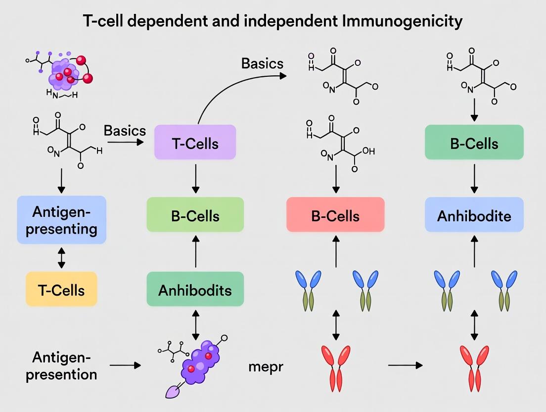
Within the broader thesis on the fundamentals of T-cell dependent (TD) and independent (TI) immunogenicity research, a comparative analysis of key adaptive immune outcomes is essential. This guide provides an in-depth technical examination of the disparate capacities of TD and TI antigens to generate immunological memory, drive affinity maturation, and induce specific antibody isotype profiles. These functional differences underpin vaccine design and therapeutic antibody development strategies.
Core Concepts and Signaling Pathways
T-Cell Dependent (TD) Antigen Recognition and B Cell Activation
TD antigens, typically proteins, require cognate help from CD4+ T follicular helper (Tfh) cells to initiate a germinal center (GC) reaction.
Diagram Title: TD B Cell Activation Pathway
T-Cell Independent (TI) Antigen Recognition and B Cell Activation
TI antigens are divided into TI-1 (mitogenic, e.g., LPS) and TI-2 (polyvalent, e.g., polysaccharides). They stimulate B cells without Tfh cell help.
Diagram Title: TI B Cell Activation Pathway
Table 1: Core Functional Outcomes of TD vs. TI Immune Responses
| Outcome Parameter | T-Cell Dependent (TD) Response | T-Cell Independent (TI) Response | Primary Experimental Evidence |
|---|---|---|---|
| Memory B Cell Formation | Robust and long-lived. GC-derived memory B cells with high BCL-6 expression. | Very weak or absent. Limited generation of true recirculating memory B cells. | Adoptive transfer experiments into naive hosts; flow cytometry for CD80/PD-L2+ memory B cells. |
| Plasma Cell Longevity | Generates both short-lived extrafollicular plasmablasts and long-lived plasma cells (LLPCs) homing to bone marrow. | Primarily short-lived plasmablasts (2-7 days). LLPCs are rare and of lower durability. | ELISPOT assays over time post-immunization; BrdU/Pyronin Y staining for proliferation. |
| Affinity Maturation | Extensive, via somatic hypermutation (SHM) and positive selection in the GC. Affinity (Ka) can increase 10-100x. | Negligible. No GC reaction, thus minimal to no SHM. Affinity remains low. | Sequencing of VH gene regions over time; Surface Plasmon Resonance (SPR) for antibody affinity. |
| Dominant Antibody Isotypes | Class switching to IgG, IgE, IgA (driven by T-cell cytokines: IFN-γ→IgG2a; IL-4→IgG1/IgE; TGF-β→IgA). | Limited class switching. Predominantly IgM. Some TI-2 can induce IgG3 (mouse) or IgG2 (human) via TLR and BAFF signals. | Isotype-specific ELISA; Intracellular cytokine staining of T cells; Cytokine knockout models. |
| Response Kinetics | Slower primary response (5-7 days), accelerated and potent secondary response. | Rapid primary response (2-3 days), but no accelerated secondary response (anamnesis). | Serum antibody titer measurement (ELISA) over time post-primary and secondary challenge. |
| Antigen Type | Proteins, peptides, hapten-carrier complexes. | TI-1: Lipopolysaccharide (LPS), bacterial DNA. TI-2: Polysaccharides, polymeric proteins. | Immunization with model antigens (e.g., NP-protein vs. NP-Ficoll). |
Detailed Experimental Protocols
Protocol: Assessing Memory B Cell Formation via Adoptive Transfer
Objective: To quantify and functionally validate memory B cells generated by TD vs. TI immunization. Materials: See "Scientist's Toolkit" below. Procedure:
- Immunization: Immunize C57BL/6 mice (n=5/group) with a TD antigen (e.g., NP-CGG, 50 µg in alum) or a TI-2 antigen (e.g., NP-Ficoll, 25 µg in PBS).
- Cell Isolation: At day 28 post-immunization, harvest spleens. Prepare a single-cell suspension and enrich for B cells via negative selection using magnetic-activated cell sorting (MACS) with a B cell isolation kit.
- Adoptive Transfer: Intravenously transfer 1 x 10^6 purified B cells from immunized donors into naïve, syngeneic recipient mice.
- Challenge: Immediately challenge recipient mice with a low, sub-immunogenic dose (5 µg) of the respective soluble antigen (NP-CGG or NP-Ficoll) in PBS.
- Readout: Measure serum anti-NP antibody titers via ELISA at days 0, 5, 7, and 14 post-challenge. A rapid, high-titer response in recipients of TD-primed B cells indicates functional memory.
- Flow Cytometry Validation: Analyze donor B cells pre-transfer for memory markers (e.g., in mouse: CD80+, PD-L2+, CD73+, CD38+ for GC-derived memory).
Protocol: Measuring Affinity Maturation by ELISA-Based Relative Affinity Assay
Objective: To compare the affinity of serum antibodies following TD vs. TI immunization. Procedure:
- Sera Collection: Collect serum from immunized mice (as in 4.1) at day 14 (peak primary) and day 60 (memory phase).
- Hapten-Conjugate Coating: Coat two identical sets of ELISA plates. One with a high-density hapten-conjugate (e.g., NP20-BSA, 20 NP molecules per carrier) and another with a low-density conjugate (NP2-BSA). Coat at 5 µg/mL in carbonate buffer overnight at 4°C.
- ELISA: Perform a standard indirect ELISA. Serial dilute serum samples across both plates. Detect bound IgG (or IgM) with isotype-specific HRP conjugates.
- Calculation: Determine the endpoint titer or half-maximal binding (EC50) for each serum sample on both NP20 and NP2 plates.
- Affinity Index: Calculate the Ratio of EC50(NP2) / EC50(NP20). High-affinity antibodies, which require minimal epitope density for binding, will have a ratio closer to 1 (similar titers on both plates). Low-affinity antibodies will have a much higher EC50 on the low-density (NP2) plate, resulting in a larger ratio. TD responses show a decreasing ratio over time (~10-3 at day 14 to ~2-1 at day 60), while TI responses show a consistently high ratio (>10).
The Scientist's Toolkit: Key Research Reagent Solutions
Table 2: Essential Reagents for Investigating TD/TI Outcomes
| Reagent / Material | Function in TD/TI Research | Example Product/Catalog # (Illustrative) |
|---|---|---|
| Model TD Antigens | Standardized protein antigens to induce GC-driven responses. | NP-Keyhole Limpet Hemocyanin (NP-KLH), Ovalbumin (OVA), Tetanus Toxoid. |
| Model TI Antigens | Defined antigens to stimulate extrafollicular, T-cell independent responses. | NP-Ficoll (TI-2), Lipopolysaccharide (LPS, TI-1), Pneumococcal Polysaccharide (PPS). |
| Adjuvants | To enhance immunogenicity and polarize response (critical for TD). | Alum (Th2 bias), Complete/Incomplete Freund's Adjuvant (CFA/IFA), AddaVax (MF59-like). |
| MACS B Cell Isolation Kits | Rapid, negative selection of untouched B cells for transfer/downstream assays. | Miltenyi Biotec Mouse Pan B Cell Isolation Kit II. |
| Fluorochrome-Labeled Antibodies | Flow cytometry phenotyping of GC, memory, and plasma cells. | Anti-mouse: CD19, B220, GL7, CD95 (Fas), CD38, CD73, CD80, PD-L2, IgG1. |
| ELISA Plates & Substrates | Quantification of antigen-specific antibody titer and isotype. | Nunc MaxiSorp plates; TMB substrate solution; anti-mouse IgM/IgG/IgG1/IgG2c HRP. |
| Hapten-Carrier Conjugates (Varying Density) | Critical for assessing antibody affinity maturation via ELISA. | NP2-BSA, NP10-BSA, NP20-BSA (Biosearch Technologies). |
| Cytokine ELISA/Kits | Measure T-cell derived cytokines directing isotype switching. | Mouse IL-4, IFN-γ, IL-21 ELISA DuoSet (R&D Systems). |
Visualizing the Divergent Pathways to Antibody Isotypes
Diagram Title: Antibody Isotype Switching Pathways
Biological and Clinical Significance of Each Pathway in Health and Disease
Understanding the mechanisms of immunogenicity is foundational to modern immunology and therapeutic development. This analysis is framed within a broader thesis on the fundamentals of T-cell dependent (TD) and T-cell independent (TI) immunogenicity research. TD responses, which require cognate help from CD4+ T helper cells, generate high-affinity antibodies, robust memory, and are critical for responses against protein antigens. In contrast, TI responses, typically triggered by repetitive epitopes on antigens like polysaccharides or lipids, can activate B cells directly or via innate immune signals, leading to rapid but limited antibody production with poor memory. The biological pathways governing these responses have profound and distinct implications for health—such as in effective vaccination—and disease—including autoimmunity, immunodeficiency, and hypersensitivity. This whitepaper provides an in-depth technical guide to the core signaling pathways involved, their clinical significance, and associated experimental methodologies.
Core Pathways in T-Cell Dependent and Independent Immunity
T-Cell Dependent B Cell Activation Pathway
This pathway is initiated when a B cell's B cell receptor (BCR) internalizes a protein antigen, processes it, and presents peptides on MHC II. A cognate CD4+ T helper cell, activated by a dendritic cell presenting the same antigen, recognizes this peptide-MHC II complex via its T cell receptor (TCR). This leads to the formation of an immunological synapse and the delivery of critical co-stimulatory signals (e.g., CD40L:CD40) and cytokines (e.g., IL-4, IL-21).
Biological Significance in Health: Drives the germinal center reaction, which is essential for somatic hypermutation, affinity maturation, class-switch recombination, and the generation of long-lived plasma cells and memory B cells. This forms the basis for durable, high-quality antibody responses to most vaccines.
Clinical Significance in Disease:
- Immunodeficiency: Mutations in genes like CD40LG (encoding CD40L) cause Hyper-IgM Syndrome, characterized by an inability to class-switch and recurrent infections.
- Autoimmunity: Dysregulation of germinal center checkpoints can lead to the production of high-affinity autoantibodies, as seen in systemic lupus erythematosus (SLE) and rheumatoid arthritis.
- Hypersensitivity: Uncontrolled Th2 responses and IgE class-switching underpin allergic diseases.
T-Cell Independent Type 1 (TI-1) Pathway
TI-1 antigens, like bacterial lipopolysaccharide (LPS), possess intrinsic mitogenic properties. They can polyclonally activate B cells through Toll-like receptors (TLR4 in the case of LPS) at high concentrations, irrespective of BCR specificity. At low concentrations, only B cells with a BCR specific for the antigen are activated synergistically via BCR and TLR.
Biological Significance in Health: Provides a rapid, first-line antibody defense against conserved microbial components, crucial in early infection before T cell responses develop.
Clinical Significance in Disease: Overactivation of TLR pathways by endogenous ligands (e.g., cell-free DNA) can drive autoreactive B cell activation, contributing to autoimmune pathologies.
T-Cell Independent Type 2 (TI-2) Pathway
TI-2 antigens, such as bacterial capsular polysaccharides, possess highly repetitive structures. These structures induce extensive cross-linking of the BCR on specific B cells, delivering a strong activation signal. Co-stimulation is provided by innate immune cells (e.g., dendritic cells, macrophages) via cytokines (BAFF, APRIL) and other surface molecules.
Biological Significance in Health: Critical for defense against encapsulated bacteria (e.g., Streptococcus pneumoniae, Haemophilus influenzae). Primarily induces extrafollicular responses, generating short-lived plasma cells producing mainly IgM and some IgG.
Clinical Significance in Disease: The inability of infants and young children to mount robust TI-2 responses explains their susceptibility to encapsulated bacteria, leading to the development of conjugate vaccines (converting the response to TD). Dysregulated BAFF/APRIL signaling is associated with autoimmune conditions like SLE.
Table 1: Comparative Features of TD and TI Immune Pathways
| Feature | T-Cell Dependent (TD) | T-Cell Independent Type 1 (TI-1) | T-Cell Independent Type 2 (TI-2) |
|---|---|---|---|
| Prototypical Antigen | Soluble proteins, viral proteins | LPS, bacterial lipoproteins | Capsular polysaccharides, viral capsids |
| Key Cellular Interaction | B cell – CD4+ T cell (cognate) | B cell – Antigen (TLR-driven) | B cell – Antigen (BCR cross-linking) |
| Co-stimulation Source | CD40L on T cells | Intrinsic mitogen (TLR signal) | Innate cells (BAFF/APRIL, TLR) |
| Germinal Center Formation | Yes | No | Rare/Limited |
| Affinity Maturation | Yes (extensive) | No | Minimal |
| Immunoglobulin Isotypes | IgG, IgA, IgE (class-switched) | IgM, IgG3 (mouse), some IgA | Primarily IgM, some IgG |
| Memory B Cell Generation | Robust | Poor/None | Limited |
| Response Kinetics | Slow (4-7 days) | Rapid (1-3 days) | Rapid (2-5 days) |
| Example in Health | Measles vaccine response | Early anti-LPS response in sepsis | Anti-pneumococcal polysaccharide response |
| Example in Disease | SLE autoantibodies | TLR-driven autoimmunity | Pediatric susceptibility to encapsulated bacteria |
Table 2: Clinical Outcomes Associated with Pathway Dysregulation
| Pathway | Deficient/Inhibited State (Disease) | Overactive/Unregulated State (Disease) |
|---|---|---|
| TD (CD40/CD40L) | Hyper-IgM Syndrome Type 1 (recurrent infections) | Potential driver of antibody-mediated autoimmunity |
| TD (GC Regulation) | Common Variable Immunodeficiency (CVID) subsets | SLE, Rheumatoid Arthritis |
| TI (TLR Signaling) | Increased susceptibility to pyogenic bacteria | Systemic inflammation, Autoimmunity (e.g., TLR7 in SLE) |
| TI (BAFF/APRIL) | Impaired TI-2 humoral immunity | SLE, Sjögren's syndrome (BAFF overexpression) |
Detailed Experimental Protocols
Protocol:In VitroTD B Cell Activation Assay
Objective: To measure antigen-specific B cell activation, proliferation, and differentiation in the presence of cognate T cell help.
- Antigen Preparation: Coat magnetic beads (e.g., Dynabeads) with a model protein antigen (e.g., NP-OVA) following manufacturer's crosslinking protocols.
- Cell Isolation: Isulate naïve B cells (CD43- B220+) and CD4+ T cells from mouse spleen or human PBMCs using magnetic-activated cell sorting (MACS) kits.
- T Cell Priming: Activate isolated CD4+ T cells for 48-72 hours with plate-bound anti-CD3 (5 µg/mL) and soluble anti-CD28 (2 µg/mL) in RPMI-1640 + 10% FBS + IL-2 (20 U/mL).
- Co-culture: Co-culture naïve B cells (1x10^5) with primed T cells (1x10^5) and NP-OVA-coated beads (bead:cell ratio 1:1) in a 96-well U-bottom plate. Include controls: B cells alone, B cells + beads, T cells alone.
- Proliferation Assay: At 72 hours, pulse cells with 1 µCi/well [3H]-thymidine for 16-18 hours. Harvest cells and measure incorporated radioactivity with a beta-counter.
- Differentiation Readout: At day 5-7, analyze culture supernatant by ELISA for antigen-specific IgG. Analyze cells by flow cytometry for surface markers (CD138, GL7, FAS) and intracellular cytokines.
Protocol: TI-2 ResponseIn VivoModel
Objective: To evaluate the humoral response to a TI-2 antigen.
- Animal Model: Use 8-12 week old wild-type C57BL/6 mice (n=5-10 per group).
- Antigen & Immunization: Prepare NP-Ficoll (a model TI-2 antigen) in sterile PBS. Immunize mice intraperitoneally with 25 µg of NP-Ficoll in 200 µL PBS. Control group receives PBS only.
- Serum Collection: Collect blood via retro-orbital bleed or tail vein at days 0 (pre-bleed), 7, 14, and 28 post-immunization. Allow blood to clot, centrifuge, and store serum at -20°C.
- Antibody Measurement: Use NP-BSA-coated ELISA plates to measure serum anti-NP antibodies. Perform serial dilutions of serum. Use isotype-specific secondary antibodies (anti-IgM, anti-IgG3) to determine the titer of each isotype. Express results as endpoint titer or as concentration relative to a standard curve.
- ELISPOT (Optional): At endpoint, isolate splenocytes and perform ELISPOT to quantify NP-specific antibody-secreting cells (ASCs) using NP-BSA-coated plates.
The Scientist's Toolkit: Key Research Reagent Solutions
Table 3: Essential Reagents for TD/TI Immunogenicity Research
| Reagent Category | Specific Example(s) | Function in Research |
|---|---|---|
| Model Antigens | NP-OVA (4-Hydroxy-3-nitrophenylacetyl-Ovalbumin), NP-Ficoll, LPS (E. coli O111:B4) | Standardized tools to probe TD (NP-OVA) vs. TI-2 (NP-Ficoll) and TI-1 (LPS) pathways in vivo and in vitro. |
| Monoclonal Antibodies (Blocking/Stimulating) | Anti-CD40L (MRI-1), Anti-CD40 (FGK4.5), Anti-BAFF (Sandy-2), Anti-TLR4/MD-2 | To agonize or antagonize specific pathway components to dissect their functional roles. |
| Cytokines & Recombinant Proteins | Recombinant murine/human BAFF, APRIL, IL-4, IL-21 | To provide specific differentiation or survival signals in cell culture assays. |
| Fluorochrome-Conjugated Antibodies (Flow Cytometry) | Anti-B220, Anti-CD138, Anti-GL7, Anti-FAS, Anti-IgM, Anti-IgG, Anti-CD4 | To identify and characterize B cell subsets (naïve, germinal center, plasma cell), T cells, and antibody isotypes. |
| Knockout/Transgenic Mouse Models | CD40KO, CD40LKO, MyD88KO, BAFF-Tg, MD4 (BCR transgenic) mice | Genetically defined models to study the in vivo consequence of ablating or overexpressing pathway components. |
| ELISA/ELISPOT Kits | Mouse IgG/IgM Total & Isotype, NP-specific ELISA kits; ELISPOT plates | To quantify total and antigen-specific antibody titers in serum or culture supernatant, and frequency of antibody-secreting cells. |
Methods and Applications: Assessing Immunogenicity Pathways in Preclinical and Clinical Development
This technical guide details core in vitro assays for evaluating humoral immunogenicity, framed within the broader thesis of T-cell dependent (TD) and T-cell independent (TI) immune response research. Understanding these pathways is critical in vaccine development, autoimmune disease research, and assessing the immunogenic risk of biologic therapeutics. This whitepaper provides detailed methodologies, key reagents, and data interpretation for three interconnected assays.
APC/T-cell Co-culture Assay
This assay models the initial cognate interaction critical for TD responses, where antigen-presenting cells (APCs) activate naïve T-cells.
Detailed Protocol
- APC Preparation: Isolate human monocytes from PBMCs via CD14+ magnetic selection. Differentiate into dendritic cells (DCs) over 6-7 days in RPMI-1640 medium supplemented with IL-4 (50 ng/mL) and GM-CSF (100 ng/mL). On day 6, mature DCs by adding LPS (100 ng/mL) or a cytokine cocktail (TNF-α, IL-1β, IL-6, PGE2). Load DCs with antigen (e.g., 10 µg/mL protein) for 4-6 hours.
- T-cell Isolation: Isolate naïve CD4+ T-cells from autologous PBMCs using a negative selection kit.
- Co-culture: Plate antigen-loaded DCs (e.g., 5x10⁴ cells/well) with naïve CD4+ T-cells (e.g., 2x10⁵ cells/well) in a U-bottom 96-well plate. Include controls: T-cells alone, DCs alone, and DCs with irrelevant antigen.
- Incubation & Analysis: Culture for 5-7 days. Measure T-cell proliferation via ³H-thymidine incorporation or CFSE dilution. Assess activation by flow cytometry for surface markers (CD25, CD69) and intracellular cytokines (IFN-γ, IL-2) after 12-24 hours of restimulation with PMA/ionomycin in the presence of a protein transport inhibitor.
Key Signaling Pathway: T-Cell Receptor (TCR) Engagement
Diagram Title: TCR and Co-stimulatory Signaling Pathways
Quantitative Data Outputs
Table 1: Typical T-cell Activation Readouts in Co-culture Assay
| Readout | Measurement Method | Baseline (No Antigen) | Antigen-Specific Response (Mean ± SD) | Key Indicator |
|---|---|---|---|---|
| Proliferation | ³H-thymidine uptake (cpm) | 500 - 2,000 cpm | 15,000 - 80,000 cpm | Stimulation Index >3-5 |
| CD25 Expression | Flow Cytometry (% positive) | 2-5% | 25-60% | Early activation |
| IFN-γ Production | ELISA (pg/mL) or ICS (% cells) | <50 pg/mL / <0.5% | 500-3000 pg/mL / 5-20% | Th1 polarization |
B-cell Activation Assay
This assay directly measures B-cell response, which can be TI (direct TLR or antigen cross-linking) or TD (requiring T-cell help).
Detailed Protocol for TI-2 (Cross-linking) Model
- B-cell Isolation: Isolate human naïve B-cells from PBMCs using a CD19+ B-cell negative selection kit.
- Stimulation: Plate purified B-cells (1-2x10⁵ cells/well) in 96-well plates. Stimulate with:
- TI-2 Antigen: Anti-IgM F(ab')₂ fragment (5-10 µg/mL) or dextran-conjugated anti-Ig.
- TI-1 Antigen: LPS (1-10 µg/mL).
- TD Signal (Control): CD40L (1 µg/mL) + IL-4 (20 ng/mL).
- Incubation: Culture for 4-6 days.
- Analysis: Measure proliferation (as above). Assess differentiation by flow cytometry for activation markers (CD86, CD83) and plasma cell marker CD138. Quantify IgM/IgG secretion in supernatant by ELISA.
Key Signaling Pathway: B-Cell Receptor (BCR) Engagement
Diagram Title: B-cell Activation Signaling Pathways: TI vs. TD
Quantitative Data Outputs
Table 2: B-cell Activation Assay Outcomes
| Stimulus (Type) | Proliferation (cpm) | CD86+ Cells (%) | Ig Secretion (ng/mL) | Primary Mechanism |
|---|---|---|---|---|
| Medium Only | 1,000 - 3,000 | 5-10% | <10 | Baseline |
| α-IgM F(ab')₂ (TI-2) | 25,000 - 75,000 | 40-70% | IgM: 100-500 | BCR cross-linking |
| LPS (TI-1) | 30,000 - 90,000 | 50-80% | IgM: 200-800 | TLR4 engagement |
| CD40L + IL-4 (TD) | 40,000 - 100,000 | 60-85% | IgG: 50-300 | CD40 & Cytokine signaling |
Cytokine Profiling
Multiplexed cytokine analysis provides a functional signature of the immune response, distinguishing TD from TI profiles.
Detailed Protocol (Luminex Multiplex Assay)
- Sample Collection: Centrifuge co-culture or B-cell assay supernatants at 500xg for 5 min. Aliquot and store at -80°C. Avoid repeated freeze-thaw.
- Assay Setup: Bring all reagents to room temperature. Prepare standards and controls in the same matrix as samples (e.g., assay medium). Add 50 µL of standard or sample to designated wells of a pre-washed magnetic bead plate.
- Incubation & Detection: Add 50 µL of the mixed antibody-bead cocktail. Incubate for 2 hours on a plate shaker. Wash twice. Add 50 µL of detection antibody. Incubate for 1 hour. Wash, add 50 µL of Streptavidin-PE. Incubate for 30 min. Wash, resuspend beads in reading buffer.
- Acquisition & Analysis: Run plate on a Luminex analyzer (e.g., MAGPIX). Use instrument software with a 5-parameter logistic curve to calculate cytokine concentrations from median fluorescence intensity (MFI).
Key Workflow: Integrated Assay Data Synthesis
Diagram Title: Integrated Immunogenicity Assay Workflow
Quantitative Data Outputs
Table 3: Differentiating Cytokine Profiles in TD vs. TI Responses
| Cytokine | Primary Source | TD Response (Range pg/mL) | TI Response (Range pg/mL) | Functional Role |
|---|---|---|---|---|
| IL-2 | Activated T-cells | 200 - 2,000 | < 50 | T-cell proliferation/survival |
| IL-4 | Th2 T-cells | 100 - 1,500 | < 30 | B-cell class switch to IgG1/IgE |
| IL-21 | Tfh/Th17 cells | 50 - 800 | < 20 | Plasma cell differentiation |
| IFN-γ | Th1 T-cells | 500 - 3,000 | < 100 | Macrophage activation, IgG2 switch |
| IL-6 | APCs, B-cells | 100 - 800 | 500 - 5,000 | Acute phase, B-cell differentiation |
| IL-10 | Bregs, Macrophages | 50 - 400 | 200 - 2,000 | Immunoregulation, limits pathology |
| TNF-α | Macrophages, T-cells | 100 - 1,000 | 300 - 4,000 | Inflammation, cell activation |
The Scientist's Toolkit: Research Reagent Solutions
Table 4: Essential Reagents for Immunogenicity Assays
| Reagent Category | Specific Example | Function in Assay | Key Consideration |
|---|---|---|---|
| Cell Isolation Kits | CD14+ microbeads (APCs); Naïve CD4+ T-cell kits; Naïve B-cell kits. | Rapid, high-purity isolation of primary immune cell subsets. | Purity (>95%) and viability (>98%) are critical for assay specificity. |
| Cell Culture Media | RPMI-1640 + 10% FBS + 1% Pen/Strep; Serum-free DC media. | Supports growth and function of primary immune cells. | Use consistent, qualified FBS batches; serum-free options reduce variability. |
| Recombinant Cytokines | Human IL-4, GM-CSF, IL-2, IL-21, CD40L. | Drives cell differentiation, survival, and assay-specific stimulation. | Use carrier protein (e.g., HSA)-free formats to avoid interference. |
| Activation Stimuli | LPS, Anti-IgM F(ab')₂, Pokeweed Mitogen, PMA/Ionomycin. | Triggers TI-1, TI-2, or polyclonal (control) activation pathways. | Use F(ab')₂ fragments to avoid Fc receptor cross-linking artifacts. |
| Detection Antibodies | Fluorochrome-conjugated anti-CD25, CD69, CD86, CD138; Cytokine capture/detection pairs for ELISA. | Enables flow cytometry phenotyping and soluble factor quantification. | Validate clones for specific applications (e.g., intracellular vs. surface staining). |
| Multiplex Bead Arrays | 25-plex Human Cytokine/Chemokine Panels (e.g., from Bio-Rad, Millipore). | Simultaneous quantification of multiple soluble analytes from limited sample volume. | Ensure assay buffer is compatible with sample matrix (e.g., culture medium). |
| Proliferation Dyes | CFSE, CellTrace Violet. | Tracks multiple rounds of cell division via flow cytometry. | Optimize dye concentration to avoid toxicity while maintaining detection sensitivity. |
This whitepaper serves as a technical guide to in vivo models central to immunogenicity research, framed within the broader thesis on the basics of T-cell dependent and independent immune responses. Predicting unintended immunogenicity—the development of anti-drug antibodies (ADAs)—is a critical hurdle in biotherapeutic development. While in silico and in vitro screens are valuable, in vivo models provide indispensable insights into the complex, integrated immune system. This document focuses on transgenic murine models and other surrogate systems that bridge the gap between preclinical studies and human clinical outcomes, detailing their application, experimental protocols, and key research tools.
Core Model Systems: Principles and Applications
Transgenic Humanized Mouse Models
These models are engineered to express human genes or immune system components, allowing for the evaluation of immune responses to human-specific therapeutics.
- HuMab Mice: Mice with human immunoglobulin loci knock-ins (e.g., Trianni, AlivaMab) that generate fully human antibody sequences against human antigens, useful for assessing immunogenicity of human protein therapeutics.
- HLA-Transgenic Mice: Mice expressing human HLA class II alleles (e.g., HLA-DR, -DQ). They are pivotal for studying T-cell dependent immunogenicity, as they present human-specific peptides to murine T-cell receptors, modeling the human CD4+ T-cell response.
- Complete Immune System Humanized Mice: Immunodeficient mice (e.g., NSG, NOG) engrafted with human CD34+ hematopoietic stem cells or peripheral blood mononuclear cells (PBMCs). These models support a more complete human immune system but are susceptible to graft-versus-host disease.
Surrogate Models for T-cell Independent Responses
For polysaccharide antigens or certain aggregated proteins, B-cell responses can occur without major histocompatibility complex (MHC) class II-mediated T-cell help.
- Wild-type Models: Specific strains of mice (e.g., BALB/c, C57BL/6) are used to study aggregate-driven or pattern-recognition receptor (PRR)-mediated B-cell activation.
- Transgenic Models with B-cell Reporters: Mice with B-cell receptors specific for a model antigen, allowing precise tracking of B-cell activation and differentiation upon challenge.
Quantitative Comparison of Key Models
Table 1: Comparative Analysis of Primary In Vivo Immunogenicity Models
| Model Type | Specific Example(s) | Key Genetic/Engraftment Feature | Primary Application (Immunogenicity) | Strengths | Limitations |
|---|---|---|---|---|---|
| HLA-Transgenic | HLA-DR4 (DRB1*04:01), HLA-DQ8 | Express human MHC class II molecules on mouse APC | Prediction of T-cell epitopes & Td immunogenicity; MHC-restricted response | Direct insight into human HLA-restriction; robust T-cell assays possible | Limited to single allele; mouse TCR repertoire may not mimic human |
| Humoral-Reporter | HuMab (Trianni), AlivaMab Mouse | Knock-in of human Ig heavy & light chain loci | De novo human ADA generation; evaluation of B-cell epitopes | Produces fully human antibodies; good for mAb discovery | Complex genetic engineering; may not reflect full tolerance mechanisms |
| Immune-Humanized | NSG-SGM3 with hu-CD34+ | IL-3, GM-CSF, SCF expression supports human myeloid engraftment | Holistic human immune response (innate & adaptive) | Functional human T, B, myeloid cells; can assess cell-mediated responses | High variability; graft-vs-host; short-lived; high cost |
| Surrogate Wild-type | BALB/c, C57BL/6 | Intact murine immune system | Screening for Ti (aggregate-driven) immunogenicity; general toxicity | Low cost, high reproducibility, well-characterized | Fully murine response; may not translate to human immune recognition |
Detailed Experimental Protocols
Protocol: T-Cell Dependent Immunogenicity Assay in HLA-Transgenic Mice
Objective: To evaluate the potential of a biotherapeutic to elicit HLA-restricted CD4+ T-cell responses.
Materials: HLA-DR4 transgenic mice (6-8 weeks old), test article (protein therapeutic), negative control (PBS or human serum albumin), positive control (keyhole limpet hemocyanin - KLH), adjuvant (e.g., Incomplete Freund's Adjuvant for priming), sterile PBS, flow cytometry reagents (anti-mouse CD4, CD44, CD62L, IFN-γ, IL-2), enzyme-linked immunosorbent spot (ELISpot) plates.
Procedure:
- Immunization: Mice (n=5-10/group) are immunized subcutaneously on day 0 with 50 µg of test article, negative control, or positive control emulsified in adjuvant.
- Rest Period: Animals rest for 10-14 days to allow for primary immune response development.
- Recall Assay: On day 14, spleens are aseptically harvested and processed into single-cell suspensions.
- T-Cell Stimulation:
- ELISpot: Splenocytes are plated at 2.5 x 10^5 cells/well with 10 µg/mL of the test article or overlapping peptides spanning its sequence. After 48h, spots for IFN-γ or IL-4 are developed and counted. A response is considered positive if the mean spot count in test wells exceeds negative control mean by >2x and is statistically significant (p<0.05).
- Intracellular Cytokine Staining (ICS): Splenocytes are stimulated with antigen/peptides for 6h in the presence of brefeldin A. Cells are surface-stained for CD4, then fixed, permeabilized, and stained for cytokines (IFN-γ, IL-2). Frequency of antigen-specific cytokine+ CD4+ T-cells is determined via flow cytometry.
- Data Analysis: Responses are reported as spot-forming units (SFU)/10^6 cells (ELISpot) or % cytokine-positive CD4+ T cells (ICS). A positive signal indicates the presence of T-cell epitopes within the therapeutic.
Protocol: Surrogate ADA Detection in HuMab Mice
Objective: To assess the in vivo immunogenic potential of a human protein therapeutic by measuring the generation of fully human anti-drug antibodies (ADAs).
Materials: HuMab mice (e.g., Trianni), test article, isotype control, PBS, Matrigel (optional), serum collection tubes, bridging electrochemiluminescence (ECL) or ELISA kit for human IgG detection.
Procedure:
- Dosing: Mice are administered the test article via a relevant route (intravenous, subcutaneous) weekly for 4 weeks. A control group receives PBS.
- Serum Collection: Blood is collected via retro-orbital or submandibular bleed pre-dose, and 7 days after the 2nd and 4th doses.
- ADA Detection via Bridging ELISA/ECL:
- Capture: A 96-well plate is coated with the test article.
- Blocking: Plates are blocked with a protein-based buffer (e.g., 3% BSA).
- Sample Incubation: Diluted mouse serum samples are added. Any human ADA (IgG) will bind to the immobilized drug.
- Detection: A biotinylated version of the test article is added, which binds to the free arm of the captured ADA. Streptavidin-HRP conjugate is then added.
- Signal Development: TMB substrate is added, reaction stopped, and absorbance read. For ECL, a ruthenylated drug and streptavidin-biotinylated drug complex is used, with signal read on a Meso Scale Discovery (MSD) instrument.
- Cut-Point Determination: The assay cut-point is established using pre-dose serum samples to define the threshold for ADA positivity (typically mean signal + 1.645*SD, for 95% specificity). Samples exceeding the cut-point are considered ADA-positive.
- Titer Analysis: Positive samples are serially diluted to determine ADA titer, reported as the highest dilution yielding a positive signal.
Visualizing Key Concepts and Workflows
Diagram 1: HLA-Transgenic Mouse Td Immunogenicity Pathway
Diagram 2: In Vivo Immunogenicity Assessment Workflow
The Scientist's Toolkit: Key Research Reagents & Materials
Table 2: Essential Reagents for In Vivo Immunogenicity Studies
| Reagent / Material | Primary Function | Example(s) & Notes |
|---|---|---|
| HLA-Transgenic Mice | Provide human MHC-II context for antigen presentation. | HLA-DR4 (DRB1*04:01), HLA-DQ6, HLA-DQ8 strains from suppliers like Jackson Lab or Taconic. |
| Immunodeficient Host Mice | Accept human immune cell engraftment for humanized models. | NOD-scid IL2Rγnull (NSG), NOG mice. SGM3 variant improves myeloid reconstitution. |
| Human Cytokines (Recombinant) | Support engraftment & survival of human cells in humanized models. | Human IL-2, SCF, FLT3-L, M-CSF. Often administered via injection or encoded in transgenic host. |
| Adjuvants | Enhance immune response to co-administered antigen for immunogenicity studies. | Incomplete Freund's Adjuvant (IFA), Alum, CpG oligonucleotides. Choice depends on response type (Th1 vs Th2). |
| Bridging Assay Kits | Detect anti-drug antibodies (ADAs) in serum. | MSD Multi-Array ECL kits, or in-house developed ELISA with biotinylated & ruthenylated drug. |
| ELISpot Kits & Plates | Quantify antigen-specific T-cell cytokine secretion at single-cell level. | Mouse IFN-γ/IL-4/IL-2 ELISpot kits from Mabtech, BD, or R&D Systems; PVDF-backed plates. |
| Flow Cytometry Antibodies | Phenotype and assess intracellular cytokines in immune cells. | Anti-mouse/human CD4, CD8, CD44, CD62L, IFN-γ, IL-17, FoxP3. Must include viability dye. |
| Overlapping Peptide Libraries | Map T-cell epitopes within a protein therapeutic sequence. | 15-mer peptides overlapping by 10-11 amino acids, covering full sequence (synthesized by JPT, Mimotopes). |
Integrating Immunogenicity Risk Assessment into the Biotherapeutic Development Workflow
The clinical success of biotherapeutics, from monoclonal antibodies to recombinant proteins and gene therapies, is critically dependent on managing immunogenicity. This guide is framed within the foundational thesis that immunogenicity arises via two primary, often interconnected, pathways: T-cell dependent (TD) and T-cell independent (TI) responses.
- T-cell Dependent Immunogenicity: The canonical adaptive immune response. Protein therapeutics are endocytosed by antigen-presenting cells (APCs), processed into peptides, and presented on MHC class II molecules. Recognition by specific CD4+ T-cell receptors activates T-helper cells, which drive B-cell maturation, affinity maturation, and the generation of high-affinity, class-switched, persistent anti-drug antibodies (ADAs).
- T-cell Independent Immunogenicity: Often driven by multivalent, repetitive epitopes (e.g., aggregated proteins) that can directly cross-link B-cell receptors (BCRs), leading to rapid but typically low-affinity, non-persistent IgM ADA responses. Certain TI pathways can also involve Toll-like receptor (TLR) activation by pathogen- or damage-associated molecular patterns (PAMPs/DAMPs).
Integrating risk assessment for both pathways throughout the development workflow is essential to mitigate ADA impacts on pharmacokinetics, pharmacodynamics, efficacy, and safety.
Quantitative Data on Immunogenicity Risk Factors
The following tables summarize key risk factors and their associated impact.
Table 1: Biotherapeutic-Specific Risk Factors & Data
| Risk Factor | Description | Quantitative Impact/Correlation |
|---|---|---|
| Sequence Relatedness | Degree of non-self vs. human sequence homology. | Proteins with <90% human homology show >50% immunogenicity incidence in clinical studies. |
| Aggregation Propensity | Tendency to form soluble/insoluble multimers. | Formulations with >1% high molecular weight species (HMWS) show a 2-5x increase in ADA rate. |
| Post-Translational Modifications (PTMs) | Non-human glycosylation, oxidation, deamidation, etc. | Specific glycan forms (e.g., α-Gal, Man3) can increase ADA incidence by 20-40%. |
| Impurities | Host cell proteins (HCPs), DNA, endotoxin. | Endotoxin levels >0.1 EU/mg can potentiate TI responses via TLR4. |
Table 2: Patient- & Treatment-Related Risk Factors
| Risk Factor | Description | Quantitative Impact/Correlation |
|---|---|---|
| Immune Status | Underlying disease (autoimmune, cancer), concomitant immunosuppression. | Patients on methotrexate show ~30-70% reduction in ADA rates to protein therapeutics. |
| Route of Administration | Subcutaneous (SC), Intravenous (IV), etc. | SC route associates with a 2-3x higher immunogenicity risk vs. IV, likely due to dendritic cell engagement. |
| Dose & Frequency | Treatment regimen intensity. | Very high or very low doses can be tolerogenic; chronic intermittent dosing often increases risk. |
Experimental Protocols for Integrated Risk Assessment
Protocol 1:In SilicoT-cell Epitope Prediction (TD Pathway Screening)
Objective: Identify potential immunogenic peptide sequences within the drug candidate. Methodology:
- Sequence Input: Input the protein's amino acid sequence into prediction algorithms.
- Algorithm Suite: Utilize a panel of tools (e.g., NetMHCIIpan, EpiMatrix, TepiTool) predicting binding affinity to a broad set of common HLA-DR, DP, and DQ alleles.
- Aggregate Scoring: Identify clusters of predicted high-affinity binders ("hot spots"). Calculate a composite score reflecting the density and strength of predicted epitopes.
- Deimmunization Design: Use output to guide protein engineering (e.g., point mutations in framework regions) to remove predicted epitopes while maintaining function.
Protocol 2:In VitroT-cell Activation Assay (TD Pathway Confirmation)
Objective: Experimentally validate the potential of the biotherapeutic to activate naive human T-cells. Methodology:
- Donor Cells: Isolate peripheral blood mononuclear cells (PBMCs) from 50-100 healthy donors representing diverse HLA alleles.
- Antigen Preparation: Prepare the biotherapeutic, its putative peptides, and controls (e.g., keyhole limpet hemocyanin - positive, human albumin - negative).
- Co-culture: Culture PBMCs with antigens for 7-12 days.
- Readout: Measure T-cell activation via:
- Proliferation: [3H]-thymidine incorporation or CFSE dilution.
- Cytokine Secretion: ELISpot for IFN-γ or IL-2.
- Data Analysis: A response is positive if significantly exceeding background in multiple donors. The response rate informs clinical risk.
Protocol 3:In VitroB-cell & Monocyte Activation Assay (TI Pathway Screening)
Objective: Assess the potential of drug aggregates or impurities to directly activate B-cells or APCs. Methodology:
- Cell Isolation: Isolate naive human B-cells (CD19+/CD27-) and monocytes (CD14+) from PBMCs.
- Stimulus Preparation: Prepare the biotherapeutic in its native, intentionally aggregated (e.g., heat-stressed), and ultra-purified forms.
- Culture & Stimulation: Culture cells with stimuli for 24-48 hours. Include controls (e.g., CpG DNA for TLR9 - positive control).
- Readout:
- B-cells: Flow cytometry for surface activation markers (e.g., CD69, CD86).
- Monocytes: Multiplex cytokine analysis (e.g., IL-1β, IL-6, TNF-α) via Luminex.
- Data Analysis: Identify formulations or conditions inducing significant activation versus native protein.
Visualizing Immunogenicity Pathways and Workflows
T-Cell Dependent ADA Pathway
T-Cell Independent Immunogenicity Pathways
Integrated Risk Assessment Workflow
The Scientist's Toolkit: Key Research Reagent Solutions
Table 3: Essential Tools for Immunogenicity Risk Assessment
| Reagent / Material | Function & Explanation |
|---|---|
| HLA-typed Human PBMCs | Provide a diverse source of immune cells (T-cells, B-cells, APCs) from multiple donors for in vitro assays, reflecting population variability. |
| Recombinant Human MHC Class II Proteins | Used in competitive binding assays (e.g., ELISA) to biochemically confirm predicted T-cell epitope binding affinity. |
| Anti-Human CD40L (CD154) Antibody | Critical for detecting antigen-specific T-cell activation via flow cytometry by marking recently activated T-cells. |
| CFSE (Carboxyfluorescein succinimidyl ester) | A fluorescent cell dye that dilutes with each cell division, allowing measurement of T- or B-cell proliferation. |
| Human IFN-γ/IL-2 ELISpot Kits | Pre-coated plates for enumerating antigen-responsive T-cells secreting specific cytokines at the single-cell level. |
| Luminex Multiplex Cytokine Panels | Bead-based arrays to quantify a broad profile of inflammatory cytokines (e.g., IL-6, TNF-α, IL-1β) from cell supernatants, assessing innate immune activation. |
| Size-Exclusion Chromatography (SEC) Standards & Columns | Essential for quantifying and characterizing protein aggregates (HMWS) in drug formulations. |
| Host Cell Protein (HCP) ELISA | Platform-specific assays to detect and quantify residual HCP impurities, which are potent immunogenicity risk factors. |
| Anti-Drug Antibody (ADA) Assay Reagents | Includes biotinylated/HRP-conjugated drug for bridging ELISA or MSD formats, critical for clinical immunogenicity monitoring. |
Application for Biologics (mAbs, Proteins), Vaccines, and Novel Modalities (e.g., AAV, mRNA)
The development of biologics, vaccines, and novel therapeutic modalities requires a fundamental understanding of immunogenicity—the unwanted immune response against the therapeutic agent. This guide is framed within the core thesis that rational immunogenicity risk assessment hinges on distinguishing between T-cell dependent (TD) and T-cell independent (TI) pathways.
- T-cell Dependent Immunogenicity: Requires CD4+ T-helper cell recognition of a peptide antigen (e.g., from a monoclonal antibody [mAb]) presented by Major Histocompatibility Complex Class II (MHC II) on antigen-presenting cells (APCs). This leads to germinal center reactions, affinity maturation, class switching, and long-lived memory B cells and plasma cells. It is the primary pathway for high-affinity, persistent anti-drug antibody (ADA) responses against protein therapeutics.
- T-cell Independent Immunogenicity: Can be triggered by repetitive epitopes (e.g., on viral capsids or aggregates) that cross-link B-cell receptors (BCR) without T-cell help. TI responses are typically rapid, induce low-affinity IgM antibodies, and lack immunologic memory. This pathway is highly relevant for vaccines (e.g., polysaccharide) and modalities like AAV vectors.
Mitigating immunogenicity demands experimental strategies tailored to predict and interrogate these distinct pathways.
T-cell Dependent Pathway Diagram
T-cell Independent (Type 2) Pathway Diagram
Key Experimental Protocols for Immunogenicity Assessment
In Vitro T-cell Activation Assay (TD Pathway)
Objective: To quantify the potential of a biologic to be processed and presented by APCs to activate CD4+ T-cells. Protocol:
- APC Preparation: Isolate monocytes from human peripheral blood mononuclear cells (PBMCs) of multiple donors (covering common HLA alleles) and differentiate into dendritic cells using GM-CSF and IL-4.
- Antigen Loading: Pulse APCs with the test biologic (mAb, protein, AAV empty capsid) at a range of concentrations (0.1-10 µg/mL). Use a known immunogenic protein (e.g., keyhole limpet hemocyanin) as a positive control and media as a negative control.
- Co-culture: Add autologous or HLA-matched CD4+ T-cells to the APCs at a ratio of ~10:1 (T-cells:APC).
- Incubation: Culture for 6-7 days in RPMI-1640 complete medium.
- Readout: Measure T-cell proliferation via [3H]-thymidine incorporation or CFSE dilution. Alternatively, quantify activation markers (CD69, CD25) or cytokine secretion (IFN-γ, IL-2) by ELISA or ELISpot after 48-72 hours. Data Interpretation: A significant, dose-dependent increase in proliferation/cytokines indicates the presence of T-cell epitopes.
B Cell Activation and Antibody-Secreting Cell (ASC) ELISpot (TI/TD Pathways)
Objective: To detect and enumerate B cells that are activated by the therapeutic to become antibody-secreting cells. Protocol:
- Cell Isolation: Isolate naïve human B cells from PBMCs using negative selection kits.
- Stimulation Culture: For TI assessment, culture B cells with the test article (e.g., AAV vector, aggregated mAb) and a TLR agonist (e.g., CpG) for 5-6 days. For TD assessment, add irradiated autologous T-cells and the test article.
- ELISpot Plate Preparation: Coat a multi-screen IP plate with the therapeutic protein (or a relevant target antigen for anti-drug antibody detection) at 5-10 µg/mL overnight.
- Cell Transfer & Incubation: Block the plate, then transfer the stimulated B cells and incubate for 24-48 hours. Secreted antibodies will bind to the captured antigen.
- Detection: Use biotinylated detection antibodies against human IgM, IgG, or IgA, followed by streptavidin-ALP and a precipitating substrate (BCIP/NBT).
- Analysis: Count the resulting spots (each representing a single ASC) using an automated ELISpot reader. Data Interpretation: High numbers of IgM spots suggest a TI response, while a strong IgG response indicates a TD pathway.
Table 1: Comparative Immunogenicity Risk Profile by Modality
| Therapeutic Modality | Primary Immunogenic Trigger | Predominant Pathway | Key Risk Factors (Quantitative Examples) |
|---|---|---|---|
| Monoclonal Antibodies | Foreign T-cell epitopes, aggregates | T-cell Dependent | Aggregate level: >1% sub-visible particles can increase risk. Sequence homology: <85% human identity confers high risk. |
| Therapeutic Proteins | Non-human sequence, modifications | T-cell Dependent | Glycosylation differences: Lack of human-like sialylation can increase clearance. |
| Polysaccharide Vaccines | Repetitive epitopes | T-cell Independent (Type 2) | Chain length: Longer polysaccharides (>20 repeat units) are more immunogenic. |
| AAV Gene Therapy Vectors | Viral capsid proteins, DNA impurities | Both TI (capsid) & TD (transgene) | Empty/Full Capsid Ratio: >20% empty capsids correlate with higher anti-AAV IgG. Pre-existing NAbs: ~30-50% of population has neutralizing antibodies to common serotypes. |
| mRNA Vaccines/Therapeutics | Double-stranded RNA (dsRNA) impurities, lipid nanoparticles (LNPs) | T-cell Dependent (strong) | dsRNA impurity: >0.1% can potently activate TLR3/RLRs. LNP component: Ionizable lipid structure dictates reactogenicity. |
Table 2: Core Assays for Immunogenicity De-risking
| Assay Name | Pathway Interrogated | Measured Output | Typical Readout Sensitivity |
|---|---|---|---|
| In Silico MHC-II Epitope Prediction | TD | Epitope burden | Predicts binding affinity (IC50 nM) for common HLA-DR alleles. |
| DC:T-cell Co-culture | TD | T-cell proliferation/cytokines | Can detect ~0.01% reactive T-cell frequency. |
| B Cell ELISpot | TI & TD | Antibody-secreting cells | 1 ASC per 1x10^6 input cells. |
| SPR/BLI for ADA Affinity | Outcome | Affinity of ADA (KD) | Measures KD from µM (low) to nM (high-affinity, TD). |
| CRISPR/Cas9 MHC-II Knockout | TD | Functional validation | Confirms loss of T-cell response in vitro. |
The Scientist's Toolkit: Essential Research Reagents
Table 3: Key Reagent Solutions for Core Immunogenicity Experiments
| Reagent / Material | Function & Application | Example Vendor(s) |
|---|---|---|
| Cryopreserved Human PBMCs (Multi-donor) | Source of primary immune cells (T, B, monocytes) for in vitro assays. Ensures coverage of diverse HLA haplotypes. | STEMCELL Tech, AllCells, Hemacare |
| Human MHC Class II Tetramers | Directly identify and isolate T-cells specific for a peptide derived from the biologic. | MBL International, ImmunoScape |
| ELISpot Kits (Human IgG/IgM) | Pre-coated or ready-to-use kits for quantifying antigen-specific antibody-secreting cells. | Mabtech, Cellular Technology Ltd. |
| TLR Agonists/Antagonists | To stimulate or inhibit pattern recognition receptors (e.g., TLR4, TLR9) when studying adjuvant effects or TI responses. | InvivoGen |
| Recombinant Human Cytokines (GM-CSF, IL-4, IL-21) | For differentiating monocytes to DCs and supporting B-cell/T-cell culture. | PeproTech, R&D Systems |
| HLA-DR Transfected Antigen-Presenting Cell Lines | Standardized cell lines (e.g., CHO or HeLa expressing a single HLA-DR allele) for consistent epitope presentation assays. | ATCC, GenHunter |
| Size Exclusion Chromatography (SEC) & Microflow Imaging (MFI) Standards | To characterize and quantify protein aggregates (key TI antigen) in drug product formulations. | Agilent, Protein Standards, Inc., Microtrac MRB |
Within the foundational thesis on T-cell dependent (TD) and T-cell independent (TI) immunogenicity research, understanding regulatory guidance is paramount. Immunogenicity—the unintended immune response to biologic therapeutics or endogenous proteins—can impact drug safety, efficacy, and pharmacokinetics. TD responses involve antigen presentation and T-helper cell activation, leading to high-affinity antibodies. TI responses, typically to repetitive epitopes, can induce rapid, lower-affinity antibody production without T-cell help. This guide details current regulatory expectations from the U.S. Food and Drug Administration (FDA), the European Medicines Agency (EMA), and the International Council for Harmonisation (ICH) for immunogenicity assessment, framing them within this core immunological context.
The FDA, EMA, and ICH provide complementary but distinct guidelines. The FDA's perspective is detailed in its 2019 guidance "Immunogenicity Testing of Therapeutic Protein Products — Developing and Validating Assays for Anti-Drug Antibody Detection." EMA's overarching principle is described in CHMP/BMWP/14327/2006 Rev 1 "Guideline on Immunogenicity assessment of therapeutic proteins" (2017). ICH contributes with broader quality and safety guidelines, notably ICH S6(R1) and ICH S8, which contextualize immunogenicity risk.
Key Guideline Comparison
The table below summarizes the core quantitative and strategic expectations from each agency.
Table 1: Comparison of Key Regulatory Expectations
| Aspect | FDA (2019 Guidance) | EMA (2017 Guideline) | ICH (S6(R1), S8) |
|---|---|---|---|
| Risk-Based Approach | Mandatory. Risk level dictates assay strategy, sampling schedule, & population. | Central to assessment. Risk based on product, patient, disease factors. | Integrated into nonclinical safety evaluation (S6). Immunotoxicity (S8). |
| Screening Assay Cut-Point | Statistical determination with 5% false positive rate. Use of pre-dose or disease-state controls. | 1% or 5% false positive rate acceptable; justification required. | Not explicitly defined; refers to regional guidelines. |
| Confirmatory Assay (Specificity) | Minimum 10-20% inhibition for positive confirmation. | Significant reduction in signal upon addition of excess drug. | Not specified. |
| Titer Reporting | Recommended for positive samples to assess magnitude. | Required for positive samples. | Not specified. |
| Neutralizing Antibody (NAb) Assays | Required for high-risk products. Use cell-based assays for drugs with mechanistic impact. | Required when ADAs are detected & drug has biological activity. Prioritize functional, cell-based assays. | Suggested based on risk (S6). |
| Assay Sensitivity | Target: detect low-positive control at 100-500 ng/mL (or lower) of affinity-purified polyclonal ADA. | Sufficient to detect clinically relevant levels. Should be justified. | Not specified. |
| Clinical Impact Assessment | Correlate ADA data (incidence, titer, NAb) with PK, PD, efficacy, & safety (hypersensitivity). | Comprehensive analysis of clinical consequences (loss of efficacy, adverse events). | Assessment of altered PK/PD and toxicity (S6). |
Experimental Protocols Within a TD/TI Framework
Regulatory-compliant immunogenicity testing requires a multi-tiered assay strategy. The following protocols are foundational.
Protocol 1: Bridging ELISA for ADA Screening (Tier 1)
This method detects ADA of different isotypes, primarily indicative of a mature, TD response.
- Plate Coating: Coat a 96-well microplate with the therapeutic protein (antigen) in PBS, pH 7.4 (2-5 µg/mL), overnight at 4°C.
- Blocking: Block nonspecific sites with a buffer containing 1% BSA or 5% non-fat dry milk for 1-2 hours at room temperature (RT).
- Sample Incubation: Add study samples, positive control (affinity-purified animal polyclonal ADA), and negative controls. Incubate 1-2 hours at RT.
- Detection: Add biotinylated therapeutic protein (1-2 µg/mL). Incubate 1 hour at RT.
- Signal Generation: Add streptavidin conjugated to horseradish peroxidase (HRP). Incubate 30-45 minutes at RT, protected from light.
- Substrate & Readout: Add chromogenic substrate (e.g., TMB). Stop reaction with acid and read absorbance at 450 nm.
- Cut-Point Analysis: Establish statistically (e.g., 95th percentile of normalized negative population) to define screening positivity.
Protocol 2: Cell-Based Neutralizing Antibody Assay (For High-Risk Products)
Essential for assessing functional impact of ADA, often disrupting TD-mediated biological activity.
- Cell Culture: Maintain a reporter cell line responsive to the drug (e.g., luciferase reporter under a drug-induced pathway).
- Sample Pre-treatment: Mix serial dilutions of ADA-positive sample with a fixed, EC80-90 concentration of the drug. Incubate 1 hour at 37°C.
- Assay Setup: Add the drug-ADA mixture to cells in a 96-well plate. Include controls: drug only (maximum response), drug with positive NAb control (inhibition), drug with negative control (no inhibition).
- Incubation: Incubate cells for a defined period (e.g., 6-24 hrs) based on pathway activation.
- Detection: Lyse cells and add luciferase substrate. Measure luminescence.
- Data Analysis: Calculate % inhibition relative to drug-only control. A sample is confirmed positive for NAb if inhibition exceeds a pre-set threshold (e.g., 30-50%).
Visualizing Immunogenicity Assessment Workflow
The following diagram outlines the logical decision flow for immunogenicity testing as per regulatory guidelines.
Title: ADA Testing Decision Flow per Regulatory Guidance
The Scientist's Toolkit: Key Research Reagent Solutions
Table 2: Essential Materials for Regulatory Immunogenicity Assays
| Item | Function in TD/TI Research & Testing |
|---|---|
| Recombinant Therapeutic Protein (Drug) | Serves as capture/detection antigen in ADA assays. The source of potential TD (processed peptides) and TI (aggregates) epitopes. |
| Positive Control Antibodies | Affinity-purified polyclonal ADA (animal origin). Critical for assay development, validation, monitoring sensitivity, & cut-point establishment. |
| Biotinylation/Labeling Kit | For creating detection reagents (biotin, ruthenium, fluorophore tags) used in bridging assay formats. |
| Reporter Cell Line | Engineered cell with drug-mechanism responsive element (e.g., luciferase). Essential for functional, cell-based NAb assays to assess TD-response impact. |
| Blocking Buffers (BSA, Serum, etc.) | Reduce nonspecific binding, a critical factor for achieving required assay sensitivity and specificity. |
| Signal Generation Substrates | TMB (colorimetric), Luciferin (luminescent), or electrochemiluminescent substrates. Enable detection of ADA-bound complexes. |
| Pre- & Post-Dose Clinical Samples | Matched sample sets are crucial for understanding individual ADA onset and titer evolution in TD responses. |
| Validated Assay Software | For statistical cut-point analysis (e.g., 95% percentile, robust method) and plate data acceptance criteria. |
Navigating FDA, EMA, and ICH immunogenicity guidelines requires a firm grounding in the principles of TD and TI immune activation. A risk-based, multi-assay approach—from screening to neutralization—is mandated to comprehensively evaluate the immune response. Integrating robust, validated experimental data with clinical outcome analysis forms the cornerstone of a successful regulatory submission, ensuring patient safety and therapeutic efficacy.
Mitigation and Optimization: Strategies to Modulate Unwanted Immunogenicity
The immunogenic potential of biotherapeutics remains a critical challenge in drug development. Within the broader thesis of T-cell dependent (Td) and T-cell independent (Ti) immunogenicity, understanding the root causes of unwanted immune responses is paramount. Td immunogenicity involves antigen processing, presentation by MHC II, and subsequent activation of helper T cells, leading to a sustained adaptive response. Ti responses, often rapid and driven by B-cell receptor cross-linking, can be triggered by repetitive epitope structures or aggregates. This guide details a systematic root cause analysis (RCA) framework for identifying the physicochemical and molecular drivers—specifically aggregates, impurities, and intrinsic sequence-dependent factors—that initiate these pathways.
Core Risk Factors: Definitions and Impact Pathways
Protein Aggregates
Aggregates are multimers of the intended product molecule, ranging from dimers to large sub-visible and visible particles. They are considered a critical quality attribute (CQA) due to their strong immunogenicity risk.
- Mechanism: Aggregates can act as repetitive antigen arrays, directly cross-linking B-cell receptors (BCRs) in a Ti manner. They may also enhance uptake by antigen-presenting cells (APCs), facilitating a potent Td response.
- Key Metric: The immunogenic risk often correlates with size, with larger, insoluble aggregates posing a higher risk.
Product-Related Impurities
These are molecular variants of the desired product formed during manufacturing or storage. They differ from aggregates as they are typically covalent modifications.
- Common Types: Oxidation, deamidation, isomerization, fragmentation, glycation, and clipping variants.
- Mechanism: Impurities can create neo-epitopes (novel T-cell or B-cell epitopes) not subject to immune tolerance, thereby triggering Td responses.
Sequence-Dependent Risk Factors
These are intrinsic properties derived from the biotherapeutic's amino acid sequence.
- T-cell Epitopes: Peptide sequences that can be bound by human MHC Class II molecules and recognized by T-helper cells, driving the Td response.
- B-cell Epitopes (linear or conformational): Regions of the protein recognized by B-cell receptors/antibodies.
- Sequence Liability Motifs: Sites prone to chemical degradation (e.g., DG for deamidation, NG for deamidation, Met for oxidation) that can generate impurities.
Table 1: Correlation Between Aggregate Levels and Anti-Drug Antibody (ADA) Incidence in Clinical Studies
| Biotherapeutic Format | Aggregate Type & Size Range | % High Molecular Weight (HMW) Species | Reported ADA Incidence (%) | Study Phase |
|---|---|---|---|---|
| Monoclonal Antibody A | Soluble, 100 nm - 1 µm | >0.5% | 45% | Phase III |
| Monoclonal Antibody B | Sub-visible, 2-10 µm | >0.1% | 28% | Phase II |
| Fusion Protein C | Soluble, <100 nm | >1.0% | 15% | Phase III |
| Enzyme Replacement D | Insoluble, >10 µm | >0.01% | 60% | Post-Marketing |
Table 2: Impact of Specific Impurities on Immunogenicity Risk
| Impurity Type | Modification Site | Relative Increase in MHC-II Binding Affinity (Predicted) | In Vitro T-cell Activation (Fold vs. Native) | In Vivo Immunogenicity Model Result |
|---|---|---|---|---|
| Oxidation | Met-255 to Met(O) | 1.8x | 3.5x | High-titer ADA in transgenic mice |
| Deamidation | Asn-67 to Asp/isoAsp | 2.5x | 4.2x | Break in immune tolerance |
| Fragmentation | C-terminal clip | Neo-epitope created | 5.1x | Enhanced APC uptake & response |
| Glycation | Lys-312 | 1.2x | 1.5x | Minimal change |
Experimental Protocols for Root Cause Analysis
Protocol for Aggregate Analysis: Asymmetrical Flow Field-Flow Fractionation (AF4) with Multi-Angle Light Scattering (MALS)
Objective: To separate and quantify soluble aggregates based on hydrodynamic size.
- System Setup: Equip an AF4 channel with a 10 kDa molecular weight cut-off (MWCO) regenerated cellulose membrane. Use a phosphate-buffered saline (PBS), pH 7.4, mobile phase.
- Separation Method:
- Focus/Injection: Inject 50-100 µg of sample over 5 minutes with a cross-flow of 1.0 mL/min.
- Elution: Employ a cross-flow gradient from 1.0 mL/min to 0.0 mL/min over 30 minutes. The channel flow is maintained at 0.5 mL/min.
- Detection: The eluent flows sequentially into UV (280 nm), MALS (detector at 18 angles), and refractive index (RI) detectors.
- Data Analysis: Use MALS and RI data with the Zimm model to calculate the absolute molecular weight and root-mean-square (rms) radius of eluting species without column calibration.
Protocol for Impurity Characterization: Peptide Mapping with LC-MS/MS
Objective: To identify and quantify site-specific post-translational modifications (PTMs).
- Sample Digestion: Denature 50 µg of protein in 8M urea. Reduce with 5 mM dithiothreitol (DTT) and alkylate with 15 mM iodoacetamide. Dilute urea to <1M and digest with trypsin (1:20 enzyme:protein ratio) overnight at 37°C.
- LC-MS/MS Analysis:
- Chromatography: Load peptides onto a C18 reversed-phase nano-column. Separate using a 90-minute gradient from 2% to 35% acetonitrile in 0.1% formic acid.
- Mass Spectrometry: Operate the mass spectrometer in data-dependent acquisition (DDA) mode. Perform a full MS1 scan (resolution 120,000), followed by higher-energy collisional dissociation (HCD) of the top 20 ions for MS2 (resolution 15,000).
- Data Processing: Search fragmentation spectra against the protein sequence using software (e.g., Byonic, Proteome Discoverer) with variable modifications for oxidation (M), deamidation (N,Q), isomerization (D), etc. Quantify modifications based on extracted ion chromatograms.
Protocol forIn SilicoT-cell Epitope Prediction
Objective: To screen the protein sequence for potential CD4+ T-cell epitopes.
- Input: Provide the full amino acid sequence of the biotherapeutic in FASTA format.
- Algorithm Selection: Use a consensus approach from multiple prediction algorithms (e.g., NetMHCIIpan, IEDB recommended 2.22) for common human HLA-DR alleles (e.g., DRB1*01:01, *03:01, *04:01, *07:01, *15:01).
- Analysis: Scan the sequence using a 15-mer peptide window. Score peptides based on predicted binding affinity (IC50, nM). Flag peptides with IC50 < 1000 nM (weak binders) and especially < 100 nM (strong binders) for further scrutiny.
- Risk Assessment: Integrate results with analysis of sequence conservation vs. human proteome and potential for processing by proteasomes.
Visualization of Pathways and Workflows
Title: Immunogenicity Risk Factor Pathways
Title: Root Cause Analysis Experimental Workflow
The Scientist's Toolkit: Key Research Reagent Solutions
Table 3: Essential Reagents and Materials for Immunogenicity RCA
| Item / Reagent | Function / Application | Key Considerations |
|---|---|---|
| Reference Standard & Stressed Samples | Well-characterized product for method development and comparison; forced degradation samples for identifying degradation pathways. | Must be from a qualified GMP batch; stress conditions should be relevant to manufacturing and storage. |
| SEC & AF4-MALS Calibration Standards | Monodisperse proteins/polymers of known molecular weight and size for column/method calibration. | Choose standards with properties (e.g., conformation, MW) similar to the product for accurate sizing. |
| Proteases for Peptide Mapping | Enzymes for specific protein digestion (e.g., Trypsin, Lys-C, Asp-N). | Sequence coverage >95% is ideal. Use mass spectrometry grade to minimize autolysis. |
| LC-MS/MS Grade Solvents | Acetonitrile, water, and formic acid for high-sensitivity peptide separation and ionization. | Low UV absorbance and minimal chemical background are critical. |
| HLA-DR Transgenic Mouse Splenocytes | Primary cells for ex vivo T-cell activation assays to confirm predicted epitopes. | Ensure the HLA allele matches the prediction and is relevant to human population coverage. |
| APC Cell Lines (e.g., THP-1, Dendritic Cells) | For in vitro assays assessing antigen uptake, processing, and presentation. | Cells should express relevant MHC II and co-stimulatory molecules; primary cells may be more physiologically relevant. |
| Epitope Prediction Software Suite | In silico tools (e.g., from IEDB, EpiVax) for screening sequence liabilities. | Use a consensus of multiple algorithms to improve prediction accuracy. |
Understanding and mitigating immunogenicity is a cornerstone of biologic drug development. This whitepaper focuses on engineering strategies to reduce T-cell dependent (TD) immune responses, a primary driver of anti-drug antibody (ADA) formation against protein therapeutics. This discussion is framed within the broader thesis that immunogenicity arises from two distinct pathways: T-cell dependent (TD) and T-cell independent (TI). TD responses require CD4+ T-helper cell recognition of foreign epitopes presented by antigen-presenting cells (APCs), leading to high-affinity, class-switched antibodies and long-lived memory B cells. In contrast, TI responses, often driven by aggregates or repetitive epitope structures, can elicit rapid but lower-affinity IgM responses without T-cell help. While both pathways are clinically relevant, TD responses pose a more significant risk for sustained ADA impact, including loss of efficacy and adverse events. Therefore, the strategic de-risking of biologic candidates necessitates a primary focus on disrupting the TD pathway through protein engineering.
Core Engineering Strategies: Mechanisms and Applications
Deimmunization
Deimmunization involves the identification and removal or alteration of T-cell epitopes within a protein therapeutic. The process targets linear peptide sequences that can be bound by MHC class II molecules and recognized by T-cell receptors (TCRs).
Mechanism: The goal is to disrupt the ternary complex formation between the peptide:MHC (pMHC) complex and the TCR, thereby preventing T-cell activation, cytokine release, and subsequent B-cell help.
Key Experimental Protocol: In Silico Epitope Mapping and In Vitro T-Cell Assay
- Epitope Prediction: Use computational tools (e.g., NetMHCIIpan, TepiTool) to scan the protein's amino acid sequence for putative human MHC class II-binding peptides (typically 9-15 amino acids).
- Peptide Synthesis: Synthesize overlapping peptides (15-mers overlapping by 10-11 amino acids) covering the full protein sequence.
- In Vitro Human T-Cell Assay:
- Isolate peripheral blood mononuclear cells (PBMCs) from healthy human donors representing diverse HLA alleles.
- Culture PBMCs with individual peptides (at 10-20 µg/mL).
- After 6-7 days, pulse cultures with [3H]-thymidine or measure proliferation via CFSE dilution to identify immunodominant epitopes.
- Confirm T-cell activation via cytokine (IFN-γ, IL-2) ELISpot or intracellular staining.
- Epitope Modification: For immunodominant epitopes, use structure-guided design oralanine scanning mutagenesis to introduce substitutions that reduce MHC II binding affinity (IC50) while maintaining protein structure and function.
- Iterative Testing: Re-test modified protein/peptides in the in vitro T-cell assay to confirm reduced immunogenicity.
Humanization
Humanization is primarily applied to non-human antibodies (e.g., murine) and involves replacing non-human sequences with human counterparts to reduce foreign epitope content.
Mechanism: Minimizes the pool of "non-self" T-cell epitopes presented by human APCs, thereby decreasing the probability of activating human T-cell clones.
Key Experimental Protocol: CDR-Grafting and Framework Optimization
- Human Template Selection: Identify a human germline antibody framework with high sequence homology to the parental non-human antibody.
- Complementarity-Determining Region (CDR) Grafting: Transplant the antigen-binding CDR loops from the parental antibody onto the selected human framework.
- Back-Mutation Analysis: Analyze the 3D model of the grafted variable region. Identify key framework residues that are:
- Critical for maintaining the CDR loop conformation ("Vernier" zone residues).
- Involved in antigen contact.
- Rare in human germline sequences.
- Revert these identified residues from the human template back to the parental sequence to preserve affinity and stability.
- Expression and Characterization: Express the humanized antibody, then characterize its antigen-binding affinity (by Surface Plasmon Resonance/Biacore), specificity, and potency (in a cell-based bioassay) compared to the parental molecule.
Glycoengineering
Glycoengineering modulates the Fc-associated N-linked glycosylation pattern of antibodies, primarily to enhance or abolish effector functions, but also to eliminate immunogenic glycans.
Mechanism: Replaces non-human glycoforms (e.g., α-Gal, Neu5Gc) with human-like structures. Alters FcγR and complement binding profiles, which can indirectly influence antigen presentation and immune complex-driven immunogenicity.
Key Experimental Protocol: Cell Line Engineering for Afucosylation or Sialylation
- Target Gene Knockout/Knockdown: For afucosylation (to enhance ADCC), use CRISPR-Cas9 or siRNA to knockout/knockdown the FUT8 (α-1,6-fucosyltransferase) gene in a CHO or HEK293 host cell line.
- Target Gene Overexpression: For increased sialylation (to reduce inflammation), stably transfect the host cell line with genes encoding β-galactoside α-2,6-sialyltransferase (ST6GAL1) and the CMP-sialic acid transporter (SLC35A1).
- Fed-Batch Cultivation: Culture engineered cells in a controlled bioreactor. Supplement media with precursors like manganese (glycosylation co-factor) and uridine (for nucleotide sugar pools).
- Glycan Analysis:
- Release N-glycans from purified antibody using PNGase F.
- Label glycans with 2-AB (2-aminobenzamide).
- Analyze by Hydrophilic Interaction Liquid Chromatography (HILIC) or Liquid Chromatography-Mass Spectrometry (LC-MS).
- Quantify the percentage of afucosylated (G0F, G1F, G2F without fucose) or sialylated (G2S1, G2S2) structures.
Table 1: Comparative Impact of Engineering Strategies on Key Parameters
| Strategy | Target | Typical Reduction in ADA Incidence* | Impact on Binding Affinity (Fold-Change) | Impact on Serum Half-Life | Primary Assay for Validation |
|---|---|---|---|---|---|
| Deimmunization | T-cell epitopes | 40-70% | Variable (± 0.5 to 5-fold) | Usually Neutral | In vitro human T-cell proliferation assay |
| Humanization | Framework sequences | 60-90% (vs. murine) | Aim for < 2-fold loss | Can improve (via human FcRn binding) | SPR/Biacore (KD), cell-based neutralization |
| Glycoengineering | Fc N-glycan | 10-30% (for non-human epitopes) | Neutral for antigen binding | Can increase (enhanced FcRn binding with specific glycans) | HILIC/UPLC for glycan profile, ADCC/CDC bioassay |
*Representative ranges from published case studies; actual values are highly molecule-dependent.
Table 2: Common In Silico Tools for Deimmunization Design
| Tool Name | Developer/Provider | Core Function | Typical Output Metric |
|---|---|---|---|
| NetMHCIIpan 4.0 | DTU Health Tech | Predicts peptide binding to a wide range of HLA-DR, DP, DQ alleles | % Rank, IC50 (nM) |
| TepiTool | IEDB / La Jolla Institute | Identifies and ranks potential T-cell epitopes within a sequence | Epitope prediction score, population coverage |
| EpiMatrix | EpiVax | Scans protein sequences for putative immunogenic epitopes | Z-score, Clustered Epitope score |
| Immune Epitope Database (IEDB) | NIAID | Repository and analysis resource for epitope data | Aggregated prediction scores from multiple tools |
Signaling Pathway and Workflow Visualizations
Diagram 1: T-Cell Dependent Antibody Response Pathway
Diagram 2: Integrated Protein Engineering Workflow for TD Mitigation
The Scientist's Toolkit: Key Research Reagent Solutions
Table 3: Essential Reagents for Immunogenicity Reduction Studies
| Reagent / Material | Vendor Examples (Illustrative) | Function in Experiments |
|---|---|---|
| Human PBMCs (Leukopaks) | STEMCELL Technologies, AllCells | Source of diverse human T-cells for in vitro immunogenicity assays. |
| HLA-Typed PBMCs | Discovery Life Sciences, Blood Centers | Enables epitope mapping in the context of specific, common HLA alleles. |
| Recombinant Human MHC Class II (DR, DQ, DP) Proteins | ImmunoPrecise, MBL International | For direct in vitro binding assays (ELISA, SPR) to quantify peptide-MHC affinity. |
| IFN-γ/IL-2 ELISpot Kits | Mabtech, R&D Systems | High-throughput measurement of T-cell activation in response to peptide/protein challenge. |
| CRISPR-Cas9 Gene Editing System for CHO Cells | Synthego, Thermo Fisher | Enables precise knockout of glycosylation genes (e.g., FUT8) for glycoengineering. |
| Glycan Labeling & HILIC Columns | Agilent, Waters | For preparation and chromatographic separation of released N-glycans (e.g., 2-AB labeled). |
| Biacore/SPR System & Chips | Cytiva | Gold-standard for real-time, label-free analysis of antigen/antibody binding kinetics (KD). |
| Human FcγR (FcγRIIIa) Expressing Cell Lines | Promega (ADCC Reporter Bioassay) | Functional cell-based assays to measure effector function changes post-glycoengineering. |
Formulation and Process Optimization to Minimize Aggregation and Particulate Formation (TI risk)
Thesis Context: Within the broader study of T-cell dependent (TD) and T-cell independent (TI) immunogenicity, the role of protein aggregates and subvisible/visible particulates is a critical, high-risk factor for TI activation. TI pathways, often involving direct B-cell receptor cross-linking or complement activation, can be triggered by multimeric antigenic structures present in drug products. This guide details the technical strategies to mitigate this key risk through formulation and process design.
Mechanisms of Aggregation and TI Immunogenicity Risk
Protein aggregates and particulates can act as repetitive, ordered antigenic arrays, providing a potent signal for B-cell activation without T-cell help. This is a hallmark of TI type 2 responses. Key pathways involved include:
- Direct B-cell Receptor (BCR) cross-linking by large, multimeric aggregates.
- Complement activation via the classical pathway (e.g., C1q binding to IgG aggregates).
- TLR engagement (e.g., by particulate matter acting as a carrier/danger signal).
Diagram Title: TI Immunogenicity Pathways Triggered by Aggregates
Critical Quality Attributes (CQAs) and Analytical Control Strategy
Monitoring and controlling species known to drive TI responses is essential. The following table summarizes key analytical methods and target specifications.
| Analytical Method | Target Species | Size Range | Key Risk for TI | Typical Control Strategy (Target) |
|---|---|---|---|---|
| Size Exclusion HPLC (SEC) | Soluble aggregates | 1 nm - 100 nm | Dimeric/oligomeric forms can cross-link BCRs. | Main monomer peak ≥98.0-99.5% |
| Micro-Flow Imaging (MFI) / Light Obscuration | Subvisible particles (SVPs) | 1 μm - 100 μm | High risk for complement & TLR activation. | Particles ≥10 μm: <6000/container (USP<787>) |
| Dynamic Light Scattering (DLS) | Polydispersity & large species | 1 nm - 1 μm | Indicator of aggregation propensity. | Polydispersity Index (PDI) <0.1 |
| Turbidity / Visual Inspection | Visible particles | >100 μm | Direct immunogenic risk; GMP requirement. | Essentially free (Ph. Eur. 2.9.20) |
Formulation Optimization Strategies
Formulation aims to maximize conformational and colloidal stability. Key excipients and their roles are listed below.
Research Reagent Toolkit: Key Formulation Components
| Reagent Category | Example | Primary Function in Mitigating Aggregation |
|---|---|---|
| Buffers | Histidine, Succinate, Phosphate | Control pH to maintain protein far from isoelectric point (pI), maximizing solubility. |
| Surfactants | Polysorbate 20/80, Poloxamer 188 | Minimize interfacial stress at air-liquid and solid-liquid interfaces during processing. |
| Sugars & Polyols | Sucrose, Trehalose, Sorbitol | Act as stabilizers via preferential exclusion, strengthening native protein conformation. |
| Amino Acids | Arginine, Glycine, Proline | Modulate viscosity, ionic strength, and specific interactions with aggregation-prone regions. |
| Antioxidants | Methionine, EDTA | Prevent oxidation-triggered aggregation, especially for methionine/cysteine-containing proteins. |
| Chelators | EDTA, DTPA | Bind metal ions that can catalyze oxidation or form unwanted bridges between proteins. |
Experimental Protocol: High-Throughput Formulation Screening with DSF
Objective: Identify formulation conditions that maximize thermal stability (Tm) as a proxy for long-term conformational stability.
Materials:
- Protein stock solution.
- Biomek or Echo liquid handler.
- 96- or 384-well PCR plates.
- Real-Time PCR system with FRET capability.
- SYPRO Orange dye (or similar).
- Library of buffers, pH conditions, and excipients.
Method:
- Sample Preparation: Using automation, prepare 50 µL samples in each well containing a fixed protein concentration (e.g., 0.5 mg/mL), a gradient of excipient concentrations (e.g., 0-10% sucrose), and a 1:1000 dilution of SYPRO Orange dye.
- Plate Setup: Seal the plate, centrifuge briefly to remove bubbles.
- DSF Run: Place plate in RT-PCR instrument. Ramp temperature from 25°C to 95°C at a rate of 0.5-1.0°C/min, monitoring fluorescence (λex ~470 nm, λem ~570 nm).
- Data Analysis: Plot fluorescence derivative vs. temperature. Determine the inflection point (
Tm) for each well. The higher theTm, the more stable the formulation.
Process Optimization to Minimize Particulate Generation
Manufacturing processes introduce multiple stress vectors. The workflow below maps unit operations to associated risks and mitigation strategies.
Diagram Title: Process Flow with Aggregation Stress Points
Experimental Protocol: Forced Degradation to Map Process Vulnerabilities
Objective: Systematically stress the drug substance to identify aggregation-prone steps and validate mitigation strategies.
Materials: Purified protein, agitator, freeze-thaw apparatus, syringe pump, spectrophotometer, SEC-HPLC, MFI instrument.
Method:
- Stress Conditions: Prepare identical aliquots of the formulated protein. Subject them to:
- Mechanical Agitation: Orbital shaking at 300 rpm for 24h at room temperature in a partially filled container.
- Freeze-Thaw: 5 cycles between -80°C and 25°C.
- Shear Stress: Repeated extrusion through a narrow-gauge needle (e.g., 27G) using a syringe pump (20 cycles).
- Thermal Stress: Incubation at 40°C for 2 weeks.
- Analysis: Post-stress, analyze each sample alongside an unstressed control using:
- SEC-HPLC: Quantify percent soluble aggregates.
- Micro-Flow Imaging: Count and image subvisible particles (≥2 µm, ≥10 µm).
- Visual Inspection: Note any visible particles or clarity change.
- Interpretation: The stressor causing the largest increase in SVPs or soluble aggregates indicates the primary process vulnerability to be addressed (e.g., if agitation causes most particles, focus on mitigating interfacial stress during filling).
An Integrated Control Strategy: The Quality by Design (QbD) Approach
A robust control strategy links material attributes and process parameters to CQAs critical for TI risk.
| Critical Process Parameter (CPP) | Link to Critical Quality Attribute (CQA) | Control Strategy to Minimize TI Risk |
|---|---|---|
| UF/DF: Final Concentration Rate | Increased protein concentration elevates collision frequency & aggregation. | Implement gradual concentration ramps; define a proven acceptable range (PAR) for max concentration factor. |
| Mixing Speed & Time during Bulk Formulation | High shear and prolonged air-liquid interface exposure generate particulates. | Use designed experiments (DoE) to define optimal mixing parameters; use baffled tanks. |
| Lyophilization: Primary Drying Temperature | Exceeding the glass transition temperature (Tg') leads to cake collapse and aggregation. | Control shelf temperature well below Tg' (determined by DSC). |
| Fill Speed & Needle Design | High-speed filling creates shear and potential for silicone oil disruption. | Validate fill parameters; use low-shear, Teflon-coated needles. |
| Container Closure Washing & Siliconization | Tungsten residues, glass flakes, or silicone oil droplets can act as nucleation sites. | Implement strict component control, alternative coatings (e.g., baked-on silicone). |
Conclusion: Minimizing TI immunogenicity risk requires a proactive, integrated strategy where formulation scientists and process engineers collaborate from early development. By understanding the mechanistic link between aggregates/particulates and TI pathways, and by implementing rigorous analytical control and QbD principles, the risk of immunogenicity from drug product-related factors can be significantly reduced. This forms a foundational pillar within the comprehensive assessment of both T-cell dependent and independent immunogenicity.
This whitepaper is framed within the broader thesis that a foundational understanding of B-cell activation pathways—specifically T-cell dependent (TD) versus T-cell independent (TI) responses—is critical for rational vaccine design. TD responses, initiated when B cells recognize antigen and receive cognate help from CD4+ T follicular helper (Tfh) cells, generate high-affinity, class-switched antibodies and long-lived plasma cells and memory B cells. These are the cornerstones of durable, effective vaccine-mediated protection. In contrast, TI responses, often stimulated by repetitive antigen structures like polysaccharides, generate rapid but lower-affinity IgM with limited memory. The strategic use of adjuvants allows vaccinologists to deliberately bias the immune system towards robust, durable TD pathways, thereby overcoming the limitations of subunit or recombinant protein antigens which are often poorly immunogenic. This guide details the technical mechanisms and methodologies for achieving this goal.
Core Mechanisms: How Adjuvants Augment TD Immunity
Modern adjuvants enhance TD responses through multiple, often synergistic, mechanisms:
- Antigen Delivery and Depot Formation: Emulsions (e.g., MF59, AS03) or particulate systems (e.g., liposomes, ISCOMs) create a local antigen reservoir, prolonging exposure and facilitating uptake by Antigen Presenting Cells (APCs).
- APC Activation and Migration: Pathogen-associated molecular pattern (PAMP) or damage-associated molecular pattern (DAMP) mimetics (e.g., MPLA, CpG, alum-induced uric acid) engage Pattern Recognition Receptors (PRRs: TLRs, NLRs, STING). This triggers:
- NF-κB and IRF signaling pathways.
- Upregulation of MHC II and co-stimulatory molecules (CD80, CD86).
- Production of polarizing cytokines (IL-12, IL-1β, IL-6, type I IFN).
- Migration to the draining lymph node.
- Tfh Cell Differentiation and Germinal Center (GC) Formation: Within the lymph node, activated APCs prime naive CD4+ T cells. Adjuvant-driven cytokine milieus (e.g., IL-6 from alum) promote differentiation into Tfh cells (expressing CXCR5, PD-1, Bcl-6). Tfh cells interact with cognate B cells at the T-B border and within the GC, providing CD40L and cytokines (IL-21, IL-4) essential for B cell proliferation, class-switch recombination, somatic hypermutation, and differentiation into memory B cells and long-lived plasma cells.
Diagram Title: Adjuvant-Driven Pathway from Innate Activation to TD Humoral Immunity
Quantitative Data: Comparative Analysis of Adjuvant Effects on TD Responses
Table 1: Impact of Selected Licensed/Clinical-Stage Adjuvants on Key TD Response Metrics in Preclinical/Clinical Studies
| Adjuvant (Class) | Representative Vaccine | Geometric Mean Titer (GMT) Fold-Increase vs. Unadjuvanted Antigen | Seroconversion Rate | IgG1/IgG2a Ratio (Mouse) | GC B Cell or Tfh Frequency Increase | Reference (Example) |
|---|---|---|---|---|---|---|
| Alum (Salt) | Hepatitis B, DTP | 5-10 fold | ~95% in adults | High (Th2 bias) | Moderate (via NLRP3/IL-1β) | HogenEsch et al., 2018 |
| MF59 (Oil-in-Water Emulsion) | Enhanced flu vaccine | 10-50 fold | Significantly higher in elderly | Intermediate | Strong (enhances APC recruitment) | O'Hagan et al., 2013 |
| AS01 (Liposome + MPLA + QS-21) | Shingrix, Malaria | 50-100+ fold | >90% in elderly | Balanced (Th1/Th2) | Very Strong (TLR4 + saponin synergy) | Didierlaurent et al., 2017 |
| CpG 1018 (TLR9 Agonist) | Heplisav-B (HepB) | 20-40 fold | >90% (vs. ~70% with alum) | Low (Th1 bias) | Strong (direct B cell/APC activation) | Halperin et al., 2019 |
| AS04 (Alum + MPLA) | Cervarix (HPV) | 10-20 fold > alum alone | ~100% | Moderate (with Th1 component) | Stronger than alum alone | Garçon et al., 2011 |
Experimental Protocols: Assessing Adjuvant-Mediated TD Responses
Protocol 1: Comprehensive Germinal Center and Tfh Cell Analysis in Draining Lymph Nodes (Mouse Model)
- Objective: Quantify the magnitude and quality of the TD response elicited by an adjuvanted vaccine.
- Materials: C57BL/6 mice (6-8 weeks), test antigen (e.g., OVA, 10μg), test adjuvant, PBS/antigen-only control, flow cytometer, antibodies for surface/intracellular staining.
- Procedure:
- Immunization: Immunize mice subcutaneously in the hind footpad or base of tail with antigen alone or antigen + adjuvant (n=5/group). Use PBS as a naive control.
- Harvest: Euthanize mice at peak GC response (typically day 7-10 post-immunization). Excise the draining lymph nodes (popliteal/inguinal).
- Single-Cell Suspension: Mechanically dissociate lymph nodes through a 70μm cell strainer.
- Flow Cytometry Staining:
- For GC B cells: Stain with anti-B220, CD95 (Fas), and GL7. GC B cells are B220+ CD95+ GL7+.
- For Tfh cells: Surface stain with anti-CD4, CXCR5, PD-1. For intracellular confirmation, perform fixation/permeabilization and stain for anti-Bcl-6.
- Include viability dye (e.g., Zombie NIR).
- Analysis: Acquire data on a flow cytometer. Analyze frequency and absolute number of GC B and Tfh cells within CD4+ T cell populations per lymph node.
Diagram Title: Experimental Workflow for GC/Tfh Cell Analysis Post-Immunization
Protocol 2: ELISA for Antigen-Specific, Class-Switched Antibody Titers
- Objective: Measure the functional humoral output of the TD response.
- Procedure:
- Serum Collection: Bleed mice retro-orbitally or terminally at multiple timepoints (e.g., day 14, 28, 56) to assess peak and long-term antibody levels.
- Coating: Coat high-binding 96-well plates with antigen (2-5μg/mL in carbonate buffer) overnight at 4°C.
- Blocking: Block with 1-5% BSA or casein in PBS-T for 1-2 hours.
- Serum Incubation: Add serially diluted serum samples (in duplicate) for 2 hours.
- Detection: Incubate with isotype-specific detection antibodies (e.g., goat anti-mouse IgG1-HRP, IgG2a/c-HRP, IgG total-HRP) for 1 hour.
- Development: Add TMB substrate, stop with sulfuric acid, read absorbance at 450nm.
- Analysis: Calculate endpoint titers or area under the curve (AUC) for comparison between groups.
The Scientist's Toolkit: Key Reagent Solutions
Table 2: Essential Research Reagents for Studying Adjuvant-Driven TD Responses
| Reagent / Material | Function / Application | Example Product/Catalog |
|---|---|---|
| TLR Agonists (MPLA, CpG, Poly(I:C)) | Defined molecular adjuvants to engage specific PRRs and study signaling pathways. | InvivoGen tlrl-mpla, tlrl-1585 (CpG-B). |
| Oil-in-Water Emulsions (MF59-like) | To study depot effect and enhanced APC antigen uptake. | Sigma Adjuvant System (similar to MF59). |
| Aluminum Hydroxide Gel (Alum) | The classic Th2-biasing adjuvant control for comparison. | Thermo Fisher 77161. |
| Fluorochrome-Labeled Antibodies (Anti-CD4, CXCR5, PD-1, Bcl-6, B220, GL7, CD95) | Essential for flow cytometric identification of Tfh and GC B cell populations. | BioLegend, BD Biosciences flow cytometry panels. |
| ELISA Kits (Mouse/Rabbit IgG/IgG1/IgG2a Isotyping) | Quantification of antigen-specific, class-switched antibody titers in serum. | SouthernBiotech Mouse IgG Isotyping ELISA. |
| Recombinant Model Antigens (OVA, KLH) | Well-characterized, immunogenic protein antigens for proof-of-concept studies. | Sigma-Aldrich OVA (A5503). |
| Intracellular Staining Buffer Set | For fixation/permeabilization to stain transcription factors (e.g., Bcl-6, FoxP3). | Thermo Fisher eBioscience Foxp3/Transcription Factor Staining Buffer Set. |
| Lymph Node Dissociation Kit | For gentle and efficient generation of single-cell suspensions from lymphoid tissues. | Miltenyi Biotec Lymph Node Dissociation Kit (C-tube). |
Co-administration with Immunomodulators and Considerations for Patient-Specific Factors
This whitepaper explores the complexities of co-administering biologic therapeutics with immunomodulators, framed within the essential context of T-cell dependent (TD) and T-cell independent (TI) immunogenicity research. A foundational understanding of these pathways is critical for predicting and managing anti-drug antibody (ADA) formation, a key risk factor in combination therapies. TD responses involve antigen presentation by dendritic cells to T-helper cells, leading to B-cell activation, affinity maturation, and durable memory. TI responses, typically triggered by repetitive antigen epitopes (e.g., Type 2 TI), directly activate B-cells without T-cell help, resulting in rapid but lower-affinity, non-memory antibody production. The strategic use of immunomodulators aims to selectively modulate these pathways to mitigate immunogenicity while preserving therapeutic efficacy, a balance heavily influenced by patient-specific factors.
Mechanisms of Action and Immunogenicity Pathways
Immunomodulators co-administered with biologics target specific nodes in immune activation cascades. Key mechanisms include:
- Cytokine Inhibition (e.g., anti-TNFα): Reduces inflammatory signals and dendritic cell maturation, attenuating the TD pathway.
- Lymphocyte Depletion/Signaling Blockade (e.g., anti-CD20, CTLA4-Ig): Directly depletes B-cells or inhibits co-stimulation required for full T-cell activation.
- Metabolic Interference (e.g., mTOR inhibitors): Alters lymphocyte proliferation and differentiation.
The impact on immunogenicity is contingent upon the specific TD/TI nature of the primary biologic's ADA response.
Diagram Title: Immunomodulator Action on TD and TI Immunogenicity Pathways
Quantitative Impact of Co-Administration on ADA Incidence
The effect of immunomodulator co-administration on ADA rates varies significantly by drug class and therapeutic area. The following table summarizes meta-analysis data from recent clinical studies (2020-2024).
Table 1: Impact of Immunomodulator Co-Therapy on ADA Rates to Biologics
| Biologic Therapeutic Area | Biologic Class (Example) | Co-Administered Immunomodulator | ADA Rate Reduction vs. Monotherapy | Key Influencing Factor |
|---|---|---|---|---|
| Inflammatory Bowel Disease | Anti-TNFα (Infliximab) | Thiopurine (AZA/6-MP) or Methotrexate | ~40-60% (from ~30% to ~12-18%) | Concurrent steroids at induction |
| Rheumatoid Arthritis | Anti-TNFα (Adalimumab) | Methotrexate | ~50-70% (from ~20% to ~6-10%) | Methotrexate dose and route |
| Multiple Sclerosis | Interferon-beta | None (standard) | N/A (Baseline high) | HLA-DR haplotype |
| Oncology (Checkpoint Inhibitors) | Anti-PD-1 (Pembrolizumab) | None recommended | N/A (ADA rare) | Underlying lymphodepletion |
| Hematology | Factor VIII (recombinant) | None (standard) | N/A (Baseline high) | TI pathway dominant; IM ineffective |
Key: ADA=Anti-Drug Antibody; AZA=Azathioprine; 6-MP=6-Mercaptopurine.
Detailed Experimental Protocol: Assessing TD/TI Contribution to ADA
This protocol is used in non-clinical studies to deconvolute the mechanistic contribution of TD versus TI pathways to a biologic's immunogenicity profile.
Title: In Vivo/In Vitro Hybrid Protocol for TD vs. TI Immunogenicity Assessment
Objective: To determine the relative contribution of T-cell dependent and independent pathways to ADA formation against a test biologic (TB).
Materials: See "The Scientist's Toolkit" below.
Methodology:
- Animal Model Preparation:
- Use 8-10 week-old, wild-type (WT) C57BL/6 mice (n=10/group).
- For TD-pathway assessment, use MHC Class II knockout (MHC-II KO) mice (n=10).
- For TI-pathway assessment, use muMT B-cell deficient mice (n=10) reconstituted with purified B-cells from WT mice.
- Dosing Regimen:
- Group 1 (WT Control): Administer TB (10 mg/kg) via subcutaneous (SC) injection on Days 0, 14, and 28.
- Group 2 (MHC-II KO): Same dosing as Group 1.
- Group 3 (TI Assessment): B-cell reconstituted muMT mice receive same TB dosing.
- Group 4 (WT + Immunomodulator): WT mice pre-treated with CTLA4-Ig (10 mg/kg, IP) on Day -1, then co-administered with TB per Group 1 schedule.
- Sample Collection:
- Collect serum via retro-orbital bleed on Days 13, 27, and 42.
- Isolate splenocytes on Day 42 for ELISpot analysis.
- ADA Analysis:
- Titer & Isotype: Measure total ADA titer using a bridging ELISA. Determine IgG (indicates TD) vs. IgM (indicates TI) isotypes via isotype-specific secondary antibodies.
- Affinity: Perform surface plasmon resonance (SPR) on pooled Day 42 sera to estimate affinity (KD). High affinity suggests TD maturation.
- T-Cell Response (ELISpot):
- Stimulate splenocytes with TB-derived MHC-II peptides.
- Perform IFN-γ ELISpot. Spot counts >2x background indicate a TD cellular response.
Diagram Title: Experimental Workflow for TD/TI Immunogenicity Assessment
The Scientist's Toolkit
Table 2: Essential Research Reagents for Immunogenicity Mechanism Studies
| Reagent / Material | Function in Protocol | Key Considerations |
|---|---|---|
| MHC Class II Knockout Mice | In vivo model to ablate canonical TD antigen presentation. | Verify strain background matches control WT. Monitor for spontaneous autoimmunity. |
| muMT or JH Knockout Mice | B-cell deficient model to assess humoral response requirement. | Requires adoptive B-cell transfer for TI-specific studies. |
| Recombinant CTLA4-Ig (Abatacept) | Immunomodulator control to inhibit T-cell co-stimulation via CD80/86 blockade. | Dose timing critical; often requires pre-treatment. |
| Anti-CD20 Depleting Antibody | Tool for selective B-cell depletion in vivo to distinguish B-cell role. | Depletion efficiency must be confirmed by flow cytometry. |
| BAFF/BLyS Recombinant Protein | Cytokine to enhance TI B-cell activation and survival in vitro. | Use in cultures to model TI conditions. |
| ELISpot Kits (IFN-γ, IL-4) | Quantify antigen-specific T-helper cell responses at single-cell level. | Must use validated TB-derived peptide pools. |
| Isotype-Specific Anti-Mouse IgG/IgM Secondary Antibodies (ELISA) | Differentiate high-affinity, class-switched (TD) vs. low-affinity, unswitched (TI) ADA. | Ensure no cross-reactivity with serum components. |
Patient-Specific Factors Influencing Co-Administration Outcomes
The efficacy of immunomodulator co-therapy is not uniform and is modulated by intrinsic patient factors that interact with TD/TI immunobiology.
1. Genetic Factors:
- HLA Haplotypes: Specific HLA-DR/DQ alleles dictate TD epitope presentation. "High-risk" haplotypes can negate immunomodulator benefits.
- Fc Gamma Receptor (FcγR) Polymorphisms: Influence immunomodulator pharmacokinetics and effector functions (e.g., ADCC for anti-CD20 agents).
2. Immunocompetence & Prior Exposure:
- Lymphocyte Reserves: Patients with prior lymphodepleting therapy may have diminished TD capacity, altering risk/benefit of co-therapy.
- Pre-existing Immunity: Cross-reactive memory T-cells from prior infections can drive TD ADA via epitope spreading, resistant to some immunomodulators.
3. Concomitant Conditions & Gut Microbiome:
- Chronic inflammatory states (e.g., obesity, subclinical infection) provide persistent cytokine signals that lower the threshold for TD activation.
- Microbiome composition influences systemic immune tone and TI responses via TLR ligands.
4. Drug-Specific Pharmacokinetics (PK):
- Patient-specific clearance rates of the primary biologic and immunomodulator must be aligned. Sub-therapeutic immunomodulator trough levels fail to suppress ADA.
Strategic co-administration with immunomodulators is a powerful tool to mitigate immunogenicity, but its success hinges on a mechanistic understanding of the underlying TD/TI ADA response and careful navigation of patient-specific variables. Future research must integrate patient immunophenotyping, epitope mapping, and PK/PD modeling to move from empirical to precision-based combination regimens, ultimately improving the safety and durability of biologic therapies.
Comparative Analysis and Validation: Benchmarking Immunogenicity Risk and Prediction
This analysis is framed within a foundational thesis on the basics of T-cell dependent (TD) and T-cell independent (TI) pathways in immunogenicity research. TD immunogenicity involves the recognition of a biologic by CD4+ T-helper cells via MHC Class II presentation of drug-derived peptides, leading to the activation and affinity maturation of B-cells and the production of high-affinity, class-switched, persistent anti-drug antibodies (ADAs). TI immunogenicity, often associated with high-risk modalities, can be driven by factors like multivalent engagement of B-cell receptors (e.g., by aggregates) or innate immune activation via toll-like receptors (TLRs), leading to rapid but lower-affinity ADA responses without extensive T-cell help. Understanding which pathway(s) a biologic engages is central to categorizing its risk and developing mitigation strategies.
Defining Risk Categories: Key Attributes
The immunogenic risk of a biologic is assessed based on intrinsic product attributes and extrinsic patient/clinical factors. High-risk biologics typically exhibit multiple high-risk attributes.
Table 1: Key Attributes Differentiating High-Risk and Low-Risk Biologics
| Attribute | High-Risk Biologics | Low-Risk Biologics |
|---|---|---|
| Structural Homology | Non-human sequences (e.g., murine, bacterial), novel scaffolds, fully human with high divergence from germline. | Fully human, high homology to endogenous human proteins. |
| Modality & Complexity | Fusion proteins, bispecifics, antibody-drug conjugates (ADCs), gene/cell therapies, enzymes. | Replacement human proteins (e.g., cytokines, growth factors), monoclonal antibodies (human/humanized). |
| Aggregation Propensity | High, due to instability, interface engineering, or conjugation. Inherently multivalent formats. | Low, stable formulation, monomeric. |
| Immunogenic Triggers | TD + TI pathways (e.g., aggregates engaging BCR and TLRs, novel T-cell epitopes). | Primarily TD, with few/no novel T-cell epitopes. |
| Impurity Profile | Higher risk of host cell proteins (HCPs), DNA, endotoxin. | Highly purified, low process-related impurities. |
| Dose & Route | High dose, subcutaneous (more immune-reactive site). | Low dose, intravenous. |
| Target Population | Patients with immune competence and pre-existing immunity. | Immunodeficient patients, or where target is immunoprivileged. |
Case Studies: Mechanistic Comparison
Case Study A: High-Risk – Murine-Derived Monoclonal Antibody (e.g., early OKT3)
- Risk Profile: High. High sequence divergence from human, presence of numerous novel T-cell epitopes.
- Immunogenic Mechanism: Strong TD pathway dominance. Murine peptides are efficiently processed and presented on MHC II, activating a robust T-helper cell response. This drives B-cell clones producing high-titer, neutralizing human anti-mouse antibodies (HAMA).
- Clinical Impact: Reduced efficacy, altered pharmacokinetics (increased clearance), potential for severe hypersensitivity.
Case Study B: High-Risk – Enzyme Replacement Therapy (e.g., agalsidase beta for Fabry disease)
- Risk Profile: High. Non-human (often recombinant human from non-human cells) glycoprotein, potential for aggregates.
- Immunogenic Mechanism: Mixed TD and TI pathways. The protein may contain novel T-cell epitopes. Crucially, aggregates or large immune complexes can act as TI-2 antigens, cross-linking B-cell receptors. They may also activate the complement system or engage TLRs on B-cells or dendritic cells, amplifying the response.
- Clinical Impact: ADA-mediated inhibition of enzymatic activity, infusion-associated reactions, potential for cross-reactive ADA against endogenous enzyme.
Case Study C: Low-Risk – Human Insulin Analog
- Risk Profile: Low. Extremely high homology to endogenous protein, small size, well-characterized.
- Immunogenic Mechanism: Low inherent immunogenicity. Any response is primarily TD and relies on rare breakdown products presenting non-self epitopes (e.g., due to formulation changes or aggregates). Immune tolerance mechanisms often prevail.
- Clinical Impact: Minimal; low-titer, non-neutralizing ADAs rarely affect efficacy.
Case Study D: Low-Risk – Fully Human Monoclonal Antibody (e.g., adalimumab)
- Risk Profile: Low/Medium (context-dependent). Fully human sequence but can contain unique CDRs that act as T-cell epitopes in some patients.
- Immunogenic Mechanism: TD pathway only. Immunogenicity is driven by individual patient MHC polymorphisms influencing the presentation of idiotypic peptides from the antibody's variable regions. The formation of anti-idiotypic antibodies is the primary outcome.
- Clinical Impact: Variable; can lead to reduced drug levels and loss of response in a subset of patients.
Experimental Protocols for Immunogenicity Assessment
Protocol 1: In Silico T-Cell Epitope Mapping (Pre-Clinical)
- Objective: Predict potential TD immunogenicity by identifying sequences that bind promiscuously to common MHC Class II alleles.
- Methodology: a. Input the protein sequence of the biologic into validated prediction algorithms (e.g., NetMHCIIpan, Immune Epitope Database tools). b. Screen against a panel of common human MHC II alleles (e.g., DRB1*01:01, *03:01, *04:01, *07:01, *15:01). c. Identify core 9-mer peptides with high predicted binding affinity (IC50 < 50 nM or percentile rank < 1%). d. De-immunize by engineering out high-affinity epitopes while maintaining function.
- Data Output: List of predicted immunogenic peptides, positional map, and a de-immunized variant sequence.
Protocol 2: Ex Vivo T-Cell Activation Assay (Pre-Clinical/Clinical)
- Objective: Measure T-cell proliferation and cytokine release in response to the biologic.
- Methodology: a. Isolate peripheral blood mononuclear cells (PBMCs) from naive or drug-exposed human donors. b. Culture PBMCs with the therapeutic protein, positive control (e.g., anti-CD3/CD28 beads), and negative control (vehicle). c. After 5-7 days, measure proliferation via 3H-thymidine incorporation or CFSE dilution. d. Collect supernatant at 48-72 hours for cytokine profiling (IFN-γ, IL-2, IL-4, IL-10) via ELISA or multiplex assay.
- Data Output: Stimulation Index (SI) for proliferation, and cytokine concentration profiles.
Protocol 3: In Vitro Dendritic Cell (DC) Activation Assay (TI Pathway Screening)
- Objective: Assess the innate immune-stimulatory potential of a biologic, particularly via TLR pathways.
- Methodology: a. Differentiate monocyte-derived DCs from human PBMCs using GM-CSF and IL-4. b. Treat mature DCs with the biologic, known TLR agonists (LPS for TLR4, CpG for TLR9), and controls. c. After 24-48 hours, analyze surface activation markers (CD80, CD83, CD86, MHC II) via flow cytometry. d. Measure secreted pro-inflammatory cytokines (IL-6, IL-12p70, TNF-α).
- Data Output: Mean Fluorescence Intensity (MFI) of activation markers and cytokine levels, indicating DC maturation.
Protocol 4: Multi-Tiered ADA Detection and Characterization (Clinical)
- Objective: Detect, confirm, and characterize ADAs in patient serum.
- Methodology:
- Tier 1 (Screening): Use a sensitive bridging ELISA or electrochemiluminescence (ECL) assay. Report results as Signal-to-Noise (S/N) or cut-point factor.
- Tier 2 (Confirmation): Compete the signal with excess soluble drug to demonstrate specificity. Report % inhibition.
- Tier 3 (Neutralization): Use a cell-based or competitive ligand-binding assay to determine if ADAs block biological activity.
- Tier 4 (Isotyping/Titer): Determine ADA isotype (IgG1-4, IgM, IgE) and report titer via serial dilution.
- Data Output: ADA incidence (% patients positive), neutralizing antibody (NAb) incidence, titer, and isotype distribution.
Visualizing Immunogenicity Pathways
Diagram 1: Core T-cell Dependent vs. Independent Immunogenicity Pathways
Diagram 2: Integrated Immunogenicity Risk Assessment Workflow
The Scientist's Toolkit: Essential Research Reagents & Materials
Table 2: Key Reagent Solutions for Immunogenicity Research
| Research Reagent / Material | Function / Application |
|---|---|
| Human PBMCs (from multiple donors) | Primary cell source for ex vivo T-cell and DC assays; provides genetic diversity in MHC haplotypes. |
| MHC Class II Tetramers (loaded with drug peptides) | Direct detection and isolation of drug-specific T-cells from patient samples. |
| Recombinant Human Cytokines (GM-CSF, IL-4, IL-2, IL-21) | Differentiation of DCs from monocytes and expansion of T-cell/B-cell cultures. |
| Anti-Human CD40L (CD154) Antibody | Blocking agent used to confirm T-cell help dependence in B-cell activation assays. |
| TLR Agonist Kit (LPS, CpG, Poly I:C, etc.) | Positive controls for innate immune activation and TI pathway studies. |
| ADA Positive Control Serum | Critical reagent for developing and validating clinical immunogenicity assays (screening/confirmatory). |
| Bridging ECL / ELISA Kit Components | Streptavidin plates, Sulfo-Tag labeled drug, biotinylated drug for sensitive ADA detection. |
| Cell-Based Reporter Assay Kit | Ready-to-use cells with luciferase/GFP readout for rapid, functional neutralizing antibody detection. |
| Peptide Libraries (overlapping 15-mers) | For mapping linear B-cell and T-cell epitopes via ELISpot or proliferation assays. |
| Protein A/G/L Beads | For purifying or detecting ADA of different immunoglobulin isotypes. |
Understanding the mechanisms of immunogenicity—the unwanted immune response against therapeutic biologics—is critical for drug safety and efficacy. This analysis is framed within a broader thesis on the fundamentals of T-cell dependent (TD) and T-cell independent (TI) immunogenicity. TD responses require CD4+ T-cell help, leading to high-affinity, class-switched antibodies and long-lived memory B cells. TI responses, typically triggered by repetitive antigen epitopes (Type 1, e.g., LPS) or extensive B-cell receptor cross-linking (Type 2), generate rapid but limited antibody responses without immunological memory. The propensity of a biologic to invoke TD versus TI pathways fundamentally impacts the nature and risk of its immunogenic profile. This guide provides a head-to-head comparison of these pathways across major drug classes, focusing on monoclonal antibodies (mAbs) and enzyme replacement therapies (ERTs).
Core Mechanisms: TD vs. TI Immunogenicity
Signaling Pathways
Diagram Title: Core Signaling Pathways for TD and TI Immunogenicity
Key Characteristics Comparison
Table 1: Fundamental Characteristics of TD vs. TI Immunogenicity
| Feature | T-Cell Dependent (TD) Response | T-Cell Independent (TI) Response |
|---|---|---|
| T Cell Help | Absolutely required (CD4+ T cells) | Not required |
| Antigen Nature | Protein antigens, requires processing | TI-1: TLR ligands (e.g., impurities). TI-2: Repetitive epitopes (e.g., polysaccharides, aggregated proteins) |
| Antigen Presentation | Required (MHC Class II) | Not required |
| B Cell Types | Follicular B cells | Marginal zone B cells, B-1 cells (TI-2) |
| Germinal Center Formation | Yes (affinity maturation, class switching) | No |
| Antibody Isotypes | IgG, IgA, IgE (class switching) | Mainly IgM, some IgG3 (limited switching) |
| Antibody Affinity | High (somatic hypermutation) | Low (no hypermutation) |
| Memory B Cell Generation | Robust | Poor or absent |
| Kinetics | Slower primary response (days) | Faster primary response (hours-days) |
| Example Drug Triggers | Anti-drug antibodies (ADAs) against unique mAb sequences. | ADA against repetitive structures on enzymes or aggregated mAbs. Immune responses to contaminant PAMPs. |
Head-to-Head Analysis by Drug Class
Monoclonal Antibodies (mAbs)
mAbs are complex proteins with unique idiotypic regions. Their immunogenicity is primarily T-cell dependent. The humanization of mAbs has reduced, but not eliminated, immunogenicity risk by minimizing foreign T-cell epitopes. TI pathways may contribute in cases of high-order aggregation (creating repetitive epitopes resembling TI-2 antigens) or host cell protein impurities acting as TI-1 stimuli.
Enzyme Replacement Therapies (ERTs)
ERTs (e.g., for lysosomal storage disorders) are often glycosylated proteins derived from non-human cells (plant, CHO). They present dual risks: TD responses against the foreign protein sequence and TI-2-like responses against exposed, repetitive carbohydrate structures (e.g., mannose residues) on the glycoprotein. This can lead to rapid clearance via mannose receptors.
Table 2: Immunogenicity Profile Comparison: mAbs vs. Enzymes
| Parameter | Monoclonal Antibody (Humanized) | Enzyme Replacement Therapy (e.g., Recombinant) |
|---|---|---|
| Dominant Pathway | Primarily T-Cell Dependent (TD) | Mixed TD and T-Cell Independent (TI-2) |
| Primary Immunogenic Motif | T-cell epitopes in CDRs/framework sequences. | Foreign protein sequence (TD) + non-human glycan patterns (TI-2). |
| Role of Aggregation | High: Can shift response towards TI-2, enhance immunogenicity. | Moderate: Adds to repetitive glycan signals. |
| Typical ADA Isotype | IgG (high-affinity, persistent). | IgM (early, TI-driven) and IgG (later, TD-driven). |
| Impact of Impurities | Host cell proteins (TD), nucleic acids (TI-1 via TLRs). | Process-related (e.g., media components, TI-1). |
| Clinical Mitigation Strategy | Humanization, de-immunization of sequences, aggregate control. | Mannose receptor uptake blockade (e.g., high mannose content adjustment), PEGylation, immune tolerance protocols. |
Experimental Protocols for Analysis
Protocol:In VitroT-Cell Assay for TD Risk Assessment
Purpose: To identify immunogenic T-cell epitopes within a biologic drug.
- Peripheral Blood Mononuclear Cell (PBMC) Isolation: Collect blood from healthy human donors (n≥50 for diverse HLA coverage). Isolate PBMCs via density gradient centrifugation (Ficoll-Paque).
- Antigen Processing & Presentation: Co-culture PBMCs with the test biologic (10-50 µg/mL). Use naïve protein and stressed/aggregated forms. Include positive control (anti-CD3/CD28 beads) and negative control (media only).
- T-Cell Activation Readout: After 7-10 days, measure activation via:
- ELISpot: Quantify IFN-γ or IL-2 secreting cells.
- Flow Cytometry: Surface CD69/CD134 (OX40) and intracellular cytokine staining.
- Proliferation Dye: e.g., CFSE dilution.
- Epitope Mapping: For positive hits, repeat assay using peptide libraries spanning the drug sequence (15-mer peptides overlapping by 10-11 aa).
Protocol:In VivoMouse Model for TI Pathway Evaluation
Purpose: To assess the potential for TI-2-like immunogenicity, particularly relevant for ERTs and aggregated mAbs.
- Animal Model: Use T-cell deficient mice (e.g., nude mice, SCID, or TCRβδ knockout). This isolates TI responses.
- Dosing Regimen: Administer test biologic (low vs. high dose, n=8/group) and a positive TI-2 control (e.g., Ficoll, 10 µg/mouse) via intravenous injection weekly for 3 weeks.
- Serum Collection: Collect serum pre-dose and 7 days post each injection.
- Analysis:
- Antigen-Specific ELISA: Measure total IgM and IgG3 anti-drug antibodies (ADAs).
- Isotype-Specific ELISA: Confirm IgM dominance.
- Luminex/Bead-Based Assay: To analyze cytokine/chemokine profiles associated with TI activation (e.g., IL-5, IL-6).
Diagram Title: Integrated Experimental Workflow for TD/TI Immunogenicity Assessment
The Scientist's Toolkit: Key Research Reagent Solutions
Table 3: Essential Reagents for TD/TI Immunogenicity Research
| Reagent Category | Specific Example(s) | Function in Analysis |
|---|---|---|
| Antigen Presentation & T Cell | Recombinant HLA-DR molecules (e.g., from Pure Protein); Peptide/MHC Tetramers. | For in vitro binding assays to predict TD epitopes. Isolate antigen-specific T cells. |
| B Cell/T Cell Isolation | Human Pan B Cell or Naïve CD4+ T Cell Isolation Kits (e.g., Miltenyi, Stemcell). | Purify specific immune cell populations for mechanistic co-culture studies. |
| Cytokine Detection | ProcartaPlex Multiplex Immunoassay Panels (Thermo Fisher); ELISpot Kits (Mabtech). | Quantify cytokine signatures differentiating TD (IL-2, IL-21) vs. TI (IL-5, IL-6) responses. |
| ADA Detection | Bridging ELISA/Ligand-Binding Assay reagents; Surface Plasmon Resonance (SPR) chips (e.g., Cytiva). | Detect and characterize anti-drug antibodies, including isotype (IgM vs. IgG) and affinity. |
| TI Pathway Agonists | TI-1: LPS, CpG ODN. TI-2: Ficoll (Ficoll-Paque), Dextran, TNP-Ficoll. | Positive controls for establishing and validating TI response assays in vivo and in vitro. |
| Animal Models | C57BL/6 (WT), NU/J (nude mice), B cell-specific KO mice (e.g., CD19-Cre). | In vivo models to dissect contributions of TD vs. TI pathways. T-cell deficient models isolate TI responses. |
| Aggregation Inducers | Protocols for stress conditions: light, heat, agitation. Size-exclusion columns (SEC-HPLC). | To generate and quantify aggregates for testing the hypothesis of aggregate-driven TI-2 responses. |
This whitepaper addresses a critical pillar within the broader thesis on the basics of T-cell dependent and independent immunogenicity research. The immunogenic potential of biotherapeutics, stemming from either adaptive (T-cell dependent) or innate (T-cell independent) immune activation, remains a major challenge in drug development. The central thesis posits that a fundamental understanding of both pathways is prerequisite for developing predictive preclinical tools. This document focuses on the rigorous validation of such tools by correlating their outputs with clinical anti-drug antibody (ADA) incidence data, thereby bridging foundational research and applied translational science.
Core Predictive Assays: Methodologies and Clinical Correlation
T-Cell Dependent Assays
Experimental Protocol: In Vitro T-Cell Assay (T-Cell Epitope Mapping & Activation)
- Peripheral Blood Mononuclear Cell (PBMC) Isolation: Collect fresh blood from ≥50 genetically diverse, healthy human donors. Isolate PBMCs via density gradient centrifugation (Ficoll-Paque).
- Antigen Processing & Presentation: Pulse autologous antigen-presenting cells (APCs), typically immature dendritic cells derived from monocytes (CD14+), with the therapeutic protein (10-50 µg/mL) for 18-24 hours.
- Co-culture: Co-culture antigen-loaded APCs with autologous CD4+ T-cells (isolated via magnetic-activated cell sorting) at a 1:10 ratio in RPMI-1640 media supplemented with IL-2 (10 U/mL).
- Proliferation Readout: After 7 days, measure T-cell proliferation via [3H]-thymidine incorporation (classic) or CFSE dilution (flow cytometry). A stimulation index (SI) ≥2 is typically considered a positive response.
- Cytokine Profiling: At day 5-6, collect supernatant and analyze for TH1 (IFN-γ, IL-2) and TH2 (IL-4, IL-5, IL-13) cytokines via multiplex ELISA or MSD assay.
Data Correlation: The frequency of responding donors and the magnitude of proliferation/cytokine release are correlated with clinical ADA rates.
T-Cell Independent Assays
Experimental Protocol: In Vitro B-Cell Activation/ TLR Reporter Assay
- TLR Transfection: Seed HEK293 cells stably transfected with human TLR4/MD-2/CD14 or TLR9 in a 96-well plate.
- Reporter Gene: Utilize a construct where NF-κB or IRF activation drives secretion of embryonic alkaline phosphatase (SEAP) or luciferase.
- Stimulation: Treat cells with the biotherapeutic (100 µg/mL - 1 mg/mL) and positive controls (LPS for TLR4, CpG-ODN for TLR9) for 18-24 hours.
- Readout: Measure SEAP colorimetrically or luciferase luminescence. Data is expressed as fold-change over negative control.
- Primary B-Cell Assay: Isolate naïve human B-cells from PBMCs. Culture with biotherapeutic and measure upregulation of activation markers (CD69, CD86) via flow cytometry at 24-48 hours.
Data Correlation: The level of innate immune activation (fold-change in reporter, %CD86+ B-cells) is correlated with early, high-titer ADA responses in patients.
Table 1: Correlation of T-Cell Assay Results with Clinical Immunogenicity
| Biotherapeutic Class | # of Molecules Studied | Mean In Vitro T-Cell Response Frequency (Range) | Corresponding Clinical ADA Incidence (%) (Range) | Correlation Coefficient (R²) |
|---|---|---|---|---|
| Humanized mAbs | 15 | 5% (0-15%) | 12% (0-25%) | 0.71 |
| Fusion Proteins | 8 | 12% (2-30%) | 25% (5-50%) | 0.68 |
| Engineered Scaffolds | 5 | 18% (10-40%) | 35% (15-60%) | 0.75 |
| Replacement Enzymes | 6 | 25% (5-50%) | 45% (10-80%) | 0.82 |
Table 2: Correlation of In Vitro Innate Immune Activation with Clinical ADA Titer/Onset
| Assay Type | Positive Cut-off | Predictive Value for High-Titer ADA (>1:1000) | Predictive Value for Early ADA (<30 days) |
|---|---|---|---|
| TLR4 Reporter (Fold-Change) | >2.5 | Sensitivity: 85%, Specificity: 70% | Sensitivity: 80%, Specificity: 65% |
| TLR9 Reporter (Fold-Change) | >3.0 | Sensitivity: 60%, Specificity: 90% | Sensitivity: 55%, Specificity: 85% |
| Primary B-Cell CD86 Upregulation | >20% CD86+ | Sensitivity: 75%, Specificity: 80% | Sensitivity: 70%, Specificity: 75% |
Visualizing Immunogenicity Pathways & Assay Workflows
T-Cell Dependent vs. Independent Immunogenicity Pathways
In Vitro T-Cell Assay Workflow
The Scientist's Toolkit: Essential Research Reagent Solutions
Table 3: Key Reagents for Predictive Immunogenicity Assays
| Item / Reagent | Function in Assay | Critical Specification |
|---|---|---|
| Cryopreserved Human PBMCs | Source of immune cells for in vitro assays. | Genetically diverse donor pool (≥50 donors), high viability post-thaw (>90%). |
| Human CD4+ T Cell Isolation Kit | Negative selection of untouched, naive CD4+ T cells. | High purity (>95%) to avoid monocyte/APC contamination. |
| Human Monocyte Isolation Kit (CD14+) | Isolation of monocytes for differentiation into dendritic cells (DCs). | High yield and purity for consistent DC generation. |
| Recombinant Human GM-CSF & IL-4 | Cytokines to differentiate monocytes into immature DCs. | Carrier-free, endotoxin-free (<0.1 EU/µg). |
| CFSE Cell Division Tracker | Fluorescent dye to measure T-cell proliferation by flow cytometry. | Consistent labeling with minimal functional impact on cells. |
| MULTI-ARRAY or MSD U-PLEX | Multiplex electrochemiluminescence platform for cytokine detection. | High sensitivity (pg/mL) for TH1/TH2 cytokines from low supernatant volumes. |
| HEK-Blue TLR4/TLR9 Reporter Cells | Cell line for detecting TLR-dependent innate immune activation. | Validated for specificity (low background) and sensitivity to controls (LPS, CpG). |
| Ultra-LEAF Purified Anti-Human CD40 | Critical co-stimulatory component in some B-cell activation assays. | Low endotoxin, azide-free formulation. |
| Endotoxin Detection Kit (LAL) | Quantifying contaminating endotoxin, a major confounder. | Sensitivity to 0.01 EU/mL, specific for relevant serotypes. |
Immunogenicity assessment is a critical component of biotherapeutic development. T-cell dependent immunogenicity, driven by CD4⁺ T-helper cell recognition of peptides presented by Major Histocompatibility Complex Class II (MHC-II) molecules, is a primary pathway for anti-drug antibody formation. This whitepaper details advanced in silico and in vitro tools for predicting and validating MHC-II epitopes, providing a framework for de-risking biologic candidates during early development phases.
In SilicoEpitope Prediction: Algorithms and Workflows
Core Prediction Algorithms
Contemporary prediction tools combine multiple algorithmic approaches to balance specificity and sensitivity.
| Algorithm Type | Representative Tools | Key Principle | Typical Accuracy (AUC) |
|---|---|---|---|
| Position-Specific Scoring Matrix (PSSM) | NetMHCIIpan, TEPITOPEpan | Binds core motif frequency analysis across aligned peptides. | 0.75 - 0.82 |
| Artificial Neural Network (ANN) | NetMHCII 2.3, MARIA | Non-linear pattern recognition trained on binding affinity data. | 0.80 - 0.86 |
| Support Vector Machine (SVM) | SVMHC, MHC2Pred | Classifies binders/non-binders using defined feature spaces. | 0.78 - 0.84 |
| Consensus/Ensemble | IEDB Consensus, MixMHC2pred | Aggregates predictions from multiple methods to improve reliability. | 0.82 - 0.88 |
Table 1: Summary of Major In Silico Epitope Prediction Algorithm Types. Accuracy ranges (Area Under Curve, AUC) are approximate and vary by allele and dataset.
Integrated Prediction Workflow
A robust prediction requires a multi-step computational pipeline to shortlist candidate epitopes for experimental validation.
Diagram 1: Integrated In Silico Epitope Prediction Pipeline (Width: 760px)
In VitroMHC-II Assays: Experimental Protocols
Key Research Reagent Solutions
| Reagent / Material | Supplier Examples | Function in MHC-II Assay |
|---|---|---|
| Purified Human MHC-II Proteins (DR, DQ, DP) | Pure Protein, BioLegend | Soluble recombinant protein for direct binding assays. |
| Antigen-Presenting Cells (APCs) (e.g., Monocyte-derived DCs, EBV-transformed B-cells) | ATCC, StemCell Technologies | Endogenous processing and presentation of full-length antigen. |
| Competitor Peptide (e.g., HA 306-318, CLIP) | GenScript, Peptide 2.0 | High-affinity labeled peptide for competitive displacement assays. |
| Fluorescent MHC-II Tetramers / Dextramers | Immudex, MBL International | Multimeric complexes for detecting antigen-specific CD4⁺ T-cells. |
| ELISA-based MHC-II Binding Kits (e.g., ProImmune REVEAL) | ProImmune, Tebu-Bio | Quantitative measurement of peptide binding affinity. |
| Cytokine Capture & Detection Reagents (IFN-γ, IL-2) | BD Biosciences, Miltenyi Biotec | Readout for T-cell activation following epitope recognition. |
Table 2: Essential Reagents for MHC-II Binding and Cellular Assays.
Detailed Protocol: Competitive MHC-II Binding Assay (ELISA-based)
Objective: Quantify the binding affinity (IC₅₀) of a test peptide to a specific human MHC-II allele.
Materials:
- MHC-II protein (e.g., HLA-DRB1*01:01) biotinylated at the β-chain.
- Streptavidin-coated ELISA plate.
- Fluorescein-labeled indicator peptide with known high affinity.
- Anti-fluorescein-HRP conjugate antibody.
- Test peptides (15-mers, overlapping the protein of interest).
- TMB substrate, stop solution, plate reader.
Methodology:
- Plate Coating: Add 50 µL/well of biotinylated MHC-II protein (0.5 µg/mL) to streptavidin plate. Incubate 2 hours at RT.
- Competition: Premix test peptide (serial dilution, 0.1 nM – 100 µM) with a fixed concentration of fluorescein-labeled indicator peptide (at its EC₈₀). Add 50 µL of mix to MHC-II coated wells. Include controls: indicator peptide alone (max signal) and well without peptide (background).
- Incubation: Seal plate, incubate 24 hours at 37°C in dark to reach equilibrium.
- Detection: Wash plate 5x. Add 50 µL/well anti-fluorescein-HRP (1:1000 dilution). Incubate 1 hour RT. Wash 5x. Add 50 µL TMB substrate, incubate 15 min.
- Quantification: Add 50 µL stop solution (1M H₃PO₄). Read absorbance at 450 nm.
- Data Analysis: Calculate % inhibition = (1 - (ODₜₑₛₜ - ODₙₒ ₚₑₚₜᵢₕₑ)/(ODₘₐₓ - ODₙₒ ₚₑₚₜᵢₜₕₑ)) * 100. Fit dose-response curve to calculate IC₅₀ (concentration causing 50% inhibition of indicator peptide binding).
Detailed Protocol: Antigen-Specific T-cell Activation Assay (ELISpot)
Objective: Detect T-cell responses (cytokine secretion) to predicted epitopes presented by autologous APCs.
Materials:
- PBMCs from healthy donors (HLA-typed).
- 96-well PVDF-backed ELISpot plates (pre-coated with anti-IFN-γ).
- Test peptides (15-mers, 10 µg/mL), positive control (PHA, 5 µg/mL), negative control (DMSO/media).
- RPMI-1640 complete medium, IL-2 (10 U/mL).
- Detection antibodies, streptavidin-AP, BCIP/NBT substrate.
- ELISpot plate reader.
Methodology:
- Plate Preparation: Pre-wet PVDF membrane with 35% ethanol, wash 5x with sterile PBS. Add 100 µL/well anti-IFN-γ capture antibody (pre-coated plates skip this step). Incubate overnight at 4°C.
- Cell & Peptide Setup: Wash plate. Add 2.5 x 10⁵ PBMCs/well in 100 µL complete medium. Add 100 µL of test peptide solution (final conc. 10 µg/mL). Run in triplicate. Include positive and negative controls. Add IL-2.
- Incubation: Incubate plate for 40-48 hours at 37°C, 5% CO₂. Do not disturb.
- Cell Removal & Detection: Discard cells, wash plate 5x with PBS/0.05% Tween-20. Add biotinylated detection anti-IFN-γ antibody (1 µg/mL). Incubate 2 hours RT. Wash. Add streptavidin-AP. Incubate 1 hour RT. Wash.
- Spot Development: Add BCIP/NBT substrate. Develop for 5-20 minutes until spots emerge. Stop by rinsing with tap water. Air dry in dark.
- Analysis: Count spots using automated ELISpot reader. A response is positive if mean spot-forming units (SFU) in test wells ≥ 2x mean negative control and ≥ 10 SFU/10⁶ PBMCs.
Diagram 2: MHC-II Presentation & T-Cell Activation Pathway (Width: 760px)
Validation Frameworks: Integrating Prediction and Assay Data
A tiered validation strategy is essential to confirm the immunogenic potential of predicted epitopes.
Validation Metrics and Benchmarks
| Validation Tier | Method(s) | Key Metric(s) | Success Criteria |
|---|---|---|---|
| Tier 1: Binding Affinity | Competitive MHC-II ELISA, SPR | IC₅₀ (nM), KD | IC₅₀ < 1000 nM (moderate-strong binder). |
| Tier 2: In Vitro Presentation | Dendritic cell (DC) assay with mass spec | Peptide spectral counts, % of total MHC-II ligandome | Detected in immunoprecipitated MHC-II eluate. |
| Tier 3: T-cell Reactivity | Primary T-cell assays (ELISpot, flow cytometry) | Spot-forming units (SFU), % cytokine⁺ CD4⁺ T-cells | Response > 2x background & > threshold (e.g., 50 SFU/10⁶ PBMCs). |
| Tier 4: In Vivo Relevance | Transgenic mouse models (HLA-II), PBMC-humanized mice | Antigen-specific IgG titers, T-cell recall responses | Correlation of epitope response with ADA in vivo. |
Table 3: Multi-Tier Validation Framework for Predicted Epitopes.
Diagram 3: Tiered Validation Workflow for Predicted Epitopes (Width: 760px)
Correlation Analysis: Predictive vs. Experimental Data
Establishing the Positive Predictive Value (PPV) of in silico tools requires systematic comparison.
| Prediction Tool | Predicted Binders Tested (n) | Experimentally Confirmed (Tier 1+) (n) | PPV (%) | Typical Use Case |
|---|---|---|---|---|
| NetMHCIIpan-4.2 | 150 | 112 | 74.7 | Broad HLA-DR allelic coverage. |
| IEDB Consensus | 120 | 90 | 75.0 | Balanced sensitivity/specificity. |
| MARIA | 100 | 82 | 82.0 | Focus on immunogenic epitopes. |
| MixMHC2pred | 80 | 68 | 85.0 | Mass spec-guided predictions. |
Table 4: Example Positive Predictive Value (PPV) of Tools (Illustrative Data). PPV = (Confirmed Binders / Predicted Binders Tested) * 100.
Integration of advanced in silico prediction with multi-tiered in vitro and in vivo validation frameworks provides a powerful, rational approach to immunogenicity risk assessment. The field is moving towards:
- Personalized immunogenicity prediction using patient-specific HLA-II haplotyping and immune repertoire sequencing.
- Integration of structural modeling (molecular dynamics) to assess pMHC-II-TCR docking geometries.
- High-throughput cellular assays using pooled peptide libraries and single-cell RNA sequencing to deconvolve epitope-specific responses. Adoption of these standardized, validated frameworks enables more confident identification and mitigation of T-cell dependent immunogenicity risks during preclinical drug development.
Immunogenicity, the unwanted immune response against biologic therapeutics, is a critical challenge in drug development. The risk is governed by T-cell dependent (TD) and T-cell independent (TI) pathways. TD immunogenicity involves antigen processing, presentation by MHC II, and activation of naïve T-helper cells, leading to B-cell activation and anti-drug antibody (ADA) production. TI pathways, less common for protein therapeutics, can involve direct B-cell activation via repetitive antigen structures or toll-like receptor (TLR) engagement. A holistic risk score must integrate predictors spanning these mechanistic bases.
Multi-omics Data Layers for Risk Assessment
A comprehensive immunogenicity profile requires the integration of multiple omics layers. Each layer provides distinct, complementary data that feed into predictive models.
Table 1: Multi-omics Data Layers for Immunogenicity Risk Assessment
| Omics Layer | Primary Data Type | Key Immunogenicity Insight | Example Technologies |
|---|---|---|---|
| Sequenomics | Amino acid sequence, DNA/RNA seq | T-cell epitope content, aggregation-prone regions, TLR ligand motifs | Next-gen sequencing, LC-MS/MS |
| Structural Proteomics | 3D protein conformation, dynamics | Neoantigen exposure, stability, MHC binding groove accessibility | HDX-MS, Cryo-EM, X-ray crystallography |
| Immunopeptidomics | MHC-associated peptide repertoires | Direct identification of presented drug-derived peptides | MHC immunopurification, LC-MS/MS |
| Cell-based Functional Omics | T-cell activation, cytokine release | Direct measure of TD immunogenic potential | TCRseq, ELISpot, single-cell RNA-seq |
Core Experimental Protocols
In VitroT-cell Activation Assay (TD Pathway)
Objective: To quantify the potential of a therapeutic protein to activate naïve CD4+ T-cells from healthy donors. Protocol:
- PBMC Isolation: Isolate peripheral blood mononuclear cells (PBMCs) from ≥50 healthy donors using density gradient centrifugation (Ficoll-Paque).
- Naïve CD4+ T-cell Purification: Isplicate naïve CD4+ T-cells (CD4+ CD45RA+ CD45RO-) from PBMCs using magnetic-activated cell sorting (MACS).
- Antigen-Presenting Cell (APC) Preparation: Generate monocyte-derived dendritic cells (moDCs) from autologous CD14+ monocytes (cultured with IL-4 and GM-CSF for 7 days).
- Co-culture: Load moDCs with the therapeutic protein (10 µg/mL) for 24h. Wash and co-culture with naïve CD4+ T-cells at a 1:10 ratio (APC:T-cell) in 96-well plates for 7 days.
- Readout: Measure T-cell proliferation via [3H]-thymidine incorporation or CFSE dilution. Quantify IFN-γ and IL-2 secretion by ELISA or multiplex Luminex.
- Data Analysis: Calculate response frequency (% of donors showing positive proliferation >2x background) and stimulation index.
MHC-Associated Immunopeptidomics (TD Pathway)
Objective: To directly identify and quantify drug-derived peptides presented on MHC II. Protocol:
- Cell Line Culture: Culture human B-lymphoblastoid cell lines (e.g., GM3107A) expressing diverse HLA-DR alleles.
- Pulse with Drug: Incubate cells with the therapeutic protein (50 µg/mL) for 24 hours.
- MHC Immunoprecipitation: Lyse cells in mild detergent buffer. Incubate lysate with pan-HLA-DR antibody (e.g., L243) conjugated to protein G beads for 16h at 4°C.
- Peptide Elution: Wash beads extensively. Elute bound peptides with 10% acetic acid at 70°C for 10 min.
- LC-MS/MS Analysis: Desalt eluted peptides using C18 stage tips. Analyze via nanoflow LC coupled to a high-resolution tandem mass spectrometer (e.g., Orbitrap Eclipse).
- Bioinformatics: Search MS/MS spectra against a custom database containing the therapeutic protein sequence using software like MaxQuant. Validate peptide identifications with FDR <1%.
In VitroTLR Activation Assay (TI Pathway)
Objective: To assess potential direct activation of innate immune pathways. Protocol:
- Reporter Cell Line Culture: Maintain HEK293 cells stably transfected with human TLR (e.g., TLR4/MD2/CD14) and an NF-κB-driven luciferase reporter gene.
- Stimulation: Seed cells in 96-well plates. The next day, stimulate with the therapeutic protein across a concentration range (0.1-100 µg/mL). Use ultrapure LPS (for TLR4) as a positive control.
- Luciferase Readout: After 6 hours, lyse cells and measure luciferase activity using a luminometer.
- Data Analysis: Calculate fold induction over untreated cells. A response >3-fold is considered positive.
AI/ML Integration for Holistic Risk Scoring
The integration of multi-omics data into a single risk score requires sophisticated machine learning (ML) pipelines.
Workflow Diagram:
AI/ML Pipeline for Immunogenicity Risk Scoring
Model Architecture: An ensemble model often performs best. For example:
- XGBoost handles tabular data from in vitro assays and computed sequence/structure features.
- A Convolutional Neural Network (CNN) processes 1D sequence data.
- A Graph Neural Network (GNN) analyzes 3D structural data represented as graphs of atomic interactions.
- A final meta-learner (e.g., logistic regression) combines the outputs into a single risk probability (0-1).
Table 2: Example Model Performance on Benchmark Dataset
| Model Type | Features Used | AUC-ROC | Precision | Recall | F1-Score |
|---|---|---|---|---|---|
| Classic Epitope Prediction (NetMHCIIPan) | Sequence only | 0.68 | 0.62 | 0.65 | 0.63 |
| Random Forest | Sequence + in vitro assay data | 0.79 | 0.71 | 0.78 | 0.74 |
| Multi-modal Deep Learning (Proposed) | All multi-omics layers | 0.92 | 0.87 | 0.89 | 0.88 |
T-cell Dependent Immunogenicity Pathway
T-cell Dependent Immunogenicity Pathway
Integrated Experimental Workflow
Integrated Multi-omics Experimental Workflow
The Scientist's Toolkit: Key Research Reagent Solutions
Table 3: Essential Research Reagents for Immunogenicity Assessment
| Reagent / Material | Supplier Examples | Function in Immunogenicity Research |
|---|---|---|
| Pan-HLA-DR Antibody (Clone L243) | BioLegend, Thermo Fisher | Immunoprecipitation of MHC II complexes for immunopeptidomics. |
| Human Recombinant IL-4 & GM-CSF | PeproTech, R&D Systems | Differentiation of monocytes into monocyte-derived dendritic cells (moDCs) for in vitro T-cell assays. |
| Naïve CD4+ T-cell Isolation Kit II (human) | Miltenyi Biotec | Negative selection magnetic beads for isolation of untouched naïve CD4+ T-cells from PBMCs. |
| HEK-Blue TLR4 Detection Cells | InvivoGen | Reporter cell line for specific, sensitive detection of TLR4 agonist activity (TI pathway). |
| IFN-γ/IL-2 ELISpot PLUS Kit (ALP) | Mabtech | High-performance kit for single-cell resolution detection of antigen-specific T-cell responses. |
| C18 StageTips (for desalting) | Thermo Fisher | Micro-columns for sample clean-up prior to LC-MS/MS analysis of immunopeptides. |
| PepMix Peptide Pools (JPT Peptides) | JPT Technologies | Overlapping 15-mer peptide pools spanning the entire therapeutic protein sequence for epitope mapping. |
| HLA-DR Tetramers (custom) | MBL International, Tetramer Shop | Direct detection and sorting of drug-specific CD4+ T-cells via flow cytometry. |
The future of immunogenicity risk prediction lies in the systematic integration of multi-scale, multi-omics data through advanced AI/ML frameworks. Moving beyond isolated in silico epitope prediction, this holistic approach unifies insights from TD and TI pathways, yielding a quantitative, interpretable risk score. Future advancements will involve real-time integration of patient-specific immunogenomics data (HLA typing, TCR repertoires) and the application of generative AI to design deimmunized protein variants with minimal risk, accelerating the development of safer biologics.
Conclusion
A thorough understanding of T-cell dependent and independent immunogenicity pathways is non-negotiable for modern drug development. Foundational knowledge of their distinct mechanisms allows for precise methodological application in risk assessment. Effective troubleshooting and mitigation strategies are critical for optimizing therapeutic safety, particularly for biologics, while robust comparative and validation frameworks are essential for accurate prediction. Future directions hinge on integrating advanced in silico tools, multi-parameter assays, and real-world evidence to build predictive models that bridge preclinical findings to clinical outcomes. This holistic approach will be pivotal in developing safer, more effective therapeutics, from next-generation biologics to innovative cell and gene therapies, ultimately improving patient care and therapeutic success rates.