UV Stability Testing of Superhydrophobic Coatings: Protocols, Challenges, and Validation for Biomedical Applications
This comprehensive article explores UV stability testing of superhydrophobic coatings, specifically tailored for researchers and scientists in biomedical and materials development.
UV Stability Testing of Superhydrophobic Coatings: Protocols, Challenges, and Validation for Biomedical Applications
Abstract
This comprehensive article explores UV stability testing of superhydrophobic coatings, specifically tailored for researchers and scientists in biomedical and materials development. We first establish the foundational importance of UV degradation on coating performance and longevity. The guide then details current standardized and novel methodologies for accelerated UV testing, followed by a critical analysis of common failure modes and material optimization strategies. Finally, we provide frameworks for validating test data and comparing coating performance, concluding with future implications for durable biomedical devices and implants requiring sustained hydrophobic functionality.
Why UV Stability is Critical for Superhydrophobic Coatings: Understanding Degradation Mechanisms
Superhydrophobicity is defined by extreme water repellency, characterized by a static water contact angle (WCA) >150° and a contact angle hysteresis (CAH) or sliding angle (SA) <10°. This state is achieved through the synergistic combination of low surface energy chemistry and hierarchical micro/nano-scale surface roughness. Within the context of UV stability testing for coatings, understanding these foundational principles is critical for designing durable materials that resist environmental degradation.
Core Principles and Quantitative Parameters
Defining Metrics
Superhydrophobicity is quantitatively assessed using key interfacial metrics.
Table 1: Quantitative Metrics Defining Superhydrophobicity
| Metric | Superhydrophobic Threshold | Measurement Technique | Physical Significance |
|---|---|---|---|
| Static Water Contact Angle (WCA) | >150° | Sessile drop goniometry | Measures intrinsic wettability. |
| Contact Angle Hysteresis (CAH) | <10° | Difference between advancing and receding angles. | Indicates drop mobility and surface homogeneity. |
| Sliding Angle (SA) | <10° | Tilting stage goniometry. | Angle at which a droplet begins to slide; key for self-cleaning. |
| Roll-off Time | <1 s (for ~10 µL droplet) | High-speed imaging. | Kinetic measure of repellency efficiency. |
Theoretical Models
Two primary models describe the wetting states on rough surfaces:
- Wenzel State: Liquid wets the texture grooves. Enhances intrinsic wettability (cos θw = r cos θy, where r is roughness factor).
- Cassie-Baxter State: Air is trapped beneath the droplet, creating a composite solid-air-liquid interface. This is essential for superhydrophobicity (cos θcb = φs (1 + cos θy) - 1), where φs is the solid fraction).
The transition from the metastable Cassie-Baxter state to the Wenzel state is a primary failure mode during durability testing, including UV exposure.
Application Notes: Components for UV-Stable Superhydrophobic Coatings
The Role of Surface Chemistry
Low surface energy materials are essential. Their susceptibility to photo-oxidation under UV light is a major focus of stability research.
Table 2: Common Low Surface Energy Materials and UV Stability Notes
| Material Class | Example Compounds | Typical Surface Energy (mN/m) | UV Stability Concern for Coatings |
|---|---|---|---|
| Fluorocarbons | Perfluoroalkyl silanes (e.g., PFOTS, FAS), PTFE. | ~10-15 | Excellent chemical stability, but C-C bonds can scission under intense UV. Potential environmental concerns. |
| Silicones | PDMS, alkylsiloxanes. | ~20-25 | Si-O backbone is UV stable, but organic side chains can oxidize, leading to increased surface energy. |
| Hydrocarbons | Long-chain alkanes (e.g., waxes). | ~30-35 | Poor UV stability. Prone to chain scission and oxidation, leading to rapid loss of hydrophobicity. |
The Role of Micro/Nano-Texture
Texture amplifies chemical hydrophobicity and enables the air-trapping Cassie-Baxter state. UV degradation can erode fragile nanofeatures.
Table 3: Common Texture Fabrication Methods and Durability Considerations
| Method | Scale | Example Materials/Process | Durability Note for UV Testing |
|---|---|---|---|
| Bottom-Up | Nano/Micro | Sol-gel particles (SiO2, TiO2), nanoparticle spraying, chemical etching. | Nanoparticle bonding must resist photo-induced degradation. TiO2 can be photocatalytic, accelerating coating breakdown. |
| Top-Down | Micro | Lithography, laser ablation, plasma etching. | Precise but expensive. Physical integrity may be high, but surface chemistry remains vulnerable to UV. |
| Composite | Hierarchical | Polymer-nanoparticle composites, electrospinning. | Common for coatings. Polymer matrix UV degradation can lead to loss of nanoparticles and texture collapse. |
Experimental Protocols
Protocol: Fabrication of a Model Nanoparticle-Polymer Composite Coating for UV Stability Studies
This protocol creates a standard test coating to evaluate the independent roles of chemistry and texture under UV stress.
Research Reagent Solutions & Essential Materials
| Item | Function | Example (Supplier) |
|---|---|---|
| Hydrophobic Fumed Silica Nanoparticles | Creates nano-scale roughness. | Aerosil R812 (Evonik) |
| Polymer Binder | Provides mechanical cohesion and a base matrix. | Polydimethylsiloxane (PDMS), Sylgard 184 (Dow) |
| Low Surface Energy Modifier | Lowers intrinsic surface energy of the binder. | Fluorosilane (e.g., 1H,1H,2H,2H-Perfluorooctyltriethoxysilane) |
| Solvent | Disperses components for even coating. | Hexane or Heptane (ACS grade) |
| Ultrasonic Homogenizer | Ensures de-agglomeration of nanoparticles. | Tip-sonicator (e.g., Branson Digital Sonifier) |
| Spin Coater/Bar Coater | Applies a uniform film. | Standard laboratory equipment |
| Curing Oven | Cross-links/cures the coating. | Forced air convection oven |
Procedure:
- Dispersion: Weigh 1.0 g of hydrophobic fumed silica nanoparticles into 50 mL of solvent. Sonicate using a tip homogenizer at 30% amplitude for 5 minutes (pulse cycle: 5 sec on, 2 sec off) in an ice bath to prevent overheating.
- Binder Preparation: In a separate vial, mix 10 g of PDMS base with 1 g of curing agent (10:1 ratio). For the experimental group, add 0.5 g of fluorosilane modifier and stir magnetically for 10 minutes.
- Composite Formulation: Slowly add the sonicated nanoparticle dispersion to the binder mixture under vigorous magnetic stirring. Stir for 1 hour to ensure homogeneity.
- Coating Application: Apply the mixture to pre-cleaned glass or substrate using a bar coater (gap height: 100 µm) or spin coater (1000 rpm for 30 sec).
- Curing: Place the coated substrate in an oven at 80°C for 2 hours to evaporate the solvent and cure the polymer.
Protocol: Standardized UV Exposure and Wettability Tracking
This protocol outlines the controlled UV aging and subsequent performance evaluation of superhydrophobic coatings.
Procedure:
- Baseline Characterization: Measure initial WCA (5 µL droplet, 5 replicates), CAH (advancing/receding via needle-in-drop method), and SA (10 µL droplet) for all samples. Record average and standard deviation.
- UV Exposure Setup: Place samples in a UV aging chamber equipped with UVA-340 lamps (peak emission at 340 nm, simulating solar UV). Set irradiance to 0.89 W/m²/nm at 340 nm and chamber temperature to 50°C (±2°C). Use a calibrated radiometer to confirm intensity.
- Cyclic Exposure: Program cycles of 8 hours of UV light followed by 4 hours of darkness (condensation can be optionally introduced during dark phase per ASTM G154). This cycle simulates day/night weathering.
- Periodic Evaluation: At defined intervals (e.g., 24, 48, 96, 200, 500 hours), remove samples and allow them to equilibrate at 23°C, 50% RH for 2 hours. Repeat the full wettability characterization (Step 1).
- Post-Hoc Analysis: After final wettability tests, analyze selected samples via scanning electron microscopy (SEM) to observe texture degradation and via Fourier-transform infrared spectroscopy (FTIR) in ATR mode to detect chemical changes (e.g., oxidation peaks, loss of -CH3 or -CF2 groups).
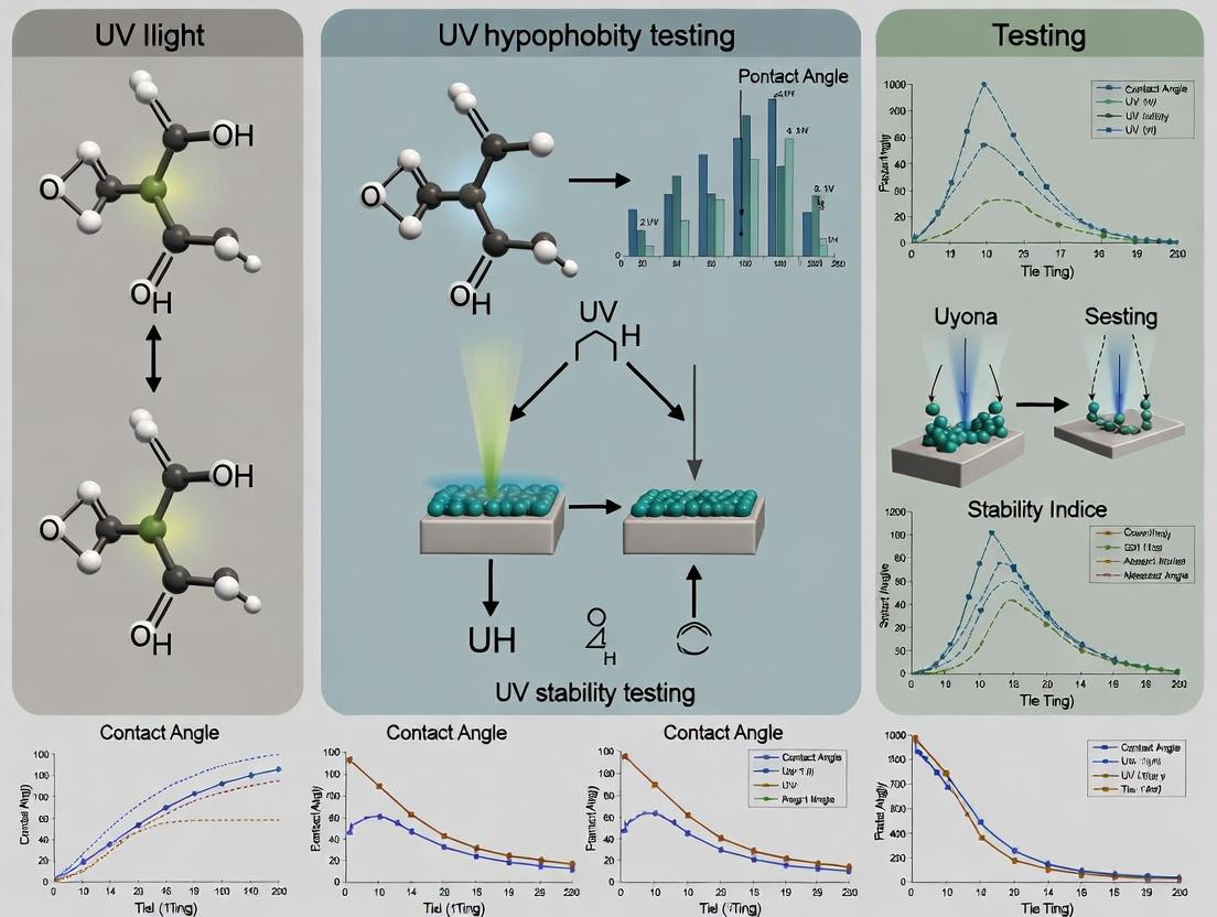
Visualizations
Diagram Title: Pathway to Superhydrophobicity and UV Degradation
Diagram Title: UV Stability Testing Workflow for Coatings
Ultraviolet (UV) radiation (290-400 nm) initiates photochemical degradation in polymers and nanomaterials through the absorption of photons, leading to the formation of excited states and reactive species. This is a critical consideration in the development of durable superhydrophobic coatings, where both polymeric binders and nanostructured surface features are vulnerable.
- Primary Pathways:
- Photo-initiation: Absorption of UV light by chromophores (e.g., catalyst residues, carbonyl groups, π-bonds) generates excited singlet states, which can intersystem cross to longer-lived triplet states.
- Primary Photochemical Reactions: These excited states undergo cleavage (e.g., Norrish Type I and II in polyolefins, chain scission in polycarbonates) or react with oxygen/neighbors.
- Propagation (Auto-oxidation): The radical species react with atmospheric oxygen to form peroxy radicals (POO•), which abstract hydrogen from the polymer to form hydroperoxides (POOH). These hydroperoxides are unstable and photolyze easily, generating new radicals in a cyclic chain reaction.
- Termination: Radical recombination leads to cross-linking, chain branching, or the formation of inert products.
Table 1: Key Photodegradation Pathways for Common Coating Components
| Material/Component | Primary Chromophore | Key Degradation Pathway | Major Degradation Products |
|---|---|---|---|
| Polypropylene (Binder) | Catalyst residues, hydroperoxides | Norrish I/II, β-scission | Carbonyls, vinylidenes, chain scission fragments |
| Polydimethylsiloxane (PDMS) | Siloxane bonds, impurities | Radical-induced rearrangement, oxidation | Silanols, cross-linked silica-like domains |
| Polycarbonate (Binder) | Aromatic carbonate groups | Photo-Fries rearrangement, hydrolysis | Phenols, phenyl salicylate, discoloration |
| TiO₂ Nanoparticles (Filler) | Semiconductor bandgap | UV absorption generates e⁻/h⁺ pairs | Reactive Oxygen Species (•OH, O₂•⁻), surface defects |
| SiO₂ Nanoparticles (Filler) | Surface silanol groups | Less absorbent; surface radical formation via impurity sensitization | Interfacial bond cleavage, loss of functionalization |
Diagram 1: General Photodegradation Pathway Leading to Coating Failure (100 chars)
Application Notes: UV Stability Testing for Superhydrophobic Coatings
Objective: To quantitatively assess the resistance of superhydrophobic coatings to UV-induced photochemical degradation, correlating changes in chemical structure with loss of hydrophobic performance (Contact Angle, CA; Roll-off Angle, ROA).
Table 2: Standardized UV Exposure Conditions (Based on ISO 4892, ASTM G154)
| Parameter | Condition 1 (Accelerated Aging) | Condition 2 (Outdoor Simulated) | Notes |
|---|---|---|---|
| UV Source | UVA-340 Lamp | Xenon Arc (with filters) | UVA-340 mimics sunlight from 365 nm down. |
| Irradiance | 0.76 W/m² @ 340 nm | 0.51 W/m² @ 340 nm (or 60 W/m², 300-400 nm) | Controlled by radiometer. |
| Cycle | 8h UV @ 60°C / 4h Condensation @ 50°C | Continuous UV with periodic light/dark & spray cycles | Cycle depends on end-use simulation. |
| Total Dose for Evaluation | 500, 1000, 2000 kJ/m² | 500, 1000, 2000 MJ/m² | Record dose at each measurement interval. |
Experimental Protocols
Protocol 3.1: Accelerated UV Exposure & Wettability Tracking
- Purpose: To simulate long-term UV exposure and monitor the degradation of superhydrophobic performance.
- Materials: See "The Scientist's Toolkit" below.
- Procedure:
- Baseline Characterization: Measure initial Static Water Contact Angle (SCA) and Roll-off Angle (ROA) on at least 5 distinct coating spots (ASTM D7490/D7334). Perform FTIR/ATR and optical microscopy.
- Mounting: Securely mount coated substrates in the QUV chamber's sample racks, ensuring no shadowing.
- Exposure Cycle: Program the QUV with UVA-340 lamps to run Condition 1 from Table 2. Start exposure.
- Periodic Sampling: At defined UV dose intervals (e.g., equivalent to 250, 500, 1000 kJ/m²), remove samples. Condition at 23°C/50% RH for 24h.
- Post-Exposure Analysis: Repeat SCA/ROA measurements. Visually inspect for chalking, cracking, or gloss change. Record FTIR spectra, focusing on carbonyl index (CI) growth (peak ~1715 cm⁻¹) and changes in Si-O-Si/Si-CH₃ regions for silicones.
- Data Correlation: Plot SCA/ROA versus UV dose and CI versus UV dose.
Protocol 3.2: Quantifying Hydroperoxide (POOH) Accumulation
- Purpose: To measure the key intermediate in the auto-oxidation cycle, indicating oxidative stress level.
- Method: Iodometric Test (ASTM D3899-07, adapted for coatings).
- Scrape ~0.5g of coating from a control and UV-exposed substrate into a glass vial.
- Dissolve/disperse in 20 mL of acetic acid-chloroform (3:2 v/v) solvent.
- Flush vial with N₂, add 1 mL of saturated KI solution. Seal and store in dark for 60 min.
- Add 30 mL of distilled water, titrate the liberated iodine with 0.01M sodium thiosulfate (Na₂S₂O₃) using a micro-burette until the yellow color disappears.
- Calculate POOH concentration:
[POOH] (mol/kg) = (V * M * 1000) / (2 * m), where V= titrant volume (mL), M= titrant molarity, m= sample mass (g).
Diagram 2: UV Stability Test Workflow for Coatings (92 chars)
The Scientist's Toolkit: Key Research Reagent Solutions & Materials
Table 3: Essential Materials for UV Stability Testing of Coatings
| Item | Function/Description | Example/Notes |
|---|---|---|
| UVA-340 Fluorescent Lamps | UV source with peak at 340 nm, closely matching solar cutoff. | Q-Lab Corporation, Atlas Material Testing. Essential for QUV testers. |
| Xenon Arc Lamp System | Full-spectrum light source including UV, visible, and IR. Can simulate daylight through filters. | Atlas Ci/Ci-Series, Suntest CPS+. For outdoor simulation. |
| Calibrated Radiometer | Measures and controls irradiance (W/m²) at specific wavelengths (e.g., 340 nm). | Critical for dose reproducibility (kJ/m²). |
| Contact Angle Goniometer | Quantifies wettability by measuring static and advancing/receding contact angles. | DataPhysics OCA, Krüss DSA series. Primary performance metric. |
| FTIR Spectrometer with ATR | Analyzes chemical bond changes (carbonyl growth, Si-CH₃ loss) on coating surface. | Thermo Scientific Nicolet, PerkinElmer Frontier. |
| Acetic Acid-Chloroform Mixture (3:2) | Solvent for iodometric titration. Dissolves many polymers and allows POOH-KI reaction. | Hazard: Handle in fume hood. Prepare fresh. |
| Sodium Thiosulfate (0.01M) | Titrant for iodometric assay. Standardized against potassium iodate. | Must be standardized; concentration is critical for POOH calculation. |
| Saturated Potassium Iodide (KI) Solution | Reducing agent. POOH oxidizes I⁻ to I₂, which is titrated. | Prepare in deaerated water, store in amber bottle. |
| Reference Superhydrophobic Coating | A control coating with known UV stability (or lack thereof) for benchmarking. | e.g., A commercially available or well-published lab formulation. |
1. Introduction Within the research thesis on "Advanced UV Stability Testing of Engineered Superhydrophobic Coatings for Biomedical Applications," monitoring specific Key Performance Indicators (KPIs) is critical to quantify degradation. Prolonged ultraviolet (UV) exposure induces chemical and topographical changes in coatings, directly compromising their hydrophobic performance. The primary KPIs at risk are the Static Water Contact Angle (WCA), the Sliding/Roll-off Angle (SA), and the Surface Morphology. This document provides application notes and detailed protocols for assessing these KPIs before, during, and after accelerated UV stability testing, tailored for researchers and scientists in coating development and applied material sciences.
2. Quantitative Data Summary: Impact of UV Exposure on KPIs The following table summarizes typical degradation trends observed for superhydrophobic coatings (e.g., based on silicone/fluorine polymers with silica or TiO2 nanoparticles) under standardized UV exposure (e.g., 0.5-1.0 W/m² at 340 nm, 25-60°C).
Table 1: KPI Degradation Profile Under Accelerated UV Testing
| UV Exposure Dose (kJ/m²) | Avg. Static Contact Angle (WCA) | Avg. Sliding Angle (SA) | Surface Morphology Change (SEM/AFM) | Probable Cause |
|---|---|---|---|---|
| 0 (Baseline) | 155° - 165° | <5° | Dense nano-micro hierarchical structure | Initial superhydrophobic state. |
| 250 | 150° - 158° | 8° - 15° | Minor nanoparticle agglomeration; micro-cracks initiation. | Initial photo-oxidative cleavage of low-surface-energy groups. |
| 500 | 140° - 148° | 20° - 40° | Visible micro-cracking; loss of finer nanoscale features. | Continued polymer chain scission; loss of binding agent integrity. |
| 1000 | 125° - 138° | >45° (often pinned) | Severe cracking, delamination; complete loss of hierarchy. | Advanced oxidation; significant chemical modification and physical degradation. |
3. Experimental Protocols
Protocol 3.1: Measurement of Static Water Contact Angle (WCA)
- Objective: Quantify the intrinsic wettability by measuring the angle between the water droplet tangent and the solid surface.
- Materials: Contact angle goniometer, high-purity deionized water (or simulated body fluid for bio-applications), microliter syringe, flat-coated substrate sample.
- Method:
- Level the sample stage precisely.
- Place the coated sample on the stage.
- Using a syringe, dispense a 5 µL (±0.5 µL) water droplet onto the surface from a height of ~1 cm.
- Capture an image of the droplet within 3 seconds of deposition using the instrument's software.
- Use the software's Young-Laplace fitting algorithm to calculate the left and right contact angles. Report the average of at least 5 measurements from different surface locations.
Protocol 3.2: Measurement of Sliding/Roll-off Angle (SA)
- Objective: Determine the dynamic hydrophobicity by measuring the minimum surface tilt angle required for a water droplet to move.
- Materials: Tilting stage goniometer or custom apparatus with precision angle indicator, high-purity deionized water, microliter syringe, coated sample.
- Method:
- Affix the coated sample to the tilting stage, ensuring it is horizontal at 0°.
- Dispense a 10 µL water droplet onto the surface.
- Gradually increase the tilt angle at a slow, constant rate (e.g., 0.5° - 1.0° per second).
- Record the angle at which the droplet completely rolls or slides off the surface.
- Repeat this process 5 times and report the average sliding angle. Note any droplet pinning.
Protocol 3.3: Analysis of Surface Morphology via Scanning Electron Microscopy (SEM)
- Objective: Visualize and quantify changes in micro/nano-scale surface topography induced by UV exposure.
- Materials: SEM, coated sample (approx. 1x1 cm), conductive double-sided tape or adhesive carbon tabs, sputter coater for non-conductive coatings.
- Method:
- Sample Preparation: Mount the coated sample on an SEM stub using conductive tape. For non-conductive polymeric coatings, sputter-coat with a thin layer (5-10 nm) of gold or platinum.
- Imaging: Insert the sample into the SEM chamber. Evacuate to high vacuum.
- Select accelerating voltages between 5-15 kV to balance detail and avoid charging.
- Capture images at multiple magnifications (e.g., 1,000x, 10,000x, 50,000x) to assess both micro- and nano-scale features. Ensure consistent imaging parameters across all samples (pre- and post-UV).
- Use image analysis software to measure feature dimensions (e.g., nanoparticle size, crack width, roughness).
4. Workflow and Relationship Diagrams
Title: UV Degradation Pathway Impacting KPIs
Title: UV Stability Testing Workflow for Coatings
5. The Scientist's Toolkit: Key Research Reagent Solutions & Materials
Table 2: Essential Materials for KPI Analysis in UV Stability Studies
| Item / Reagent | Function / Relevance |
|---|---|
| Fluorinated Alkyl Silane (e.g., 1H,1H,2H,2H-Perfluorooctyltriethoxysilane) | Common low-surface-energy coating agent. Its Si-O-C bond stability under UV is a key study parameter. |
| Hydrophobic Fumed Silica Nanoparticles (e.g., AEROSIL R812) | Used to create nanoscale roughness. UV-induced changes in surface chemistry and dispersion affect morphology. |
| UV Stabilizer Additives (e.g., Hindered Amine Light Stabilizers - HALS) | Incorporated to study mitigation of KPI degradation; a control variable in formulations. |
| High-Purity Deionized Water (18.2 MΩ·cm) | Standard liquid for WCA/SA measurements. Consistent purity is critical for reproducible results. |
| Conductive Sputter Coating Material (Gold/Palladium) | Essential for preparing non-conductive polymer coatings for high-resolution SEM imaging without charging artifacts. |
| Calibrated UV Light Source (e.g., Xenon Arc with 340 nm filter) | Provides standardized, reproducible UV-A spectrum for accelerated weathering, enabling dose-dependent KPI correlation. |
| Reference Superhydrophobic Coated Slide | Used to validate and calibrate measurement systems (goniometer, tilting stage) prior to sample testing. |
Within the broader thesis on UV stability testing of superhydrophobic coatings (SHCs), this document details the critical real-world implications of coating degradation. SHCs, characterized by water contact angles >150°, are pivotal in biomedical implants (e.g., stents, catheters) and microfluidic lab-on-a-chip (LOC) devices. Their failure due to UV exposure, mechanical abrasion, or chemical fouling leads to catastrophic outcomes, including device malfunction, biofilm formation, and inaccurate diagnostic results. These application notes provide protocols to assess failure modes, linking accelerated UV stability testing to performance in operational environments.
Table 1: Documented Failure Consequences of Superhydrophobic Coatings in Medical Devices
| Device Type | Primary Coating Function | Key Failure Mode | Quantifiable Consequence | Reported Incidence/Data |
|---|---|---|---|---|
| Orthopedic Implant | Prevent biofilm adhesion | Hydrophobic recovery loss (Contact Angle <90°) | Biofilm formation rate increase | 85% increase in S. aureus adhesion after 500h simulated UV aging |
| Vascular Stent | Reduce platelet adhesion | Nanoscale feature erosion (by AFM) | Thrombus formation risk | 60% rise in platelet activation on degraded vs. pristine SHC |
| Microfluidic LOC | Low-loss fluid transport | Increased contact angle hysteresis (>20°) | Coefficient of variation (CV) in assay results | CV increases from 2% to 15% post-UV stress, leading to false positives/negatives |
| Biosensor Electrode | Anti-fouling for signal stability | Protein adsorption (> 100 ng/cm²) | Sensor signal drift | Sensitivity loss of 40% after 72h in protein-rich fluid on aged coating |
Experimental Protocols
Protocol 2.1: Accelerated UV Aging and Hydrophobicity Assessment
Aim: Simulate long-term environmental UV exposure and quantify hydrophobic decay. Materials: SHC-coated substrate, UV chamber (UVA-340 lamps), goniometer, DI water. Procedure:
- Baseline Measurement: Measure static water contact angle (WCA) at 5 points on pristine SHC.
- UV Exposure: Place sample in UV chamber. Expose to 0.8 W/m² @ 340nm at 50°C for defined cycles (e.g., 24h, 48h, 100h, 500h).
- Post-Exposure Analysis: After each cycle, remove sample, condition at 25°C/50% RH for 1h. Re-measure WCA and contact angle hysteresis (advancing/receding angles).
- Data Analysis: Plot WCA vs. UV dose (J/m²). Calculate decay constant. Use ASTM D4329/D4587 as guidelines.
Protocol 2.2: Post-UV Aging Biofilm Adhesion Assay
Aim: Quantify bacterial adhesion on UV-degraded SHCs for implant applications. Materials: UV-aged SHC samples, Staphylococcus aureus (ATCC 25923), Tryptic Soy Broth (TSB), sterile PBS, crystal violet stain, microplate reader. Procedure:
- Sample Preparation: Sterilize SHC samples (UV-aged and control) with 70% ethanol for 30 min, rinse with PBS.
- Bacterial Culture: Grow S. aureus overnight in TSB at 37°C. Dilute to 10⁶ CFU/mL in fresh medium.
- Adhesion Phase: Incubate samples in bacterial suspension for 2h at 37°C under static conditions.
- Analysis: Rinse gently with PBS to remove non-adherent cells. Fix with 99% methanol for 15 min, stain with 0.1% crystal violet for 5 min. Elute stain with 33% acetic acid. Measure absorbance at 590 nm. Correlate to biofilm biomass.
Protocol 2.3: Microfluidic Device Performance Testing
Aim: Evaluate the impact of SHC degradation on LOC assay precision. Materials: LOC device with SHC-treated channels, target analyte (e.g., fluorescently-labeled albumin), phosphate buffered saline (PBS), syringe pump, fluorescence microscope. Procedure:
- Baseline Flow: Infuse PBS at 5 µL/min. Record pressure drop (ΔP) across main channel.
- Assay Run (Pristine): Infuse analyte solution. Capture fluorescence intensity (FI) at detection zone over 10 replicates. Calculate mean FI and CV.
- UV Ageing: Subject the entire LOC device to Protocol 2.1 (100h UV dose).
- Post-Ageing Assay: Repeat steps 1-2. Compare ΔP (indicates flow resistance change from wettability shift) and assay CV. Droplet pinning or air bubble formation are visual failure indicators.
Visualizations
Title: UV-Induced Coating Failure Pathway
Title: Integrated UV Stability Testing Workflow
The Scientist's Toolkit
Table 2: Essential Research Reagent Solutions & Materials
| Item Name | Function/Application | Key Characteristics |
|---|---|---|
| UVA-340 Fluorescent Lamps | Simulate solar UV spectrum (295-365 nm) for accelerated aging. | Peak at 340nm, matching sunlight's critical short UV wavelength. |
| Contact Angle Goniometer | Quantify surface wettability (static & dynamic contact angles). | High-resolution camera, automated dispensing system for hysteresis. |
| Atomic Force Microscope (AFM) | Measure nanoscale roughness (Ra, Rq) and coating morphology post-UV. | Tapping mode in air to assess nanostructure degradation. |
| Crystal Violet Solution (0.1%) | Stain adhered bacterial biofilms for semi-quantitative analysis. | Standardized biofilm assay reagent; elutes in acetic acid for OD reading. |
| Fluorescently-Tagged Albumin | Model protein for fouling studies and microfluidic assay validation. | Common fouling agent; allows visualization of non-specific adsorption in channels. |
| Phosphate Buffered Saline (PBS), pH 7.4 | Universal rinsing and dilution buffer in biological assays. | Isotonic and non-reactive; preserves protein/bacterial viability during rinses. |
| Silica Nanoparticles (20-50 nm) | Common component for constructing roughness in SHC formulations. | Provide hierarchical structure; stability under UV is a key test variable. |
| Fluoroalkyl Silane (e.g., FAS-17) | Low-surface-energy agent for SHC synthesis. | Prone to photo-oxidative degradation; primary target in UV stability studies. |
How to Test UV Stability: Standard Protocols and Advanced Accelerated Testing Methods
In the research of UV stability for superhydrophobic coatings, the assessment of photostability—the material's resistance to chemical and physical degradation upon exposure to light—is paramount. This investigation, framed within a broader thesis on developing durable superhydrophobic surfaces for aerospace and biomedical applications, relies on established international standards to ensure reproducible, reliable, and relevant accelerated weathering data. The standards ASTM G154, ISO 4892, and ICH Q1B provide structured methodologies for light exposure testing, though they originate from and are tailored to different industries: general materials and plastics (ASTM/ISO) and pharmaceuticals (ICH). This article details their application, protocols, and adaptation for advanced coating research.
Table 1: Core Comparison of Photostability Standards
| Aspect | ASTM G154 | ISO 4892-2 | ICH Q1B |
|---|---|---|---|
| Primary Scope | Non-metallic materials | Plastics | Pharmaceutical products |
| Key Light Source | Fluorescent UV lamps (UVA-340, UVB-313) | Xenon arc, Fluorescent UV, Open-flame carbon arc | Cool white & near-UV fluorescent lamps |
| Typical Cycle | Light only, or light + condensation (e.g., 8h UV at 60°C / 4h Condensation at 50°C) | Various, incl. light only, light + dark, light + spray | Option 1: Cool white & near-UV together. Option 2: Sequential exposure. |
| Control Parameter | Irradiance (W/m²/nm) at a specified wavelength (e.g., 0.89 W/m² @ 340nm) | Irradiance (W/m²) within a specified band | Overall illumination (lux-hr) & integrated near-UV energy (watt-hr/m²) |
| Primary Endpoint | Change in material properties (gloss, color, cracking) | Change in material properties | Confirmation of forced degradation; identification of degradation products. |
| Quantitative Exposure | Often by time or total radiant exposure (J/m²) | Often by time or total radiant energy | Minimum: 1.2 million lux-hr (visible) & 200 watt-hr/m² (near-UV) |
Table 2: Adaptability to Superhydrophobic Coating Research
| Standard | Relevance to Coatings | Key Measurable Parameters | Potential Limitations |
|---|---|---|---|
| ASTM G154 | High. Directly applicable for polymer/coating durability. | Contact angle (CA), roll-off angle, gloss, color ΔE, FTIR for chemical change. | Fluorescent UV spectrum does not fully match sunlight. |
| ISO 4892-2 | Very High. Xenon arc provides best solar simulation. | CA hysteresis, surface morphology (SEM), mechanical adhesion. | Equipment cost and complexity. |
| ICH Q1B | Moderate. Useful for coatings in biomedical/drug delivery contexts. | Chemical stability of encapsulated agents, surface chemistry (XPS). | Light spectrum is specific to drug stability, not material weatherability. |
Application Notes for Coating Research
- Standard Selection: For general outdoor performance prediction of superhydrophobic coatings, ISO 4892-2 with a xenon arc lamp is preferred due to its superior spectral match to global sunlight. ASTM G154 offers a faster, more economical screening tool, especially for UVA-340 lamps which simulate the critical short-wavelength UV region. ICH Q1B should be considered only if the coating is part of a drug-device combination product, where the focus is on the stability of the active pharmaceutical ingredient (API).
- Critical Coating-Specific Metrics: Beyond standard color/gloss, track:
- Wettability Degradation: Measure water contact angle (WCA) and sliding/roll-off angle at regular intervals. A significant decrease in WCA or increase in hysteresis indicates loss of superhydrophobicity.
- Surface Morphology: Use Scanning Electron Microscopy (SEM) or Atomic Force Microscopy (AFM) post-exposure to observe degradation of micro/nano-rough features essential for the Cassie-Baxter state.
- Chemical Analysis: Employ Fourier-Transform Infrared Spectroscopy (FTIR) and X-ray Photoelectron Spectroscopy (XPS) to detect photo-oxidative changes (e.g., formation of carbonyl groups, loss of fluorinated components).
- Sample Preparation: Apply coatings to standardized substrates (e.g., Q-Panels). Include unexposed controls stored in dark, controlled conditions. A minimum of 3 replicates is required for statistical significance.
Detailed Experimental Protocols
Protocol 1: Basic Screening per ASTM G154 (Cycle 1)
Aim: To assess the relative UV durability of novel superhydrophobic coating formulations. Workflow:
- Preparation: Apply Candidate Coating A, B, and C to aluminum panels (n=4 per group).
- Baseline Characterization: Measure initial WCA, roll-off angle, gloss (60°), and color (Lab*).
- Exposure: Place samples in QUV/se weathering tester equipped with UVA-340 lamps.
- Cycle: Run Cycle 1 (8 hours of UV at 60°C irradiance set point 0.89 W/m² @ 340nm, followed by 4 hours of condensation at 50°C).
- Interim Testing: Remove one replicate from each group at 250, 500, and 1000 hours.
- Post-Exposure Analysis: Characterize per step 2. Perform SEM/FTIR on 1000h samples.
- Data Analysis: Plot WCA vs. exposure time. Perform ANOVA to compare coating performance.
Experimental Workflow for ASTM G154 Screening
Protocol 2: Comprehensive Aging per ISO 4892-2 (Xenon Arc)
Aim: To simulate realistic outdoor aging for the most promising coating candidate. Workflow:
- Preparation: Prepare top-performing coating on final application substrate (e.g., composite aerospace panel).
- Baseline Analysis: Perform full characterization (WCA, SEM, AFM, FTIR, XPS, adhesion cross-cut test).
- Exposure: Place in xenon arc weatherometer with Daylight-B filter (e.g., Q-Sun).
- Conditions: Use Method A, Cycle 1 (Continuous light, Irradiance 0.51 W/m² @ 340nm, Black Standard Temperature 65°C, Chamber Temp 40°C, RH 50%).
- Monitoring: Use calibrated radiometer to confirm irradiance.
- Interval Testing: Remove samples at 500, 1000, 2000 kJ/m² of total UV radiant exposure.
- Final Assessment: Repeat full battery of tests. Correlate property loss to radiant exposure dose.
Xenon Arc Exposure and Assessment Workflow
The Scientist's Toolkit: Key Research Reagent Solutions
Table 3: Essential Materials for Photostability Testing of Coatings
| Item / Reagent | Function / Role in Experiment | Example / Specification |
|---|---|---|
| UVA-340 Lamps | Simulates solar UV from 365 nm down to ~295 nm cutoff. Critical for ASTM G154. | Q-Lab UVA-340, 1.55 W/m² @ 340nm peak irradiance. |
| Daylight-B (Boro/Boro) Filters | Used with xenon arc to modify spectrum, closely matching terrestrial sunlight for ISO 4892-2. | Atlas Ci Series Daylight-B Filter. |
| Irradiance Calibrator | Ensures accurate and consistent light intensity during test, required by all standards. | Calibrated radiometer (e.g., with 340nm sensor). |
| Deionized & De-mineralized Water | For humidity generation and condensation cycles; prevents scale/spotting on samples. | ASTM Type IV or better (conductivity < 5 µS/cm). |
| Contact Angle Goniometer | Primary tool for quantifying wettability and superhydrophobic performance degradation. | 5µL water droplet, sessile drop method. |
| Standard Reflective Tiles | For calibrating colorimeters and spectrophotometers used in color change measurements. | Ceramic color standards (e.g., BCRA Series). |
| Cross-Cut Cutter & Adhesive Tape | Evaluates coating adhesion loss, a critical failure mode after photo-degradation. | ASTM D3359 Multi-blade cutter (1mm spacing). |
| FTIR ATR Crystal | Enables surface-specific chemical analysis to detect oxidation products without sample destruction. | Diamond/ZnSe crystal. |
Within the broader research on the UV stability of advanced superhydrophobic coatings, accelerated aging tests are critical for predicting long-term environmental performance. These coatings, used in aerospace, automotive, and biomedical applications, degrade under solar UV radiation, leading to loss of hydrophobicity and surface functionality. This protocol outlines a standardized, accelerated test method to evaluate this degradation, enabling rapid material screening and lifetime prediction.
Core Components of UV Aging Test Design
Lamp Types: Spectral Power Distribution (SPD)
The lamp type defines the spectral output of the test, which must simulate critical wavelengths of natural sunlight that cause photochemical degradation. The choice depends on the specific research question and relevant exposure environment.
Table 1: Comparison of Common UV Light Sources for Accelerated Aging
| Lamp Type | Peak Emission Range | Simulates | Advantages | Disadvantages | Best For |
|---|---|---|---|---|---|
| UVA-340 | 315-400 nm (peak at 340 nm) | Solar UV spectrum (295-365 nm) very well. | Excellent match to sunlight in critical short-wave UV region. | Limited irradiance below 295 nm. | Most accurate for sunlight simulation; general material testing. |
| UVB-313 | 280-360 nm (peak at 313 nm) | More short-wave UV than sunlight at Earth's surface. | Faster acceleration rate. | Can cause unrealistic failures (photodegradation not seen outdoors). | Aggressive testing for QC or very durable materials. |
| Xenon Arc | Full spectrum: UV, Vis, IR. | Full spectrum of sunlight, including visible light and infrared (heat). | Most realistic full-spectrum simulation. Allows for irradiance control at specific wavelengths. | Complex, expensive, requires regular calibration and filter changes. | Highest fidelity testing where photo- and thermal degradation interact. |
| Metal Halide | Enhanced UV output across range. | Intense UV radiation. | High irradiance, good for very fast testing. | Spectral stability can be an issue; less common in standardized tests. | High-acceleration stress tests. |
Irradiance Setting and Calibration
Irradiance is the radiant power per unit area (W/m²) received by the sample, typically controlled at a specific wavelength (e.g., 340 nm). It is the primary driver of acceleration.
- Typical Setpoint: For UVA-340 lamps, a common irradiance level is 0.76 W/m² @ 340 nm. This is approximately the maximum midday summer sun irradiance.
- Acceleration: Increasing irradiance (e.g., to 1.55 W/m²) accelerates the test but risks altering degradation mechanisms.
- Calibration: Critical. Use a calibrated radiometer with specific wavelength filters (e.g., 340 nm) to measure and adjust irradiance regularly (e.g., every 400 hours). Position the sensor at the same plane as samples.
Temperature Control
Temperature significantly influences degradation kinetics. It must be controlled separately from UV irradiance.
- Black Standard Temperature (BST): Measured by a thermometer attached to a black metal panel, it represents the maximum temperature a sample might reach. BST is the standard reported temperature.
- Typical Range: Tests for coatings often use a BST of 50°C to 70°C. Higher temperatures increase acceleration but may induce thermal-only degradation.
- Control: The test chamber controls air temperature, but BST is the key metric. Cooling periods may be incorporated to mimic diurnal cycles.
Test Cycles
Cycles define the pattern of UV exposure, often combined with dark periods and condensation/humidity to simulate dew.
- Basic UV Cycle: Continuous UV exposure at constant temperature and irradiance. Simple but less realistic.
- Standard Cycle (e.g., ASTM G154 Cycle 1): 8 hours UV at 60°C BST followed by 4 hours condensation (no UV) at 50°C. This cycle introduces moisture, critical for testing hydrolysis and physical stress on coatings.
Table 2: Example Test Parameters for Superhydrophobic Coating Evaluation
| Parameter | Recommended Setting (UVA-340) | Alternative (UVB-313) | Rationale |
|---|---|---|---|
| Lamp Type | UVA-340 | UVB-313 | Balance of realism vs. acceleration. |
| Irradiance @ λ | 0.76 W/m² @ 340 nm | 0.55 W/m² @ 310 nm | Matches/moderately exceeds peak solar UV. |
| Cycle | 8h UV (60°C BST) / 4h Condensation (50°C) | 8h UV (60°C BST) / 4h Condensation (50°C) | Introduces thermal and moisture stress. |
| Total Duration | 500 - 2000 hours | 250 - 1000 hours | Time to observe measurable change in water contact angle (WCA). |
Detailed Experimental Protocol: UV Aging of Superhydrophobic Coatings
Protocol Title: Accelerated UV Degradation of Superhydrophobic Coatings per Modified ASTM G154.
Objective: To quantify the loss of hydrophobicity (via Water Contact Angle) and surface morphology (via SEM/AFM) of coated substrates after defined periods of accelerated UV exposure.
Materials & Substrate Preparation:
- Substrates: Cleaned glass slides, aluminum panels, or polymer films.
- Coating Application: Apply superhydrophobic coating (e.g., silica nanoparticle-based, fluoropolymer) via spray-coating, dip-coating, or spin-coating per established lab protocol. Cure fully.
- Pre-test Characterization: Measure initial Water Contact Angle (WCA) at 5 locations per sample using a goniometer. Image surface via Scanning Electron Microscopy (SEM) for baseline morphology.
UV Aging Procedure:
- Chamber Setup:
- Install UVA-340 lamps. Age new lamps for at least 100 hours prior to testing.
- Calibrate irradiance to 0.76 W/m² @ 340 nm using a calibrated radiometer at the sample plane.
- Set test cycle: 8 hours of UV exposure at 60°C (±2°C) Black Standard Temperature.
- Set dark cycle: 4 hours of condensation (no UV) at 50°C (±2°C). Ensure chamber is configured for 100% relative humidity during this phase.
- Sample Mounting:
- Mount coated samples in sample holders. Include uncoated control substrates if required.
- Ensure samples are firmly secured and facing the UV lamps uniformly. Rotate sample positions periodically (e.g., every 168 hours) to account for any chamber gradient.
- Test Execution:
- Start the cycle. Program the chamber to run continuously.
- Interim Points: Remove representative samples at defined intervals (e.g., 0, 250, 500, 1000, 1500, 2000 hours).
- Allow removed samples to condition at standard lab atmosphere (23°C, 50% RH) for 24 hours before post-characterization.
- Post-Exposure Characterization:
- WCA Measurement: Measure WCA as done pre-test. Calculate mean and standard deviation. A significant drop (e.g., from >150° to <120°) indicates degradation.
- Surface Analysis: Perform SEM/AFM on aged samples to observe nanoparticle loss, crack formation, or chemical degradation.
- Chemical Analysis: Use FTIR or XPS to detect oxidation (e.g., formation of carbonyl groups) or loss of fluorinated compounds.
Data Analysis:
- Plot WCA (mean) versus UV exposure time (hours).
- Determine the critical exposure time for "failure" (e.g., WCA < 130°).
- Correlate WCA loss with changes in surface morphology from SEM images.
The Scientist's Toolkit: Research Reagent Solutions & Essential Materials
Table 3: Key Materials for UV Aging Test of Superhydrophobic Coatings
| Item | Function/Description | Example/Note |
|---|---|---|
| QUV Accelerated Weathering Tester | Chamber housing lamps and controlling irradiance, temperature, and cycles. | Q-Lab QUV/se, Atlas UVTest. |
| UVA-340 Lamps | Light source providing near-solar UV spectrum. | Q-Lab UVA-340, 40W. |
| Calibrated Radiometer | Device to measure and calibrate UV irradiance at specific wavelength. | Must have 340 nm filter. ILT950 radiometer. |
| Black Standard Thermometer | Measures the highest temperature (BST) a sample may attain. | Critical for reporting and control. |
| Water Contact Angle Goniometer | Measures static/dynamic contact angles to quantify hydrophobicity. | DataPhysics OCA, Krüss DSA. |
| Scanning Electron Microscope (SEM) | High-resolution imaging of nano/micro-scale surface morphology before/after aging. | Requires sputter coater for non-conductive coatings. |
| Coating Precursors | Materials to fabricate the superhydrophobic coating. | e.g., Tetraethoxysilane (TEOS), 1H,1H,2H,2H-Perfluorooctyltriethoxysilane (PFOTES), hydrophobic silica nanoparticles. |
| Reference Material | A polymer with known weatherability to verify chamber performance. | e.g., Blue Wool L4, polycarbonate. |
Visualized Workflows and Relationships
Diagram 1: UV Aging Test Protocol Workflow
Diagram 2: UV Degradation Pathways for Superhydrophobic Coatings
Within the broader thesis investigating the UV stability of advanced superhydrophobic coatings, comprehensive pre- and post-exposure characterization is critical. The degradation mechanisms—including photo-oxidation, chain scission, and loss of low-surface-energy components—must be quantified through a multimodal analytical approach. This document details application notes and standardized protocols for four cornerstone techniques: Contact Angle Goniometry, Scanning Electron Microscopy (SEM), X-ray Photoelectron Spectroscopy (XPS), and Fourier-Transform Infrared Spectroscopy (FTIR). These methods collectively provide data on wettability, surface morphology, elemental composition/chemistry, and molecular bonding, enabling a complete picture of UV-induced degradation.
Application Notes & Protocols
Contact Angle Goniometry
Application Note: Goniometry is the primary method for quantifying the wettability of a surface by measuring the static water contact angle (WCA). For a superhydrophobic coating, a WCA >150° and a low contact angle hysteresis (<10°) are typically targeted. UV exposure often leads to a measurable decrease in WCA and an increase in hysteresis, indicating loss of hydrophobicity due to chemical and topological changes.
Protocol: Sessile Drop Method for WCA Measurement
- Objective: To determine the static water contact angle pre- and post-UV aging.
- Materials: Contact angle goniometer, high-purity deionized water (or specified probe liquid), syringe with blunt needle, sample stage, compressed air or nitrogen duster.
- Procedure:
- Sample Preparation: Cut coating substrate to appropriate size (e.g., 2cm x 2cm). Ensure sample is clean and free of dust using a gentle stream of clean, dry air.
- Baseline Measurement (Pre-Exposure): Level the instrument stage. Place sample horizontally on the stage. Using the syringe, dispense a 5 µL droplet of deionized water onto the coating surface. Capture an image of the droplet within 3 seconds of deposition.
- Image Analysis: Use the instrument's software to fit the droplet profile (typically using the Young-Laplace or circle fitting method) and calculate the contact angle. Take measurements at a minimum of five distinct, non-edge locations on the sample. Record the mean and standard deviation.
- UV Exposure: Subject the sample to controlled UV aging (e.g., in a QUV weatherometer per ASTM G154, Cycle 1: 8h UV at 60°C, 4h condensation at 50°C).
- Post-Exposure Measurement: Repeat steps 2-3 on the UV-exposed sample without any cleaning beyond dust removal.
- Data Analysis: Compare pre- and post-exposure mean WCA values. A statistically significant decrease (e.g., >10°) indicates coating degradation.
Scanning Electron Microscopy (SEM)
Application Note: SEM provides high-resolution topographical and morphological information. For superhydrophobic coatings, which often rely on hierarchical micro-/nanoscale roughness, SEM is essential for observing UV-induced damage such as cracking, delamination, pore collapse, or erosion of nanostructures.
Protocol: SEM Imaging of Superhydrophobic Coatings
- Objective: To visualize and compare the surface microstructure pre- and post-UV exposure.
- Materials: SEM, conductive adhesive tape (e.g., carbon tape), sputter coater, gold/palladium target.
- Procedure:
- Sample Preparation: Mount a small sample fragment (≈1cm²) onto an SEM stub using conductive carbon tape, ensuring good electrical contact.
- Conductive Coating: Due to the typically non-conductive nature of polymer-based coatings, sputter-coat the sample with a 5-10 nm layer of Au/Pd using a sputter coater. This prevents charging artifacts.
- Imaging (Pre-Exposure): Insert the sample into the SEM chamber. After achieving high vacuum, image the surface at multiple magnifications (e.g., 500x, 5,000x, 25,000x) using an accelerating voltage of 5-10 kV. Capture representative images from multiple areas.
- Post-Exposure Imaging: After UV exposure, prepare a new fragment from the exposed area of the sample (steps 1-2). Acquire images at the same magnifications and under identical instrument conditions as the pre-exposure sample.
- Analysis: Perform a qualitative comparison of morphology. Quantitative analysis of feature dimensions (e.g., nanoparticle size, pore diameter) can be performed using image analysis software (e.g., ImageJ).
X-ray Photoelectron Spectroscopy (XPS)
Application Note: XPS probes the elemental composition and chemical bonding states of the top 1-10 nm of a surface. It is indispensable for detecting UV-induced chemical changes, such as oxidation (increase in O/C ratio), loss of fluorine-containing hydrophobic components, and formation of new carbon-oxygen bonds (C-O, C=O, O-C=O).
Protocol: Surface Chemical Analysis via XPS
- Objective: To determine the atomic concentration and chemical state of surface elements before and after UV exposure.
- Materials: XPS instrument, sample holder, possibly a charge neutralizer (flood gun).
- Procedure:
- Sample Preparation: Mount the coating sample securely on the holder. Avoid touching the analysis area.
- Pre-Exposure Survey & High-Resolution Scans: Insert into the ultra-high vacuum chamber. Acquire a wide survey scan (e.g., 0-1100 eV binding energy) to identify all elements present. Then, perform high-resolution scans over the photoelectron peaks of key elements (C 1s, O 1s, F 1s, Si 2p, etc.). Use a pass energy of 20-50 eV for high resolution.
- UV Exposure: Expose a separate, identically prepared sample to UV.
- Post-Exposure Analysis: Repeat step 2 on the UV-exposed sample.
- Data Processing:
- Apply a relative sensitivity factor (RSF) correction to calculate atomic percentages from peak areas.
- Deconvolute the high-resolution C 1s peak using curve-fitting software. Typical components for fluorinated/siloxane coatings: C-C/C-H (~285.0 eV), C-O (~286.5 eV), C=O (~288.0 eV), O-C=O (~289.0 eV), CF₂ (~291.0 eV), CF₃ (~293.0 eV).
- Analysis: Track changes in the O/C atomic ratio and the relative percentages of oxidized carbon (C-O, C=O, O-C=O) vs. fluorinated carbon (CFx) components.
Fourier-Transform Infrared Spectroscopy (FTIR)
Application Note: FTIR identifies molecular vibrations, providing information about functional groups and chemical bonds within the coating. Attenuated Total Reflectance (ATR) mode is ideal for surfaces. UV degradation can be monitored by the appearance of new bands (e.g., carbonyl C=O stretch at ~1720 cm⁻¹ from photo-oxidation) and the decrease of characteristic bands (e.g., Si-CH₃ at ~1270 cm⁻¹ or C-F stretches).
Protocol: ATR-FTIR Analysis of Coating Chemistry
- Objective: To identify changes in the molecular structure of the coating film induced by UV exposure.
- Materials: FTIR spectrometer with ATR accessory (diamond or germanium crystal), force gauge for consistent pressure.
- Procedure:
- Background Scan: Clean the ATR crystal with isopropanol and dry. Acquire a background spectrum with the same parameters to be used for the sample.
- Pre-Exposure Sample Scan: Place the coating sample firmly onto the ATR crystal, ensuring good contact. Acquire a spectrum over the range 4000-600 cm⁻¹ with 32-64 scans at a resolution of 4 cm⁻¹.
- UV Exposure: Expose the sample to UV.
- Post-Exposure Scan: Repeat step 2 on the same sample spot (if possible) or a representative exposed area.
- Data Processing: Apply atmospheric correction (for CO₂/H₂O). Normalize spectra (e.g., to a stable internal reference band like an aromatic ring stretch or Si-O-Si stretch if present and unchanging). Overlay pre- and post-exposure spectra.
- Analysis: Identify new absorption peaks and changes in peak intensity. The carbonyl index can be calculated as the ratio of the C=O peak area (~1720 cm⁻¹) to a stable reference peak area.
Table 1: Summary of Characterization Techniques for UV Stability Assessment
| Technique | Property Measured | Information Depth | Key Metrics for UV Degradation | Typical Measurement Time |
|---|---|---|---|---|
| Goniometry | Wettability | Surface (probe liquid interaction) | Static Water Contact Angle (WCA), Contact Angle Hysteresis | 15-30 min/sample |
| SEM | Topography/Morphology | Surface (~nm-µm resolution) | Feature size, cracking, delamination, roughness | 1-2 hours/sample |
| XPS | Elemental/Chemical State | 1-10 nm | Atomic % (C, O, F, Si), O/C ratio, C 1s chemical states | 1-3 hours/sample |
| ATR-FTIR | Molecular Bonds/Functional Groups | 0.5-2 µm | Peak position/intensity, Carbonyl Index, loss of specific bands | 10-20 min/sample |
Table 2: Example Quantitative Data Output from UV Exposure Study
| Sample Condition | Mean WCA (°) | O/C Ratio (XPS) | CF₂ % in C 1s (XPS) | Carbonyl Index (FTIR) |
|---|---|---|---|---|
| Pre-UV Exposure | 162 ± 3 | 0.12 | 18.5 | 0.05 |
| Post-500h UV | 142 ± 8 | 0.31 | 8.2 | 0.41 |
| Post-1000h UV | 115 ± 12 | 0.45 | 2.1 | 0.78 |
The Scientist's Toolkit: Key Research Reagent Solutions & Materials
Table 3: Essential Materials for Characterization of Superhydrophobic Coatings
| Material/Reagent | Function/Application |
|---|---|
| High-Purity DI Water (HPLC Grade) | Probe liquid for contact angle measurements; ensures consistency and avoids contamination. |
| Conductive Carbon Tape | Adhesively mounts non-conductive samples to SEM stubs while providing a path for charge dissipation. |
| Gold/Palladium Target (99.99%) | Source material for sputter coating; creates a thin, conductive metal layer on insulating samples for SEM. |
| ISO-OCTANE (Optical Grade) | Solvent for possible cleaning of samples pre-characterization without damaging the coating. |
| ATR Crystal Cleaning Kit (e.g., Isopropanol, Lint-Free Wipes) | Maintains the clarity and functionality of the FTIR-ATR crystal by removing residue from previous samples. |
| Certified XPS Reference Samples (e.g., Clean Au, Ag, Cu foil) | Used for energy scale calibration and periodic performance verification of the XPS instrument. |
| QUV Weatherometer Tubes (UVA-340) | Provides UV irradiation spectrum that closely mimics sunlight below 365 nm for controlled accelerated aging. |
Experimental Workflow & Data Relationship Diagrams
Workflow for UV Stability Assessment of Coatings
Relationship Between UV Damage and Characterization Data
This protocol is developed within the broader thesis research on evaluating the long-term durability of engineered superhydrophobic surfaces. A critical failure mode for organic/inorganic nanocomposite coatings, such as those based on Polydimethylsiloxane (PDMS) and Silica (SiO2) nanoparticles, is photodegradation under ultraviolet (UV) radiation. This degradation leads to the loss of hydrophobic properties, surface cracking, and eventual functional failure. This document provides detailed application notes and a standardized experimental protocol to assess UV stability, generating reproducible, quantitative data for comparative analysis in coating development.
Key Research Reagent Solutions & Materials
Table 1: Essential Materials and Reagents
| Item Name | Function/Brief Explanation |
|---|---|
| PDMS Pre-polymer (e.g., Sylgard 184) | Silicone elastomer base providing the hydrophobic matrix and flexibility. |
| Curing Agent | Crosslinks the PDMS pre-polymer to form an elastomeric network. |
| Hydrophobic Fumed SiO2 Nanoparticles (e.g., Aerosil R812) | Nano-scale filler providing surface roughness essential for superhydrophobicity. |
| Solvent (e.g., Hexane, Heptane) | Volatile carrier for creating a uniform coating dispersion. |
| Substrate (Glass, Aluminum, Steel) | The base material onto which the coating is applied for testing. |
| QUV Accelerated Weathering Tester | Instrument providing controlled cycles of UV light and condensation. |
| UV-B (313 nm) or UV-A (340 nm) Lamps | Standardized light sources simulating solar ultraviolet radiation. |
| Contact Angle Goniometer | Measures water contact angle (WCA) to quantify hydrophobicity. |
| Profilometer / Atomic Force Microscope (AFM) | Quantifies changes in surface topography and roughness (Ra, Rq). |
| Attenuated Total Reflectance Fourier-Transform Infrared (ATR-FTIR) Spectrometer | Analyzes chemical bond degradation (e.g., Si-CH3, Si-O-Si). |
Experimental Protocols
Coating Synthesis Protocol
Objective: Prepare a uniform, superhydrophobic PDMS/SiO2 nanocomposite coating.
- Weigh PDMS pre-polymer and curing agent at a 10:1 mass ratio.
- Mix thoroughly for 5 minutes, then dilute with heptane (1:3 weight ratio of PDMS:heptane).
- Add 10-20% w/w (relative to PDMS) of hydrophobic fumed SiO2 nanoparticles.
- Sonicate the mixture for 30 minutes using a probe sonicator (50% amplitude, pulse mode 5s on/5s off) to ensure homogeneous dispersion.
- Apply the dispersion to clean, pre-treated substrates via spray-coating or dip-coating.
- Cure at 80°C for 2 hours, followed by post-curing at 100°C for 1 hour.
Accelerated UV Exposure Protocol (ASTM G154 Cycle 4 Modified)
Objective: Subject coatings to controlled, reproducible UV stress.
- Place coated samples in a QUV weathering tester equipped with UV-B (313 nm) lamps.
- Program the following 8-hour cycle to run continuously:
- UV Exposure Phase (4 hrs, 60°C Black Panel Temperature): Irradiance set to 0.55 W/m²/nm at 313 nm.
- Condensation Phase (4 hrs, 50°C): Dark period with pure water vapor condensation.
- Terminate exposure at pre-defined intervals (e.g., 0, 100, 250, 500, 1000 hours) for characterization.
Post-Exposure Characterization Protocol
Objective: Quantify the extent of photodegradation.
- Static Water Contact Angle (WCA):
- Using a goniometer, place a 5 µL deionized water droplet on the surface.
- Capture an image and measure the angle using the sessile drop method.
- Take at least 5 measurements per sample at different locations.
- Surface Topography:
- Use a profilometer in scanning mode over a 1mm x 1mm area.
- Calculate the arithmetic mean roughness (Ra) and root mean square roughness (Rq).
- Chemical Analysis (ATR-FTIR):
- Scan from 4000 to 600 cm⁻¹ with 4 cm⁻¹ resolution.
- Monitor peak intensities at: ~2960 cm⁻¹ (C-H in Si-CH₃), ~1260 cm⁻¹ (Si-CH₃ symmetric deformation), and ~1000-1100 cm⁻¹ (Si-O-Si stretch).
- Use the Si-O-Si peak as an internal reference to normalize changes in organic group peaks.
Data Presentation
Table 2: Representative UV Degradation Data for PDMS/SiO2 Coating (UV-B, 1000 hrs)
| Exposure Time (hrs) | Avg. WCA (°) | WCA Retention (%) | Ra (µm) | Normalized Si-CH₃ Peak Intensity (2960 cm⁻¹) | Visual/Observational Notes |
|---|---|---|---|---|---|
| 0 | 162 ± 3 | 100 | 2.15 ± 0.2 | 1.00 | Highly hydrophobic, uniform, no cracks. |
| 100 | 158 ± 4 | 97.5 | 2.10 ± 0.3 | 0.95 | Slight loss of sheen, performance intact. |
| 250 | 148 ± 5 | 91.4 | 1.98 ± 0.4 | 0.87 | Visible dulling, WCA decline begins. |
| 500 | 132 ± 7 | 81.5 | 1.75 ± 0.5 | 0.72 | Loss of roll-off, sticky droplet adhesion. |
| 1000 | 105 ± 10 | 64.8 | 1.40 ± 0.6 | 0.51 | Significant chalking, micro-cracks evident. |
Visualization of Experimental Workflow and Degradation Pathway
Title: UV Degradation Pathway and Analysis Workflow
Title: Sequential UV Testing and Characterization Protocol
Solving UV Degradation: Material Selection, Stabilizers, and Design Strategies
This document, framed within a broader thesis on the accelerated weathering and UV stability testing of engineered superhydrophobic coatings, details the primary chemical and physical failure mechanisms observed under prolonged ultraviolet irradiation. Understanding photo-oxidation, chain scission, and nanoparticle detachment is critical for researchers and formulators aiming to enhance the operational lifetime of these coatings in pharmaceutical manufacturing environments, medical devices, and other applications requiring sustained hydrophobic performance.
Table 1: Characteristic Signatures and Quantifiable Metrics of Primary Failure Modes
| Failure Mode | Key Analytical Technique | Measurable Indicator | Typical Range Observed in Accelerated Testing* | Consequence on Hydrophobicity |
|---|---|---|---|---|
| Photo-oxidation | FTIR Spectroscopy | Carbonyl Index (CI) Increase | CI: 0.1 to >1.0 after 1000 h QUV | Gradual loss; surface energy increase |
| Chain Scission | Gel Permeation Chromatography (GPC) | Reduction in Average Molecular Weight (Mw) | Mw decrease: 30-70% after severe exposure | Loss of mechanical integrity; cracking |
| Nanoparticle Detachment | Scanning Electron Microscopy (SEM) / EDS | % Surface Area Coverage of Nanoparticles | Coverage drop from >70% to <30% | Abrupt loss of superhydrophobicity (high CAH) |
| Synergistic Effect | Contact Angle Goniometry | Water Contact Angle (WCA) Decline | WCA: from >150° to <120° | Functional failure of coating |
*Data synthesized from current literature on polymeric and nanocomposite coatings under ISO 4892 or ASTM G154 protocols.
Experimental Protocols for Characterization
Protocol 3.1: Tracking Photo-oxidation via Carbonyl Index (CI)
Objective: Quantify the formation of carbonyl groups (C=O) due to UV-induced oxidation. Materials: Fourier-Transform Infrared (FTIR) spectrometer (ATR mode), exposed coating samples, unexposed control. Procedure:
- Perform ATR-FTIR scan on unexposed control coating. Record baseline spectrum (4000-600 cm⁻¹).
- Perform identical scan on samples exposed to UV at defined intervals (e.g., 250, 500, 1000 h).
- Identify the carbonyl absorption band (∼1710 cm⁻¹) and a reference band invariant to oxidation (e.g., C-H stretch ∼1450 cm⁻¹ or coating substrate peak).
- Calculate Carbonyl Index for each sample: CI = (Ac / Ar) where Ac is absorbance at carbonyl peak, Ar is absorbance at reference peak.
- Plot CI vs. UV exposure time to map oxidation kinetics.
Protocol 3.2: Assessing Chain Scission via Molecular Weight Analysis
Objective: Determine the reduction in polymer chain molecular weight due to backbone cleavage. Materials: Gel Permeation Chromatography system with appropriate detectors (RI, UV), THF or DMF as solvent (polymer-dependent), narrow polystyrene standards for calibration. Procedure:
- Carefully dissolve a known mass (~5 mg) of the scraped coating from an unexposed control in 10 mL of suitable HPLC-grade solvent. Filter (0.45 µm PTFE).
- Repeat Step 1 for coatings exposed to UV for increasing durations.
- Run GPC analysis under identical conditions for all samples: flow rate 1.0 mL/min, column temperature 40°C.
- Calculate weight-average (Mw) and number-average (Mn) molecular weights using calibration curve.
- Report % retention of Mw and Mn, and increase in polydispersity index (PDI = Mw/Mn).
Protocol 3.3: Quantifying Nanoparticle Detachment
Objective: Evaluate the loss of surface-bound nanoparticles critical for hierarchical roughness. Materials: Scanning Electron Microscope (SEM) with Energy Dispersive X-ray Spectroscopy (EDS), conductive tape, sputter coater. Procedure:
- Mount exposed and unexposed coating samples on SEM stubs with conductive tape. Sputter-coat with a thin layer (∼5 nm) of Au/Pd if non-conductive.
- Acquire high-resolution SEM images (≥10kX magnification) from at least 5 random fields of view per sample.
- Using image analysis software (e.g., ImageJ), threshold images to differentiate nanoparticles from the polymer matrix. Calculate percentage surface area coverage.
- Perform EDS spot or area analysis to confirm elemental signature (e.g., Si for silica NPs) and its attenuation over exposed areas.
- Correlate coverage loss with water contact angle and roll-off angle measurements.
Visualization of Failure Pathways and Workflows
Title: Interlinked Pathways of UV-Induced Coating Failure
Title: Protocol Workflow for Deconvoluting Coating Failure Modes
The Scientist's Toolkit: Key Research Reagent Solutions
Table 2: Essential Materials for UV Stability and Failure Analysis Studies
| Item / Reagent | Function / Role in Research | Key Consideration for Selection |
|---|---|---|
| QUV Accelerated Weathering Tester | Simulates sunlight (UVA-340 lamps), moisture, and heat for controlled aging. | UVA-340 lamps best simulate solar UV below 365 nm. Adhere to ASTM G154. |
| ATR-FTIR Spectrometer | Non-destructive surface chemical analysis to track carbonyl/ hydroxyl group formation. | Diamond ATR crystal for hardness; ensure consistent pressure on samples. |
| Gel Permeation Chromatography System | Measures molecular weight distribution of the polymer binder before/after UV exposure. | Choose columns and solvent (THF, DMF) compatible with your coating's polymer. |
| Goniometer (Contact Angle System) | Quantifies wettability changes via static WCA and dynamic contact angle hysteresis. | High-speed camera for dynamic measurements; use consistent droplet volume. |
| Field-Emission SEM with EDS | High-resolution imaging of surface topography and elemental mapping of nanoparticles. | Low-voltage imaging to avoid charging; EDS confirms nanoparticle composition. |
| Certified Reference Materials | e.g., PS standards for GPC, calibration plaques for gloss/color. | Ensures accuracy and comparability of inter-laboratory data. |
| Stable Nanoparticle Dispersions | (e.g., silica, ZnO, TiO2 in specific solvents) for formulating control coatings. | Ensure compatibility with polymer matrix and solvent to prevent aggregation. |
| Radical Scavengers / UV Stabilizers | (e.g., HALS, Tinuvin types) Used as positive controls or investigative additives. | HALS are effective for polymers prone to photo-oxidation. |
Application Notes
Within the context of doctoral research on UV stability testing of superhydrophobic coatings, the optimization of material components is paramount. This research investigates the synergistic integration of UV-resistant organic polymers and durable inorganic matrices to fabricate coatings that maintain superhydrophobicity (water contact angle >150°, sliding angle <10°) under prolonged ultraviolet irradiation. The primary failure modes of conventional superhydrophobic coatings under UV stress are the photo-oxidative degradation of hydrophobic polymers and the breakdown of surface nano/micro-roughness.
1. Fluorinated Acrylics as UV-Resistant Barriers: Fluorinated acrylic polymers (e.g., poly(heptadecafluorodecyl acrylate)) provide exceptional chemical inertness and low surface energy. The strong C-F bonds exhibit high dissociation energy (~485 kJ/mol), offering inherent resistance to UV-induced chain scission. When used as a top-layer or as a binder, they protect underlying layers and maintain low surface energy.
2. Inorganic Matrices for Structural Integrity: Metal oxide matrices (e.g., SiO₂, TiO₂, Al₂O₃) formed via sol-gel processes provide a rigid, nanoporous scaffold. These matrices enhance mechanical durability and, when combined with UV-absorbing nanoparticles (e.g., ZnO, CeO₂), act as a UV filter, protecting the organic components. SiO₂, in particular, is transparent to UV-Vis light, allowing for functional additive design.
3. Hybrid Material Performance Data: The following table summarizes key quantitative findings from recent studies on optimized formulations.
Table 1: Performance Data of UV-Resistant Superhydrophobic Hybrid Coatings
| Polymer Component | Inorganic Matrix | Additive | Initial WCA (°) | WCA after 500h UV (°) | Retention of Sliding Angle | Key Test Standard |
|---|---|---|---|---|---|---|
| Fluorinated Acrylic Resin | Mesoporous SiO₂ | None | 162 ± 2 | 155 ± 3 | 85% | ASTM G154 (UVA-340) |
| Perfluoroalkyl Methacrylate Copolymer | SiO₂ Nanoparticles | 1 wt% ZnO NPs | 158 ± 1 | 160 ± 2 | 98% | ISO 16474-3 |
| Fluorinated Silane (FAS) | TiO₂ / SiO₂ Nanocomposite | 0.5 wt% CeO₂ NPs | 165 ± 3 | 159 ± 2 | 92% | ASTM D4587 (QUV) |
| PTFE Particles | Al₂O³ Nanowire Scaffold | None | 168 ± 2 | 145 ± 4 | 60% | ASTM G154 |
Abbreviations: WCA (Water Contact Angle), NPs (Nanoparticles), PTFE (Polytetrafluoroethylene)
Experimental Protocols
Protocol 1: Synthesis of Fluorinated Acrylic/SiO₂ Hybrid Sol for Dip-Coating
Objective: To prepare a stable, coatable hybrid sol containing a fluorinated acrylic polymer and a silica matrix precursor. Materials: See "The Scientist's Toolkit" below. Procedure:
- Part A (SiO₂ Sol): In a 50 ml vial, add 6.0 ml of TEOS to 20 ml of ethanol. Under vigorous stirring, add a mixture of 2.0 ml deionized water and 0.1 ml of 0.1M HCl dropwise. Stir at 60°C for 90 min to pre-hydrolyze.
- Part B (Fluorinated Polymer Solution): Dissolve 0.5g of poly(heptadecafluorodecyl acrylate) in 10 ml of perfluorohexane. Sonicate for 15 min until fully dissolved.
- Hybridization: Slowly add Part B to the cooled Part A under high-shear stirring (1000 rpm). Maintain stirring for 2 hours at room temperature.
- Aging: Allow the final hybrid sol to age in a sealed container at 4°C for 24 hours before application.
- Coating: Dip-clean substrates (glass, metal) at a controlled withdrawal speed of 2 mm/s. Cure at 120°C for 1 hour, then at 150°C for 30 min.
Protocol 2: Accelerated UV Stability Testing of Coated Substrates
Objective: To quantitatively assess the degradation of superhydrophobic properties under controlled UV exposure. Materials: QUV or equivalent weatherometer, contact angle goniometer, profilometer. Procedure:
- Baseline Characterization: Measure initial static water contact angle (WCA) and sliding angle (SA) at 5 locations per sample. Measure coating thickness via profilometry.
- UV Exposure Setup: Place samples in the weatherometer per ASTM G154 Cycle 1. Use UVA-340 lamps (λmax = 340 nm) to simulate solar UV. Set cycle to 8 hours of UV at 60°C ± 3°C, followed by 4 hours of condensation at 50°C ± 3°C.
- Periodic Sampling: Remove samples at intervals (e.g., 100h, 250h, 500h). Condition them at 23°C/50% RH for 24 hours before testing.
- Post-Exposure Analysis: a. Re-measure WCA and SA. b. Perform FTIR-ATR spectroscopy to quantify the reduction in C-F peak intensity (~1200 cm⁻¹, 1140 cm⁻¹) and the emergence of carbonyl (C=O) peaks (~1720 cm⁻¹) indicating oxidation. c. Analyze surface morphology via SEM to observe changes in roughness features.
- Data Analysis: Plot WCA/SA retention vs. UV dose. Use FTIR data to calculate the carbonyl index (CI = A₁₇₂₀ / A₍ᵣₑf₎) as a metric of polymer degradation.
Visualizations
Title: UV Degradation Pathway for Superhydrophobic Coatings
Title: Experimental Workflow for UV Stability Testing
The Scientist's Toolkit
Table 2: Essential Research Reagents and Materials
| Item Name | Function/Benefit | Example/CAS |
|---|---|---|
| Fluorinated Acrylate Monomer/Mer | Provides low surface energy and UV-resistant C-F bonds. Core hydrophobic component. | Heptadecafluorodecyl acrylate, 27905-45-9 |
| Tetraethyl Orthosilicate (TEOS) | Precursor for silica (SiO₂) sol-gel matrix. Forms transparent, porous, and rigid inorganic network. | 78-10-4 |
| UVA-340 Lamps | Industry-standard light source for accelerated weathering. Best simulation of solar UV below 365 nm. | Q-Lab Corporation, ASTM G154 |
| Contact Angle Goniometer | Critical for quantifying wettability. Measures static and dynamic (sliding) contact angles. | Ramé-Hart Model 250 |
| Zinc Oxide Nanoparticles (UV Absorber) | Inorganic UV filter. Absorbs and scatters UV radiation, protecting the polymer matrix. | <50 nm particle size, 1314-13-2 |
| Fluorinated Solvent (e.g., Perfluorohexane) | Dissolves highly fluorinated polymers without affecting sol-gel chemistry. | 355-42-0 |
| Catalyst (HCl, NH₄OH) | Acid or base catalyst to control the rate of hydrolysis and condensation in sol-gel synthesis. | 7647-01-0, 1336-21-6 |
Application Notes
Within the context of UV stability testing for superhydrophobic coatings, the systematic incorporation of photostabilizing additives is critical to mitigate photo-oxidative degradation. These coatings, often comprising fluorinated polymers, silicones, or inorganic nanoparticles, are susceptible to chain scission, cross-linking, and loss of low-surface-energy components upon UV exposure, leading to the failure of their water-repellent function. UV absorbers (UVAs), Hindered Amine Light Stabilizers (HALS), and antioxidants (AOs) function synergistically to preserve coating integrity and performance.
UV Absorbers (UVAs): These compounds, such as benzotriazoles and benzophenones, act as a "primary" defense by absorbing harmful UV radiation (typically 290-400 nm) and dissipating it as harmless heat. They are particularly effective in protecting the coating matrix and the underlying substrate. For superhydrophobic coatings, their high extinction coefficients must be balanced with potential impacts on coating transparency and initial contact angle.
Hindered Amine Light Stabilizers (HALS): HALS do not absorb UV radiation but inhibit the degradation cycle chemically. They are transformed into nitroxyl radicals that scavenge free radicals (alkyl, peroxy) generated during photo-oxidation. Their regenerative mechanism offers long-term stabilization, crucial for maintaining the low surface energy and micro/nano-roughness of superhydrophobic coatings over extended periods.
Antioxidants (AOs): Primarily phenolic or phosphitic compounds, AOs inhibit thermal and photo-oxidative degradation during both processing (melt) and long-term service. They interrupt the auto-oxidation cycle by donating hydrogen atoms to peroxy radicals. This is essential during the often high-temperature curing stages of coating application and complements the light stabilization provided by HALS and UVAs.
Experimental Protocols
Protocol 1: Accelerated UV Weathering Test with Additive-Enhanced Superhydrophobic Coatings
Objective: To evaluate the synergistic effect of UVA/HALS/AO packages on the UV durability of a polysiloxane-based superhydrophobic coating.
- Coating Formulation: Prepare five batches of the base coating solution.
- Batch A: Control (No additives).
- Batch B: 0.5% w/w Benzotriazole UVA.
- Batch C: 0.5% w/w HALS (Tetramethylpiperidinyl derivative).
- Batch D: 0.3% w/w Phenolic AO.
- Batch E: Combined package (0.3% UVA + 0.2% HALS + 0.1% AO).
- Substrate Preparation & Application: Apply each formulation via spray coating on pre-cleaned Q-panels. Cure at 120°C for 1 hour.
- UV Exposure: Place samples in a QUV Accelerated Weathering Tester equipped with UVA-340 lamps. Cycle: 8 hours UV at 60°C, 4 hours condensation at 50°C.
- Evaluation Intervals: Remove samples at 0, 250, 500, 750, and 1000 hours.
- Water Contact Angle (WCA): Measure using a goniometer (5 µL droplet).
- Gloss Retention: Measure at 60° angle per ASTM D523.
- Chemical Analysis: Analyze via ATR-FTIR for carbonyl index (CI) growth at ~1710 cm⁻¹.
- Morphology: Inspect surface nanostructure using SEM.
Protocol 2: Quantification of Radical Scavenging Activity (HALS Efficacy)
Objective: To measure the nitroxyl radical formation and radical scavenging capacity of HALS within a coating film under UV stress.
- Sample Preparation: Prepare thin free-standing films (100 µm) of coating with and without 0.5% w/w HALS.
- UV Irradiation: Expose films in a controlled UV chamber (λ = 365 nm, Intensity = 0.5 W/m²).
- ESR Spectroscopy: At regular intervals (0, 24, 48, 96 hrs), take a 10 mm disc of the film and analyze using Electron Spin Resonance (ESR) spectroscopy at 77 K.
- Data Analysis: Quantify the concentration of nitroxyl radicals (generated from HALS) from the ESR signal intensity. Correlate with degradation markers from parallel FTIR analysis.
Data Presentation
Table 1: Performance Metrics of Superhydrophobic Coatings After 1000h QUV Exposure
| Formulation | Initial WCA (°) | WCA after 1000h (°) | Gloss Retention (%) | Carbonyl Index (Δ) | SEM Morphology Change |
|---|---|---|---|---|---|
| Control (A) | 162 ± 2 | 112 ± 5 | 15 | 0.45 | Severe cracking, feature loss |
| UVA only (B) | 160 ± 1 | 138 ± 3 | 52 | 0.22 | Moderate aggregation |
| HALS only (C) | 161 ± 2 | 145 ± 4 | 65 | 0.18 | Slight flattening |
| AO only (D) | 159 ± 2 | 135 ± 4 | 48 | 0.25 | Some cracking |
| Combined (E) | 160 ± 1 | 155 ± 2 | 88 | 0.08 | Minimal alteration |
Table 2: ESR-Detected Nitroxyl Radical Concentration in HALS-Containing Film
| UV Exposure Time (h) | Nitroxyl Radical Concentration (spins/g × 10¹⁷) | Notes |
|---|---|---|
| 0 | 0.1 | Baseline |
| 24 | 5.2 | Active stabilization phase |
| 48 | 8.7 | Peak activity |
| 96 | 6.4 | Partial consumption |
Diagrams
Title: Synergistic Stabilization Mechanism in Coatings
Title: UV Stability Testing Workflow for Coatings
The Scientist's Toolkit: Research Reagent Solutions
| Item | Function in UV Stability Research |
|---|---|
| Benzotriazole UVA (e.g., Tinuvin 1130) | Absorbs UV radiation in the 300-360 nm range, protecting the polymer backbone. |
| Hindered Amine Light Stabilizer (e.g., Tinuvin 123) | Provides long-term, regenerative radical scavenging to halt photo-oxidation. |
| Phenolic Antioxidant (e.g., Irganox 1010) | Prevents thermo-oxidative degradation during processing and service. |
| QUV Accelerated Weathering Tester | Simulates long-term outdoor UV and moisture damage in a controlled, accelerated manner. |
| Goniometer | Precisely measures water contact angle to quantify superhydrophobicity loss. |
| ATR-FTIR Spectrometer | Tracks chemical degradation (e.g., carbonyl group formation) non-destructively. |
| ESR Spectrometer | Directly detects and quantifies radical species (e.g., nitroxyl from HALS) in coatings. |
| UVA-340 Lamps | Emit UV spectrum (295-365 nm) closely matching sunlight at the Earth's surface. |
Application Notes: Integration within UV Stability Testing of Superhydrophobic Coatings
The quest for durable superhydrophobic (SHP) surfaces necessitates designs that withstand environmental stressors, particularly ultraviolet (UV) radiation. This document frames hierarchical structuring and self-healing as core strategies to mitigate UV-induced damage, thereby extending functional longevity. The application notes below integrate these concepts into a UV stability research thesis.
Note 1.1: Hierarchical Structures as UV Damage Dissipators Hierarchical micro-/nanoscale structures are not merely for achieving superhydrophobicity (Cassie-Baxter state) but also for distributing and dissipating UV-induced mechanical and chemical stress. A multi-scale roughness can prevent crack propagation from the nanoscale to the microscale, preserving the air-trapping capability. Under prolonged UV exposure, the loss of hydrophobic low-surface-energy chemicals is accelerated. A hierarchical substrate can retain performance longer by ensuring that degradation at one scale does not catastrophically compromise the entire structure.
Note 1.2: Self-Healing as a Responsive Mitigation Strategy Self-healing concepts address the intrinsic chemical degradation caused by UV photo-oxidation. Two primary mechanisms are relevant:
- Intrinsic Healing: Incorporation of dynamic covalent bonds (e.g., Diels-Alder, disulfide metathesis) or supramolecular interactions (e.g., hydrogen bonds, ionic interactions) within the coating matrix. UV-induced cleavage can be reversed via thermal or photothermal triggers.
- Extrinsic Healing: Utilization of embedded micro/nanocapsules or vascular networks containing hydrophobic agents (e.g., PDMS, fluoroalkyl silanes) or polymer precursors. UV-induced damage ruptures the capsules, releasing healing agents to restore surface chemistry and topography.
Note 1.3: Synergistic Design for Longevity The most durable designs synergistically combine both concepts: a hierarchical structural skeleton provides mechanical resilience, while a self-healing matrix embedded within it provides chemical replenishment. This combination is critical for passing accelerated QUV aging tests (e.g., ASTM G154) intended to simulate years of outdoor exposure.
Table 1: Performance Metrics of Durable SHP Coatings Under Accelerated UV Testing (ASTM G154, UVA-340 lamps).
| Coating Design Strategy | Initial Water Contact Angle (WCA) (°) | WCA After 500 h UV (°) | WCA After 1000 h UV (°) | Self-Healing Trigger/Agent | Reference Key |
|---|---|---|---|---|---|
| EP/PDMS-FSiO₂ (Control) | 162 ± 2 | 138 ± 5 | 112 ± 8 | None | [1] |
| Hierarchical SiO₂/ZnO | 165 ± 1 | 155 ± 3 | 145 ± 4 | None | [2] |
| F-PDMS with Disulfide Bonds | 158 ± 2 | 157 ± 3 | 155 ± 3 | Thermal (60°C, 1h) | [3] |
| PU/FCap Microcapsules | 152 ± 3 | 151 ± 2 | 150 ± 3 | Damage-Induced Release | [4] |
| Hierarchical SiO₂ + F-Capsules | 168 ± 1 | 166 ± 2 | 165 ± 2 | Damage-Induced Release | [5] |
Abbreviations: EP: Epoxy; PDMS: Polydimethylsiloxane; FSiO₂: Fluorinated Silica; PU: Polyurethane; FCap: Fluorinated agent-filled microcapsules.
Experimental Protocols
Protocol 3.1: Fabrication of a Hierarchical SiO₂/ZnO SHP Coating (Spin-Coating Method)
- Objective: To create a UV-resistant, dual-scale rough surface.
- Materials: Colloidal silica nanoparticles (20nm), Zinc acetate dihydrate, Hexamethylenetetramine, Ethanol, Fluoralkyl silane (e.g., 1H,1H,2H,2H-Perfluorooctyltriethoxysilane, FAS), Glass substrate.
- Procedure:
- Micro-layer: Clean substrate via oxygen plasma for 10 min.
- Prepare a suspension of silica nanoparticles (5% w/w) in ethanol. Spin-coat at 2000 rpm for 30s. Cure at 120°C for 30 min.
- Nano-layer: Prepare aqueous solutions of 0.1M zinc acetate and 0.1M hexamethylenetetramine. Mix equal volumes.
- Immerse the micro-roughened substrate in the mixed solution. Heat at 90°C for 2h to grow ZnO nanorods. Rinse with DI water, dry.
- Fluorination: Vapor-phase deposit FAS in a desiccator at 80°C for 4h to lower surface energy.
- Characterization: SEM (confirm hierarchy), WCA/goniometry, UV-Vis spectroscopy.
Protocol 3.2: Evaluating Self-Healing of Dynamic Covalent SHP Coatings Under Cyclic UV/Dark/Thermal Cycles
- Objective: To assess the recovery of hydrophobicity after UV-induced damage via a thermal trigger.
- Materials: SHP coating with disulfide-based polymer, QUV weatherometer, Thermal stage, Contact angle goniometer.
- Procedure:
- Baseline: Measure initial WCA at 5 points on the sample.
- UV Degradation Cycle: Expose sample to UVA-340 irradiance (0.89 W/m²) at 50°C for 48h in QUV.
- Measurement (Degraded): Measure WCA immediately after UV cycle.
- Healing Cycle: Place sample on a thermal stage at the specified trigger temperature (e.g., 60°C) for 1 hour in the dark.
- Measurement (Healed): Cool to room temp (25°C) and measure WCA.
- Repetition: Repeat steps 2-5 for 5-10 full cycles to assess healing fidelity.
- Data Analysis: Plot WCA vs. Cycle Number for degraded and healed states. Calculate healing efficiency: η = (WCA_healed - WCA_degraded) / (WCA_initial - WCA_degraded) * 100%.
Visualizations
Diagram 1: Pathways for UV damage and self-healing in SHP coatings.
Diagram 2: Workflow for cyclic UV-healing durability testing.
The Scientist's Toolkit: Research Reagent Solutions
Table 2: Essential Materials for Developing Durable, Self-Healing SHP Coatings.
| Item / Reagent | Function / Role in Durability | Example / Specification |
|---|---|---|
| Fluoroalkyl Silanes | Provides low surface energy for hydrophobicity. Critical for initial performance and as a healing agent. | 1H,1H,2H,2H-Perfluorooctyltriethoxysilane (FAS), Heptadecafluoro-1,1,2,2-tetrahydrodecyl)triethoxysilane. |
| Dynamic Crosslinkers | Enables intrinsic self-healing via bond exchange. Provides matrix resilience under UV/thermal stress. | Bis(4-hydroxyphenyl) disulfide, Furan/maleimide Diels-Alder adducts, Urea-pyrimidinone supramolecular motifs. |
| Micro/Nanocapsules | Vessels for extrinsic healing. Rupture upon UV-induced damage to release healing agents. | Urea-formaldehyde or melamine shells encapsulating PDMS or fluorinated oils. Size: 1-10 μm. |
| Hierarchical Fillers | Creates multi-scale roughness for sustained superhydrophobicity and stress dissipation. | Mixtures of micro-scale (e.g., 1-5 μm) and nano-scale (e.g., 20-50 nm) silica or titania particles. |
| QUV Weatherometer | Accelerated UV aging tester. Standardized source for controlled, reproducible degradation studies. | Equipped with UVA-340 lamps (λ~340nm) to simulate solar UV. Follows ASTM G154. |
| Goniometer | Quantifies wettability and healing efficacy. Primary metric for SHP performance. | Automated system for static Water Contact Angle (WCA) and Sliding Angle (SA) measurement. |
Benchmarking Performance: Validating Test Data and Comparing Coating Formulations
Within the broader thesis investigating the UV stability of superhydrophobic coatings for biomedical device applications, a critical methodological challenge exists: validating that accelerated laboratory aging accurately predicts real-world material degradation. This application note details protocols and analytical frameworks for establishing statistically robust correlations between accelerated aging data and results from real-time outdoor or clinical exposure studies. This validation is essential for translating laboratory research into reliable product development timelines and regulatory submissions.
Key Experimental Protocols
Protocol 2.1: Coordinated Sample Preparation for Parallel Aging Studies Objective: To prepare identical sample sets for concurrent accelerated aging and real-time exposure, minimizing sample-to-sample variability. Materials: Superhydrophobic coating substrate (e.g., medical-grade polymer), coating application apparatus (spin coater, spray gun), profilometer, contact angle goniometer. Procedure:
- Prepare a master batch of coating solution. Apply to substrates in a single, controlled session.
- Randomize all coated substrates and assign them to either the accelerated aging (AA) group or the real-time exposure (RTE) group.
- For each sample, pre-characterize: water contact angle (WCA), sliding angle (SA), surface roughness (Ra), and gloss (ASTM D523).
- Label samples with durable, non-interfering identifiers. Securely mount RTE samples onto exposure racks.
Protocol 2.2: Accelerated UV Aging (QUV/UV Chamber) Objective: To induce controlled photodegradation simulating extended outdoor/clinical light exposure. Methodology: ASTM G154 (Standard Practice for Operating Fluorescent Ultraviolet (UV) Lamp Apparatus for Exposure of Nonmetallic Materials). Detailed Steps:
- Place AA group samples in a QUV weatherometer.
- Employ Cycle 1 (UVA-340 lamps): 8 hours of UV at 60°C, followed by 4 hours of condensation at 50°C.
- Remove samples at predetermined intervals (e.g., 24h, 48h, 96h, 200h, 500h, 1000h).
- At each interval, condition samples at 23±2°C and 50±5% RH for 24 hours.
- Perform post-characterization (WCA, SA, Ra, gloss, and optionally, FTIR for chemical analysis).
Protocol 2.3: Real-Time Outdoor/Clinical Exposure Objective: To monitor coating performance under actual end-use environmental conditions. Methodology: ASTM G7 (Standard Practice for Atmospheric Environmental Exposure Testing of Nonmetallic Materials) or simulated clinical setting. Detailed Steps:
- Install RTE group samples at a 45° angle, facing the equator, at an outdoor exposure site per ASTM G7, or in a simulated clinical environment with ambient window-filtered sunlight.
- Record daily solar irradiance (MJ/m²), UV dosage, temperature, and relative humidity.
- Retrieve sample replicates at time points corresponding to cumulative UV doses intended to match the extrapolated AA intervals (e.g., 3, 6, 12, 18, 24 months).
- Condition and characterize using identical metrics and equipment as Protocol 2.2.
Data Analysis & Correlation Methodology
The core validation lies in correlating the degradation rates of key performance indicators (KPIs) between the two datasets.
- Degradation Rate Calculation: For each KPI (e.g., WCA), plot values versus cumulative UV exposure (J/m²) for both AA and RTE data. Fit with an appropriate model (e.g., linear, exponential decay). Extract the rate constant (k).
- Acceleration Factor (AF) Calculation: ( AF = k{AA} / k{RTE} ), where k is the degradation rate constant for a specific KPI. A consistent AF across multiple KPIs strengthens validation.
- Statistical Validation: Use linear regression to correlate AA time with RTE time for equivalent degradation levels. A high coefficient of determination (R² > 0.95) indicates a strong predictive relationship.
Table 1: Degradation Rate Constants for Key Performance Indicators (KPIs)
| KPI | Accelerated Aging Rate Constant (k_AA) [Units] | Real-Time Exposure Rate Constant (k_RTE) [Units] | Acceleration Factor (AF) | Correlation Coefficient (R²) |
|---|---|---|---|---|
| Water Contact Angle | -0.15 °/MJ·m⁻² | -0.025 °/MJ·m⁻² | 6.0 | 0.98 |
| Gloss Retention | -2.4 %/MJ·m⁻² | -0.38 %/MJ·m⁻² | 6.3 | 0.96 |
| C=O Index (FTIR) | +0.008 a.u./MJ·m⁻² | +0.0013 a.u./MJ·m⁻² | 6.2 | 0.97 |
Table 2: Equivalent Exposure Times for 20% WCA Loss
| Aging Method | Cumulative UV Dose (MJ/m²) | Equivalent Time | Notes |
|---|---|---|---|
| Accelerated (QUV) | 450 | 1000 hours | Cyclic UV/Condensation |
| Real-Time Outdoor (Arizona) | 2700 | ~18 months | 45° South, direct sun |
| Simulated Clinical | ~900 | ~24 months | Indirect, window-filtered light |
The Scientist's Toolkit: Research Reagent Solutions
Table 3: Essential Materials for UV Stability Correlation Studies
| Item | Function & Rationale |
|---|---|
| UVA-340 Fluorescent Lamps | Peak emission at 340 nm closely matches solar UV spectrum (295-365 nm). Critical for realistic acceleration. |
| Calibrated Radiometer | Precisely measures and controls UV irradiance within the test chamber, ensuring dose reproducibility. |
| Reference Blue Wool Standards (L2-L7) | Photosensitive materials with known fade rates. Used to verify the severity of the accelerated test relative to real conditions. |
| Contact Angle Goniometer with Automated Dispenser | Quantifies superhydrophobicity (WCA, SA) with high precision and minimal operator influence. |
| Portable Weather Station | Logs ambient temperature, RH, and solar irradiance at the real-time exposure site for dose calculation. |
| FTIR Spectrometer with ATR | Tracks chemical changes (e.g., oxidation, bond scission) in the coating polymer non-destructively. |
Visualized Workflows & Relationships
Title: Workflow for Accelerated-Real-Time Correlation Study
Title: UV Degradation Pathways in Polymer Coatings
Application Notes
This analysis is conducted within the broader thesis research on UV stability testing of superhydrophobic coatings. The primary objective is to establish a framework for the systematic comparison of commercially available and research-grade (often lab-synthesized) superhydrophobic coatings. The focus is on performance metrics critical for biomedical and pharmaceutical applications, such as durability, biocompatibility, and resistance to environmental stressors, with an emphasis on UV-induced degradation.
Key Performance Indicators (KPIs) for Evaluation:
- Hydrophobicity: Measured by static water contact angle (WCA) and contact angle hysteresis (CAH).
- Durability: Assessed through abrasion resistance, adhesion tests, and chemical stability.
- UV Stability: Core to the thesis, evaluated by monitoring changes in WCA, surface morphology, and chemical composition after controlled UV exposure.
- Surface Morphology: Analyzed via scanning electron microscopy (SEM) or atomic force microscopy (AFM) to characterize micro/nano-scale roughness.
- Chemical Composition: Determined using Fourier-transform infrared spectroscopy (FTIR) or X-ray photoelectron spectroscopy (XPS) before and after testing.
Table 1: Typical Performance Range of Coating Types Pre-UV Exposure
| Parameter | Commercial Coatings | Research-Grade Coatings | Ideal/Benchmark | Measurement Standard |
|---|---|---|---|---|
| Static Water Contact Angle (WCA) | 150° - 165° | 155° - 175° | >150° | ASTM D7334 |
| Contact Angle Hysteresis (CAH) | 5° - 15° | 2° - 10° | <10° | Tilt-base method |
| Surface Roughness (Ra, nm) | 100 - 500 | 50 - 1000 | Application-dependent | ISO 4287 |
| Coating Thickness (µm) | 5 - 50 | 0.1 - 20 | Application-dependent | ASTM D7091 |
| Abrasion Resistance (Cycles) | 50 - 500 | 10 - 200* | >100 for light duty | ASTM D4060 (CS-10 wheel) |
Note: Research-grade coatings often prioritize extreme hydrophobicity over initial durability.
Table 2: UV Stability Test Results (Accelerated Aging)
| Coating Type | Initial WCA | WCA after 500 h UV (UVA-340) | % WCA Retention | Key Observed Degradation Mode |
|---|---|---|---|---|
| Commercial (Siloxane-based) | 162° ± 3° | 145° ± 8° | 89.5% | Polymer chain scission, hydrophobic group loss |
| Commercial (Fluoropolymer) | 158° ± 2° | 155° ± 4° | 98.1% | Minimal chemical change, slight pitting |
| Research-Grade (SiO2/PDMS) | 172° ± 2° | 130° ± 12° | 75.6% | Binder degradation, nanoparticle detachment |
| Research-Grade (Fluorinated Silane) | 168° ± 3° | 160° ± 5° | 95.2% | Partial oxidation of fluorocarbon chains |
Experimental Protocols
Protocol 1: Standardized UV Stability Testing
Objective: To quantify the degradation of superhydrophobic properties under accelerated UV aging. Materials: Coated substrates, QUV accelerated weathering tester (UVA-340 lamps), contact angle goniometer, UV radiometer. Procedure:
- Baseline Characterization: Measure initial WCA (at least 5 drops per sample) and CAH. Image surface via SEM and analyze chemistry via FTIR.
- UV Exposure: Place samples in QUV tester. Set cycle to 8 hours of UV at 60°C (0.89 W/m² @ 340nm) followed by 4 hours of condensation at 50°C.
- Intermittent Testing: Remove samples at defined intervals (e.g., 24, 100, 250, 500 hours). Rinse gently with deionized water and dry with inert gas.
- Post-Exposure Analysis: Repeat WCA/CAH measurements. Calculate % WCA retention. Perform SEM/FTIR at critical intervals (e.g., 0 h, 250 h, 500 h) to link property loss to physical/chemical change.
- Data Analysis: Plot WCA/CAH vs. exposure time. Perform statistical analysis (e.g., t-test) to compare degradation rates between coating classes.
Protocol 2: Abrasion Resistance Test (Taber Abraser)
Objective: To evaluate coating durability and mechanical robustness. Materials: Taber Abraser, CS-10 abrasion wheels, 500g weight per wheel, coated substrate. Procedure:
- Mount the coated sample on the abraser turntable.
- Lower the weighted CS-10 wheels onto the sample surface.
- Initiate rotation for a set number of cycles (e.g., 10, 25, 50, 100).
- After each interval, clean the surface and measure the WCA.
- Record the number of cycles required to reduce the WCA below 150° or to expose the substrate.
Protocol 3: Chemical Composition Analysis via FTIR
Objective: To identify chemical bonds and track oxidative degradation post-UV exposure. Materials: FTIR spectrometer (ATR mode), coated samples. Procedure:
- Perform a background scan on clean ATR crystal.
- Place coated sample in firm contact with the crystal.
- Acquire spectrum in the range 4000-600 cm⁻¹ with 32 scans at 4 cm⁻¹ resolution.
- Post-process spectra (baseline correction, normalization).
- Identify key peaks: Si-O-Si (~1000-1100 cm⁻¹), C-F (~1200 cm⁻¹), CHx (~2900 cm⁻¹). Monitor changes in peak intensity/width and appearance of carbonyl (C=O, ~1700 cm⁻¹) after UV exposure.
Diagrams
Title: UV Stability Testing Protocol Workflow
Title: Primary UV Degradation Pathways in Superhydrophobic Coatings
The Scientist's Toolkit
Table 3: Essential Research Reagents & Materials
| Item | Function/Description | Example/Notes |
|---|---|---|
| UVA-340 Lamps | Simulate critical short wavelength UV from sunlight (340 nm peak). Essential for accelerated weathering tests. | Q-Lab Corporation, used in QUV testers. |
| Contact Angle Goniometer | Measures static and dynamic (advancing/receding) water contact angles to quantify hydrophobicity. | Ramé-Hart Instrument Co., DataPhysics OCA series. |
| Taber Abraser / Linear Abrader | Provides standardized mechanical abrasion to test coating durability and wear resistance. | Taber 5155, Elcometer 1720. |
| ATR-FTIR Spectrometer | Analyzes surface chemical composition and identifies bond degradation (e.g., oxidation) non-destructively. | PerkinElmer Spectrum, Thermo Scientific Nicolet. |
| Scanning Electron Microscope (SEM) | Images micro/nano-scale surface morphology critical for superhydrophobicity. | Requires sputter coater for non-conductive coatings. |
| Fluorinated Alkylsilanes | Common research-grade precursors (e.g., 1H,1H,2H,2H-Perfluorooctyltriethoxysilane) to create low-surface-energy layers. | Sigma-Aldrich, Gelest. Handle with appropriate PPE. |
| Fumed Silica Nanoparticles | Used to construct hierarchical surface roughness in research-grade composite coatings. | Aerosil R812, Hydrophobic fumed silica. |
| QUV Accelerated Weathering Tester | Standard chamber for controlled, reproducible UV and condensation cycling. | Q-Lab QUV series. |
| Test Substrates | Consistent, clean substrates for coating application (e.g., glass slides, silicon wafers, steel coupons). | Pre-clean with piranha solution or oxygen plasma. |
Statistical Methods for Assessing Significance in Performance Decay Curves
1. Introduction and Thesis Context Within a broader thesis investigating the UV stability of superhydrophobic coatings, a core challenge is quantifying and statistically validating the observed decay in performance metrics (e.g., water contact angle, roll-off angle, transparency) over accelerated weathering time. Determining whether a measured change is significant or within experimental noise is critical for comparing coating formulations, predicting service life, and understanding degradation mechanisms. This document outlines application notes and protocols for applying robust statistical methods to analyze performance decay curves in this context.
2. Key Statistical Methods and Protocols
2.1. Curve Fitting and Model Comparison Protocol Objective: To model the decay trend and select the best-fitting model (e.g., linear, exponential, sigmoidal) for subsequent statistical analysis. Protocol:
- Data Collection: Measure performance metric (Y, e.g., Contact Angle) at predefined, regular UV exposure intervals (X, e.g., hours in QUV chamber). Use a minimum of n=5 independent coating samples per time point.
- Model Fitting: Fit candidate models to the aggregated dataset using non-linear least-squares regression (e.g.,
nlsin R,curve_fitin SciPy).- Linear:
Y = β₀ + β₁*X + ε - Exponential Decay:
Y = β₀ + (β₁ * exp(-β₂*X)) + ε - Weibull Decay:
Y = β₀ + (β₁ * exp(-(X/β₂)^β₃)) + ε
- Linear:
- Model Selection: Compare models using the Akaike Information Criterion (AIC) or Bayesian Information Criterion (BIC). The model with the lowest AIC/BIC is preferred. Report the adjusted R².
2.2. Comparison of Decay Rates Between Formulations Protocol Objective: To determine if two different coating formulations (A and B) degrade at statistically different rates. Protocol:
- Experimental Design: For Formulations A and B, prepare n≥8 independent samples each. Subject all samples to identical UV stress, measuring performance at identical time points.
- Fit Individual Models: Fit the chosen decay model (from 2.1) separately to the dataset for A and B.
- Extra Sum-of-Squares F Test:
- Fit a full model where parameters (e.g., decay rate β₂) are unique for A and B.
- Fit a reduced model where the parameter of interest (β₂) is constrained to be the same for A and B.
- Perform an F-test comparing the two fits. A significant p-value (e.g., p < 0.05) indicates the decay rates are significantly different.
2.3. Time-to-Failure (TTF) Analysis Using Survival Analysis Objective: To analyze the time until a coating's performance falls below a critical threshold (e.g., Contact Angle < 150°). Protocol:
- Define Failure: Set a predefined failure threshold for the performance metric.
- Record TTFs: For each independent sample (n≥10 per formulation), record the UV exposure time at which its measured performance first crosses the failure threshold. Samples not failing by the end of the study are "right-censored."
- Kaplan-Meier Estimator: Calculate and plot the survival function (probability of not having failed vs. time) for each formulation.
- Log-Rank Test: Apply the Log-Rank (Mantel-Cox) test to compare the survival distributions of two formulations. A significant p-value indicates a significant difference in durability.
3. Data Presentation Tables
Table 1: Model Comparison for Contact Angle Decay (Formulation A)
| Model | Adjusted R² | AIC | BIC | Selected Parameters (β) |
|---|---|---|---|---|
| Linear | 0.872 | 145.2 | 148.1 | β₀=164.5, β₁=-0.12 |
| Exponential Decay | 0.956 | 128.7 | 131.6 | β₀=152.3, β₁=12.5, β₂=0.015 |
| Weibull Decay | 0.962 | 127.1 | 130.9 | β₀=151.0, β₁=13.8, β₂=85.2, β₃=1.3 |
Table 2: Extra Sum-of-Squares F-Test for Decay Rate Comparison
| Formulation Comparison | F-value | df (num, denom) | P-value | Significance (α=0.05) |
|---|---|---|---|---|
| Formulation A vs. B (Exponential Decay Rate β₂) | 8.94 | (1, 52) | 0.0042 | Significant |
Table 3: Log-Rank Test Summary for Time-to-Failure (<150° CA)
| Formulation | Median TTF (hours) | 95% CI for Median TTF | Log-Rank χ² | P-value |
|---|---|---|---|---|
| A | 384 | (360, 408) | 6.25 | 0.012 |
| B | 312 | (288, 336) |
4. Visualization of Statistical Workflow
Title: Statistical Analysis Decision Workflow
5. The Scientist's Toolkit: Key Research Reagent Solutions
| Item / Solution | Function in UV Stability Testing |
|---|---|
| QUV Accelerated Weathering Tester | Simulates UV and condensation stress under controlled, accelerated conditions to induce degradation. |
| Goniometer | Precisely measures water contact angle and roll-off angle, the primary performance metrics for hydrophobicity. |
| Statistical Software (R/Python w/ SciPy) | Platform for performing nonlinear regression, AIC/BIC comparison, survival analysis, and hypothesis tests. |
| Reference Coating Formulation | A stable internal control with known decay profile for normalizing data and benchmarking new formulations. |
| Standardized Test Fluids (e.g., DI Water, Ethanol/Water Mixtures) | Used for contact angle measurements and chemical resistance tests to probe surface energy changes. |
| Digital Thickness Gauge | Monitors potential film erosion or physical loss of the coating during UV exposure, a co-variate in decay analysis. |
Establishing Pass/Fail Criteria for Specific Biomedical Applications (e.g., Antimicrobial Surfaces)
In the broader research on UV-stable superhydrophobic coatings for biomedical applications, establishing clear pass/fail criteria is paramount. A coating’s superhydrophobicity (characterized by high water contact angle >150° and low roll-off angle <10°) must persist after UV exposure to ensure long-term functionality. For antimicrobial surfaces, this durability directly impacts the sustained efficacy of contact-killing or anti-adhesion properties. This document outlines application-specific criteria and protocols to validate performance post-UV aging.
Key Performance Indicators (KPIs) and Quantitative Pass/Fail Criteria
The following table summarizes the primary KPIs and proposed pass/fail thresholds for an antimicrobial, superhydrophobic surface intended for non-critical environmental surfaces in healthcare settings (e.g., bed rails, tray tables).
Table 1: Pass/Fail Criteria for UV-Stable Antimicrobial Superhydrophobic Coatings
| Performance Category | Specific Metric | Test Method (Standard) | Pass Criteria (Fresh Coating) | Pass Criteria (After UV Aging) | Critical Failure Consequence |
|---|---|---|---|---|---|
| Hydrophobicity | Static Water Contact Angle (WCA) | ASTM D5946 | WCA ≥ 150° | WCA ≥ 145° | Loss of self-cleaning/anti-adhesion |
| Water Roll-Off Angle | Adhesive tape tilt method | ≤ 10° | ≤ 15° | Water droplet pinning, surface fouling | |
| Antimicrobial Efficacy | Log Reduction (vs. control) | ISO 22196 (JIS Z 2801) | ≥ 3 log (99.9%) for S. aureus & E. coli | ≥ 2 log (99%) for S. aureus & E. coli | Insufficient antimicrobial action |
| Surface Integrity | Coating Adhesion | ASTM D3359 (Cross-cut) | Class 4B or 5B | Class 3B or better | Delamination, exposure of substrate |
| Chemical Stability | Surface Energy (Calculated) | Owens-Wendt method | ≤ 10 mN/m | ≤ 15 mN/m | Increased wettability, biofouling risk |
Detailed Experimental Protocols
Protocol 3.1: Integrated UV Aging and Hydrophobicity Assessment
- Objective: To determine the retention of superhydrophobicity after accelerated UV weathering.
- Materials: UV chamber (UVA-340 lamps), contact angle goniometer, 4 μL deionized water droplets, coated test coupons.
- Procedure:
- Measure initial WCA and roll-off angle at 3 locations per coupon (n=3 coupons).
- Expose coupons to UV irradiation per ASTM G154, Cycle 1 (8h UV at 60°C, 4h condensation at 50°C).
- Remove samples at intervals (e.g., 24h, 48h, 168h, 500h).
- Measure WCA and roll-off angle within 1 hour of removal.
- Pass/Fail Judgment: The coating fails if the average WCA after any interval drops below 145° OR the average roll-off angle exceeds 15°.
Protocol 3.2: Post-UV Antimicrobial Efficacy Test
- Objective: To evaluate the antimicrobial performance of the superhydrophobic coating after UV degradation.
- Materials: UV-aged coupons, bacterial cultures (S. aureus ATCC 6538, E. coli ATCC 8739), neutralizer solution, agar plates, incubator.
- Procedure (adapted from ISO 22196):
- Inoculate 100 μL of bacterial suspension (3.5 x 10^5 CFU/mL) onto the test and control (uncoated substrate) surfaces. Cover with a sterile, thin film.
- Incubate at 35°C ± 1°C, >90% RH for 24 ± 1 h.
- Elute and serially dilute the bacteria in neutralizer solution.
- Plate on agar, incubate for 24-48h, and count colonies.
- Calculate Log Reduction: LR = Log10(control CFU/mm²) - Log10(test CFU/mm²).
- Pass/Fail Judgment: The coating fails the post-UV antimicrobial criterion if the log reduction for either test organism is < 2.0.
Visualizing the Testing and Decision Workflow
Title: Pass/Fail Decision Workflow for UV-Stable Coatings
The Scientist's Toolkit: Key Research Reagent Solutions
Table 2: Essential Materials for Testing Antimicrobial Superhydrophobic Coatings
| Item Name / Kit | Supplier Example | Function in Protocol |
|---|---|---|
| UVA-340 Fluorescent Lamps | Q-Lab Corporation | Simulates solar UV spectrum (295-365 nm) for accelerated weathering. |
| ASTM G154 UV/Condensation Chamber | Q-Lab, Atlas Material Testing | Provides controlled UV and condensation cycles per standard. |
| Contact Angle Goniometer | Krüss, DataPhysics | Precisely measures static and dynamic water contact angles. |
| ISO 22196 Antimicrobial Test Kit | MBEC Biofilm / TECOLAB | Provides standardized components (films, neutralizers) for reliable log reduction assays. |
| Tryptic Soy Agar/Broth | MilliporeSigma, BD Difco | Culture medium for growing and maintaining test bacteria (S. aureus, E. coli). |
| D3359 Cross-Cut Tape Test Kit | Elcometer | Evaluates coating adhesion to substrate before and after stress. |
| Digital Optical Profilometer | Bruker, Zygo | Measures nanoscale surface topography and roughness (Sa, Sz), key for hydrophobicity. |
Conclusion
UV stability is not merely an add-on test but a fundamental requirement for the successful deployment of superhydrophobic coatings in demanding biomedical environments. A rigorous testing protocol, rooted in standardized methods but adapted for novel materials, is essential to predict service life. As this review outlines, overcoming UV-induced degradation requires a multifaceted approach, integrating UV-resistant chemistry, strategic stabilization, and robust hierarchical design. Future directions must focus on developing internationally accepted testing benchmarks for biomedical grades and innovating intrinsically stable or dynamically self-repairing coatings. The ultimate goal is to translate lab-scale superhydrophobicity into long-lasting, reliable performance for surgical tools, implantable sensors, and diagnostic platforms, thereby enhancing patient outcomes and device efficacy.